Molecular Identification of Bloodmeal Source in Ixodes ricinus Ticks
Using 12S rDNA As a Genetic Marker
PIERRE-FRANC¸OIS HUMAIR,1VE´RONIQUE DOUET,1FRANCISCA MORA´N CADENAS,1
LEO M. SCHOULS,2INGRID VAN DE POL,2ANDLISE GERN1,3
J. Med. Entomol. 44(5): 869Ð880 (2007)
ABSTRACT We developed an efÞcient molecular method for the identiÞcation of the bloodmeal sources in the tick Ixodes ricinus (L.), the European vector of the agents of Lyme borreliosis and tick-borne encephalitis. A ⬇145-bp orthologous fragment of the vertebrate mitochondrial 12S rDNA was used as a molecular marker to discriminate host vertebrate species. The method consists of a single run polymerase chain reaction ampliÞcation of the 12S rDNA molecular marker by using nondegenerate primers followed by a reverse line blot hybridization assay by using speciÞc oligonucleotide probes. The palette of probes allowed us to distinguish major groups of host vertebrates (e.g., mammals, small rodents, artiodactyls, birds, lizards) and to identify the blood-meal sources at the genus or species level. External primers were designed and used to sequence the 12S rDNA molecular marker of a broad range of known or potential host vertebrate species (n⫽ 60), including mammal (n ⫽ 28), bird (n ⫽ 31), and reptile (n ⫽ 1) species. The use of this technique coupled with known methods for identiÞcation of tick-borne pathogens (e.g., Borrelia
burgdorferisensu lato) allowed us to determine the source of infective bloodmeal and to identify reservoir species. The present method was successfully used to identify the source of bloodmeals in all feeding I. ricinus ticks and in half of questing Þeld-collected I. ricinus ticks. Moreover, the bloodmeal source was identiÞed in 65% of ticks infected with B. burgdorferi sensu lato. Further development of this technique may be envisaged for the detection of other vector-borne patho-gens and their reservoir hosts.
KEY WORDS bloodmeal, host identiÞcation, tick, Ixodes ricinus, 12S rDNA
To develop efÞcient control strategies toward vec-tor-borne zoonoses, a clear understanding of the transmission dynamics of pathogens is needed. This requires an exhaustive identiÞcation of reservoir host species of these agents and an assessment on their respective role in habitats of interest. Among tick-borne zoonoses, Lyme borreliosis shows the most frequent occurrence in the Northern Hemi-sphere. In Europe, the etiological agent of Lyme borreliosis, Borrelia burgdorferi sensu lato (s.l.), is mainly vectored by the tick Ixodes ricinus (L.), and this pathogen is maintained through cycles involv-ing ticks and vertebrate hosts such as mammals and birds (Gern and Humair 2002). European reservoirs of B. burgdorferi s.l. have been largely investigated since the Þrst description of the pathogen in I.
ricinus(Burgdorfer et al. 1983), and a provisional list of recognized reservoir and nonreservoir species has been drawn up (Gern et al. 1998, Gern and Humair 2002). However, this list is probably not exhaustive, and it may contain some inaccuracies
due to the host identiÞcation procedure adopted. As a gold standard, reservoir identiÞcation implies an-imal trappings, temporary maintenance in captivity and use of tick xenodiagnosis. This procedure may increase the importance of small rodents as reser-voirs, because these animals can be very easily trapped and maintained in captivity compared with birds and larger mammals. Indeed, tick xenodiag-nosis has been largely used with small rodents (Ae-schlimann et al. 1986; Matuschka et al. 1992; De Boer et al. 1993; Humair et al. 1993, 1999) but less fre-quently used with medium-sized mammals, birds, and reptiles (Matuschka et al. 1994, 1997; Kahl and Geue 1995; Craine et al. 1997; Gern et al. 1997; Humair and Gern 1998; Humair et al. 1998; Kurte-nbach et al. 1998; Dsouli et al. 2006). As an alter-native method, the assessment of the prevalence of borrelial infection in host feeding I. ricinus larvae or in derived nymphs (compared with the infection prevalence in questing larvae) may give indicative information on the infectivity of a particular verte-brate species for ticks, as shown for some species (Ta¨lleklint and Jaenson 1994, Poupon et al. 2006). Alternatively, the analysis of bloodmeals in vectors is an elegant and effective approach that was Þrst explored for hematophagous Diptera, in particular, for 1Laboratory of Eco-Epidemiology, Institute of Biology, University
of Neuchaˆtel, 2009 Neuchaˆtel, Switzerland.
2Laboratory for Vaccine-Preventable Diseases, National Institute for Public Health and the Environment, Bilthoven, The Netherlands.
3Corresponding author, e-mail: lise.gern@unine.ch.
mosquitoes, black ßies and tsetse ßies, by using im-munological techniques (Tempelis 1975, Beier et al. 1988, Hunter and Bayly 1991, Clausen et al. 1998). Then, the use of polymerase chain reaction (PCR) techniques, such as heteroduplex analysis or multi-plexed PCR, was introduced (Boakye et al. 1999, Lee et al. 2002, Ngo and Kramer 2003, Kent and Norris 2005). Methods for bloodmeal analysis in ticks have been developed with the use of a PCR ampliÞcation targeting the cytochrome b gene coupled with se-quencing (Tobolewski et al. 1992) or with a reverse line blot assay (RLB) (Kirstein and Gray 1996). The bloodmeal analysis in ticks faces the problem of ac-cessibility of free-living ticks, which have recently fed. After their molt, questing ticks occur on the vegeta-tion, and they may remain there for months, seeking for a host. Consequently, the quality and the quantity of bloodmeal remnants in questing ticks are both very poor. The sensitivity of the technique turned out to be crucial for the identiÞcation of bloodmeal source in ticks. Kirstein and Gray (1996) were able to detect host DNA in ticks up to 280 d postengorgement. More recently, Pichon et al. (2003, 2005) developed a similar technique targeting the nuclear 18S rRNA gene to discriminate major groups of vertebrate hosts.
We describe herein an efÞcient molecular method for the identiÞcation of I. ricinus bloodmeals by using a variable fragment of the mitochondrial 12S rRNA gene as a genetic marker. This marker allows at the same time to discriminate major groups of vertebrate hosts (e.g., mammals, birds, small rodents, artiodactyls, and lizards) and to identify the bloodmeal source at the genus or species level.
Materials and Methods
Search for an Adequate Molecular Marker. After
assessment of various mitochondrial genes, 12S rDNA sequences of various mammal and bird species that are known or potential hosts for I. ricinus ticks were re-trieved from the GenBank database and aligned using ClustalX multi-alignment software. The interspeciÞc or intergeneric genetic variability of the amplicon, the nondegenerate character of primers, and the small size of the amplicon were the three major criteria in the choice of an adequate molecular marker. Forward and reverse nondegenerate primers (6F and 12S-9R) were designed in conserved regions ßanking a variable region (Table 1). The amplicon size was 143Ð 150 bp. External nondegenerate primers (12S-12F and 12S-13R) also were designed for the complete se-quencing of the 12S rDNA marker of various verte-brate species (Table 1). Using the external primers, the amplicon was 612Ð 635 bp in size and included the 12S rDNA target sequence.
Collection of Vertebrate Tissues. A large collection
of vertebrate tissue samples was established during this study thanks to several collaborators (see Ac-knowledgments). This collection includes samples from various species of mammals and birds, and one species of reptile, which all belong to the vertebrate fauna of Switzerland and are known or potential hosts
for I. ricinus. When possible, samples from different individuals coming from different locations in Swit-zerland were sequenced for each species to assess the intraspeciÞc genetic variability of the 12S rDNA marker.
Samples were taken from dead and frozen animals by using sterile scalpel blades and sterilized forceps. Tissue samples were generally taken from muscles or skin. For some species of small mammals, liver samples kept in alcohol were available. Tissue samples were subjected to DNA extraction, PCR ampliÞcation, and sequencing.
Collection of Field-Derived Feeding and Questing Ticks. Feeding ticks were collected from Þve species
of mammals (Apodemus sylvaticus, Clethrionomys
glareolus, Sciurus vulgaris, Capreolus capreolus, and
Erinaceus europaeus) and from four species of birds (Turdus merula, Turdus philomelos, Sitta europaea, and
Parus major). Ticks were identiÞed to species, stage, and sex, and they were kept frozen at ⫺80⬚C until DNA extraction.
Questing nymph and adult I. ricinus ticks were col-lected by ßagging the vegetation in a woodland in Neuchaˆtel, Switzerland, in spring 2005. Ticks were identiÞed to species, stage, and sex, and they were maintained at relative humidity close to saturation (RH ⬎ 95%) and at room temperature until DNA extraction. Questing ticks were analyzed for both host DNA and Borrelia identiÞcations.
DNA Extraction. DNA was extracted from
verte-brate muscle, skin, or liver tissues by using a DNeasy tissue kit (QIAGEN, Basel, Switzerland) according to the manufacturerÕs protocol. DNA was eluted in 200l of elution buffer (QIAGEN), the DNA concentration was measured with a spectrophotometer, and DNA extracts were stored at⫺20⬚C until further use.
Before DNA isolation, ticks were soaked in ethanol 70% and air-dried. DNA was extracted from unfed and feeding ticks by using a protocol described previously (Guy and Stanek 1991, Rijpkema et al. 1995). Brießy, feeding ticks were individually homogenized in 100l of 0.7 M ammonium hydroxide by using sterile Þlter tips or micropestles. Questing Þeld-collected ticks were not homogenized, and they were placed as entire ticks in ammonium hydroxide. Tubes were incubated at 100⬚C for 15 min. After a quick cooling, tubes were left open and incubated at 100⬚C for 15 min to evap-orate the ammonia. Negative controls (0.7 M ammo-nium hydroxide without tick) were included during each DNA extraction procedure from ticks. Tick ly-sates were stored at⫺20⬚C until use for PCR ampli-Þcation.
PCR Amplification. Forward and reverse
nonde-generate primers (12S-6F and B-12S-9R) (Table 1) were used to amplify the⬇145-bp 12S rDNA fragment that acts as molecular marker for the discrimination of vertebrates. PCR ampliÞcation was performed in a 50-l reaction volume containing a total of 3.0 mM MgCl2, 0.2 mM dNTPs, 0.8M of each primers, 1⫻ Taq
buffer, 1.25 U of TaqDNA polymerase (QIAGEN) and ultraÞltrated H2O. Twenty microliters of tick lysates
Touch-down PCR conditions were used: initial dena-turation step for 3 min at 94⬚C, Þrst cycle of 20 s at 94⬚C, 30 s at 60⬚C, and 30 s at 72⬚C; the following cycles are identical except for the annealing temperature, which was lowered by 1⬚C at each cycle until it reached 52⬚C, then 20 cycles (for vertebrate DNA extracts) or 40 cycles (for tick lysates) were carried out (20 s at 94⬚C, 30 s at 52⬚C, and 30 s at 72⬚C) and a Þnal extension step at 72⬚C for 7 min. PCR products were stored at 4⬚C until use for reverse line blotting.
For sequencing, external forward and reverse prim-ers (12S-12 F and 12S-13R) (Table 1) were used to amplify the⬇600-bp 12S rDNA fragment, including the complete molecular marker. The PCR ampliÞ-cation was carried out for 25 cycles (denaturation at 94⬚C for 30 s, primer annealing at 60⬚C for 30 s, and extension at 72⬚C for 1 min) with initial
denatur-ation step at 94⬚C for 2 min and a Þnal extension step at 72⬚C for 10 min. After ampliÞcation, the presence of PCR products was checked on a 1% agarose gel. PCR products were stored at⫺20⬚C until use for sequencing.
All PCR ampliÞcations were performed in a TGra-dient thermocycler (Biometra, Goettingen, Ger-many). For each PCR reaction, negative and posi-tive controls were included. Great care was exercised to prevent contaminations. PCR setup, DNA extraction, sample additions, and PCR ampli-Þcation and post-PCR analyses were performed in four separate rooms. PCR setup was done in a cab-inet with built-in UV lamps. PCR setup and DNA extraction were performed in rooms restricted to this project only. Each area had its own dedicated set of pipettes and sterile Þlter tips.
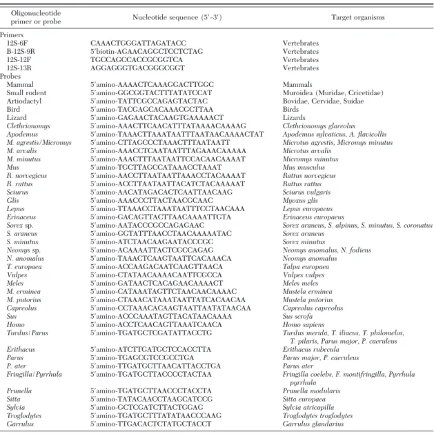
Table 1. Oligonucleotide sequences of primers and probes used in PCR amplification and RLB assays
Oligonucleotide
primer or probe Nucleotide sequence (5⬘Ð3⬘) Target organisms
Primers 12S-6F CAAACTGGGATTAGATACC Vertebrates B-12S-9R 5⬘biotin-AGAACAGGCTCCTCTAG Vertebrates 12S-12F TGCCAGCCACCGCGGTCA Vertebrates 12S-13R AGGAGGGTGACGGGCGGT Vertebrates Probes
Mammal 5⬘amino-AAAACTCAAAGGACTTGGC Mammals
Small rodent 5⬘amino-GGCGGTACTTTATATCCAT Muroidea (Muridae, Cricetidae)
Artiodactyl 5⬘amino-TATTCGCCAGAGTACTAC Bovidae, Cervidae, Suidae
Bird 5⬘amino-TACGAGCACAAACGCTTAA Birds
Lizard 5⬘amino-GAGAACTACAAGTGAAAAACT Lizards
Clethrionomys 5⬘amino-AAACTTCAACATTTATAAAACAAAAG Clethrionomys glareolus Apodemus 5⬘amino-TAAACTTAAATAATTTAATAACAAAACTAT Apodemus sylvaticus, A. flavicollis M. agrestis/Micromys 5⬘amino-CTTAGCCCTAAACTTTAATAATT Microtus agrestis, Micromys minutus
M. arvalis 5⬘amino-AAACCTCAATAATTTAGAAACAAAAA Microtus arvalis
M. minutus 5⬘amino-AAACTTTAATAATTCCACAACAAAAT Micromys minutus
Mus 5⬘amino-TGCTTAGCCATAAACCTAAAT Mus musculus
R. norvegicus 5⬘amino-AACCTTAATAATTAAACCTACAAAAT Rattus norvegicus
R. rattus 5⬘amino-ACCTTAATAATTACATCTACAAAAAT Rattus rattus
Sciurus 5⬘amino-AACATAGACACTCAATTAACAAG Sciurus vulgaris
Glis 5⬘amino-AAACCCTTACTAACGCAAC Myoxus glis
Lepus 5⬘amino-TTAAACCTAAATAATTTCCTAACAAA Lepus europaeus
Erinaceus 5⬘amino-GACAGTTACTTAACAAAATTGTA Erinaceus europaeus
Sorexsp. 5⬘amino-AATACCCGCCAGAGAAC Sorex araneus, S. alpinus, S. minutus, S. coronatus
S. araneus 5⬘amino-GGTATTTAACCTAACAAAAATAC Sorex araneus
S. minutus 5⬘amino-ATCTAACAAGAATACCCGC Sorex minutus
Neomyssp. 5⬘amino-ACAAAATTACTCGCCAGAG Neomys anomalus, N. fodiens
N. anomalus 5⬘amino-TAAACTCAAGTAATTCACAAACA Neomys anomalus
T. europaea 5⬘amino-ACCAAGACAATCAAGTTAACA Talpa europaea
Vulpes 5⬘amino-CTATAACAAAACAATTCGCCA Vulpes vulpes
Meles 5⬘amino-GATAACTCACAGAACAAAACT Meles meles
M. erminea 5⬘amino-CATAAATAGTTCTAACAACAAAAC Mustela erminea
M. putorius 5⬘amino-CTAAACATAAATAATTATCACAACAA Mustela putorius
Capreolus 5⬘amino-CCTAAACACAAGTAATTAATATAACAA Capreolus capreolus
Sus 5⬘amino-ACCCAAATAGTTACATAACAAAA Sus scrofa
Homo 5⬘amino-ACCTCAACAGTTAAATCAACA Homo sapiens
Turdus/Parus 5⬘amino-TGATGCTCGATATTACCTG Turdus merula, T. iliacus, T. philomelos, T. pilaris, Parus major, P. caeruleus
Erithacus 5⬘amino-ATCTTGATGCTCCACCTTA Erithacus rubecula
Parus 5⬘amino-TGAGCGTCCGCCTGA Parus major, P. caeruleus
P. ater 5⬘amino-TTGATGCTTAACATTACCTGA Parus ater
Fringilla/Pyrrhula 5⬘amino-TGATGCTTACCCCTACTAA Fringilla coelebs, F. montifringilla, Pyrrhula pyrrhula
Prunella 5⬘amino-TGATGCTTAACCCTACCTA Prunella modularis
Sitta 5⬘amino-TATACAACCTAAGCATCCG Sitta europaea
Sylvia 5⬘amino-GCTCGATCTTACTGGAG Sylvia atricapilla
Troglodytes 5⬘amino-TGATGCTTTATATAACCCAAG Troglodytes troglodytes
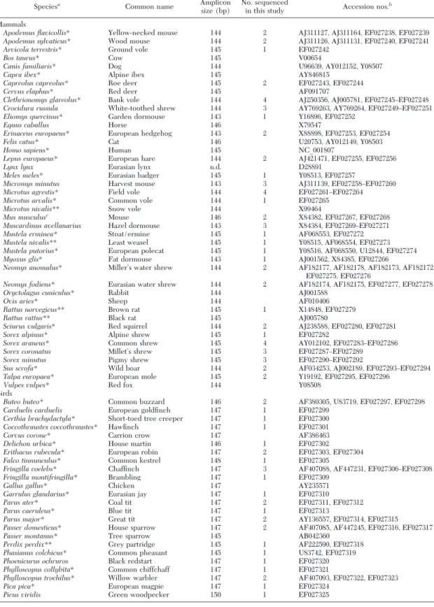
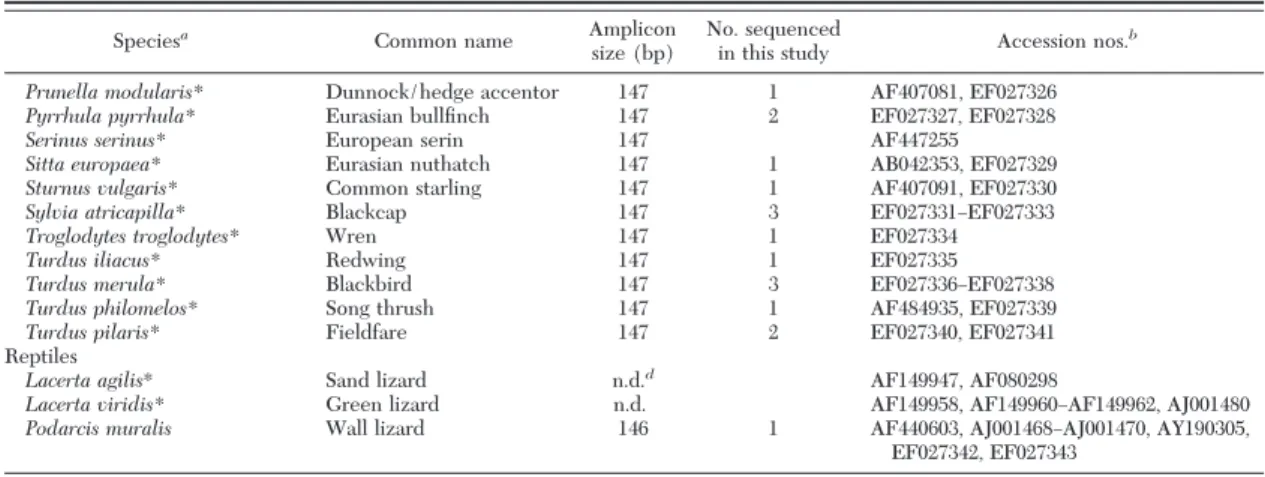
Table 2. Vertebrate species 12S rDNA sequences subjected to multi-alignment for search of molecular marker and probe design, and GenBank accession numbers
Speciesa
Common name Amplicon size (bp)
No. sequenced
in this study Accession nos.
b
Mammals
Apodemus flavicollis* Yellow-necked mouse 144 2 AJ311127, AJ311164, EF027238, EF027239
Apodemus sylvaticus* Wood mouse 144 2 AJ311126, AJ311131, EF027240, EF027241
Arvicola terrestris* Ground vole 145 1 EF027242
Bos taurus* Cow 145 V00654
Canis familiaris* Dog 144 U96639, AY012152, Y08507
Capra ibex* Alpine ibex 145 AY846815
Capreolus capreolus* Roe deer 145 2 EF027243, EF027244
Cervus elaphus* Red deer 145 AF091707
Clethrionomys glareolus* Bank vole 144 4 AJ250356, AJ005781, EF027245ÐEF027248
Crocidura russula White-toothed shrew 144 3 AY769263, AY769264, EF027249ÐEF027251
Eliomys quercinus* Garden dormouse 143 1 Y16896, EF027252
Equus caballus Horse 146 X79547
Erinaceus europaeus* European hedgehog 143 2 X88898, EF027253, EF027254
Felis catus* Cat 146 U20753, AY012149, Y08503
Homo sapiens* Human 145 NC_001807
Lepus europaeus* European hare 144 2 AJ421471, EF027255, EF027256
Lynx lynx Eurasian lynx n.d. D28891
Meles meles* Eurasian badger 145 1 Y08513, EF027257
Micromys minutus Harvest mouse 143 3 AJ311139, EF027258ÐEF027260
Microtus agrestis* Field vole 144 4 EF027261ÐEF027264
Microtus arvalis* Common vole 144 1 EF027265
Microtus nivalis** Snow vole 144 X99464
Mus musculusc Mouse 146 2 X84382, EF027267, EF027268
Muscardinus avellanarius Hazel dormouse 143 3 X84384, EF027269ÐEF027271
Mustela erminea* Stoat/ermine 145 1 AF068553, EF027272
Mustela nivalis** Least weasel 145 1 Y08515, AF068554, EF027273
Mustela putorius* European polecat 145 1 Y08516, AF068550, U12844, EF027274
Myoxus glis* Fat dormouse 143 1 AJ001562, X84385, EF027266
Neomys anomalus* MillerÕs water shrew 144 2 AF182177, AF182178, AF182173, AF182172,
EF027275, EF027276
Neomys fodiens* Eurasian water shrew 144 2 AF182174, AF182175, EF027277, EF027278
Oryctolagus cuniculus* Rabbit 144 AJ001588
Ovis aries* Sheep 144 AF010406
Rattus norvegicus** Brown rat 145 1 X14848, EF027279
Rattus rattus** Black rat 145 AJ005780
Sciurus vulgaris* Red squirrel 144 2 AJ238588, EF027280, EF027281
Sorex alpinus* Alpine shrew 145 1 EF027282
Sorex araneus* Common shrew 145 4 AY012102, EF027283ÐEF027286
Sorex coronatus MilletÕs shrew 145 3 EF027287ÐEF027289
Sorex minutus Pigmy shrew 145 3 EF027290ÐEF027292
Sus scrofa* Wild boar 144 2 AF034253, AJ002189, EF027293ÐEF027294
Talpa europaea* European mole 145 2 Y19192, EF027295, EF027296
Vulpes vulpes* Red fox 144 Y08508
Birds
Buteo buteo* Common buzzard 146 2 AF380305, U83719, EF027297, EF027298
Carduelis carduelis European goldÞnch 147 1 EF027299
Certhia brachydactyla* Short-toed tree creeper 147 1 EF027300
Coccothraustes coccothraustes* HawÞnch 147 1 EF027301
Corvus corone* Carrion crow 147 AF386463
Delichon urbica* House martin 146 1 EF027302
Erithacus rubecula* European robin 147 2 EF027303, EF027304
Falco tinnunculus* Common kestrel 148 1 EF027305
Fringilla coelebs* ChafÞnch 147 3 AF407088, AF447231, EF027306ÐEF027308
Fringilla montifringilla* Brambling 147 1 EF027309
Gallus gallus* Chicken 147 AY235571
Garrulus glandarius* Eurasian jay 147 1 EF027310
Parus ater* Coal tit 147 2 EF027311, EF027312
Parus caeruleus* Blue tit 147 1 EF027313
Parus major* Great tit 147 2 AY136557, EF027314, EF027315
Passer domesticus* House sparrow 147 2 AF407085, AF447245, EF027316, EF027317
Passer montanus* Tree sparrow 145 AB042360
Perdix perdix** Grey partridge 145 1 AF222590, EF027318
Phasianus colchicus* Common pheasant 145 1 U83742, EF027319
Phoenicurus ochruros Black redstart 147 1 EF027320
Phylloscopus collybita* Common chiffchaff 147 1 EF027321
Phylloscopus trochilus* Willow warbler 147 2 AF407093, EF027322, EF027323
Pica pica* European magpie 147 1 EF027324
Picus viridis Green woodpecker 150 1 EF027325
Sequencing of Vertebrate 12S rDNA Molecular Marker. The ⬇600-bp PCR products were puriÞed
with QIAquick PCR puriÞcation kit (QIAGEN). Cy-cle sequencing was performed using the dideoxy chain termination method by using an ABI Prism BigDye Terminator cycle sequencing kit (Applied Biosystems, Rotkreuz, Switzerland). Cycle sequencing parameters were as follows: denaturation at 96⬚C for 15 s, primer annealing at 53⬚C for 15 s, and extension at 60⬚C for 4 min. The cycle sequencing products were puriÞed either by ethanol precipitation or with DyeEx spin columns (QIAGEN) and applied to an ABI Prism 310 genetic analyzer (Applied Biosystems). DNA se-quences were checked with Sequence Navigator soft-ware (Applied Biosystems). Sequences from the same species were aligned with each other and with re-spective sequences from the GenBank database when available. The 12S rDNA marker sequences of various vertebrates obtained in this study are available in the GenBank database under respective accession num-bers (Table 2).
Sequence Analysis and Probe Design. The 143Ð150-bp
12S rDNA marker sequences (newly sequenced or retrieved from the GenBank database) of various ver-tebrate species hosts for I. ricinus ticks were aligned using ClustalX multi-alignment software embl.net. Ar-eas of genetic variability between species or between groups of vertebrates were used to design oligonucleo-tide probes using the Oligo software Primers 3.
Reverse Line Blot Hybridization. The reverse line
blotting method for the identiÞcation of host DNA in ticks developed herein is based on the RLB technique described by Rijpkema et al. (1995)). Brießy, the probes were covalently linked to an activated Biodyne C membrane (Pall, Dreieich, Germany) by their 5⬘ amino group. For this purpose, the membrane was placed in a miniblotter system (Immunetics, Cam-bridge, MA), and line slots were Þlled with the dif-ferent probes (100 Ð500 pmol). After a quick
incuba-tion (at least 1 min at room temperature), the excess of probes was removed, and the membrane was inac-tivated in 100 mM NaOH for 10 min (maximum) and washed in 2⫻ SSPE buffer/0.1% SDS (SSPE; Invitro-gen, Basel, Switzerland) for 5 min at 60⬚C. Then, the membrane was placed in the miniblotter, but the po-sition of the membrane was 90⬚ rotated compared with the previous position. The slots were Þlled with the biotin-labeled heat-denatured PCR products, and the membrane was incubated for 60 min at 42⬚C. Then, the PCR product solutions were removed, and the membrane was washed twice in 100 ml of 2⫻ SSPE/ 0.5% SDS for 10 min at 55⬚C and incubated with streptavidin-peroxidase conjugate (Roche Diagnos-tics, Rotkreuz, Switzerland) at 42⬚C for 30 min. After a washing step, the membrane was incubated for 1Ð2 min. with enhanced chemiluminescence detection liquid (GE Healthcare, OtelÞngen, Switzerland) and exposed to X-ray Þlm HyperÞlm (GE Healthcare).
Detection and Identification of Borrelia Species in Ticks. For the detection and identiÞcation of B. burg-dorferis.l. species in ticks, we used the PCR and RLB hybridization method described previously (Alekseev et al. 2001, Burri et al. 2007). Oligonucleotide probes used are described in Poupon et al. (2006).
Statistical Analysis. Statistics were calculated with
S-Plus 7.0 for Windows (Insightful Corp., Seattle, WA). The Fisher exact test was used to assess the success of the host DNA identiÞcation method be-tween nymphs and adults (statistical signiÞcance, Pⱕ 0.05).
Results
Sequencing and Alignment of 12S rDNA Marker of Various Vertebrate Species. Using external primers,
the 12S rDNA molecular marker was sequenced for a broad range of vertebrate species belonging to the fauna of Switzerland: 28 species of mammals, 31
spe-Table 2. continued
Speciesa
Common name Amplicon size (bp)
No. sequenced
in this study Accession nos.
b
Prunella modularis* Dunnock/hedge accentor 147 1 AF407081, EF027326
Pyrrhula pyrrhula* Eurasian bullÞnch 147 2 EF027327, EF027328
Serinus serinus* European serin 147 AF447255
Sitta europaea* Eurasian nuthatch 147 1 AB042353, EF027329
Sturnus vulgaris* Common starling 147 1 AF407091, EF027330
Sylvia atricapilla* Blackcap 147 3 EF027331ÐEF027333
Troglodytes troglodytes* Wren 147 1 EF027334
Turdus iliacus* Redwing 147 1 EF027335
Turdus merula* Blackbird 147 3 EF027336ÐEF027338
Turdus philomelos* Song thrush 147 1 AF484935, EF027339
Turdus pilaris* Fieldfare 147 2 EF027340, EF027341
Reptiles
Lacerta agilis* Sand lizard n.d.d AF149947, AF080298
Lacerta viridis* Green lizard n.d. AF149958, AF149960ÐAF149962, AJ001480
Podarcis muralis Wall lizard 146 1 AF440603, AJ001468ÐAJ001470, AY190305,
EF027342, EF027343
aSpecies marked with an asterisk (*) are known hosts for I. ricinus ticks in Switzerland (Aeschlimann 1972, Papadopoulos et al. 2002); species
marked with 2 asterisks are known hosts for I. ricinus ticks in Europe (Arthur 1963, Matuschka et al. 1997).
bSequences obtained from GenBank database or obtained in this study (accession numbers starting with EF). cLaboratory host species.
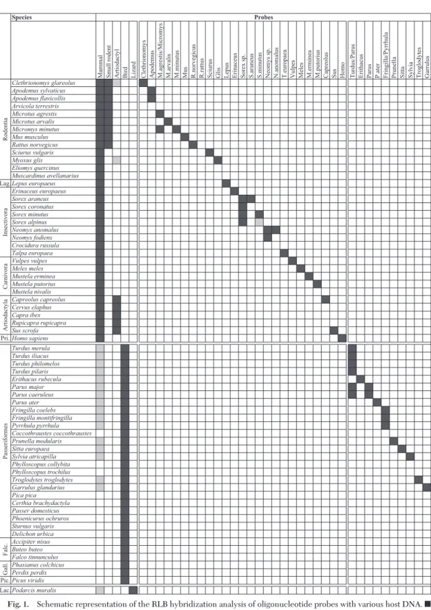
Fig. 1. Schematic representation of the RLB hybridization analysis of oligonucleotide probes with various host DNA. f, strong hybridization; u, slight hybridization may occur. Falc., Falconiformes; Gall., Galliformes; Lac., Lacertidae; Lag. Lagomorpha; Pic. Piciformes; and Pri., Primates.
cies of birds, and one species of reptiles (Table 2). Some species are known hosts for I. ricinus ticks in Switzerland (Aeschlimann 1972, Papadopoulos et al. 2002, Poupon et al. 2006) and in Europe. If possible, sequences from two or more individuals of each spe-cies originating from different sites in Switzerland were obtained and aligned with sequences retrieved from the GenBank database if available. For a few species, only sequences from GenBank database were available. Overall, we aligned 12S rDNA sequences from a large spectrum of vertebrate species including 42 species of mammals, 35 species of birds, and three species of reptiles (Table 2). According to the list of known host species of I. ricinus in Switzerland (Ae-schlimann 1972, Papadopoulos et al. 2002, Poupon et al. 2006), only three species of mammals (Martes
martes, Martes foina,and Rupicapra rupicapra) and 20 species of birds were missing.
The 12S rDNA molecular marker shows very low intraspecies genetic variability. For genetic variability between sequences retrieved from GenBank and those obtained in this study, only sequences derived from vertebrates collected in Switzerland were con-sidered for the consensus sequence.
Design of Specific Probes. The partial 12S rDNA
consensus sequences of 42 species of mammals, 35
species of birds and three species of reptiles were multi-aligned to design speciÞc probes. Five probes were designed to discriminate main groups of ver-tebrate hosts: mammals, birds, small rodents (Mu-roidea), lizards, and artiodactyls, which include Bo-vidae, CerBo-vidae, and Suidae (Table 1). Twenty-four and 10 probes, respectively, were designed to iden-tify major mammal and bird I. ricinus hosts at the genus or species level (Table 1). The design of species-speciÞc probes seemed to be impossible for the discrimination of some species (e.g., Apodemus spp. and Turdus spp.).
Identification of Vertebrate DNA by RLB. To assess
the speciÞcity of the Þve group-speciÞc probes and the 34 genus- or species-speciÞc probes (Table 1), iden-tiÞcation of vertebrate DNA by RLB was performed using DNA extracted from the aforementioned Swiss fauna tissue collection (n⫽ 60) and from one labo-ratory species (Mus musculus) (Fig. 1). Overall, all probes showed a strong hybridization with the re-spective host DNA (Fig. 1). In a few cases, a weak cross-hybridization occurred, in particular with group-speciÞc probes. The artiodactyl probe showed a weak cross-hybridization with vole DNA (C.
glareo-lus) and dormouse DNA (Myoxus glis). The mammal probe showed a strong hybridization with all tested
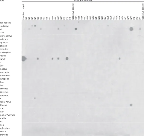
Fig. 2. Reverse line blot assay for the identiÞcation of vertebrate host species in host samples by using group- and species/genus-speciÞc probes.
mammalian DNA, but turned out to be less speciÞc because it weakly hybridized with DNA from 16 bird species as well as with DNA from a lizard species,
Podarcis muralis.In contrast, the bird probe was highly speciÞc and allowed to unambiguously discriminate avian hosts from nonavian hosts. The lizard probe was also highly speciÞc. The other 34 probes allowed to clearly identify vertebrate hosts either at the genus level (e.g., the Apodemus probe) or at the species level (e.g., the M. putorius probe) (Fig. 1). The Turdus/
Parusprobe and the Parus probe had to be used both to discriminate Turdus sp. DNA from Parus sp. DNA. In fact, the Turdus/Parus probe reacted to Turdus sp. and Parus sp., whereas the Parus probe reacted to
Parussp. only. Figure 2 shows the RLB hybridization patterns for some host species.
Host Identification in Feeding Ticks. Host
identi-Þcation was performed in nine I. ricinus ticks collected while feeding on Þve mammal and four bird species
(A. sylvaticus (one larva), C. glareolus (one larva), S.
vulgaris(one nymph), E. europaeus (one female), C.
capreolus(one female), T. merula (one nymph), T.
phi-lomelos(one nymph), P. major (one nymph), and S.
europaea(one nymph). In all nine ticks, host DNA was present and showed a perfect hybridization pattern ac-cording to the probe speciÞcity.
Host and Borrelia Identifications in Unfed Field-Collected Ticks. To assess the utility of the present
host identiÞcation method under Þeld conditions, nymphal and adult ticks collected in Neuchaˆtel were tested for the presence of host and Borrelia DNA (Fig. 3). The detection of host DNA in questing ticks required the use of a high number of PCR ampliÞca-tion cycles, and as a result, contaminaampliÞca-tion with human DNA could not be avoided. Therefore, mammal and
Homoprobes for detection of mammalian and human DNA, respectively, were removed from our set of probes.
Fig. 3. Reverse line blot assay for the identiÞcation of vertebrate host species in questing I. ricinus ticks (Neuchaˆtel, Switzerland) using group- and species/genus- speciÞc probes. N, nymph; F, female; M, male; X and H, different batches of ticks.
In total, 55 nymphal, 28 male, and 26 female I. ricinus ticks were tested for host and Borrelia identiÞcations. Host DNA could be detected in half of questing I.
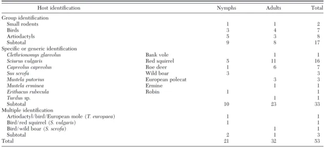
ricinusticks (53/109; 48.6%). Detection was signiÞ-cantly higher in adults (32/54; 59.3%) than in nymphs (21/55; 38.2%) (P ⫽ 0.035; Fisher exact test) and slightly varied between sexes (15/28 males; 53.6% and 17/26 females; 65.4%). For one third of the questing ticks with a bloodmeal identiÞed (17/53; 32.1%), host identiÞcation was possible only at the group level (small rodents [Muroidea], birds, or artiodactyls) (Table 3). For virtually two thirds of the I. ricinus ticks with identiÞed host DNA (33/53; 62.3%), host iden-tiÞcation could be achieved at the species or genus level (Table 3). In three questing I. ricinus ticks, the presence of a mixture of DNA from two or three different hosts was observed (Table 3). Red squirrel (S. vulgaris) followed by roe deer (C. capreolus) were the species from which DNA was most frequently detected in questing ticks (Table 3). DNA from ar-tiodactyls (n ⫽ 20) (including eight artiodactyls, seven C. capreolus, three S. scrofa and two multiple identiÞcations), and rodents (n⫽ 20) (including two Muroidea, 16 S. vulgaris, one C. glareolus, and one multiple identiÞcation) were most frequently de-tected followed by birds (n⫽ 12) (including seven birds, one E. rubecula, one Turdus sp., and three mul-tiple identiÞcations) and carnivores (n⫽ 4) (includ-ing three M. putorius and one M. erminea).
Borreliainfection was detected in 23/109 (21.1%) questing I. ricinus ticks. In 15 of these 23 infected ticks (65.2%), identiÞcation of the blood source was also possible (Table 4). In these ticks, single infection with
B. burgdorferisensu stricto (n⫽ 5), Borrelia afzelii (n⫽ 2), Borrelia garinii (n ⫽ 1), Borrelia valaisiana (n⫽ 5), and untypeable Borrelia (n ⫽ 2) was observed (Table 4). No multiple infections were detected. B.
burgdorferiss and B. afzelii infections were observed with squirrel DNA, and B. valaisiana was observed with bird DNA. In ticks containing blood remnants
from artiodactyls, DNA from B. afzelii, B. burgdorferi s.s., and B. valaisiana was detected (Table 4).
Discussion
This report describes the development of an efÞ-cient new technique for the identiÞcation of blood-meal sources in the tick I. ricinus, the European vector of the agents of Lyme borreliosis and tick-borne en-cephalitis. The method consists of a single-run PCR ampliÞcation of the 12S rDNA molecular marker using a single set of nondegenerate primers followed by RLB hybridization assay by using speciÞc probes. This method allowed identifying the bloodmeal source in half of questing I. ricinus ticks collected from vegeta-tion (49%). This sensitivity was similar to those ob-tained in previous studies using 18S rDNA as target (Pichon et al. 2003, 2005; Estrada-Pen˜a et al. 2005). The identiÞcation to the genus or species level oc-curred for two thirds of ticks with bloodmeal identi-Þcation. This represents a more precise identiÞcation compared with previous studies that identiÞed host
Table 3. Host DNA identification in field-collected questing I. ricinus ticks by RLB assay (Neuchâtel, Switzerland)
Host identiÞcation Nymphs Adults Total
Group identiÞcation
Small rodents 1 1 2
Birds 3 4 7
Artiodactyls 5 3 8
Subtotal 9 8 17
SpeciÞc or generic identiÞcation
Clethrionomys glareolus Bank vole 1 1
Sciurus vulgaris Red squirrel 5 11 16
Capreolus capreolus Roe deer 1 6 7
Sus scrofa Wild boar 3 3
Mustela putorius European polecat 3 3
Mustela erminea Ermine 1 1
Erithacus rubecula Robin 1 1
Turdussp. 1 1
Subtotal 10 23 33
Multiple identiÞcation
Artiodactyl/bird/European mole (T. europaea) 1 1
Bird/red squirrel (S. vulgaris) 1 1
Bird/wild boar (S. scrofa) 1 1
Subtotal 2 1 3
Total 21 32 53
Table 4. Bloodmeal source identification in B. burgdorferi s.l.-infected questing I. ricinus ticks (Neuchâtel, Switzerland)
Host identiÞcation
No. infected I. ricinus ticksa
Bbs.s. Ba Bg Bv Borrelia sp. Red squirrel 2N, 1Mb 1N 1N European polecat 1F Small rodents 1F Birds 2N, 1M Artiodactyls 1N 1N 1N Bird/wild boar 1F Artiodactyl/bird/mole 1N Total 5 2 1 5 2 a
Ba, B. afzelii; Bbs.s., B. burgdorferi sensu stricto; Bg, B. garinii; Bv,
B. valaisiana;and Borrelia sp., untypeable Borrelia.
b
groups rather than host species (Pichon et al. 2003, 2005).
That questing ticks were collected at the beginning of the tick activity season (spring) suggests that the previous bloodmeal occurred during the previous tick activity season and that the method is sensitive enough to detect and identify the source of bloodmeals, which are several months old. A better sensitivity might be expected with the summer/autumnal tick cohort, as observed by Pichon et al. (2005). The identiÞcation of DNA from two or three different hosts in a single tick suggests that interrupted feeding may occur in I.
ric-inus.Possibly, this may contribute to the transfer of tick-borne pathogens. However, such transmission probably rarely occurs.
Coupled with the RLB method for the detection and identiÞcation of B. burgdorferi s.l. (Alekseev et al. 2001, Poupon et al. 2006, Burri et al. 2007), the host identiÞcation technique could conÞrm the existence of associations between hosts and Borrelia genospe-cies, notably between red squirrels, B. burgdorferi s.s., and B. afzelii (Humair and Gern 1998) as well as between birds, B. garinii, and B. valaisiana (Humair et al. 1998). Currently, our sample sizes are too small to conÞrm previous observations. The presence of B.
afzelii, B. valaisiana,and B. burgdorferi s.s. in ticks in which artiodactyl blood remnants were detected was more surprising, because these hosts are generally considered zooprophylactic (Gern and Humair 2002). One explanation may be that these ticks have been infected through cofeeding transmission of the patho-gens from infected to uninfected ticks feeding on uninfected artiodactyls (Hu et al. 2003). The overall prevalence of Borrelia infection in ticks (21.1%) in this study was similar to prevalences observed in other tick populations in Switzerland (Jouda et al. 2003, 2004a,b; Burri et al. 2007).
The target gene for the identiÞcation of bloodmeal sources is the 12S rRNA gene, which encodes for the small subunit ribosomal RNA in mitochondria. Mito-chondrial DNA (mtDNA) presents peculiar features, such as lack of recombination, maternal inheritance, and presence of orthologous genes, and it has been used extensively in phylogenetic and evolutionary studies. In addition, mtDNA occurs in high copy num-bers in a eukaryote cell; therefore, is a molecule of choice when analyzing small quantity of degraded biological material, as in the case of bloodmeal rem-nants in tick midguts. The 12S rRNA gene was inves-tigated because it is described with some tRNAs as the most conserved regions of the mitochondrial genome (Saccone et al. 1999). Various authors have used the cytochrome b gene as a molecular marker to identify bloodmeals in Diptera (Boakye et al. 1999, Lee et al. 2002, Ngo and Kramer 2003) and in ticks (Tobolewski et al. 1992, Kirstein and Gray 1996). A preliminary analysis (unpublished) demonstrated that the cyto-chrome b gene is subject to a high inter- and intraspe-ciÞc genetic variability, which could have lead to a problematic probe and primer design for the large spectrum of I. ricinus hosts. However, the nuclear 18S rRNA gene, used in other studies (Pichon et al. 2003,
2005; Estrada-Pen˜a et al. 2005), is too much conserved to identify the genus or species origin of tick blood-meals.
The small size of the molecular marker is crucial for the host identiÞcation in questing ticks. In the case of degraded DNA, as occurs in questing ticks, an inverse correlation exists between the efÞciency of PCR ampliÞcation and the size of the amplicon (Kirstein and Gray 1996). The nondegenerate character of primers avoids mispriming and enhances the spec-iÞcity of DNA ampliÞcation reaction. High inter-species or intergenus genetic variability and a low intraspecies polymorphism of the molecular marker allow a precise identiÞcation of the hosts that ticks have been feeding on.
The palette of oligonucleotide probes used in the RLB allowed at the same time to distinguish major groups of host vertebrates and to identify bloodmeal sources at the genus or species level. Forty oligonu-cleotide probes were designed to identify groups, gen-era or species that may serve as natural or laboratory hosts for I. ricinus. The analysis of various vertebrate DNA samples from our tissue library demonstrated that the speciÞcity of the method was very good and allowed a correct identiÞcation of a large range of host species. However, in some cases, the precise identiÞ-cation at the species level was not possible. For mam-mals, for example, the Apodemus probe did not permit to distinguish A. flavicollis from A. sylvaticus, and the
M. agrestis/Micromysprobe did not discriminate M.
agrestis and M. minutus. For some avian hosts, the speciÞcity was even lower as in the case of the Turdus/
Parusprobe that reacts to four Turdus species and two
Parusspecies. The concurrent use of the Parus probe is necessary to distinguish Turdus sp. from Parus sp.
The sensitivity of the technique becomes crucial when analyzing host-seeking I. ricinus ticks collected from the vegetation. Host blood remnants in Þeld-collected tick midguts come from the previous instar, may be several months old and are present in small quantity and in a degraded state. To enhance the techniqueÕs sensitivity, the number of PCR ampliÞca-tion cycles was increased. The use of nested PCR was avoided because of the high risk of contaminations with products from the Þrst PCR run. Using the 40-cycle PCR procedure, human contamination was very frequent. Therefore, the mammal and the Homo probes were discarded when analyzing Þeld-collected ticks. In addition, to avoid any other contamination, extremely strict conditions were applied to the method. For example, PCR setup and DNA extraction were performed in rooms dedicated to this use and restricted to this project.
In conclusion, the present technique based on PCR ampliÞcation of a 12S rDNA molecular marker and RLB hybridization assays provides a rapid and accu-rate method for determining the source of bloodmeals in I. ricinus ticks collected in the Þeld. This method could be used to identify new host species of arthro-pod vectors. Coupled with methods for identiÞcation of vector-borne pathogens, this technique should al-low researchers to determine the source of infective
bloodmeal, identify reservoir hosts at the species level, and assess the importance of a host species in a habitat. Further development of this technique may be envis-aged for other arthropod vectors.
Acknowledgments
We thank Martin Zimmerli and Blaise Mulhauser (Muse´um dÕHistoire Naturelle, Neuchaˆtel); Michel Beaud (Muse´e dÕHistoire Naturelle, Fribourg); Peter Vogel, Jacques Hausser, and Patrick Basset (De´partement dÕEcologie et dÕEvolution, Lausanne); Marie-Ange`le Poupon and Ilona Loubry (Institut de Biologie, Neuchaˆtel); Georg Brosi and Erwin Eggenberger (Amt fu¨r Jagd und Fischerei, Grau-bu¨nden); Arthur Fiechter (Service de la Faune, Neuchaˆtel); Claude Fischer, Matisse, and the Zoo de la Garenne for help in the collection of vertebrate tissue samples. We thank Yves Cheminade for technical assistance, the Laboratoire de Bota-nique e´volutive (University of Neuchaˆtel) for the use of the sequencer, and Yong-Min Yuan for help with the sequencer. This project was supported by the Swiss National Science Foundation 3200-057098 and 3200B0-100657. V.D. and P.-F.H. were supported by the Swiss National Science Foun-dation. P.-F.H. was also supported by the Roche Foundation and the Novartis Foundation for Medicine and Biology.
References Cited
Aeschlimann, A. 1972. Ixodes ricinus, Linne´, 1758
(Ix-odoidea: Ixodidae). Essai pre´liminaire de synthe`se sur la biologie de cette espe`ce en Suisse. Acta Trop. 29: 321Ð340.
Aeschlimann, A., E. Chamot, F. Gigon, J. P. Jeanneret, D. Kesseler, and C. Walther. 1986. Borrelia burgdorferi in
Switzerland. Zentbl. Bakt. Hyg. A. 263: 450 Ð 458.
Alekseev, A. N., H. V. Dubinina, I. Van de Pol, and L. M. Schouls. 2001. IdentiÞcation of Ehrlichia spp. and
Bor-relia burgdorferiin Ixodes ticks in the Baltic regions of Russia. J. Clin. Microbiol. 39: 2237Ð2242.
Arthur, D. R. 1963. British ticks. Butterworths, London,
United Kingdom.
Beier, J. C., P. V. Perkins, R. A. Wirtz, J. Koros, D. Diggs, T. P. Gargan II, and D. K. Koech. 1988. Bloodmeal
identiÞ-cation by direct enzyme-linked immunosorbent assay (ELISA), tested on Anopheles (Diptera: Culicidae) in Kenya. J. Med. Entomol. 25: 9 Ð16.
Boakye, D. A., J. Tang, P. Truc, A. Merriweather, and T. R. Unnasch. 1999. IdentiÞcation of bloodmeals in
haema-tophagous Diptera by cytochrome B heteroduplex anal-ysis. Med. Vet. Entomol. 13: 282Ð287.
Burgdorfer, W., A. G. Barbour, S. F. Hayes, O. Pe´ter, and A. Aeschlimann. 1983. Erythema chronicum migransÐa
tick-borne spirochetosis. Acta Trop. 40: 79 Ð 83.
Burri, C., F. Mora´n Cadenas, V. Douet, J. Moret, and L. Gern. 2007. Ixodes ricinus density and infection prevalence
with Borrelia burgdorferi sensu lato along a north-facing altitudinal gradient in the Rhoˆne Valley (Switzerland). Vector Borne Zoonotic Dis. 7: 50 Ð58.
Clausen, P. H., I. Adeyemi, B. Bauer, M. Breloeer, F. Salchow, and C. Staak. 1998. Host preferences of tsetse (Diptera:
Glossinidae) based on bloodmeal identiÞcations. Med. Vet. Entomol. 12: 169 Ð180.
Craine, N. G., P. A. Nuttall, A. C. Marriott, and S. E. Randolph. 1997. Role of grey squirrels and pheasants in the
transmis-sion of Borrelia burgdorferi sensu lato, the Lyme disease spirochaete, in the UK. Folia Parasitol. 44: 155Ð160.
De Boer, R., K. E. Hovius, M. K. Nohlmans, and J. S. Gray. 1993. The woodmouse (Apodemus sylvaticus) as a
reser-voir of tick-transmitted spirochetes (Borrelia burgdor-feri) in The Netherlands. Zentralbl. Bakteriol. 279: 404 Ð 416.
Dsouli, N., H. Younsi-Kabachii, D. Postic, S. Nouira, L. Gern, and A. Bouattour. 2006. Reservoir role of lizard
Psam-modromus algirusin transmission cycle of Borrelia burg-dorferisensu lato (Spirochaetaceae) in Tunisia. J. Med. Entomol. 43: 737Ð742.
Estrada-Pen˜a, A., J. J. Osa´car, B. Pichon, and J. S. Gray. 2005.
Host and pathogen detection for immature stages of Ix-odes ricinus(Acari: Ixodidae) in north-central Spain. Exp. Appl. Acarol. 37: 257Ð268.
Gern, L., E. Rouvinez, L. N. Toutoungi, and E. Godfroid. 1997. Transmission cycles of Borrelia burgdorferi sensu
lato involving Ixodes ricinus and/or I. hexagonus ticks and the European hedgehog, Erinaceus europaeus, in subur-ban and ursubur-ban areas in Switzerland. Folia Parasitol. 44: 309 Ð314.
Gern, L., A. Estrada-Pen˜a, F. Frandsen, J. S. Gray, T. G. Jaenson, F. Jongejan, O. Kahl, E. Korenberg, R. Mehl, and P. A. Nuttall. 1998. European reservoir hosts of Borrelia
burgdorferisensu lato. Zentralbl. Bakteriol. 287: 196 Ð204.
Gern, L., and P.-F. Humair. 2002. Ecology of Borrelia
burg-dorferisensu lato in Europe, pp. 149 Ð174. In J. S. Gray, O. Kahl, R. S. Lane, and G. Stanek [eds.], Lyme borreliosis: biology, epidemiology, and control. CAB International, Wallingford, Oxon, United Kingdom.
Guy, E. C., and G. Stanek. 1991. Detection of Borrelia
burg-dorferiin patients with Lyme disease by the polymerase chain reaction. J. Clin. Pathol. 44: 610 Ð 611.
Hu, C. M., Y. Cheminade, J.-L. Perret, V. Weynants, Y. Lobet, and L. Gern. 2003. Early detection of Borrelia
burgdor-ferisensu lato infection in Balb/c mice by co-feeding Ixodes ricinusticks. Int. J. Med. Microbiol. 293: 421Ð 426.
Humair, P. F., N. Turrian, A. Aeschlimann, and L. Gern. 1993. Borrelia burgdorferi in a focus of Lyme borreliosis:
epizootiologic contribution of small mammals. Folia Para-sitol. 40: 65Ð70.
Humair, P.-F., and L. Gern. 1998. Relationship between
Borrelia burgdorferi sensu lato species, red squirrels (Sciurus vulgaris) and Ixodes ricinus in enzootic areas in Switzerland. Acta Trop. 69: 213Ð227.
Humair, P.-F., D. Postic, R. Wallich, and L. Gern. 1998. An
avian reservoir (Turdus merula) of the Lyme borreliosis spirochetes. Zentralbl. Bakteriol. 287: 521Ð538.
Humair, P. F., O. Rais, and L. Gern. 1999. Transmission of
B. afzeliifrom Apodemus mice and Clethrionomys voles to Ixodes ricinusticks: differential transmission pattern and overwintering maintenance. Parasitology 118: 33Ð 42.
Hunter, F. F., and R. Bayly. 1991. ELISA for identiÞcation
of blood meal source in black ßies (Diptera: Simuliidae). J. Med. Entomol. 28: 527Ð532.
Jouda, F., M. Crippa, J.-L. Perret, and L. Gern. 2003.
Dis-tribution and prevalence of Borrelia burgdorferi sensu lato in Ixodes ricinus ticks of canton Ticino (Switzerland). Eur. J. Epidemiol. 18: 907Ð912.
Jouda, F., J.-L. Perret, and L. Gern. 2004a. Ixodes ricinus
density, and distribution and prevalence of Borrelia burg-dorferisensu lato infection along an altitudinal gradient. J. Med. Entomol. 41: 162Ð169.
Jouda, F., J. L. Perret, and L. Gern. 2004b. Density of
quest-ing Ixodes ricinus nymphs and adults infected by Borrelia burgdorferi sensu lato in Switzerland: spatio-temporal pattern at a regional scale. Vector Borne Zoonotic Dis. 4: 23Ð32.
Kahl, O., and L. Geue. 1995. Laboratory study on the role of
the European fox, Vulpes vulpes, as a possible reservoir of Borrelia burgdorferis.l., pp. 239. In Proceedings, 2nd
In-ternational Conference on Tick-Borne Pathogens at the Host-Vector Interface: a global perspective, 28 AugustÐ1 September 1995, Kruger National Park, South Africa.
Kent, R. J., and D. E. Norris. 2005. IdentiÞcation of
mam-malian blood meals in mosquitoes by a multiplexed poly-merase chain reaction targeting cytochrome B. Am. J. Trop. Med. Hyg. 73: 336 Ð342.
Kirstein, F., and J. S. Gray. 1996. A molecular marker for the
identiÞcation of the zoonotic reservoirs of Lyme borre-liosis by analysis of the blood meal in its European vector Ixodes ricinus.Appl. Environ. Microbiol. 62: 4060 Ð 4065.
Kurtenbach, K., D. Carey, A. N. Hoodless, P. A. Nuttall, and S. E. Randolph. 1998. Competence of pheasants as
res-ervoirs for Lyme disease spirochetes. J. Med. Entomol. 35: 77Ð 81.
Lee, J. H., H. Hassan, G. Hill, E. W. Cupp, T. B. Higazi, C. J. Mitchell, M. S. Godsey, Jr., and T. R. Unnasch. 2002.
IdentiÞcation of mosquito avian-derived blood meals by polymerase chain reaction-heteroduplex analysis. Am. J. Trop. Med. Hyg. 66: 599 Ð 604.
Matuschka, F. R., P. Fischer, M. Heiler, D. Richter, and A. Spielman. 1992. Capacity of European animals as
reser-voir hosts for the Lyme disease spirochete. J. Infect. Dis. 165: 479 Ð 483.
Matuschka, F.-R., H. Eiffert, A. Ohlenbusch, and A. Spiel-man. 1994. Amplifying role of edible dormice in Lyme
disease transmission in central Europe. J. Infect. Dis. 170: 122Ð127.
Matuschka, F. R., S. Endepols, D. Richter, and A. Spielman. 1997. Competence of urban rats as reservoir hosts for
Lyme disease spirochetes. J. Med. Entomol. 34: 489 Ð 493.
Ngo, K. A., and L. D. Kramer. 2003. IdentiÞcation of
mos-quito bloodmeals using polymerase chain reaction (PCR) with order-speciÞc primers. J. Med. Entomol. 40: 215Ð222.
Papadopoulos, B., P. F. Humair, A. Aeschlimann, C. Vaucher, and W. Bu¨ttiker. 2002. Ticks on birds in Switzerland.
Acarologia 42: 3Ð19.
Pichon, B., D. Egan, M. Rogers, and J. Gray. 2003. Detection
and identiÞcation of pathogens and host DNA in unfed host-seeking Ixodes ricinus L. (Acari: Ixodidae). J. Med. Entomol. 40: 723Ð731.
Pichon, B., M. Rogers, D. Egan, and J. Gray. 2005.
Blood-meal analysis for the identiÞcation of reservoir hosts of tick-borne pathogens in Ireland. Vector-Borne Zoonotic Dis. 5: 172Ð180.
Poupon, M.-A., E. Lommano, P.-F. Humair, V. Douet, O. Rais, M. Schaad, L. Jenni, and L. Gern. 2006. Prevalence
of Borrelia burgdorferi sensu lato in ticks collected from migratory birds in Switzerland. Appl. Environ. Microbiol. 72: 976 Ð979.
Rijpkema, S. G., M. J. Molkenboer, L. M. Schouls, F. Jongejan, and J. F. Schellekens. 1995. Simultaneous detection and
genotyping of three genomic groups of Borrelia burgdor-ferisensu lato in Dutch Ixodes ricinus ticks by character-ization of the ampliÞed intergenic spacer region between 5S and 23S rRNA genes. J. Clin. Microbiol. 33: 3091Ð3095.
Saccone, C., C. De Giorgi, C. Gissi, G. Pesole, and A. Reyes. 1999. Evolutionary genomics in Metazoa: the
mitochon-drial DNA as a model system. Gene 238: 195Ð209.
Ta¨lleklint, L., and T.G.T. Jaenson. 1994. Transmission of
Borrelia burgdorferis.l. from mammal reservoirs to the primary vector of Lyme borreliosis, Ixodes ricinus (Acari: Ixodidae), in Sweden. J. Med. Entomol. 31: 880 Ð 886.
Tempelis, C. H. 1975. Host-feeding patterns of mosquitoes,
with a review of advances in analysis of blood meals by serology. J. Med. Entomol. 11: 635Ð 653.
Tobolewski, J., M. J. Kaliszewski, R. K. Colwell, and J. H. Oliver, Jr. 1992. Detection and identiÞcation of
mam-malian DNA from the gut of museum specimens of ticks. J. Med. Entomol. 29: 1049 Ð1051.