University of Dundee
Following the Formation of Synaptonemal Complex Formation in Wheat and Barley by High-Resolution Microscopy
Darrier, Benoit; Arrieta, Mikel; Mittmann, Sybille U.; Sourdille, Pierre; Ramsay, Luke; Waugh, Robbie Published in: Plant Meiosis DOI: 10.1007/978-1-4939-9818-0_15 Publication date: 2020 Document Version
Peer reviewed version
Link to publication in Discovery Research Portal
Citation for published version (APA):
Darrier, B., Arrieta, M., Mittmann, S. U., Sourdille, P., Ramsay, L., Waugh, R., & Colas, I. (2020). Following the Formation of Synaptonemal Complex Formation in Wheat and Barley by High-Resolution Microscopy. In M. Pradillo, & S. Heckmann (Eds.), Plant Meiosis: Methods and Protocols (pp. 207-215). (Methods in Molecular Biology; Vol. 2061). Humana Press. https://doi.org/10.1007/978-1-4939-9818-0_15
General rights
Copyright and moral rights for the publications made accessible in Discovery Research Portal are retained by the authors and/or other copyright owners and it is a condition of accessing publications that users recognise and abide by the legal requirements associated with these rights.
• Users may download and print one copy of any publication from Discovery Research Portal for the purpose of private study or research. • You may not further distribute the material or use it for any profit-making activity or commercial gain.
• You may freely distribute the URL identifying the publication in the public portal.
Take down policy
If you believe that this document breaches copyright please contact us providing details, and we will remove access to the work immediately and investigate your claim.
Title: Following the formation of synaptonemal complex formation in wheat and barley by
high resolution microscopy.
Running Head: 3D-SIM of the cereals synaptonemal complex
Authors: Benoit Darrier3,4, Mikel Arrieta1, Sybille U. Mittmann1,2 , Pierre Sourdille3,4 , Luke
Ramsay1, Robbie Waugh1,2 and Isabelle Colas1.
Authors Affiliation:
1- Cell and Molecular Sciences, The James Hutton Institute, Invergowrie, Dundee, Scotland DD2 5DA, UK.
2- Division of Plant Sciences, University of Dundee at The James Hutton Institute, Invergowrie, Dundee, Scotland DD2 5DA, UK.
3- INRA UMR 1095, Génétique, Diversité & Ecophysiologie des Céréales, Domaine de Crouël, 5, Chemin de Beaulieu, 63039 Clermont-Ferrand FRANCE
4- School of Agriculture, Food and Wine, The University of Adelaide, Hartley Grove, Waite Campus, Urrbrae SA 5064
Emails : Isabelle Colas : Isabelle.Colas@hutton.ac.uk
Benoit Darrier: benoit.darrier@adelaide.edu.au
Sybille Mittmann: Sybille.Mittmann@kws.com Mikel Arrieta: Mikel.Arrieta@hutton.ac.uk Robbie Waugh: r.waugh@dundee.ac.uk
Pierre Sourdille: Pierre.SOURDILLE@clermont.inra.fr Luke Ramsay: Luke.Ramsay@hutton.ac.uk
Corresponding author(s): Isabelle Colas: Isabelle.Colas@hutton.ac.uk
This is a post-peer-review, pre-copyedit version of a chapter published in Plant Meiosis: Methods and Protocols. The final authenticated version is available online at: https://doi.org/10.1007/978-1-4939-9818-0_15.
Summary
Wheat and barley have large genomes of 15 Gb and 5.1 Gb respectively which is much larger than the Human genome (3.3 Gb). The release of their respective genomes has been a tremendous advance the understanding of the genome organization and the ability for deeper functional analysis in particular meiosis. Meiosis is the cell division required during sexual reproduction. One major event of meiosis is called recombination, or the formation of crossing over, a tight link between homologous chromosomes, ensuring gene exchange and faithful chromosome segregation. Recombination is a major driver of genetic diversity but in these large genome crops, the vast majority of these events is constrained at the end of their chromosomes. It is estimated that in barley, about 30% of the genes are located within the poor recombining centromeric regions, making important traits, such as resistance to pest and disease for example, difficult to access. Increasing recombination in these crops has the potential to speed up breeding program and requires a good understand of the meiotic mechanism. However, most research on recombination in plant has been carried in Arabidopsis
thaliana which despite many of the advantages it brings for plant research, has a small genome
and more spread out of recombination compare to barley or wheat. Advance in microscopy and cytological procedures have emerged in the last few years, allowing to follow meiotic events in these crops. This protocol provides the practical’s steps required for cytological preparation of barley and wheat pollen mother cells for light microscopy, highlighting some of the difference between the two cereals.
1. Introduction
There is a vast number of cytological and immunochemical methods to study meiosis in model organisms but if all are based on the same principles, some work better in one organisms than another. In plant, cell wall is also complicating the penetration of probes or antibodies which requires extra steps of permeabilization or digestion (1-2). Meiosis is the specific cell division that produce male and female gamete and is highly controlled (3-4) and cytology is a powerful method for functional analysis of this process. The duration of meiosis in barley is about 43 hours (5-6), while in bread wheat, it lasts for 24 hours despite having to process 3 times more genetic material (7). An important event in meiosis is chromosome synapsis, that aligns the homologues together which allows for homologous recombination (8-10). Any defect in chromosome synapsis leads to reduced recombination and disastrous chromosome segregation (11-14). This protocol takes advantage of 3D Structured Illumination Microscopy (3D-SIM) to
follow the events of synapsis in both barley and wheat. We are using TaASY1 (Asynaptic 1) (15), an antibody raised in wheat and developed by Boden et al (16) to monitor the formation
of chromosome axes during the first step of the synaptonemal complex formation. To monitor synapsis, we are using a custom antibody made in our lab against the barley protein HvZYP1 (10, 12). The protocol details the steps to prepare pollen mother cells isolation in both barley
and wheat, for investigating the progression of synapsis, highlighting some of the difference between the two organisms and associated difficulties.
2. Materials
2.1 Plant1. Barley cv. Bowman seeds 2. Wheat cv Chinese spring seeds
2.2 Meiosis Staging and plant fixation
1. Stereomicroscope Microtec HM-4TR; equipped with 10x focusing eyepiece with graticule 10:100 (HM-415FG STG) and HM-3L4N LED stand (see Note 1)
2. Microscope Microtec LM-2TR equipped with wide field eyepiece 10x/FN 20 with graticule 10:100 (LM-E10FG) and calibrating stage micrometer 1 mm in 100 parts (PM-STG) and objective 10x and 40x. (see Note 1)
3. Dissecting tools: Very Fine forceps (or insulin needles), single edge razor blade 4. Glass Microscope slides and 18x18 glass coverslip
5. Aceto-carmine: Dissolve 0.5g of Carmine in 45% acetic acid, bring to boil and stir for 15-20 minutes. Removed from heat, let it cool and keep in the fridge overnight. Filter to remove any precipitates and store in a dark bottle (see Note 2)
6. Fixative: 4% formaldehyde fixative was made by dissolving 1 g of paraformaldehyde in 12.5 ml of water, heat up to 60°C for few minutes and add 2-3 drops of 5M NaOH until the solution clears up. Let it cool on ice and add 12.5 ml of 2X PBS (see Note 3)
2.3 Slide preparation and Immunostaining
1. Working buffer: 1X PBS containing 0.5% TritonTM X100, 0.5% Tween® 20. Also add 0.5% Lipsol if working with wheat.
2. Embryo Dish (or cavity slide) and Glass rod.
3. Microscope Polysine® slide .
5. PAP pen liquid blocker.
6. Blocking solution: Working solution containing 5% Goat Serum Albumin and 5% Donkey Serum Albumin. Make it fresh. (see Note 4)
7. Primary antibodies: TaASY1 (Agrisera) and HzZYP1 (10) are used at 1:1000 and 1:500 respectively in blocking buffer.
8. Secondary antibodies: Secondary Antibody, Alexa Fluor® 488 or 568 conjugate (Thermo Fisher Scientific) against rat and rabbit are used at 1:300 in blocking buffer (10).
9. Antifading agent: Vectashield antifade mounting medium containing DAPI (Vector Laboratories, catalogue number: H-1200).
10. Humidity chamber (see Fig. 1)
11. Glass Coplin staining jar (any brand or size)
12. Glass coverslip, 22x44 mm, thickness No 1.5 (VWR or similar) and Coverslip made of para-film (or similar)
3. Methods
3.1 Plant materialBoth barley and wheat were grown from seeds (3 per pot) in compost-based pots (7 inches diameter) in temperature-controlled glasshouse under the following conditions: 16h of light at 18-20°C and 8h of dark at 14-16°C (see Note 5). The humidity was kept between 65-70%. Barley staging started from 6 weeks of growing by looking at spikes of 1.2 to 2 cm (see Note 6). Wheat staging started at around 8 weeks, looking for spikes of about 5 to 6 cm (see Note
7). In both species, the later stage of meiosis is in the middle of the spike with the earlier stages
toward top and bottom and this floret was used to define the spike meiotic stage. 3.2 Meiosis Staging and plant fixation
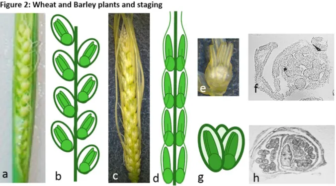
2. Even though the morphology of barley and wheat is a bit different (Fig. 2 b, d), each developing spikelet has three anthers of identical size (Fig. 2 e-h). Wheat will require a bit more dissection than barley where anthers are not embedded within plant tissue.
3. Under the stereomicroscope, remove one of the three developing anthers and place it onto a microscope slide.
4. Add a drop of aceto-carmine and squash it slightly under a glass coverslip.
5. Under the microscope, locate the anther with 10X objective and identify the meiotic stage by using the 40x Objectives.
6. Transfer the remaining 2 anthers in an embryo dish containing 200 µl of working buffer at room temperature or on ice in case of warm weather. Pool the anthers of at least half of the spike
7. Replace the working buffer by fixative and incubate for 20 and 30 minutes for barley and wheat respectively. (see Note 7)
8. Wash the sample twice for 5 minutes with 1X PBS and proceed to the slide preparation (see Note 8)
3.3 Slide preparation and Immunostaining
1. On a polysine slide, make a well in the centre with the PAP pen and let it dry fully. 2. Remove most of the working buffer and tap out the fixed anthers repeatedly with a glass
rod to release the meiocytes. Add 30 µl of working buffer, mix gently and transfer 25 µl of the solution onto the prepared slide.
3. Let it dry slowly and add 30 µl of the primary antibody freshly prepared in blocking solution.
4. Add a soft plastic coverslip and leave for 30 minutes (up to 1h) at RT in the dark wet chamber, then move the box to a cold room (or fridge) for at least 24 h (up to 48 h).
5. Warm up the slides at RT for at least 30 minutes (up to 1 h), then wash them twice 5 minutes in 1X PBS on orbital shaker.
6. Apply 40 µl of secondary antibody solution, cover with a soft plastic coverslip and leave to incubate at RT for 90 minutes to 2 h.
7. Wash the slides as previously in step 6, remove the excess PBS and add one drop of Vectashield containing DAPI.
8. Add a full-length glass cover slip, press gently upside down on paper towel to remove the Vectashield excess, and seel the 4 slides with nail varnish.
9. Let the varnish to dry and store in the dark at 4C.
3.4 Imaging
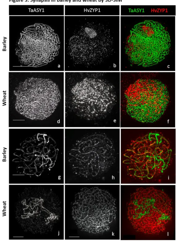
1. 3D-SIM images (Fig. 3) were acquired on a DeltaVision OMX Blaze (GE Healthcare) fitted with an Olympus PlanApo N 60x 1.42 NA oil objective. Settings for image acquisition were used as in (12); At least 32-bits 1024x1024 image (z=0.125).
2. The volume of the wheat meiocytes was 2.5x larger than barley meiocytes. It takes about 5-7 minutes per cell to acquire the whole barley cell, at least twice as much for wheat nuclei. For example, a total of 93 total images (31 focal planes per channel; 3 wavelengths) were taken for barley meiocyte at leptotene/zygotene stage images could be taken with but 399 images (133 focal planes per channel; 3 wavelength) were needed for wheat meiocytes at the same stage.
3. Imaging Software used were Fiji (ImageJ) and Imaris 8.0.2.
3.1 In Fiji, images were opened using the BioFomat importer, which is opening any type of microscope images (Leica, Zeiss, Nikkon, Deltavision,…).
3.2 Select the image to open; in the new Window you could choose the option “split channel” to open the 3 channels in separate windows.
3.4 To change the channel colour, go to Image/color/channels tool and choose your colour. 3.5 To merge multiple channel together, go to Image/color/merge channels.
3.6 To save your image, go to File/save as and choose your extension.
3.7 If images needs deconvolution, we used the plugin DeconvolutionLab. OMX images don’t need deconvolution.
4. In imaris, images were opened in view mode that allows 3D visualisation. This imaging software is very intuitive, and z-projection were done using “Easy 3D” for each channel or merged channels. Images were saved using the “snapshot” option choosing for the correct dimension (example 512x512) and image printing resolution (600 dpi at least).
4. Notes
1. Over brands can be use if having equivalent eyepieces and objectives.
2. The preparation of aceto-carmine must be done under fume hood. The use of aceto-carmine does not require a fume hood if used in a well-ventilated area for a short period of time (no longer than 2 hours per day).
3. Formaldehyde is a carcinogen and should be worked under fume hood at all time. Para-formaldehyde won’t dissolve without NaOH and will lose efficiency if boiled. Check the local rules for disposal by professional chemical waste disposal compagnies.
4. Goat and donkey serums are stored in the freezer but avoid freeze-thaw cycle.
5. Different growing conditions (even a small difference of temperature) might affect growing time and therefore meiosis start.
6. For prophase no flag leaf has emerged but for metaphase stages, flag leaf is emerging in wheat. 7. The whole spike can also be fixed by increasing each time by 10 minutes and sample can be stored
for a couple of days before slide preparation although we recommend to use the sample immediately.
8. Washing steps can be conducted using a combination of buffers with different stringencies depending on the species.
5. Acknowledgements
The research leading to these results has received funding from the European Community's Seventh Framework Programme FP7/2007-2013 under grant agreement n° 222883 MeioSys and ERC advanced grant “Shuffle” (Project ID: 669182). Use of the OMX microscope was supported by the Euro-BioImaging PCS and through the MRC Next Generation Optical Microscopy Award (Ref: MR/K015869/1) and part of this work was performed in the frame of the Proof of Concept Studies (PCS) for the ESFRI research infrastructure project Euro-BioImaging at the PCS facility OMX (WTB Dundee). L.R. and R.W. were funded from the Scottish Government’s Rural and Environment Science and Analytical Services Division Work Program 5.2. B.D. was funded by INRA and Auvergne and his training at the James Hutton Institute was supported by a funding from INRA-DARESE (Direction de l'Action Régionale, de l'Enseignement Supérieur et de l'Europe) in the course of EIR-A (Ecole Internationale de Recherche d'Agreenium). S.M. was funded by Biotechnology and Biological Science Research Council EASTBIO PhD studentship program and M.A. was supported by the European Community's Seventh Framework Programme FP7-PEOPLE-2013-ITN COMREC- 606956,
6. References
1. Bass, H.W. and Birchler, J. (2011) Plant cytogenetics : Genome Structure and Chromosome Function. Springer, New York. 350 pages
2. Pawlowski, W., Grelon, M., Armstrong, S. (2013) Plant Meiosis: methods and Protocols, Methods in Molecular Biology, 990, Springer, New York, 237 pages 3. Baudat, F., Imai, Y., and de Massy, B. (2013) Meiotic recombination in mammals:
localization and regulation. Nat. Rev. Gen. 14(11), 794-806.
4. Mercier, R., Mézard, C., Eric Jenczewski, E. et al. (2014) The Molecular Biology of Meiosis in Plants. Annu. Rev. Plant Biol. 66,297-327
5. Finch RA and Bennett MD (1972) The duration of meiosis in diploid and autotetraploid barley. Canadian Journal of Genetics and Cytology 14(3): 507-515
6. Higgins, J.D., Perry R.M.., Barakate A., Ramsay L, Waugh R, Halpin C, Susan J.Armstrong SJ, and Franklin FCH (2012) Spatiotemporal Asymmetry of the Meiotic Program Underlies the Predominantly Distal Distribution of Meiotic Crossovers in Barley The Plant Cell 24(10):4096-109.
7. Bennett MD, Chapman Vand Riley R (1971) The Duration of Meiosis in Pollen Mother Cells of Wheat, Rye and Triticale . Proceedings of the Royal Society of
London. Series B, Biological Sciences 178(1052): 259-275
8. Zickler, D. and N. Kleckner, (1999) Meiotic chromosomes: Integrating structure
and function. Annual Review of Genetics 33: 603-754.
9. Phillips, D., Nibau, C., Wnetrzak, J. et al. (2012) High Resolution Analysis of Meiotic Chromosome Structure and Behaviour in Barley (Hordeum vulgare L.).
Plos One, 7(6): e39539
10. Colas I, Darrier B, Arrieta M, Mittmann SU, Ramsay L, Sourdille P and Waugh R. (2017). Observation of Extensive Chromosome Axis Remodeling during the “Diffuse-Phase” of Meiosis in Large Genome Cereals. Observation of extensive chromosome axis remodelling during the ‘diffuse-phase’ of meiosis in large genome cereals. Frontiers in Plant Sciences. 8:1235.
doi: 10.3389/fpls.2017.01235.
11. Barakate, A., Higgins, J.D., Vivera, S., et al. (2014) The synaptonemal complex protein ZYP1 is required for imposition of meiotic crossovers in barley. Plant Cell 26(2), 729-40.
12. Colas, I., Macaulay, M., Higgins, J.D.et al. (2016) A spontaneous mutation in MutL-Homolog 3 (HvMLH3) affects synapsis and crossover resolution in the barley desynaptic mutant des10. New Phyt 212(3), 693-707.
13. Golubovskaya, I.N., Wang, C.J., Timofejeva, L. et al. (2011) Maize meiotic mutants with improper or non-homologous synapsis due to problems in pairing or synaptonemal complex formation. J. Exp. Bot. 62(5), 1533-1544.
14. Pawlowski, W.P., Wang, C.J., Golubovskaya,I.N. et al., (2009) Maize AMEIOTIC1 is essential for multiple early meiotic processes and likely required for the initiation of meiosis. PNAS 106(9), 3603-3608
15. Armstrong, S.J., A.P. Caryl, G.H. Jones, and F.C. Franklin (2002). Asy1, a protein required for meiotic chromosome synapsis, localizes to axis-associated chromatin in Arabidopsis and Brassica. J Cell Sci 115:3645-3655.
16. Boden, S.A., P. Langridge, G. Spangenberg, and J.A. Able. (2009) TaASY1 promotes homologous chromosome interactions and is affected by deletion of Ph1.
Plant J 57:487-497.
17. Schindelin, J., I. Arganda-Carreras, E. Frise, V. Kaynig, M. Longair, T. Pietzsch, S. Preibisch, C. Rueden, S. Saalfeld, B. Schmid, J.Y. Tinevez, D.J. White, V. Hartenstein, K. Eliceiri, P. Tomancak, and A. Cardona. (2012) Fiji: an open-source platform for biological-image analysis. Nature Methods 9:676-682.
Figure 1: Example of humid Chambers
(a) Humid chamber from an old Boekel Scientific InSlide Out™ Slide Hybridizer, 241000; (b) converted lunch box from supermarket; (C) polystyrene humid chamber box for hybridization.
Figure 2: Wheat and Barley plants and staging
(a) Wheat spike and (b) corresponding diagram of the florets organization; (c) Barley spike and (d) corresponding diagram of the florets organization; (e) barley spikelet; (f) Cross section of barley flower; (g) diagram of a wheat spikelet; (h) cross section of wheat spikelet.
Figure 3: Synapsis in barley and wheat by 3D-SIM
(a-c) Barley meiocyte at the beginning of synapsis, leptotene/zygotene stage (d-f) Wheat meiocyte at the beginning of synapsis, leptotene/zygotene stage. (g-i) Barley meiocyte almost fully synapsed, late zygotene (j-l) Wheat meiocyte almost fully synapsed, late zygotene. TaASY1 (green), HvZYP1 (Red). Scale bar 5µm