HAL Id: hal-02168876
https://hal.archives-ouvertes.fr/hal-02168876
Submitted on 29 Jun 2019HAL is a multi-disciplinary open access archive for the deposit and dissemination of sci-entific research documents, whether they are pub-lished or not. The documents may come from teaching and research institutions in France or abroad, or from public or private research centers.
L’archive ouverte pluridisciplinaire HAL, est destinée au dépôt et à la diffusion de documents scientifiques de niveau recherche, publiés ou non, émanant des établissements d’enseignement et de recherche français ou étrangers, des laboratoires publics ou privés.
Antiproliferative effect of the histone demethylase
inhibitor GSK-J4 in chondrosarcomas
Eva Lhuissier, Juliette Aury-Landas, Lyess Allas, Martine Boittin, Karim
Boumediene, Catherine Baugé
To cite this version:
Eva Lhuissier, Juliette Aury-Landas, Lyess Allas, Martine Boittin, Karim Boumediene, et al.. An-tiproliferative effect of the histone demethylase inhibitor GSK-J4 in chondrosarcomas. IUBMB Life, Wiley, 2019, pp.1711-1719. �10.1002/iub.2110�. �hal-02168876�
- 1 -
Antiproliferative effect of the histone demethylase inhibitor GSK-J4 in chondrosarcomas
Eva Lhuissier1, Juliette Aury-Landas1, Lyess Allas1, Martine Boittin1, Karim Boumediene1,
Catherine Baugé1#
1 Normandie Univ, UNICAEN, EA7451 BioConnecT, Caen, France
# Address for correspondence:
Catherine Baugé EA7451 BioConnecT
Université de Caen Normandie 14032 CAEN
France
Phone: +33 (0)2 31 06 82 18 Fax: +33 (0)2 31 06 82 24
- 2 - Summary
Chondrosarcoma (CS) is the second most common malignant bone sarcoma. Its treatment remains an issue, since this tumor is radio- and chemo-resistant. In the present study, we investigated the antitumoral potential of GSK-J4, a small molecule described as an inhibitor of histone demethylases UTX and JMJD3 (KDM6A and KDM6B), alone or in combination with cisplatin in chondrosarcomas.
Human chondrosarcoma-derived cell lines were treated with GSK-J4 in the presence or not of cisplatin. Survival curves were established and cell proliferation and cycle were evaluated by flow cytometry using dividing cell tracking technique utilizing CFSE labelling, or DNA staining by propidium iodide. Apoptosis and senescence were also investigated.
GSK-J4 decreased proliferation of chondrosarcoma cells. Additionally, it induced apoptosis in CH2879 and JJ012 cells, but not in SW1353 chondrosarcomas. In addition, its association with cisplatin decreased cell proliferation more than drugs alone, whereas it did not increase apoptosis compared to cisplatin alone. Interestingly, GSK-J4 alone as well as in association with cisplatin did not affect chondrocyte survival nor proliferation.
In conclusion, this study suggests that demethylase inhibitors may be useful in improving therapy for chondrosarcoma in reducing its proliferation.
- 3 - Introduction
Chondrosarcoma (CS) is a rare bone tumor characterized by the production of a cartilage-like matrix, occurring most often in pelvis and long bones, and affects mainly adults. The treatment has very little changed over the past 40 years because this tumor is resistant to chemotherapy and radiotherapy (1). It consists of chirurgical tumor resection (2). The identification of new therapies is therefore urgently required.
Recently KDM6 family members (KDM6A/UTX and KDM6B/JMJD3) have been identified as histone demethylases playing important roles in tumoral processes, and their inhibition has been reported to reduce tumoral cell proliferation (3, 4). Thereby, these genes could potentially represent novel candidate targets for intervention in chondrosarcomas.
KDM6A, also called Ubiquitously transcribed tetratricopeptide Repeat X-linked Protein (UTX) is an ubiquitously expressed protein that controls basal levels of H3K27me3 (5, 6). It regulates the induction of ectoderm and mesoderm differentiation (7, 8) and is essential for reprogramming(9). KDM6B, also called Jumonji D3 (JMJD3), is the second enzyme able to demethylate H3K27me3. Its expression is induced upon inflammation(10), viral and oncogenic stimuli (10–12). It controls neuronal and epidermal differentiation (13, 14) and inhibits reprogramming(15).
Interestingly, the treatment of cells with GSK-J4, a pharmacologic inhibitor of UTX and JMJD3, reduces cell viability and increases apoptosis in pediatric brain stem glioma (3), acute lymphoblastic leukemia (4), and B-cell lymphoma (16). It is a small-molecule catalytic site inhibitor that is selective for the H3K27me3-specific JMJ subfamily, which acts by mimicking α-ketoglutarate binding at active site of JMJD3 and UTX (17).
In the present study, we evaluated the impact of GSK-J4 in chondrosarcomas and their associated normal cells, namely chondrocytes. We used three different cell lines (SW1353, JJ012 and CH2879) with different genetic profiles, namely concerning IDH genes, in order to take into account the genetic diversity of chondrosarcomas. Indeed, it has been previously
- 4 -
shown that IDH mutations inhibit DNA and histone demethylation which could affect cell response to GSK-J4.
Herein, we showed that GSK-J4 reduces proliferation in chondrosarcoma cells. It also induces apoptosis in CH2879 and JJ012, but not in SW1353 cells. Whereas its association to cisplatin reduces chondrosarcoma proliferation, it was not able to induce more apoptosis than drugs alone. In chondrocytes, GSK-J4 treatment has no effect on proliferation nor survival of these normal cells, suggesting that this pharmacological drug may be an efficient treatment to reduce progression of chondrosarcomas.
Experimental Procedures Drugs
GSK-J4 and cisplatin were provided by Clinisciences (MedChemExpress Europe, Sweden) and by Sigma (St Quentin Fallavier, France), respectively, and dissolved in dimethylsulfoxyde (DMSO).
Cell culture
Human chondrosarcoma cell line SW1353 was purchased from the American Type Culture Collection (ATCC, Manassas, VA, USA). The human JJ012 cell line were kindly provided by Dr. Joel A. Block (Rush University medical center) (18, 19). They were cultured in Dulbecco’s Modified Eagle Medium (DMEM) supplemented with 10% fetal bovine serum (FBS) (Invitrogen, Cergy-Pontoise, France) and antibiotics. The cell line CH2879 was kindly provided by Pr. A. Llombart-Bosch (University of Valencia, Spain), cultured in Roswell Park Memorial Institute 1640’s medium (Lonza AG, Verviers, Belgium) supplemented with 10% FBS (Invitrogen) and antibiotics (20). Chondrocytes were obtained and cultured as previously described (21). They were cultured in DMEM, supplemented with 10% FBS and antibiotics. All cells were incubated at 37°C in a humidified atmosphere containing 5% CO2. Cell cultures were regularly tested for
- 5 - Cell viability assay
Cells were seeded in 6-well plates, at 10,000 cells per well, and cultured in the presence of GSK-J4 and/or cisplatin. Relative numbers of viable cells were determined by cell count after excluding trypan blue staining. Each count was performed twice, and independent experiments were done three times.
Cell proliferation assay
Cell proliferation was assessed through dividing cell tracking technique utilizing carboxyfluorescein succinimidyl ester (CFSE) staining. Cells were labelled with 20 µM CFSE (Affymetrix eBioscience) for 20 min at room temperature, allowing its diffusion into cells, and its binding to intracellular proteins. Five volumes of cold completed medium were then added to stop the staining reaction. The cells were then washed three times with PBS to remove the excess of the staining. Then, cells were treated with GSK-J4 and/or cisplatin. After 72 h, samples were detached and analyzed by a Gallios cytometer (Beckman Coulter, Villepinte, France) on the technical platform of SFR 146 (Structure Federative de Recherche 146, Caen, France) with a FL1 (FITC) detector. The CFSE monitors the distinct generations of the proliferating cells by dye dilution resulting in a decrease of green fluorescence. Consequently, every generation of cells appears as a different peak of decreasing fluorescent intensity on a flow cytometric histogram.
Cell cycle analysis
Cells were washed with PBS, treated with trypsin-EDTA (Lonza AG, Verviers, Belgium) and fixed with 70% ethanol at -20°C and conserved at 4°C. For analysis, cells were washed twice with PBS and resuspended in 10 µg/mL RNase (Invitrogen, Cergy-Pontoise, France) and 50 µg/µL propidium iodide (IP) (Sigma Aldrich, St Quentin Fallavier, France) to label DNA. Fluorescence (DNA content) was measuring using Gallios (Beckman Coulter, Villepinte,
- 6 -
France) on the technical platform of SFR 146 (Structure Federative de Recherche 146, Caen, France). A minimum of 10,000 events were analysed using Kaluza 1.5a software.
Apoptosis assay
Treated and control cells were immunostained with APO2.7-PE conjugated antibody, as described by the manufacturer (Beckman Coulter), and analysed by cytometer. Apo2.7 specifically detects the Mr 38,000 mitochondrial membrane antigen 7A6, which is exclusively exposed on the cell membrane of apoptotic cells and can therefore be used as a late apoptotic marker in non permeabilized cells. A minimum of 10,000 events were analysed using Kaluza 1.5a software.
Caspase activation
Caspase-Glo 3/7 Assay (Promega) was used. Briefly, 50 µL reagent was added to treated or control cells in 100 µL of culture medium. After 30 minutes incubation at room temperature, luminescence was measured using a Varioskan LUX plate reader (ThermoScientific).
Senescence analysis
Cellular Senescence Assay kit (Meck Millipore) was used according to the manufacturer protocol. Briefly, cells were fixed with 1mL of fixative solution for 15 min at room temperature and incubated with SA-β-Gal staining solution at 37°C without CO2. The SA-β-Gal positive cells
were detected under a microscope (Scope.A1 Zeiss) and considered as positive when blue staining was evident in cytoplasm.
Protein extraction and western-blot
Total proteins were extracted using a
RIPA lysis buffer (Tris-HCl 50 mM pH 7.5 ; IGEPAL 1% ;
NaCl 150 mM ; EGTA 1 mM ; NaF 1 mM) supplemented with a cocktail of phosphatase (NA
3VO
4- 7 -
and phenylmethylsulfonyl fluoride 4 µL/mL). 30 µg of proteins extracts were resolved by 12%
SDS-PAGE, and transferred to polyvinylidene difluoride (PVDF) membranes (Bio-Rad). After
probing with primary antibodies, the membranes were incubated with horseradish
peroxidase-conjugated secondary antibodies, and signals visualized by Western Lightning®
Plus-ECL (Perkin Elmer) using ChemiDoc XRS+ system with Image Lab software v5.2 (Bio-Rad).
Antibodies specific for p21 (sc-397) and β-actin (sc-47778) were provided by Santa Cruz
Biotechnology.
Statistical analysis
For each analysis, three different experiments were performed. The values are means ± SEM. Statistical significance was determined with Student test. P-values were considered as statistically significant when less than 0.05.
Results
GSK-J4 reduced survival in chondrosarcomas but not in chondrocytes
First, we investigated cell growth in three chondrosarcoma cell lines (CH2879, SW1353, JJ012) cultured in the presence of GSK-J4 (10 µM) for 6 days. These cells have a strong genetic heterogeneity (suppl figure 1). Treatment clearly reduced the growth of chondrosarcomas (figure 1a). However, the number of cells decreased, compared to day 0, only for two cell lines (CH2879 and JJ012), suggesting that GSK-J4 induced death in these both cell lines. In SW1353, the number of cells was lower in GSK-J4 treated cells than untreated cells, but it was not reduced compared to day 0, suggesting that GSK-J4 may impair proliferation in these cells, without inducing death. Next, we treated chondrosarcoma cells and chondrocytes (HAC) with GSK-J4 for four days. The treatment reduced cell number in all chondrosarcoma cells but not in normal chondrocytes (figure 1b).
- 8 -
GSK-J4 reduced cell proliferation in chondrosarcomas
To study the GSK-J4 effect on cell proliferation, we labelled cells with CFSE (5(6)-Carboxyfluorescein N-hydroxysuccinimidyl ester) before incubating them with GSK-J4 for three days. CFSE fluorescence is stable for a long time allowing the study of cell proliferation by observation of dye diffusion. Indeed, covalently bound CFSE to intracellular proteins is divided equally between daughter cells, allowing discrimination of successive rounds of cell division.
In each chondrosarcoma cell line, we observed that CFSE fluorescence was higher in GSK-J4 treated-cells than in untreated cells (figure 2a) and consequently the dye dilution was lower in treated cells (figure 2b), indicating that GSK-J4 reduced cell division number, and consequently proliferation.
Next, we analysed cells for DNA content. GSK-J4 induced a blockage of the S-phase in CH2879 and JJ012 chondrosarcoma cell lines. Similar effect was observed in SW1353 cells, but the difference was not statistically significant (figure 2c).
GSK-J4 induced apoptosis and (rarely) senescence in CH2879 and JJ012 cells, but not in SW1353
To elucidate whether GSK-J4 induced cell death, we analysed apoptosis and senescence. First, we observed a significant increase of apoptosis demonstrated by the activation of caspases, the increase of sub-G1 cell fraction and Apo2.7 staining, in CH2879 and JJ012 cells, but not in SW1353 chondrosarcomas (figure 3a-c).
Furthermore, GSK-J4 is able to induce senescence in CH2879 and JJ012 cells, since we could observe some SA-β-gal positive cells (which appeared blue upon microscopic observation) after GSK-J4 incubation (figure 3d). However, it seems to be a marginal process because less than 2 % were senescent in our culture condition (J4 10 µM, 4 days). Additionally, GSK-J4 increased p21 expression in CH2879 (figure 3e). At contrary, no SA-β-gal positive-cells nor increase of p21 expression could be observed in SW1353, suggesting the absence of GSK-J4-induced senescence in this cell line.
- 9 -
GSK-J4/cisplatin combination reduced cell proliferation but did not induce more apoptosis than drugs alone in chondrosarcomas
Furthermore, we investigated whether GSK-J4 is able to increase cytotoxity of standard drugs, such as cisplatin. Chondrosarcomas and chondrocytes were treated with GSK-J4 or/and cisplatin for 3 days before assaying cell survival, proliferation and apoptosis.
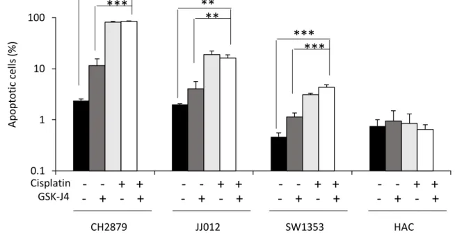
GSK-J4 combined to cisplatin decreased cell number more than each drug alone in chondrosarcoma cells (figure 4a). It reduced their proliferation (figure 4b) without inducing more apoptosis than cisplatin alone (figure 5). In chondrocytes, GSK-J4 led no reduction of cell number nor apoptosis, even in the presence of cisplatin.
Discussion
Chondrosarcoma treatment is challenging because this bone tumor is resistant to conventional chemotherapy and radiotherapy. In the present study, we show that GSK-J4, alone as well as in association with cisplatin, reduces chondrosarcoma proliferation. In addition, this inhibitor of histone demethylases is able to induce cell death in some chondrosarcoma cells.
First, we demonstrated that in chondrosarcomas but not in normal chondrocytes, GSK-J4 decreases cell proliferation. This is in agreement with anti-proliferative effect of this inhibitor observed in other tumoral cells, such as pediatric brainstem glioma (3), acute lymphoblastic leukemia (4), and B-cell lymphoma (16). Associated to this reduction of proliferation, we observed an accumulation of cells in S-phase. These effects were consistent with the literature since GSK-J4 has been previously showed to reduce proliferation and induce accumulation of cells in S-phase in differentiating embryoid body (22). Additionally, other treatments such as taurine, epigallocatechin gallate or genistein has been reported to inhibit proliferation while increasing the percentage of cells in S-phase (23).
Whereas the drug induced death by apoptosis and more moderately the senescence in some chondrosarcomas (JJ012 and CH2879), it could not induce apoptosis in SW1353, even in the presence of cisplatin. This was surprising because GSK-J4 was previously described to be
- 10 -
able to induce death in chemo-resistant tumoral cells, and to sensitize cells to standard chemotherapy agents in vitro and in vivo (16, 24).
Interestingly, we could observe a correlation between the response to cisplatin and GSK-J4. Indeed, no apoptosis could be detected after treatment with both drugs in SW1353, whereas cell death was observed in JJ012 and CH2879 cells after GSK-J4 as well as cisplatin treatment. In addition, we did not observe more apoptosis in cells treated with the combination of GSK-J4 and cisplatin than with cisplatin alone. Together, these observations suggest that death mechanisms induced by cisplatin and GSK-J4 might be similar in chondrosarcomas. GSK-J4 was initially described as a specific JMJD3 and UTX inhibitor (17). However, the antitumoral effect of this drug is likely due to its ability to inhibit JMJD3 (but not UTX) activity. Indeed, in acute lymphoblastic leukemia, only JMJD3 knockdown leads to a reduction of tumoral cell viability, while UTX silencing enhances cell proliferation, showing that both proteins with similar enzymatic function can play opposing roles in the context of the same disease (4). Also, UTX has been described as a tumor suppressor in several solid tumors(25– 29). In addition, a recent study of GSK-J4 activity indicated that histone demethylase KDM5B (JARID1B), which removes H3K4me3 marks is also a target of GSK-J4, although with a lower affinity (30).
GSK-J4 inhibits histone demethylases though interaction with α-ketoglutarate (α-KG)binding at active site of these histone demethylases (17). Consequently, IDH1 or IDH2 mutants, which have been found in more than half of chondrosarcoma samples (31), might affect GSK-J4 sensibility in chondrosarcoma. Indeed, IDH1/2 mutants robustly modulate the status of α-KG by catalyzing α-KG to D-2 hydroxyglutarate (D-2HG), an antagonist of α-KG (32). Thus, increased levels of D-2-HG have been found in cartilage tumours with an IDH mutation (33), and are able to competitively inhibit α-KG dependent histone demethylases (32), affecting trimethylation of the H3K27 as well as H3K4 (34–36). However, no difference in levels of H3K27 and H3K4 trimethylation was observed between IDH mutant and IDH wildtype chondrosarcomas (31). Additionally, previous studies and sequencing performed in the lab
- 11 -
showed that CH2879 harbors IDH1/2 wild-type, JJ012 IDH1-mutant (p.Arg132Gly), and SW1353 IDH2-mutant (p.Arg172Ser), while the current study show effective induction of apoptosis in CH2879 and JJ012 by GSK-J4, but not in SW1353. These results demonstrated that IDH1/2 or α-KG status did not affect the effectiveness of GSK-J4, indicating that there should be another mechanism (not interaction with α-KG) in inhibition of cell proliferation by the drug. Consequently, the molecular mechanism of GSK-J4 responsible of the reduction of chondrosarcoma survival need further analysis, and its understanding may allow the development of new drugs with higher efficiency.
In conclusion, this study shows that GSK-J4 reduce cell proliferation in chondrosarcomas and is able to induce death in some of them in vitro, suggesting that this pharmacological drug may be an efficient treatment to reduce progression of chondrosarcomas.
Acknowledgments
We thank Joel BLOCK (Rush University Medical Center, Chicago, USA) and Antonio LLOMBART-BOSH (University of Valencia, Valencia, Spain) for gracious chondrosarcoma gift. We also thank Marion BERTHELOT (BioConnecT, Unicaen, Caen, France) and Marilyne GUILLAMIN (SFR 146 ICORE, Unicaen, Caen) for technical assistance for in vitro experiments and flow cytometry, respectively.
Juliette Aury-Landas was recipient of a fellowship from Agence Nationale de la Recherche [ANR-15-CE14-0002-01], Région Normandie, and European Union [FEDER/FSE 2014-2020 – 16E00779/16P03685]. Eva Lhuissier and Lyess Allas were recipient of PhD stipends from Ministère de la Recherche/Université de Caen.
- 12 - The authors have no conflict of interest.
References
1. Rosenberg, A. E., Nielsen, G. P., Keel, S. B., Renard, L. G., Fitzek, M. M., Munzenrider, J. E., and Liebsch, N. J. (1999) Chondrosarcoma of the base of the skull: a clinicopathologic study of 200 cases with emphasis on its distinction from chordoma. Am. J. Surg. Pathol. 23, 1370–1378 2. Kubo, T., Sugita, T., Shimose, S., Matsuo, T., Arihiro, K., and Ochi, M. (2008) Expression of
hypoxia-inducible factor-1alpha and its relationship to tumour angiogenesis and cell proliferation in cartilage tumours. J. Bone Joint Surg. Br. 90, 364–370
3. Hashizume, R., Andor, N., Ihara, Y., Lerner, R., Gan, H., Chen, X., Fang, D., Huang, X., Tom, M. W., Ngo, V., Solomon, D., Mueller, S., Paris, P. L., Zhang, Z., Petritsch, C., Gupta, N., Waldman, T. A., and James, C. D. (2014) Pharmacologic inhibition of histone demethylation as a therapy for pediatric brainstem glioma. Nat. Med. 20, 1394–1396
4. Ntziachristos, P., Tsirigos, A., Welstead, G., Trimarchi, T., Bakogianni, S., Xu, L., Loizou, E., Holmfeldt, L., Strikoudis, A., King, B., Mullanders, J., Becksfort, J., Nedjic, J., Paietta, E., Tallman, M. S., Rowe, J. M., Tonon, G., Satoh, T., Kruidenier, L., Prinjha, R., Akira, S., Van Vlierberghe, P., Ferrando, A. A., Jaenisch, R., Mullighan, C. G., and Aifantis, I. (2014) Contrasting roles for histone 3 lysine 27 demethylases in acute lymphoblastic leukemia. Nature. 514, 513–517 5. Hübner, M. R., and Spector, D. L. (2010) Role of H3K27 Demethylases Jmjd3 and UTX in
Transcriptional Regulation. Cold Spring Harb. Symp. Quant. Biol. 75, 43–49
6. Kooistra, S. M., and Helin, K. (2012) Molecular mechanisms and potential functions of histone demethylases. Nat. Rev. Mol. Cell Biol. 13, 297–311
7. Torres, C. M., Laugesen, A., and Helin, K. (2013) Utx Is Required for Proper Induction of
Ectoderm and Mesoderm during Differentiation of Embryonic Stem Cells. PLOS ONE. 8, e60020 8. Wang, C., Lee, J.-E., Cho, Y.-W., Xiao, Y., Jin, Q., Liu, C., and Ge, K. (2012) UTX regulates
mesoderm differentiation of embryonic stem cells independent of H3K27 demethylase activity. Proc. Natl. Acad. Sci. U. S. A. 109, 15324–15329
9. Mansour, A. A., Gafni, O., Weinberger, L., Zviran, A., Ayyash, M., Rais, Y., Krupalnik, V., Zerbib, M., Amann-Zalcenstein, D., Maza, I., Geula, S., Viukov, S., Holtzman, L., Pribluda, A., Canaani, E., Horn-Saban, S., Amit, I., Novershtern, N., and Hanna, J. H. (2012) The H3K27 demethylase Utx regulates somatic and germ cell epigenetic reprogramming. Nature. 488, 409–413
10. Santa, F. D., Totaro, M. G., Prosperini, E., Notarbartolo, S., Testa, G., and Natoli, G. (2007) The Histone H3 Lysine-27 Demethylase Jmjd3 Links Inflammation to Inhibition of Polycomb-Mediated Gene Silencing. Cell. 130, 1083–1094
11. Agger, K., Cloos, P. A. C., Rudkjær, L., Williams, K., Andersen, G., Christensen, J., and Helin, K. (2009) The H3K27me3 demethylase JMJD3 contributes to the activation of the INK4A–ARF locus in response to oncogene- and stress-induced senescence. Genes Dev. 23, 1171–1176
12. Barradas, M., Anderton, E., Acosta, J. C., Li, S., Banito, A., Rodriguez-Niedenführ, M., Maertens, G., Banck, M., Zhou, M.-M., Walsh, M. J., Peters, G., and Gil, J. (2009) Histone demethylase JMJD3 contributes to epigenetic control of INK4a/ARF by oncogenic RAS. Genes Dev. 23, 1177– 1182
13. Sen, G. L., Webster, D. E., Barragan, D. I., Chang, H. Y., and Khavari, P. A. (2008) Control of differentiation in a self-renewing mammalian tissue by the histone demethylase JMJD3. Genes Dev. 22, 1865–1870
14. Jepsen, K., Solum, D., Zhou, T., McEvilly, R. J., Kim, H.-J., Glass, C. K., Hermanson, O., and Rosenfeld, M. G. (2007) SMRT-mediated repression of an H3K27 demethylase in progression from neural stem cell to neuron. Nature. 450, 415–419
- 13 -
15. Zhao, W., Li, Q., Ayers, S., Gu, Y., Shi, Z., Chen, Y., Wang, H. Y., and Wang, R.-F. (2013) Jmjd3 Negatively Regulates Reprogramming Through Histone Demethylase Activity Dependent and -Independent Pathways. Cell. 152, 1037–1050
16. Mathur, R., Sehgal, L., Havranek, O., Köhrer, S., Khashab, T., Jain, N., Burger, J. A., Neelapu, S. S., Davis, R. E., and Samaniego, F. (2017) Inhibition of demethylase KDM6B sensitizes diffuse large B-cell lymphoma to chemotherapeutic drugs. Haematologica. 102, 373–380
17. Kruidenier, L., Chung, C., Cheng, Z., Liddle, J., Che, K., Joberty, G., Bantscheff, M., Bountra, C., Bridges, A., Diallo, H., Eberhard, D., Hutchinson, S., Jones, E., Katso, R., Leveridge, M., Mander, P. K., Mosley, J., Ramirez-Molina, C., Rowland, P., Schofield, C. J., Sheppard, R. J., Smith, J. E., Swales, C., Tanner, R., Thomas, P., Tumber, A., Drewes, G., Oppermann, U., Patel, D. J., Lee, K., and Wilson, D. M. (2012) A selective jumonji H3K27 demethylase inhibitor modulates the proinflammatory macrophage response. Nature. 488, 404–408
18. Jagasia, A. A., Block, J. A., Diaz, M. O., Nobori, T., Gitelis, S., Inerot, S. E., and Iyer, A. P. (1996) Partial deletions of the CDKN2 and MTS2 putative tumor suppressor genes in a myxoid chondrosarcoma. Cancer Lett. 105, 77–90
19. Scully, S. P., Berend, K. R., Toth, A., Qi, W. N., Qi, Z., and Block, J. A. (2000) Marshall Urist Award. Interstitial collagenase gene expression correlates with in vitro invasion in human chondrosarcoma. Clin. Orthop.
20. Gil-Benso, R., Lopez-Gines, C., López-Guerrero, J. A., Carda, C., Callaghan, R. C., Navarro, S., Ferrer, J., Pellín, A., and Llombart-Bosch, A. (2003) Establishment and characterization of a continuous human chondrosarcoma cell line, ch-2879: comparative histologic and genetic studies with its tumor of origin. Lab. Investig. J. Tech. Methods Pathol. 83, 877–887
21. Duval, E., Bigot, N., Hervieu, M., Kou, I., Leclercq, S., Galéra, P., Boumediene, K., and Baugé, C. (2011) Asporin Expression Is Highly Regulated in Human Chondrocytes. Mol. Med. 17, 816–823 22. Mandal, C., Kim, S. H., Kang, S. C., Chai, J. C., Lee, Y. S., Jung, K. H., and Chai, Y. G. (2017)
GSK-J4-Mediated Transcriptomic Alterations in Differentiating Embryoid Bodies. Mol. Cells. 40, 737– 751
23. Luo, Y., Huo, Y., Song, P., Zhang, X., and Liao, M. (2019) Validation and functional analysis of the critical proteins in combination with taurine, epigallocatechin gallate and genistein against liver fibrosis in rats. Biomed. Pharmacother. 115, 108975
24. Dalvi, M. P., Wang, L., Zhong, R., Kollipara, R. K., Park, H., Bayo, J., Yenerall, P., Zhou, Y.,
Timmons, B. C., Rodriguez-Canales, J., Behrens, C., Mino, B., Villalobos, P., Parra, E. R., Suraokar, M., Pataer, A., Swisher, S. G., Kalhor, N., Bhanu, N. V., Garcia, B. A., Heymach, J. V., Coombes, K., Xie, Y., Girard, L., Gazdar, A. F., Kittler, R., Wistuba, I. I., Minna, J. D., and Martinez, E. D. (2017) Taxane-Platin-Resistant Lung Cancers Co-develop Hypersensitivity to JumonjiC Demethylase Inhibitors. Cell Rep. 19, 1669–1684
25. Thieme, S., Gyárfás, T., Richter, C., Özhan, G., Fu, J., Alexopoulou, D., Muders, M. H., Michalk, I., Jakob, C., Dahl, A., Klink, B., Bandoła, J., Bachmann, M., Schröck, E., Buchholz, F., Stewart, A. F., Weidinger, G., Anastassiadis, K., and Brenner, S. (2013) The histone demethylase UTX regulates stem cell migration and hematopoiesis. Blood. 121, 2462–2473
26. Mar, B., Bullinger, L., Basu, E., Schlis, K., Silverman, L., Döhner, K., and Armstrong, S. (2012) Sequencing Histone Modifying Enzymes Identifies UTX mutations in Acute Lymphoblastic Leukemia. Leukemia. 26, 1881–1883
27. Jankowska, A. M., Makishima, H., Tiu, R. V., Szpurka, H., Huang, Y., Traina, F., Visconte, V., Sugimoto, Y., Prince, C., O’Keefe, C., Hsi, E. D., List, A., Sekeres, M. A., Rao, A., McDevitt, M. A., and Maciejewski, J. P. (2011) Mutational spectrum analysis of chronic myelomonocytic leukemia includes genes associated with epigenetic regulation: UTX, EZH2, and DNMT3A. Blood. 118, 3932–3941
28. Wang, J. K., Tsai, M.-C., Poulin, G., Adler, A. S., Chen, S., Liu, H., Shi, Y., and Chang, H. Y. (2010) The histone demethylase UTX enables RB-dependent cell fate control. Genes Dev. 24, 327–332 29. van Haaften, G., Dalgliesh, G. L., Davies, H., Chen, L., Bignell, G., Greenman, C., Edkins, S., Hardy,
- 14 -
D., Buck, G., Campbell, P. J., Cole, J., Dunmore, R., Forbes, S., Jia, M., Jones, D., Kok, C. Y., Leroy, C., Lin, M.-L., McBride, D. J., Maddison, M., Maquire, S., McLay, K., Menzies, A., Mironenko, T., Lee, M., Mudie, L., Pleasance, E., Shepherd, R., Smith, R., Stebbings, L., Stephens, P., Tang, G., Tarpey, P. S., Turner, R., Turrell, K., Varian, J., West, S., Widaa, S., Wray, P., Collins, V. P., Ichimura, K., Law, S., Wong, J., Yuen, S. T., Leung, S. Y., Tonon, G., DePinho, R. A., Tai, Y.-T., Anderson, K. C., Kahnoski, R. J., Massie, A., Khoo, S. K., Teh, B. T., Stratton, M. R., and Futreal, P. A. (2009) Somatic mutations of the histone H3K27 demethylase, UTX, in human cancer. Nat. Genet. 41, 521–523
30. Heinemann, B., Nielsen, J. M., Hudlebusch, H. R., Lees, M. J., Larsen, D. V., Boesen, T., Labelle, M., Gerlach, L.-O., Birk, P., and Helin, K. (2014) Inhibition of demethylases by GSK-J1/J4. Nature. 514, E1-2
31. Cleven, A. H. G., Suijker, J., Agrogiannis, G., Briaire-de Bruijn, I. H., Frizzell, N., Hoekstra, A. S., Wijers-Koster, P. M., Cleton-Jansen, A.-M., and Bovée, J. V. M. G. (2017) IDH1 or -2 mutations do not predict outcome and do not cause loss of 5-hydroxymethylcytosine or altered histone modifications in central chondrosarcomas. Clin. Sarcoma Res. 10.1186/s13569-017-0074-6 32. Xu, W., Yang, H., Liu, Y., Yang, Y., Wang, P., Kim, S.-H., Ito, S., Yang, C., Wang, P., Xiao, M.-T., Liu,
L., Jiang, W., Liu, J., Zhang, J., Wang, B., Frye, S., Zhang, Y., Xu, Y., Lei, Q., Guan, K.-L., Zhao, S., and Xiong, Y. (2011) Oncometabolite 2-Hydroxyglutarate Is a Competitive Inhibitor of α-Ketoglutarate-Dependent Dioxygenases. Cancer Cell. 19, 17–30
33. Amary, M. F., Damato, S., Halai, D., Eskandarpour, M., Berisha, F., Bonar, F., McCarthy, S., Fantin, V. R., Straley, K. S., Lobo, S., Aston, W., Green, C. L., Gale, R. E., Tirabosco, R., Futreal, A., Campbell, P., Presneau, N., and Flanagan, A. M. (2011) Ollier disease and Maffucci syndrome are caused by somatic mosaic mutations of IDH1 and IDH2. Nat. Genet. 43, 1262–1265
34. Sasaki, M., Knobbe, C. B., Munger, J. C., Lind, E. F., Brenner, D., Brüstle, A., Harris, I. S., Holmes, R., Wakeham, A., Haight, J., You-Ten, A., Li, W. Y., Schalm, S., Su, S. M., Virtanen, C.,
Reifenberger, G., Ohashi, P. S., Barber, D. L., Figueroa, M. E., Melnick, A., Zúñiga-Pflücker, J.-C., and Mak, T. W. (2012) IDH1(R132H) mutation increases murine haematopoietic progenitors and alters epigenetics. Nature. 488, 656–659
35. Koch, C. M., Andrews, R. M., Flicek, P., Dillon, S. C., Karaöz, U., Clelland, G. K., Wilcox, S., Beare, D. M., Fowler, J. C., Couttet, P., James, K. D., Lefebvre, G. C., Bruce, A. W., Dovey, O. M., Ellis, P. D., Dhami, P., Langford, C. F., Weng, Z., Birney, E., Carter, N. P., Vetrie, D., and Dunham, I. (2007) The landscape of histone modifications across 1% of the human genome in five human cell lines. Genome Res. 17, 691–707
36. Barski, A., Cuddapah, S., Cui, K., Roh, T.-Y., Schones, D. E., Wang, Z., Wei, G., Chepelev, I., and Zhao, K. (2007) High-resolution profiling of histone methylations in the human genome. Cell. 129, 823–837
Figure legends
Figure 1: GSK-J4 decreased cell survival in chondrosarcoma cells.
(a) CH2879, JJ012 and SW1353 chondrosarcoma cells were treated with GSK-J4 (10 µM) for 6 days. Viable cells were regularly counted. Values were normalized to day 0. Data are expressed as means ± SEM (n=3).
(b) Chondrosarcoma cells and human articular chondrocytes (HAC) were treated with GSK-J4 (10 µM) for 4 days. At the end of treatment, adherent cells were counted after trypan blue
- 15 -
exclusion of dead cells. Values are normalized to untreated cells. Data are expressed as means ± SEM (n=3).
Figure 2: GSK-J4 reduced cell proliferation in chondrosarcomas.
(a-b) Cells were labelled with CFSE, and then treated with GSK-J4 (10 µM) for 3 days. CFSE fluorescence were tracked by flow cytometry (a). Dye dilution was calculated for each experiments and showed in histograms. Data are expressed as means ± SEM (n=3) (b). (c) Cells were treated with 10 µM GSK-J4 for 4 days. Then, DNA content was stained by propidium iodide, and cell cycle determined by flow cytometry. Histograms represent G0-G1, S and G2-M phase percentages. Data are expressed as means ± SEM (n=3).
Figure 3: GSK-J4 induced cell death in CH2879 and JJ012 cells, but not in SW1353. (a) Chondrosarcomas were treated with 10 µM GSK-J4 for 2 days. Then, caspase-3 and -7 activity were measured. Histograms represent activity relative to untreated cells. Data are expressed as means ± SEM (n=5).
(b) Cells were treated with 10 µM GSK-J4 for 4 days. Then, cells were fixed and cell cycle determined by flow cytometry. Histograms represent fraction of cells in Sub-G1 phase. Data are expressed as means ± SEM (n=3).
(c) Cells were treated as previously. Then, they were stained with Apo 2.7 antibody coupled to phycoerythrin and analysed by flow cytometry (n=3).
(d) After treatment, senescent cells were stained with SA- β-gal. Representative pictures are shown. Magnification: x20 (n=3).
(e) p21 expression was evaluated by western blot.
Figure 4: Association GSK-J4/cisplatin decreased cell growth and proliferation more than drugs alone in chondrosarcoma cells.
- 16 -
(a) Viable cells were counted. Data normalized to untreated condition are expressed as means ± SEM (n=3).
(b) Cells were stained with CFSE, then treated as previously. Dye dilution was analysed by flow cytometry (n=3).
Figure 5: GSK-J4 in combination with cisplatin did not increase apoptosis more than cisplatin alone in chondrosarcoma cells.
Cells were treated with GSK-J4 (10µM) or/and cisplatin (5µM) for 3 days, then stained with Apo 2.7 antibody coupled to phycoerythrin and analysed by flow cytometry. APO2.7+ cells were considered as apoptotic (n=3).
Figure 1
B
0.1 1 10 100 0 2 Days 4 6 JJ012 ** * * 0.1 1 10 100 0 2 Days 4 6 0µM GSKJ4 10µM GSKJ4 SW1353 * ** *A
0.1 1 10 100 0 2 4 6 Ce ll g ro w th (r ela tiv e u nit ) Days CH2879 *** * * ** Control GSK-J4 0 0.2 0.4 0.6 0.8 1 1.2 CH2879 JJ012 SW1353 HAC Ce ll n um be r (r el at iv e to u nt rea ted c el ls) Control GSK-J4Figure 2
CFSE Ev en ts DMSO GSK-J4CFSE positive control
A
CFSE CFSE CH2879 JJ012 SW1353 0% 20% 40% 60% 80% 100% 120% 0 10 0 10 0 10 GSK-J4 (µM) GSK-J4 (µM) GSK-J4 (µM) Ce ll f ra ct io n G2-M S G0-G1 ** *** * * GSK-J4 (10µM) - + - + - + CH2879 JJ012 SW1353 Control 0 10 20 30 40 50 60 CH2879 JJ012 SW1353 CF SE d ilu tio n Control GSK-J4C
B
*
***
*
*
*
0 1 2 3 CH2879 JJ012 SW1353 Ca sp ase 3/ 7 ac tiv ity (r elat iv e unit ) Ctrl GSK-J4 Control GSK-J4 Control GSK-J4 (10µM) JJ012 CH2879 SW1353Figure 3
B
C
D
0 10 20 30 40 CH2879 JJ012 SW1353 Ce lls in S ub G 1 ( % ) 0µM 10µM*
*
Control GSK-J4 0 5 10 15 20 25 CH2879 JJ012 SW1353 FS090 Apo 2.7 po sit iv e c ells (%) 0µM 10µM**
**
Control GSK-J4A
p21 (21 kDa) actin (43 kDa) GSK-J4 - + - + - + CH2879 JJ012 SW1353E
Figure 4
0.01 0.1 1 Ce ll g ro w th (r ela tiv e u nit )*
*
A
B
***
*
*
*
***
*
*
***
-SW1353-
+ +
- + - +
-JJ012-
+ +
- + - +
-CH2879-
+ +
- + - +
Cisplatin GSK-J4 1 10 100 CF SE d ilu tio n*
**
***
*
**
**
**
***
**
-HAC-
+ +
- + - +
-SW1353-
+ +
- + - +
-JJ012-
+ +
- + - +
-CH2879-
+ +
- + - +
Cisplatin GSK-J4Figure 5
0.1 1 10 100 1 2 3 5 Ap op to tic c ells (% ) -HAC-
+ +
- + - +
-SW1353-
+ +
- + - +
-JJ012-
+ +
- + - +
-CH2879-
+ +
- + - +
***
***
**
**
***
***
Cisplatin GSK-J4Suppl data
Supp figure 1 :
Suppl figure 1: Venn diagram showing the number of mutated genes identified by whole-exome sequencing in each cell lines
Material and methods
Genomic DNA was isolated from CH2879, SW1353, JJ012 chondrosarcoma cell lines using NucleoSpin Tissue (Macherey-Nagel, Hoerdt, France), according to the manufacturer’s instructions and quantified on a NanoDrop 2000 spectrophotometer (ThermoFisher Scientific). DNA integrity was checked on 1.2% agarose gel. Next, whole exome sequencing was performed by Integragen (Evry, France). Exome was captured using SureSelect Human All Exon V5+UTR (Agilent technologies, Santa Clara, CA, USA) following the manufacturer’s instructions. Paired-end sequencing was performed on HiSeq4000 (Illumina, Inc, San Diego, CA, USA). Base calling, sequencing reads alignment to the human reference genome GRCh38/hg38 and variant calling were processed using the Broad Institute’s GATK Haplotype Caller GVCF tool v3.7. Variants were annotated using Annovar. Filtering was performed following strict criteria and consisted of removing any low confidence variants (QPhred < 30 and read depth < 10) and excluding variants with a minor allele frequency (MAF) > 0.001 reported in 1000 Genomes Project, in NHLBI GO Exome Sequencing Project (ESP) or in Exome Aggregation Consortium (ExAC) datasets. Variants present in the Integragen Reference Database with a MAF > 0.01 were excluded as they must correspond to false positives related to the technology. Only missense or loss of function variants, corresponding to nonsense, stop loss, indels or essential splice acceptor/donor site variants, were considered. Pathogenicity was predicted using SIFT, MutationTaster and PolyPhen-2 tools. Venn diagram was constructed with genes with deleterious coding mutations with a tool available online at http://bioinformatics.psb.ugent.be/webtools/Venn/.