RESEARCH ARTICLE
Mms19 promotes spindle microtubule
assembly in Drosophila neural stem cells
Rohan ChippalkattiID1,2, Boris EggerID3, Beat SuterID1*
1 Cell Biology, University of Bern, Berne, Switzerland, 2 Graduate School for Cellular and Biomedical
Sciences, University of Bern, Berne, Switzerland, 3 Department of Biology, University of Fribourg, Fribourg, Switzerland
*Beat.Suter@izb.unibe.ch
Abstract
Mitotic divisions depend on the timely assembly and proper orientation of the mitotic spindle. Malfunctioning of these processes can considerably delay mitosis, thereby compromising tissue growth and homeostasis, and leading to chromosomal instability. Loss of functional
Mms19 drastically affects the growth and development of mitotic tissues in Drosophila
lar-vae and we now demonstrate that Mms19 is an important factor that promotes spindle and astral microtubule (MT) growth, and MT stability and bundling. Mms19 function is needed for the coordination of mitotic events and for the rapid progression through mitosis that is characteristic of neural stem cells. Surprisingly, Mms19 performs its mitotic activities through two different pathways. By stimulating the mitotic kinase cascade, it triggers the localization of the MT regulatory complex TACC/Msps (Transforming Acidic Coiled Coil/Min-ispindles, the homolog of human ch-TOG) to the centrosome. This activity of Mms19 can be rescued by stimulating the mitotic kinase cascade. However, other aspects of the Mms19 phenotypes cannot be rescued in this way, pointing to an additional mechanism of Mms19 action. We provide evidence that Mms19 binds directly to MTs and that this stimulates MT stability and bundling.
Author summary
Mitosis is a fundamental process that segregates replicated chromosomes into daughter cells, allowing organ growth and development in multicellular organisms. To properly distribute the genetic material, the mitotic spindle, an organelle consisting of extended microtubules, microtubule motors, and additional microtubule-associated proteins needs to be built in a coordinated, robust, but still dynamic way. Failure to set up these spindles properly leads to chromosomal instability or differentiation defects, and this can lead to tumor formation, reduced organ growth, or lack of specific cell types. Whereas Mms19 protein performs activities unrelated to mitosis, we found thatDrosophila Mms19 is also crucial for mitotic progression and organ growth. This led us to discover that Mms19 had been repurposed to also assist in the formation of stable spindle microtubules. By regulat-ing spindle architecture,Mms19 allows neural stem cells to timely progress through mito-sis to build the normal brain. Surprisingly,Mms19 exerts its spindle regulatory function a1111111111 a1111111111 a1111111111 a1111111111 a1111111111 OPEN ACCESS
Citation: Chippalkatti R, Egger B, Suter B (2020)
Mms19 promotes spindle microtubule assembly in Drosophila neural stem cells. PLoS Genet 16(11): e1008913.https://doi.org/10.1371/journal. pgen.1008913
Editor: Jean-Rene´ Huynh, College de France CNRS,
FRANCE
Received: June 1, 2020 Accepted: October 13, 2020 Published: November 19, 2020
Peer Review History: PLOS recognizes the
benefits of transparency in the peer review process; therefore, we enable the publication of all of the content of peer review and author responses alongside final, published articles. The editorial history of this article is available here:
https://doi.org/10.1371/journal.pgen.1008913 Copyright:© 2020 Chippalkatti et al. This is an open access article distributed under the terms of theCreative Commons Attribution License, which permits unrestricted use, distribution, and reproduction in any medium, provided the original author and source are credited.
Data Availability Statement: All relevant data are
within the manuscript and itsSupporting Informationfiles.
Funding: This work was supported by funding
again through different activities. It stimulates microtubule assembly through a mitotic kinase cascade consisting of 3 kinases to activate microtubule organizer proteins. Addi-tional evidence suggests that it is capable of interacting with microtubules and promotes microtubule bundling and that this is also important to form a functional mitotic spindle.
Introduction
The fidelity of chromosome segregation during mitotic divisions depends on the proper regu-lation of the structure and dynamics of spindle microtubules. Numerous mitotic factors, including different mitotic kinases and microtubule (MT) associated proteins, meticulously regulate the formation, orientation, polymerization, and catastrophe of spindle MTs to ensure that the chromatids are evenly delivered to the two products of the division process, the daugh-ter cells. The mitotic spindle not only regulates chromosome segregation, its proper spindle architecture, and orientation profoundly impacts development and cellular physiology. Spin-dle size and architecture dynamically adapt to changes in cellular morphology within and between organisms. Early embryonic divisions in organisms such asC. elegans and X. laevis occur in the large volumes of the egg and blastomeres whereas in the later stages of embryo-genesis cell sizes are dramatically smaller [1,2]. The mitotic spindle is scaled according to the cell size by MT-regulatory proteins, indicating that cell division is in tune with the develop-mental progression of these organisms [3]. The MT associated protein Tpx2 mediates spindle architecture variations in mouse neuronal stem cells (NSCs), where astral MTs predominate the early developmental stages and inner spindle MTs predominate during the later develop-mental stages [4]. This points to the importance of MT regulatory proteins in regulating spin-dle architecture during development. Studying such novel proteins can reveal structural and dynamic aspects of spindle assembly and their role in development and cellular physiology.
Mms19 was identified as a gene required for nucleotide excision repair (NER) [5]. The pro-tein Mms19 can be found as part of the Cytoplasmic Iron-Sulfur Assembly complex (CIA), which mediates the incorporation of iron-sulfur clusters into NER proteins such as Xpd [6,7]. However, a distinct, apparently DNA repair-independent, function ofMms19 came to light because its knock-down caused numerous mitotic spindle abnormalities in human cells [8]. Downregulation ofMms19 in young Drosophila embryos also revealed spindle abnormalities and chromosome segregation defects, and these phenotypes were linked to the mitotic control pathway when results by Nag and co-workers revealed that Mms19 acts as a positive regulator of the Cdk Activating Kinase (CAK) activity [9]. CAK has dual roles during the cell cycle. It activates the mitotic Cdk1 during mitosis but is recruited by Xpd to form the holoTFIIH com-plex during interphase [10]. The incorporation into TFIIH causes CAK to phosphorylate an entirely different set of substrates and allows CAK to perform its functions in transcription. Nag et al proposed that Mms19 binds to Xpd during mitosis and that this binding competes with the binding of Xpd to the TFIIH subunits. Mms19 binding to Xpd could thereby release CAK to activate its mitotic targets. In this model, reduced levels of Mms19 prevent sufficient dissociation of the Xpd-CAK complexes, hindering the establishment of the required levels of the mitotic CAK activity. Indeed, overexpressing CAK complex components in theMms19 loss-of-function (Mms19P) background rescued the mitotic defects to a large degree [9].
The remarkable findings by Nag et al uncovered a novel mitotic pathway forMms19. But this study mostly focused on youngDrosophila embryos, which are unusual in that all somatic nuclei share a common cytoplasm, in which the mitotic divisions take place. Furthermore, their cell cycle consists of only S and M phases, without intervening G phases. In this situation
PLOS GENETICS Spindle assembly by Mms19 (project grant 31003A_173188;www.snf.ch) and
the University of Bern (www.unibe.ch) to BS. The funders had no role in study design, data collection and analysis, decision to publish, or preparation of the manuscript.
Competing interests: The authors have declared
with the shared cytoplasm, the absence of Mms19 often causes microtubules emanating from one spindle pole to contact the chromosomes of a neighboring nucleus. It is therefore difficult to extrapolate these findings to mononuclear, diploid cells that are isolated from their neigh-bors by plasma membranes and go through a full cell cycle with G phases. Furthermore, even though the spindle abnormalities observed in the absence of Mms19 could be linked to com-promised CAK activity, the pathway acting downstream of CAK is not understood. Finally, overexpression of the CAK complex brought about only a partial rescue of theMms19Pdefects, pointing towards additional, possibly CAK-independent spindle regulatory roles ofMms19.
The objective of this study was thus to investigate the mitotic function ofMms19 in normal diploid cells with the goal of dissecting the precise pathway through whichMms19 acts to regu-late mitotic spindle assembly and cell cycle progression. BecauseMms19Plarvae lack imaginal discs, we chose the larval brain neuroblasts (NBs) as a model to analyze the mitotic roles of Mms19 in cells with a full cycle. The newly identified Mms19 phenotypes allowed us to pin-point steps in the pathways that requireMms19 activity. We found that Mms19 is required in NBs for timely progression through the cell cycle and consequently for establishing normal cell numbers in the NB lineage.Mms19 is also required for the growth and assembly of spindle and astral microtubules (MTs). Our results connect these defects to the mis-localization of the microtubule regulator TACC, suggesting that TACC is a crucial downstream target of the mitotic kinase cascade through CAK-Cdk1-Aurora kinases. Additionally, and apparently unrelated to the CAK-Cdk1 axis, we also identified a direct interactionin vitro between Mms19 and microtubules and found that Mms19 promotes microtubule stability and bundle formation.
Results
Mms19
Pbrains show a microcephaly phenotype
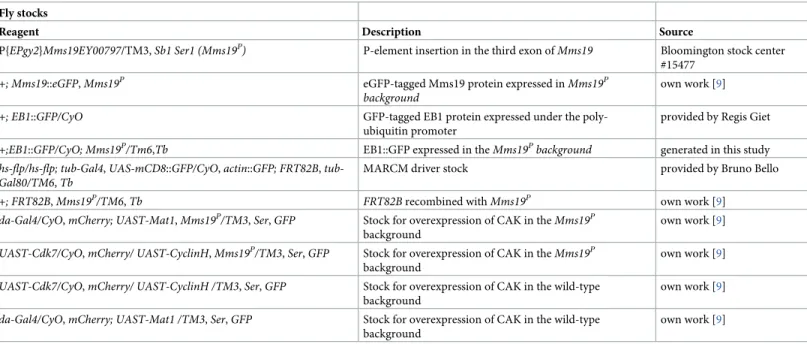
NormalDrosophila larvae spend on average 5–7 days at 25˚C to pass through the larval stages. In contrast,Mms19Plarvae take not only at least 8–10 days to reach the size of outgrown WT third instar larvae, but they also spend a total of around 15 days in the 3rdlarval instar stage before they die (seeTable 1for details about the fly stocks used). These larvae display a typical mitotic phenotype without recognizable imaginal disc tissues [9]. Even though outgrown Mms19Plarvae lack imaginal discs, their brain is still present, although it is much smaller, dis-playing a microcephaly phenotype (Fig 1A–1B). Additionally, the optic lobe (OL) appears deformed and underdeveloped. Compared to the wild type, the total volume of theMms19P brains and the volumes of the central brains (CBs) and OLs, too, are drastically reduced (Fig 1D–1F). We also stained the brains of all genotypes with antibodies against Miranda (Mira, which marks CB NBs; seeTable 2for details about all antibodies used for staining), to count the number of CB NBs. Surprisingly, however, the number of CB NBs per brain lobe did not significantly change between the controls and theMms19Pbrains (Fig 1C). This implies that theMms19PNBs have been properly determined and are present. It was reported previously that aneuploidy resulting from defective spindle checkpoint function causes premature differ-entiation of NBs and a reduction in brain size [11]. We performed fluorescentin situ hybrid-ization to screen for aneuploid NBs in the WT as well asMms19Pbrains but did not find significantly elevated levels of aneuploidy in the mutant (S1 Fig;P = 0.1493). The reduced cen-tral brain size might then result from dying differentiating neuronal cells or, more likely, from NBs that divide only slowly, thereby contributing fewer Ganglion Mother Cells (GMCs) and neurons to the CB. The OL develops from the invagination of a group of ectodermal cells in the head region during late embryogenesis [12]. In the wild type, these cells, called neuroe-pithelial (NE) cells, start proliferating after larval hatching and generate the distinct regions of
the OL after several rounds of divisions [13]. The drastic reduction of the OL volume seen in Mms19Plarvae contributes the largest part to the overall reduction of the brain size seen. Although this points to a requirement forMms19 also in these highly proliferative NE cells, we focused on the better studied CB NBs as these cells are large and very well suited to analyze mitotic phases and possible defects in spindle assembly.
To verify the requirement for Mms19 for normal brain development, we expressed wild-type Mms19 fused to eGFP (Mms19::eGFP) under the control of the endogenousMms19 pro-moter in theMms19Pbackground to test whether the observed phenotype can be rescued by Mms19 activity. Mms19::eGFP fully rescued the brain morphology, the volumes of the whole brain lobe as well as the CB and OL sizes (Fig 1A, 1D–1F). This not only confirmed that the defects observed in the mutant are indeed due to the absence ofMms19 activity and not due to a second site mutation on this chromosome, it also showed that the Mms19::eGFP fusion pro-tein is functional.
A mitotic activity ofMms19 was described to promote the Cdk-activating kinase activity of the Cdk7/CycH/Mat1 complex (CAK complex; [9]). The responsible mechanism appears to be a competitive binding of Mms19 to Xpd, which would otherwise recruit CAK to TFIIH, where it assumes a different substrate specificity and is unable to activate the M-Cdk Cdk1. The key result that led to this model was that the lack of imaginal discs caused by the absence ofMms19 activity could be rescued considerably by expressing the CAK complex components under the control of the Upstream Activating Sequence (UAS) enhancer, using the daughterless(da)-Gal4 driver in theMms19Pbackground (da>CAK,Mms19P) [9]. We, therefore, tested whether it was possible to rescue the small brain phenotype by the identical strategy. This was not the case, as in da>CAK,Mms19Pbrains both CBs and OLs remained smaller (Fig 1A, 1D–1F), the number of CB NBs was similar as in the wild type (Fig 1C) and the overall brain volume was reduced. These observations indicated that the mitotic cells in theMms19Pbrains probably did not proliferate enough to produce the normal amount of neuronal tissue and that this defect might not only be caused by insufficient CAK activity. We also considered whether the expres-sion of CAK underda-Gal4 control might cause the abnormalities leading to the reduced
Table 1. Fly stocks used for these studies. Fly stocks
Reagent Description Source
P{EPgy2}Mms19EY00797/TM3, Sb1 Ser1 (Mms19P) P-element insertion in the third exon ofMms19 Bloomington stock center
#15477 +; Mms19::eGFP, Mms19P eGFP-tagged Mms19 protein expressed inMms19P
background
own work [9]
+; EB1::GFP/CyO GFP-tagged EB1 protein expressed under the poly-ubiquitin promoter
provided by Regis Giet
+;EB1::GFP/CyO; Mms19P/Tm6,Tb EB1::GFP expressed in theMms19Pbackground generated in this study
hs-flp/hs-flp; Gal4, UAS-mCD8::GFP/CyO, actin::GFP; FRT82B, tub-Gal80/TM6, Tb
MARCM driver stock provided by Bruno Bello
+; FRT82B, Mms19P/TM6, Tb FRT82B recombined with Mms19P own work [9] da-Gal4/CyO, mCherry; UAST-Mat1, Mms19P/TM3, Ser, GFP Stock for overexpression of CAK in theMms19P
background
own work [9]
UAST-Cdk7/CyO, mCherry/ UAST-CyclinH, Mms19P/TM3, Ser, GFP Stock for overexpression of CAK in theMms19P
background
own work [9]
UAST-Cdk7/CyO, mCherry/ UAST-CyclinH /TM3, Ser, GFP Stock for overexpression of CAK in the wild-type background
own work [9]
da-Gal4/CyO, mCherry; UAST-Mat1 /TM3, Ser, GFP Stock for overexpression of CAK in the wild-type background
own work [9]
https://doi.org/10.1371/journal.pgen.1008913.t001
Fig 1.Mms19Pbrains display a microcephaly (small brain) phenotype. 3rdinstar larval brain NBs were visualized by
staining for Miranda (red, cytoplasmic). They were also stained for pH3 (white, nuclear) and DNA (blue, Hoechst 33342 dye). (A)-(B) A WT brain lobe can be subdivided into the OL (shaded green) and the CB (shaded red).Mms19P
brains are significantly smaller, with a diminished OL. Overexpressing the CAK complex components driven by daughterless-Gal4 (da>CAK) in theMms19Pbackground appeared to slightly rescue this phenotype but this rescue
was not statistically significant (brain volume:P>0.99; OL volume: P>0.99). Mms19::eGFP expressed in the Mms19P
background rescued theMms19Pphenotype. Daughterless driven CAK expression in the WT background did not seem to bring about any noticeable defects in brain size or morphology. Counting the number of CB NBs (in the Red shaded area) staining positively for Mira revealed that the number of NBs per brain lobe is similar across all genotypes (C) (WT: n = 15 brains, 30 lobes, 3 experiments;Mms19P: n = 15 brains, 30 lobes, 3 experiments; da>CAK,Mms19P:
n = 9 brains, 18 lobes, 3 experiments; Mms19::eGFP,Mms19P: n = 9 brains, 18 lobes, 3 experiments; da>CAK; WT: n = 6 brains, 12 lobes, 2 experiments). (D) the volume of the brain is significantly reduced in theMms19Pand da>CAK,Mms19Pbrains. Furthermore, segmentation and volume measurement of the OL and CB revealed a
significant reduction inMms19Pand da>CAK,Mms19Pbrains (E)-(F). Brain volume and morphology were restored
to WT levels when Mms19::eGFP was expressed inMms19Pbrains (D)-(F). (WT: n = 14 brains, 28 lobes, 3
brain size. For this, we dissected brains from larvae expressing da>CAK in the wild-type back-ground. However, we did not find significant changes in either the brain morphology (Fig 1A), the CB NB numbers (Fig 1C), or the CB and OL volumes (Fig 1E and 1F). This result, therefore, points to the possibility that Mms19 acts through two different pathways to achieve normal organ size.
In
Mms19
Pmutant brains a higher proportion of NBs are in mitosis
In order to better understand the microcephaly phenotype, we performed 5-Ethynyl-2’-deox-yuridine (EdU) incorporation assays coupled with phosphorylated Histone H3 (pH3) staining and examined defects in cell cycle progression in the NBs. Based on pH3 and EdU staining, the cells can be allocated to one of the following phases: only EdU = S, i.e. cells in S phase; pH3 without EdU = M, i.e. cells undergoing mitosis; both EdU and pH3 = G2/M, i.e. cells transiting from S to G2/M phase; and neither EdU nor pH3 (= G1/G0, i.e. Gap phase;S2A Fig). As it is difficult to determine from pH3 staining alone, whether the cells are in the G2 phase or in M, we combined the double-positive cells as well as the pH3 positive cells in the same category and referred to this as ‘mitotic phase’. CB NBs were additionally marked with antibodies against Miranda (Mira) and the number of cells was counted in each class. The results are rep-resented as a percentage of the total number of NBs per brain lobe. We observed that about half the NBs were not labeled (i.e. G1/G0 cells: 47–56%;S2B Fig) and the relative differences between the genotypes were small. However, lack ofMms19 caused a clear increase in the frac-tion of cells in the mitotic phase (37% compared to 28% in the wild type;S2D Fig). This result could either mean that more cells undergo divisions or that theMms19PNBs are either trapped in M phase or proceed more slowly through it.
experiments; Mms19::eGFP,Mms19P: n = 14 brains, 28 lobes, 3 experiments; da>CAK; WT: n = 8 brains, 16 lobes, 2
experiments). Statistical significance (SS) was determined by the Kruskal-Wallis test. Multiple columns were compared using Dunn’s post test,����(P<0.0001),���(P<0.001), scale = 50μm.
https://doi.org/10.1371/journal.pgen.1008913.g001
Table 2. Primary and secondary antibodies used for immunostainings. Primary antibodies
Antibody Manufacturer Catalog number Dilution
Rat anti-Miranda Abcam Ab197788 1:250
Rabbit anti-alpha Tubulin Abcam Ab18251 1:500
Mouse anti-alpha Tubulin Sigma T6199 1:500
Rabbit anti-GFP Immunokontakt 210-PS-1GFP 1:500
Rabbit anti-pH3 Cell Signaling 9701 1:200
Rabbit anti-Mms19 Genscript Custom antibody 1:500
Mouse anti-γ-Tubulin Sigma T6557 1:1,000
Rabbit anti-TACC provided by Jordan Raff - 1:1,000
Rabbit anti-Msps Provided by Hiro Ohkura - 1:1,000
Rabbit anti-Aurora A Provided by Jurgen Knoblich - 1:200
Secondary antibodies
Goat anti-rat Cy3 Jackson Immuno 112-165-167 1:150
Goat anti-mouse Alexa 488 Invitrogen R37120 1:500
Goat anti-rabbit Alexa 488 Invitrogen A27034 1:500
Goat anti-mouse Alexa 647 Invitrogen A21235 1:500
https://doi.org/10.1371/journal.pgen.1008913.t002
NBs depend on
Mms19 for timely and coordinated spindle assembly and
orientation
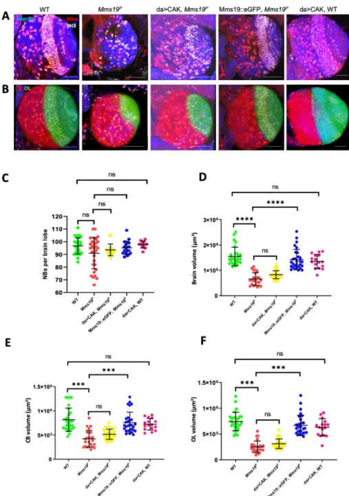
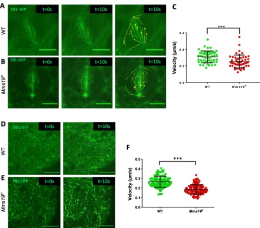
To study howMms19 contributes to spindle assembly and progression through mitosis, we utilized a transgene that expresses EB1::GFP, a MT plus end binding and tracking protein that labels growing MT ends [14]. Live imaging of NBs expressing EB1::GFP revealed the dynamics of the formation of the mitotic spindle and allowed us to measure progression through the early part of the M-phase, including the period from Nuclear Envelope Break-Down (NEBD) to the onset of anaphase B. NEBD was determined by the appearance of the GFP signal in the nuclear region, which lacks a GFP signal until NEBD. To determine the onset of anaphase B, we analyzed the distribution of EB1::GFP and the spindle morphology throughout mitosis. EB1 localization and behavior during anaphase B has been studied previously [15–17]. At the beginning of anaphase B, the spindle starts to elongate and this elongation is accompanied by an overall reduction in EB1::GFP fluorescence. We observed this pattern in WT NBs, where at 7 min post NEBD, the spindle slightly elongated, concomitant with a decrease in EB1::GFP fluorescence. NBs expressing EB1::GFP in the wild-type background started anaphase B approximately 6–8 min after NEBD (Fig 2A and 2C;S1 mov). On the other hand, NBs express-ing EB1::GFP in anMms19Pbackground reached anaphase B onset only around 16–20 min after NEBD (Fig 2B and 2C;S3 mov).
Interestingly, in some of the EB1::GFP expressingMms19PNBs, the spindle formation started before the two centrosomes had finished migrating to the opposite sides of the nucleus (Fig 2B;S3 mov). As a result, at 3 min, a kinked spindle was observed, which eventually straightened out at 5 mins. To quantify defects in centrosome migration/positioning, we used a method described previously [18] to measure the angle between the 2 centrosomes just before NEBD (Fig 2D). In all of the WT cells, the angle between the centrosomes was in the range of 135˚ to 180˚. But in around 20% ofMms19PNBs, severe centrosome mispositioning was noted as the angles fell into the range of 45˚ to 135˚ (E). In one case of anMms19PNB spindle, only one centrosome started nucleating MTs (S5C Fig;S8 mov). The spindle remained monopolar until 6 min post-NEBD and only then, a bipolar spindle became apparent. Further, around 20% of the spindles inMms19PNBs changed their orientation during the course of mitosis (Fig 2B;S3 mov), while all wild-type spindles examined remained firmly anchored at the cortex and did not change their orientation (Fig 2A).
Drosophila larval NBs are characterized by a defined apical-basal polarity [19] where the atypical Protein Kinase C (aPKC)-Bazooka-Pins complex localizes to the apical cortex while Miranda localizes to the basal cortex (Fig 2F). Retention of NB identity and self-renewal depends on the inheritance of the aPKC-containing apical complex whereas differentiation of the GMC relies on the inheritance of the Mira-bound cargo of the basal cortex [23]. Therefore, the orientation of the mitotic spindle is tightly coupled to this apical-basal polarity axis [20,21]. We therefore further examined the spindle orientation defects in fixed cells labeled with the polarity marker Miranda (Mira), which localizes to the basal cortex of the NB, forming a cres-cent-like pattern. We found that most of the wild-type spindles form at an angle of 10 degrees or lower relative to Mira (Fig 2G, 2H and 2J). On the other hand,Mms19PNBs more fre-quently failed to align their spindles within 10 degrees (Fig 2G, 2I and 2J) and the largest tilt that we observed was around 60˚. However, a marked effect on cell fate determination is only observed when the spindle angle gets close to 90˚ [22,23]. In such cases, even though both daughter cells inherit a part of the Mira-containing basal cargo, this is not sufficient to drive differentiation. Instead, both cells assume a NB identity, leading to an increase in NB numbers per brain lobe [23]. Consistent with this spindle orientation defect being insufficient to cause major NB amplifications, we did not see a significant difference in NB numbers per brain lobe
Fig 2. NBs depend onMms19 for timely and coordinated spindle assembly and orientation. (A-C) WT NBs
typically start anaphase B 6–7 min after the onset of NEBD. (B)Mms19PNBs require on average 15–20 min to reach anaphase B onset. (B-C) n = 30 NBs per genotype, 1 experiment. The quantitative assessment of the duration of WT andMms19Pmitoses is compared in (C). SS was determined by an unpaired t-test (���P<0.001); Scale = 5μm. In this
particularMms19PNB (B) the spindle poles are not fully separated at NEBD and the spindle initially appears kinked. (D) To analyze centrosome positioning defects, we measured the angle between the two centrosomes relative to the center of the nucleus, just before NEBD. (E) In most WT NBs, the angle between the centrosomes fell between 135˚ and 180˚. However, 20% of the mutant NBs displayed centrosome separation defects as the angle was lower than 135˚. N = 25 per genotype, 1 experiment. Additionally, in some of theMms19PNBs analyzed, the spindles changed their orientation throughout the course of mitosis, indicating spindle orientation defects. (F) Spindle orientation in the WT
between the wild type andMms19P(Fig 1C). Therefore, the relatively minor spindle orienta-tion defect due to lack ofMms19 does not appear to lead to differentiation problems but impedes efficient mitosis. We conclude that in the absence ofMms19, spindle formation is not properly coordinated with cell cycle progression. Furthermore, defects in centrosome migra-tion and in spindle assembly and orientamigra-tion contribute to mitotic delays inMms19PNBs.
Mms19 is cell autonomously required to maintain normal cell numbers
In the experiments with theMms19Pmutants, Mms19 was absent from all larval cells. In this situation, the mitotic delay could be due to a systemic effect or due to the lack of a cell-autono-mous activity of Mms19. To test whetherMms19 is specifically required in the mitotic cells for the timely progression through mitosis, we generatedMms19Pmosaic NB clones in another-wise wild-type background. Mosaic clones were induced in NBs 24hrs after larval hatching (ALH) and the expansion of these clones was analyzed by dissecting the brains in mid-third instar larvae 72hrs ALH. For this experiment, we focused on the type I NBs on the ventral side. On average, 45 cells per clone were found in control clones, but only around 30–35 cells in Mms19Pclones (P<0.05;S3A–S3C Fig), indicating that the cell-autonomous loss ofMms19 activity hinders the establishment of normal cell numbers in the NB lineage. This observation reaffirms our conclusion that the absence ofMms19 delays mitosis in NBs and that this mitotic delay probably causes moreMms19PNBs to linger in M-phase (S2 Fig).
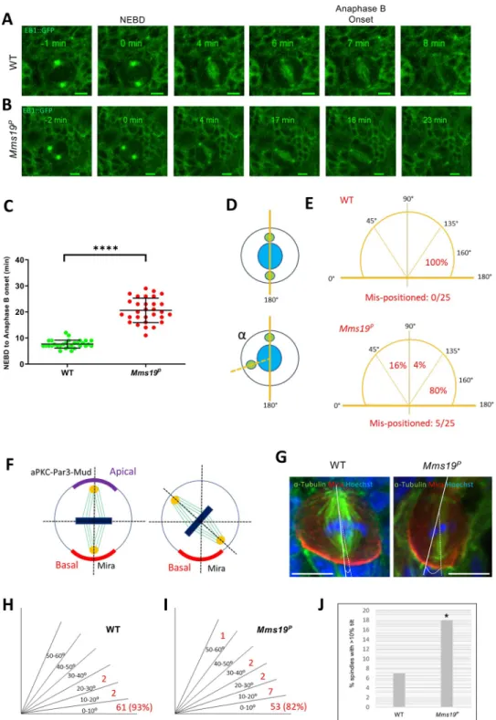
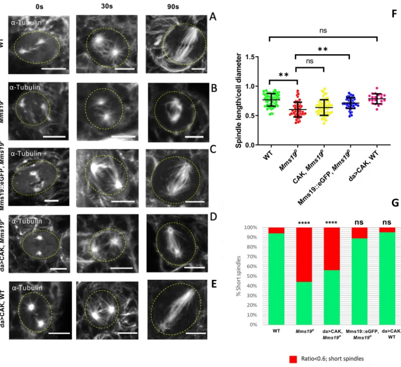
Mms19 is required to form spindles of normal length and density
Mms19Pspindles were found to be generally shorter than the wild-type ones (Fig 3A–3D). In normal NB spindles, the centrosomes were anchored close to the cell cortex, with spindle MTs emanating from them and extending to the chromosomes (Fig 3A and 3A’). But in around a quarter of the mutant cells, even though the chromosomes were aligned at the metaphase plate, the centrosomes were not connected to the cell cortex and were only a short distance away from the metaphase plate (Fig 3B and 3B’). In order to quantify this defect, we measured the length of the spindles across all genotypes and found that the spindles inMms19Pand da>CAK,Mms19Pwere generally shorter than the wild-type control spindles (S3 Table). As the NBs were also considerably smaller inMms19Pand da>CAK,Mms19Pbrains, we addi-tionally displayed the spindle length relative to the cell diameter (Fig 3C). A ratio closer to 1 indicates that the centrosomes were anchored close to the cell cortex, as in a healthy spindle. On the other hand, if the ratio was equal to or lower than 0.6, the spindle was defined as a ‘short spindle’ because the centrosomes were further inside the cell. We found a significant reduction in this ratio inMms19Pand da>CAK,Mms19PNB spindles (Fig 3C and 3D). Mms19PNBs contained around 20% short spindles while this number went up to 30% for da>CAK,Mms19P(Fig 3D). The failure of CAK to rescue theMms19Pphenotype was not due to mitotic defects induced by over-active CAK; because NBs overexpressing CAK in the wild-type background displayed a spindle length/ cell diameter ratio comparable to wild-wild-type NBs (Fig 3C and 3D). On the other hand, expression of Mms19::eGFP in theMms19Pbackground rescued the short spindle phenotype and only 3% of the NBs displayed it. These findings once
NBs is tightly coupled to the apical-basal polarity axis and this is reflected by the angle formed by the spindle with respect to the basal Mira crescent. (G) In a fraction ofMms19PNBs the spindle appears misoriented with respect to (w.
r.t) the Mira crescent localization. (H-J) Quantification of the spindle angle revealed that a considerably higher number ofMms19PNBs are oriented at an angle of more than 10˚ w.r.t Mira. Significance was calculated using Fisher’s exact
test,�(P = 0.0309). n = 65 NBs per genotype, 3 experiments, scale = 5μm.
Fig 3.Mms19 is required to form a spindle of normal length and density. (A-A’) Z projection of a typical bipolar
spindle in a wild-type (WT) NB with the spindle poles anchored to the cell cortex. (B-B’) short spindle found in Mms19PNBs. The spindle is abnormally short and the spindle poles are detached from the cortex. (C) In order to
quantify spindle elaboration, we normalized the spindle length to the cell diameter and calculated the ratio. This ratio decreases significantly forMms19PNBs. (��P = 0.0027). This phenotype is rescued by expressing Mms19::eGFP in the
Mms19Pbackground (��P = 0.0067) but da>CAK fails to rescue the short spindles (P>0.99). SS was calculated using
Kruskal-Wallis test, columns were compared using Dunn’s post test. (D) If this ratio was equal to or less than 0.6, we considered the spindle as ‘short spindle’. The graph compares the percentage of ‘short spindles’ across different genotypes. Significance was calculated using Fisher’s exact test, (����P<0.0001,��P = 0.0025). WT; Mms19P; da>CAK,
Mms19P; Mms19::eGFP,Mms19P: n = 90 NBs, 3 experiments. da>CAK, WT: n = 30, 2 experiments. (E-F) Relative density of astral MTs was quantified and compared across all genotypes. Maximum intensity projections of mitotic
again highlight the possibility of a CAK independent function of Mms19 in regulating spindle architecture.
To determine the effect ofMms19 on MT formation and stability, we measured astral as well as inner spindle MT density. For astral MT quantification (Fig 3F), we used the method described by Yang and co-workers [24] and found a significant reduction in theMms19PNBs as compared to the wild type (Fig 3E–3G). The inner spindle density was also reduced in the Mms19PNBs (Fig 3H). This phenotype appeared to be slightly rescued by CAK overexpression and was fully rescued by expressing Mms19::eGFP in theMms19Pbackground. Reduced astral MT stability could be linked to the spindle positioning and orientation defects described previ-ously (Fig 2B,2E,2G–2J) as astral MTs were shown to contact the cell cortex and regulate spin-dle positioning [25].Mms19 is thus necessary for the formation of fully assembled spindles and astral MTs.
Mms19 assists MT polymerization in vivo
To assess whether the abnormal spindle phenotypes inMms19PNBs could arise due to defects in MT growthin vivo, we studied NBs expressing EB1::GFP. Live imaging of EB1::GFP revealed the path of elongation of single MTs, akin to that of a comet, and the movement speed of the GFP signal reflects the growth speed of the MT plus ends. Wild-type andMms19P larval brains expressing EB1::GFP were dissected and live imaging was performed. The speed of the EB1::GFP particles was then tracked manually using ImageJ. The measurements indi-cated that the spindle MTs of wild-type NBs polymerized at a rate that is significantly higher than the rate observed inMms19PNBs (Fig 4A–4C; WT-S4 mov;Mms19P-S5 mov). To assess whether the MT assembly function ofMms19 is restricted to mitotic spindles, we also tested whether the absence ofMms19 also affects MT polymerization in the post-mitotic glia cells. Measuring EB1 comet speeds in glia cells showed a decrease in MT growth forMms19Pglia as compared to wild-type glia (Fig 4D–4F; WT-S6 mov;Mms19P-S7 mov). These results estab-lished thatMms19 assists MT polymerization and growth in vivo in mitotic NBs and in post-mitotic glia. The reduced MT growth rate described here seems to be a major factor for the delay in spindle assembly observed inMms19Pbrain NBs. Whereas approximately 3 minutes after the onset of NEBD, wild-type spindles had fully assembled (Fig 2A;S1 mov), most Mms19P‘spindles’ appeared to be short at 3min post-NEBD, and their MT density seemed much lower than in the wild type (S2 mov;S8 mov).
Mms19 is required for spindle re-assembly
To learn more about theMms19 function in spindle MT growth, we performed the in vivo spindle re-growth assay described by Gallaud and co-workers [18]. With this procedure, the NB spindles were depolymerized by incubation on ice for 30 min (Fig 5A, left panel). Large centrosomal asters were observed in the WT NBs after they were shifted back to 25˚C for 30s
NBs were obtained (E) and the relative density of astral MTs was quantified by first calculating the inner spindle MT density (inner dotted ellipse) and then subtracting this from the MT intensity from the entire cell (outer dotted circle). This value was then divided by the inner spindle intensity to obtain the relative astral MT density (F). (G) Astral MT density and (H) inner spindle fluorescent intensity was measured in fixed NBs immunostained forα-Tubulin and compared across all the genotypes. Both astral and inner spindle densities were decreased significantly inMms19PNBs
as compared to WT NBs. Additional expression of da>CAK appeared to partially rescue the phenotype, but this rescue was not statistically significant (astral:P = 0.2633; inner spindle: P = 0.1789). Mms19::eGFP, when expressed in the Mms19Pbackground, rescued the phenotypes. WT;Mms19P; da>CAK,Mms19P; Mms19::eGFP,Mms19P: n = 60 NBs,
3 experiments. da>CAK, WT: n = 27, 2 experiments. SS was calculated using Kruskal-Wallis test, columns were compared using Dunn’s post test.����P<0.0001,���P<0.001;��P<0.01,�P<0.05, scale = 5μm.
(Fig 5A, central panel). MT fibers also appeared to be nucleated around the chromatin (i.e. in the central region between the centrosomes;Fig 5A, central panel). Wild-type NB spindles then regained their standard size and morphology within 90 sec after being shifted back to 25˚C (Fig 5A, right panel). InMms19PNBs, we also observed a weak astral MT mesh at 30s. However, the spindles failed to re-form to the normal shape after 90 sec. Instead, they remained abnormally short (Fig 5B and 5F). Interestingly, whereas the Mms19::eGFP fusion protein was able to rescue this phenotype, CAK overexpression was unable to do so (Fig 5C and 5D). To validate this short spindle phenotype, we calculated the spindle sizes relative to the cell diameter after 90 sec incubation at 25˚C (Fig 5F). Even thoughMms19Pcells are smaller, their normalized spindle size was still significantly smaller than the wild-type one. When CAK was overexpressed inMms19PNBs, the normalized spindle length did not show a significant rescue even though Mms19::eGFP was able to rescue theMms19Pmutant pheno-type (Fig 5F and 5G). The lack of rescue by CAK overexpression does not seem to be caused by a CAK gain-of-function activity, because, again, wild-type NBs that overexpressed CAK did not display such defects in spindle length (Fig 5E–5G). This result shows thatMms19 has an
Fig 4.Mms19 assists MT polymerization in vivo. (A-B) WT and Mms19PNBs expressing EB1::GFP were imaged live,
and time-lapse movies were acquired to calculate the velocity of EB1::GFP labelled MT ‘plus’ ends. The left panel shows the image of a single time frame. The central panel shows a projection of 20 time points (taken over 10s). In the far right panel, individual EB1::GFP comets are tracked. The velocities represented in (C) show reduced MT growth velocity inMms19PNB spindles. n = 11 cells per genotype, 4–5 tips analyzed from each cell, 1 experiment. (D-E) EB1::
GFP expressing surface glia cells were imaged live to analyze the MT plus end velocities. The left panel shows a single time frame and the right-side panel shows a projection of 20 time points (taken over 10s). The velocities represented in (F) show reduced MT growth velocity inMms19Pglia. n = 15 brains from each genotype, 7–8 tips analyzed from each brain, 1 experiment. SS for C and F was calculated using unpaired t-test, (���P<0.001), scale bar = 5μm.
https://doi.org/10.1371/journal.pgen.1008913.g004
Fig 5.Mms19 is required for spindle re-assembly. After cold treatment, spindle re-assembly was analyzed at 0 sec (i.e. immediately after cold treatment; left panel), 30 sec
(central panel), and 90 sec (right panel) after shifting them to 25˚C. For this, the tissue was fixed and immunostained forα-tubulin. At 0 sec in the WT NBs (A), only centrosomes were visible, but after incubation at 30 sec, few fibers had nucleated from the centrosomes and around the chromatin. At 90 sec, the spindle regained its normal shape and density. InMms19PNBs (B), spindles did not regain the normal shape after 90 sec. Additionally, the microtubule density was reduced in these stunted
spindles. Expression of Mms19::eGFP in the mutant background (C), rescued spindle reformation, but overexpression of CAK (D) rescued only slightly and the length and density of MT still appeared reduced (B,D,F:P>0.99). (E) Overexpression of CAK in the WT background did not affect spindle re-growth. (F) The scatter plot shows the spindle length relative to the cell diameter after 90 sec incubation at 25˚C. The normalization eliminates variations due to varying cell sizes. SS was calculated by Kruskal-Wallis test, columns were compared by Dunn’s post test (���P<0.001), (��P<0.01). (G) The graph compares the percentage of ‘short spindles’ across different genotypes.
SS was calculated using Fisher’s exact test (����P<0.001). Scale = 5μm. WT; Mms19P; da>CAK,Mms19P; Mms19::eGFP,Mms19P: n = 60 NBs, 3 experiments. da>CAK,
WT: n = 20, 2 experiments.
important role in establishing proper spindle MT length and that a CAK independent activity of Mms19 is also involved in this.
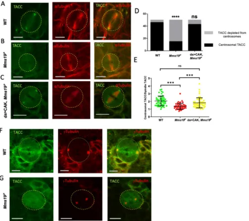
Centrosomal localization of the MT regulator TACC depends on
Mms19
Transforming Acidic Coiled-Coil (TACC), a downstream target of Aurora A kinase, is a criti-cal regulator of centrosomal MTs. During mitosis, Aurora A phosphorylates TACC and stimu-lates its localization on the centrosomes, where TACC further recruits mini-spindles (Msps). Loss-of-function mutations in eitherAurora A, TACC, or msps show drastic abnormalities in astral and spindle MTs [26,27]. AsMms19PNBs display defects in centrosomal separation, spindle length, and astral MTs, we examined whether Aurora A and TACC might be involved in the same process as Mms19 and if the absence of functionalMms19 impedes TACC localiza-tion and funclocaliza-tion. We, therefore, stained wild-type andMms19PNBs for TACC and observed a strong signal at the wild-type centrosomes (Fig 6A). On the other hand, in >50% ofMms19P NBs, TACC did not show any enrichment on the centrosomes (Fig 6B and 6D). Similar results were also obtained with the TACC interactor Msps (S4A–S4D Fig). As TACC acts downstreamFig 6. Centrosomal localization of the MT regulator TACC depends onMms19. (A) A mitotic NB shows TACC
localization at the spindle poles. (B, D) >50% of analyzedMms19pNBs fail to localize TACC to spindle poles. SS was
calculated using Fisher’s exact test (����P<0.0001). (C, D) TACC localization was restored upon expression of
additional CAK subunits (Cdk7, CycH, and Mat1) in theMms19Pbackground. (E) The amount of TACC localized on
the centrosomes was quantified by comparing the fluorescent intensity of the centrosomal TACC signal to the TACC signal on the spindles. A ratio greater than 1 was considered as clear centrosomal TACC, while a ratio equal to or less than 1 indicated unlocalized TACC. SS was calculated by Kruskal-Wallis test, columns were compared by Dunn’s post test (���P<0.001), scale = 5μm. n = 25 NBs per genotype, 3 experiments. (F, G) NBs were stained with antibodies
againstγ-Tubulin to test for centrosomal localization. (F) TACC co-colocalized with γ-Tubulin on wild-type centrosomes. (G) In the mutant NB, TACC fails to concentrate atγ-Tubulin foci.
https://doi.org/10.1371/journal.pgen.1008913.g006
of the Aurora A kinase, we also examined the localization of Aurora A and found it to be depleted from centrosomes inMms19PNBs (S4E–S4G Fig). Because Aurora A itself acts downstream of Cdk1 [28], this defect might be caused by insufficient CAK activity inMms19 mutants [9]. We tested this hypothesis by over-expressing the three CAK components in the Mms19Pbackground (da>CAK,Mms19P). Indeed, upon CAK overexpression, the fraction of spindles displaying centrosomal TACC almost reached wild-type levels (Fig 6C and 6D). To quantify the enrichment of TACC on centrosomes, we compared the fluorescence intensity of centrosomal TACC to the TACC fluorescence intensity on the spindle. This ratio decreased in Mms19PNBs but was rescued in da>CAK,Mms19PNBs (Fig 6E). It was also reported that Aurora A loss-of-function can cause centrosome fragmentation [26]. Co-stainingMms19P NBs with antibodies against the centrosomal proteinγ-Tubulin showed that even when the TACC signal was not seen on centrosomes, the centrosomalγ-Tubulin was still present, indi-cating that TACC mislocalization is not caused by centrosome fragmentation (Fig 6F and 6G). These findings indicate that centrosomal localization of TACC and its stimulation of astral and spindle MT stability or growth is at least partially dependent onMms19 and CAK activity.
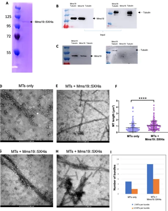
Mms19 binds to MTs and stimulates MT assembly
To explore the possibility that Mms19 also functions through different activities, we prepared protein extracts from flies expressing Mms19::eGFP driven by its endogenous promoter [9]. We subjected them to immunoprecipitations (IP) and analyzed co-purifying proteins by Mass spectrometry (S1 Table). Adult wild-type flies and Imp::eGFP expressing flies, respectively, served as controls to exclude any non-specific binding to beads and GFP, respectively. Amongst the proteins exclusively bound to Mms19::eGFP were the CIA proteins Mip18, Ciao1, and Ant2, which form a complex with Mms19 to mediate Fe-S cluster delivery [6,7]. The fact that we recovered these proteins efficiently, indicated that the purification was effi-cient. BecauseMms19 functions on microtubules and our second control, IMP::eGFP, is also involved in MT dependent processes, we additionally inspected the data for tubulin and MT binding proteins that are enriched by the Mms19::eGFP IP compared to the wild-type control without an eGFP tag (S2 Table). This comparison revealed a clear enrichment of tubulin and several Microtubule Associated Proteins (MAPs). Some of the associated proteins were not present in the wild type control, some were present in the Mms19::eGFP and IMP::eGFP frac-tions, and others exclusively in the Mms19::eGFP fraction. These results, therefore, suggested that Mms19 might directly or indirectly bind to MTs.
To validate the interaction of Mms19 with tubulin, we next tested whether purified Mms19::5xHis andα/β-Tubulin dimers interact in vitro. Mms19::5xHis was purified from E. coli to avoid co-purification of MAPs, and this purified protein fraction showed a single band when separated by SDS-PAGE and stained with Coomassie Blue (Fig 7A). Mms19::5XHis was incubated at an equimolar ratio with purified porcine brainα/β-Tubulin and then bound to the Ni-NTA resin. All incubations and washing steps were carried out at 4˚C. Copurifying pro-teins were then assessed by western blotting (seeTable 3for details regarding the antibodies used). A band corresponding toα-Tubulin was observed when tubulin was incubated with Mms19::5xHis (Fig 7B and 7C), but tubulin alone did not bind to the resin, pointing to a direct interaction between tubulin and Mms19::5xHis.
To better understand the mechanism of modulation of the MT dynamics by Mms19, we complemented the biochemical experiments with a high-resolution optical method. Polymer-ized MTs were incubated at room temperature (RT) with either BRB80/solvent buffer or with Mms19::5xHis, purified upon expression inE. coli (Fig 7A). These samples were then visual-ized by negative stain electron microscopy (EM). MT fibers in general appeared to be less
Fig 7. Mms19::5xHis binds to tubulin and stimulates MT assemblyin vitro. (A) Mms19 tagged with 5X Histidine
was purified fromE. coli. A single band at approximately 100KDa is seen when the purified protein fraction is separated by SDS-PAGE and stained with Coomassie Blue. Mms19::5xHis-tubulin interaction was investigated using a pull-down assay with Ni-NTA resin. Tubulin binding to Mms19::5xHis is expected to result in co-purification of tubulin with Mms19::5xHis. (B) Input panels show tubulin added, either alone or with Mms19. (C) Tubulin was found binding to the resin only upon prior incubation with Mms19::5xHis (tubulin added alone did not bind to the resin). (D) MTs only (without Mms19::5xHis added) were visualized by negative stain EM. Here, an MT fiber can be seen depolymerizing (indicated by arrow). (E) In an MT-Mms19::5xHis mixture, discreet particles can be seen binding laterally along the surface of MTs. (F) Measuring the length of MT fibers revealed that MTs were generally longer when incubated together with Mms19::5xHis compared to MTs to which only the BRB80 buffer was added. n = 150 MTs per condition, 3 experiments, ss was calculated using student’s t-test,����P<0.0001. MT bundles containing 2 (G) or more
than 2 MTs (H) were more frequently observed upon the addition of Mms19::5xHis to MTs. The number of bundles observed in each condition is quantified in (I).
https://doi.org/10.1371/journal.pgen.1008913.g007
stable upon the addition of BRB80/solvent alone (at RT) as some MT fibers were observed undergoing de-polymerization (Fig 7D) under these conditions. Remarkably, MTs incubated with Mms19::5xHis were comparatively longer (Fig 7E and 7F) and more bundled (Fig 7G, 7H and 7I). Furthermore, the Mms19::5xHis containing samples contained particles not found in the samples without Mms19 and these particles appeared along the lateral MT surface (Fig 7E and 7H). It thus appears that Mms19 might bind along the surface of MTs, facilitating their assembly or stabilizing MTs, and possibly stimulating inter-MT contacts.
Discussion
Mms19 was initially identified as a gene that acts in the nucleotide excision repair (NER) path-way. In this DNA repair pathway, it provides an Fe-S cluster to other NER enzymes. When evi-dence emerged about a possible NER independent mitotic activity ofMms19 [8,9], this warranted further investigations into the precise role ofMms19 during mitotic spindle assem-bly in diploid cells. We now report novel, important roles ofMms19 for astral MT assembly, spindle establishment, and orientation, and mitotic timing inDrosophila NBs. We provide strong evidence that Mms19 regulates spindle assembly by promoting the activity of the Cdk activating kinase CAK, and we identified Aurora A and TACC as its downstream targets in this pathway. Further,in vitro data points to a CAK-independent activity where Mms19 directly binds to tubulin and thereby regulates MT assembly and stability.
The trimeric CAK complex is an essential activator of entry into and progression through the initial phase of mitosis because an activating T-loop phosphorylation by Cdk7 is required to fully activate Cdk1 [29,30]. In this activation, Mms19 seems to perform a crucial task of sequestering Xpd in order to allow CAK to fully activate Cdk1 (S6A Fig). This is evident from the rescue of imaginal disc morphology, the rescue of the TACC localization to the centro-somes, and possibly by a slight rescue of the NB spindle phenotypes by CAK overexpression in theMms19Pbackground [9] (Fig 3G and 3H;Fig 6). Cdk1 along with other mitotic kinases such as Aurora A and Polo regulate a multitude of mitotic processes including centrosome maturation, bipolar spindle assembly, and mitotic checkpoint activation [31,32]. Centrosome maturation and entry into mitosis were known to be driven by positive feedback loops between Cdk1, Aurora A, and Polo [32,33]. Additionally, it was shown that Cdk1 acts upstream of Aurora A as it activates Aurora A during the G2/M transition [28]. Cdk1 was also shown pre-viously to regulate the cytoplasmic localization of Bora, an activator of Aurora A, inDrosophila sensory organ precursors [34]. Our demonstration thatMms19 is required for the localization of Aurora A to the centrosomes (S4E–S4G Fig) nicely fits into this model becauseMms19 pro-motes the mitotic kinases cascade through CAK-Cdk1 [9], and one function of Cdk1 is to acti-vate Aurora A. Further, Aurora A directly phosphorylates TACC thereby triggering its centrosomal localization [26]. Given that the failure of TACC localization in the absence of Mms19 can be rescued by CAK over-expression, we now propose TACC as a downstream tar-get of the Mms19-CAK-Cdk1 axis. In this process, the modulation of the CAK activity seems
Table 3. Antibodies used for probing western blots. Primary antibodies
Antibody Manufacturer Catalog number Dilution
Rabbit anti-Mms19 Synthesized by Genscript Inc. Custom antibody 1:2,000
Rabbit anti-alpha-Tubulin Abcam Ab18251 1:2,000
Secondary antibodies
Goat anti-rabbit HRP Thermo Fischer Scientific 65–6120 1:10,000
to be the main mode of action of Mms19 towards TACC. The best-characterized function of TACC is its interaction with Msps which leads to the stabilization of astral MTs [35]. This activity involves their recruitment to the centrosomes, and we found that not only the centro-somal localization of TACC (Fig 6) but also the one of Msps depends onMms19 and elevated CAK activity (S4A–S4D Fig). Additionally, it has been reported that Aurora A also mediates the timely degradation of Cyclin B and that NBs lacking functionalAurora A spend more time in mitosis due to delayed Cyclin B degradation [36]. Apart from the spindle assembly defects this effect could also contribute to the mitotic delay observed inMms19PNBs. The identified pathway downstream of Mms19 might thus promote spindle assembly and regulate mitotic duration.
The phenomenon of centrosome asymmetry is well documented inDrosophila NBs where, after centriole duplication, the daughter centriole retains MT nucleation activity and is anchored to the apical cortex whereas the mother centriole generates an aster just before NEBD [37]. Interestingly, despite this asymmetry, aster formation from both centrosomes happens synchronously in WT NBs. Conversely, a striking feature of some of theMms19PNBs is the delay in MT assembly from the ‘mother’ centrosome destined for the GMC (S5B and S5C Fig;S3 mov;S8 mov). In the example presented inS8 mov, the ‘mother’ centrosome seems to be unable to nucleate MTs until 7–8 minutes post NEBD. Importantly, even in this case, the spindle is bi-astral, indicating that the centrioles have duplicated normally, producing a functional pair of centrosomes. From this, we can narrow down the function ofMms19 to the assembly of centrosomal MTs. The astral MTs are likely a main target, and their drastic loss in theMms19 mutants the main cause of the observed defects in centrosome migration and spindle orientation, because both these processes require contact of long astral MTs with the cortex [25,38].
It has been established that mitotic spindle assembly in mammalian cells or inDrosophila NBs is driven by both centrosomal MTs and MTs emanating from the chromatin [39,40]. We focused our studies mainly on the MTs emanating from the centrosomes because we were able to link theMms19 activity to the localization of TACC and Msps to this place. However, the formation of MTs around the chromatin might also be affected in theMms19PNBs. Spindle assembly normally also initiates around the chromatin, and an MT meshwork is seen forming in the region around the chromatin [40]. In the MT re-growth assay in theMms19PNBs (Fig 5B), we observed a MT mesh forming in the region around the chromatin at t = 30s. But com-pared to wild-type NBs, the length and density of MTs in this region remained abnormally short at t = 90s. Additionally, measuring the inner spindle density (Fig 3H) revealed a signifi-cant decrease inMms19PNBs. As chromatin-nucleated MTs would also contribute to the MT density in the inner spindle, these results indicate defects in the assembly of MTs around chro-matin. Furthermore, live imaging ofMms19PNBs (S2 mov,S8 mov) showed that the overall spindle MT mesh was initially very sparse, and only after 7-8min post NEBD, a fully assembled spindle with MT density comparable with wild-type NB spindles became visible. These find-ings thus indicate thatMms19 is needed for MT formation from both centrosomes and from around the chromatin.
CAK overexpression rescued TACC localization, but it could not fully rescue the spindle assembly/short spindle and the microcephaly phenotypes (Figs1,3and5). Earlier reports [9,41] suggested a direct interaction between Mms19 and MTs as Mms19 was shown to par-tially co-localize with spindles in mammalian cells andDrosophila embryos. We made similar observations in NBs expressing Mms19::eGFP (S5D Fig). Mms19::eGFP partially co-localizes with the spindles and seems to be enriched on astral MTs. Additionally, Mms19::eGFP also co-localizes with the MT bundles in the neurite of cultured neurons (S5E and S5F Fig). In this study, we report for the first time a direct interaction between Mms19 and tubulin, and we
linked this interaction to an activity of Mms19 in stimulating MT assembly and stability (Fig 7). Even though more work is needed to understand this activity, the first results on this addi-tional function are intriguing and call for its further exploration.
Recently, it was shown that Mip18/Galla-2 and Xpd, which are binding partners of Mms19 [8,9], form a complex called ‘CGX’ (Crumbs/Galla-2/Xpd), and this complex recruits Kpl61F, the fly homolog of Kinesin-5, to the embryonic spindle [42,43]. Kinesin-5 activity is crucial for centrosome separation and bipolar spindle assembly [44]. Even though a direct interaction between Mms19 and Kinesin-5/Kpl61F has not been reported [43] (this work) and we do not see the typical monopolar spindle defect associated withKpl61F [40] in theMms19PNBs, it is possible that the absence ofMms19 may compromise the activity of the CGX complex, too, and that the misregulated Kinesin-5/Kpl61F activity might contribute to the observed spindle defects.
Based on our findings and published data [9,45,46], we propose a model that outlines the CAK dependent and independent activities of Mms19 required for efficient mitotic spindle assembly in diploid cells. In the CAK dependent pathway model (S6A and S6B Fig), Mms19 competes with the CAK complex for binding to Xpd [8,9]. We hypothesize that CAK is mostly associated with Xpd during interphase, and integrated into the TFIIH complex, where its mitotic activity is inhibited. Although basal levels of CAK activity might persist, they would not be sufficient for full activation of Cdk1 (S6A Fig). During mitosis, binding of Mms19 to Xpd prevents the interaction between Xpd and the CAK complex and between Xpd and the core TFIIH. This releases the inhibition of the Cdk-activating kinase activity of CAK by Xpd and allows CAK to fully activate Cdk1 to drive mitosis [9,45]. Upon knockdown or inactiva-tion ofMms19, more CAK would remain bound to Xpd and TFIIH, preventing it from opti-mally activating Cdk1 (S6B Fig). Nevertheless, basal levels of free CAK would remain present. Such reduced levels of CAK activity might still allow the cells to enter mitosis but would be unable to activate adequate levels of Cdk1. The low levels ofCdk1 activity would thereby affect the downstream pathways, such as the localization of TACC and Msps through Aurora A (S6B Fig). Additionally, we now found evidence for a CAK-independent activity where Mms19 directly binds to MTs, promotes MT stability, and stimulates inter-MT contacts (Fig 7,S6C
Fig).
Mms19, a gene initially described as a NER regulator, evidently has clear and essential addi-tional roles as a mitotic gene and as an MT regulator. Given the critical roles it fulfills, the proper control of Mms19 expression and localization must be crucial for the proper function-ing ofMms19. Future studies should therefore address the transcriptional and post-transla-tional control of Mms19 expression and localization and how this impacts its functions in cell physiology, development, and diseases.
Materials and methods
Dissection and immunostaining of larval brains
Larval brains were dissected and stained as described [47]. Briefly, wandering third instar lar-vae were dissected in PBS and fixed for 15min in 4% paraformaldehyde supplemented with 0.3% Triton X-100, 100mM PIPES (pH 6.9), 1mM MgSO42and 1mM EGTA (to stabilize
MTs). After 3 washes in PBS+0.1% TritonX-100 (PBST), primary antibodies were added (Table 2). After overnight incubation, the brains were washed 3x with PBST, stained with sec-ondary antibodies (Table 2), and mounted on glass slides in Aqua Polymount mounting medium (Polysciences; for details on reagents and kits, seeTable 4).
For MT regrowth assays, brains were dissected in Schneider’s medium (supplemented with 10% fetal bovine serum) at 25˚C and incubated on ice for 30 min to depolymerize MTs. Brains
were then incubated for different time points in a 25˚C water bath, followed by fixation and immunostaining. This experiment was performed three times and 20 spindles were analyzed in each iteration. Slides were imaged on a Leica TCS-SP8 microscope (Leica Microsystems) equipped with a 63X, NA 1.4 Plan Apochromat objective. Images were acquired using LAS X software and analyzed using Fiji/ImageJ [48] (seeTable 5for details regarding the special soft-ware used).
Measuring brain compartment volumes
Whole brain lobes were immunostained with antibodies against Miranda (Mira) and phos-pho-Histone 3 (pH3), and DNA was visualized with Hoechst 33342. Z stacks were acquired with a TCS-SP8 confocal microscope on a 63X, 1.4NA Plan-Apochromat objective with 1μm spacing between each optical section. Segmentation, volume measurement, and 3D recon-struction was performed by using the TrackEM2 plugin in Fiji [49].
MARCM crosses
For generating mosaic clones we used the methods described previously [50] with modifica-tions. Briefly, the driver stockhs-flp; tub-Gal4, UAS-mCD8::GFP/CyO, actin::GFP; FRT82B, tub-Gal80/TM6, Tb was crossed to +; FRT82B, Mms19P/TM6, Tb. GFP-Balancer negative 24hr
Table 4. Kits and reagents used for these studies.
Reagent/Kit Manufacturer Catalog
number
Aqua Poly/Mount mounting medium Polysciences Inc 18606–20
Click-it EdU incorporation kit, Alexa Flour 647 Thermo Fischer Scientific C10340
Paclitaxel (Taxol) Sigma T7402
Purified porcine tubulin Cytoskeleton Inc T240-B
Schneider’s Drosophila medium ThermoFischer Scientific 21720–024
Hoechst 33342 ThermoFischer Scientific H3570
Protein G Mag Sepharose Xtra GE life sciences 28967066
Ni-NTA agarose Qiagen 30210
Collagenase I Sigma-Aldrich C0130-100MG
Phenylmethylsulfonyl fluoride (PMSF) Sigma-Aldrich 10837091001
cOmplete, Mini, EDTA-free Protease Inhibitor Cocktail Sigma-Aldrich 4693159001
Concanavalin A Sigma-Aldrich C7898
Formamide Sigma-Aldrich F9037-100ml
Uranyl acetate Electron microscopy
sciences
22400
1,4-Piperazinediethanesulfonic acid, Piperazine-1,4-bis(2-ethanesulfonic acid), Piperazine-N,N0-bis
(2-ethanesulfonic acid) (PIPES)
Sigma-Aldrich P6757
5’ Cy5 labeled Oligonucleotide probe: 5’ Cy5-AACACAACACAACACAACACAACACAACACAACAC Microsynth AG ChrII
Aphidicolin Sigma-ALdrich A0781
https://doi.org/10.1371/journal.pgen.1008913.t004
Table 5. Software used.
Software Source Version
Fiji (ImageJ) https://imagej.net/Fiji
-Leica Application Suite (LAS X) Leica microsystems
-PRISM Graph pad software Version 5
https://doi.org/10.1371/journal.pgen.1008913.t005
old larvae were selected and heat shocked at 37˚C in a glass vial submerged in a water bath for 15min. Larvae were then returned to 25˚C and the brains of the non-Tubby larvae were dis-sected 48hrs later.
Live imaging
Brains expressing EB1::GFP were dissected and mounted on stainless steel chambers as described in [51]. Brains were then imaged using a 100X, NA 1.3 oil immersion objective on a Visiscope Spinning disk microscope (Visitron GmbH) fitted with Nikon Ni2 stand, a CSU-W1 scanner unit, and a Photometrics Evolve 512 EMCCD camera. Images were acquired for 60 seconds with 500ms time intervals at 200ms exposure at 60% laser power (488nm). To track the particle velocities, the particles were manually traced in Fiji/ImageJ [48]. Probably due to limited resolution, it was not possible to unambiguously account for merging or splitting events, and therefore only particles with a linear trajectory, which did neither split nor merge, were analyzed. At least 4–5 particles from each spindle were analyzed from a total of 11 cells per genotype. For surface glia quantification, at least 7–8 particles were analyzed from each of the 15 brains. Stacks were exported to.AVI movies at 14 frames per second.
To measure the NEBD to Anaphase B duration, EB1::GFP expressing brains were mounted as described above and imaged at 40% laser power with a 63X, 1.3 NA objective of a Nikon W1 LIPSI spinning disk microscope fitted with a Photometrics Prime 95B CMOS camera. Movies were acquired on Nikon’s NIS elements software for 2hrs with an interval of 1 min at 200 ms exposure. Z-stacks were acquired simultaneously with 2μm distance between successive optical sections. Movies were analyzed and processed with Fiji/ImageJ. Stacks were exported to.AVI movies at 12 frames per second.
Quantification of spindle orientation
The orientation of the mitotic spindle was examined with respect to the basal Mira crescent. A reference line was drawn passing approximately through the center of the Mira crescent and the angle between the spindle and this reference line was determined using the ImageJ angle tool.
EdU incorporation
Click-it EdU kit (Invitrogen) was used to measure EdU incorporation. Brains were dissected in PBS and incubated in Schneider’s medium supplemented with 10μM 5-Ethynyl-2’-deoxyur-idine (EdU) for 2hrs at 25˚C. EdU is an analog of Thym5-Ethynyl-2’-deoxyur-idine and is incorporated by S phase cells during DNA replication. EdU is then detected due to its binding to a dye-azide conjugate. Brains were subsequently fixed with 4% PFA and incubated with primary antibodies (anti-Mira to mark NBs and anti-pH3 to mark mitotic cells) and secondary antibodies. Subse-quently, they were processed for EdU detection following the manufacturer instructions.
Preparation of whole-fly extract
1g of flies were collected in Eppendorf tubes and frozen in liquid nitrogen. Frozen flies were crushed into a fine powder using a pre-cooled mortar pestle. The powder was incubated in lysis buffer (25mM Hepes, 150mM NaCl, 1mM EDTA, 0.1% TritonX-100, 1mM phenyl-methylsulfonyl fluoride (PMSF), 1 complete EDTA-free protease inhibitor tablet (Roche/ Sigma-Aldrich) for 30 min and then centrifuged in an Eppendorf tube for 30 min at 16,000 g and 4˚C. The supernatant was saved and snap-frozen in liquid nitrogen.
Immunoprecipitation to prepare extract for Mass Spectrometry
ProteinG-Mag Sepharose (GE) beads were washed 3X with PBS, incubated for 2hrs with anti-GFP antibody (3E6, provided by Anne Marcil). Beads were then incubated with the crude extracts for 6hrs at 4˚C. Following 3 washes with the wash buffer (25mM Hepes, 150mM Nacl, 1mM EDTA, 1mM PMSF, 1 tablet complete EDTA free protease inhibitor tablet), bound pro-teins were eluted by 15 min incubation in urea elution buffer (6–8 M Urea, 20 mM Tris pH 7.5, and 100 mM NaCl) or glycine elution buffer (100mM Glycine, pH 2.6. These eluates were neutralized by adding 150mM Tris-Cl, pH 8.8).
Mass-spectrometry
Eluted proteins in 8M urea were processed essentially as described by Engel and colleagues [52]. Briefly, proteins were reduced by the addition of 1/10 volume of 0.1 M DTT and incu-bated for 30 min at 37˚C, followed by alkylation with a five-fold molar excess of iodoacetamide and incubation for 30 min at 37˚C. Proteins were precipitated at -20˚C by the addition of 5 volumes cold acetone and incubation for 30 min at -20˚C. All liquid was carefully removed, and the pellet dried in ambient air for 15 min before reconstitution of the proteins in 8 M urea, 50 mM Tris-HCl pH 8.0 to a final protein concentration between 0.2–0.3 mg/mL. Protein concentration was determined by Bradford assay. An aliquot corresponding to 5μg protein was diluted to a final urea concentration of 2 M urea with 20 mM Tris-HCl pH 8.0, and 2 mM CaCl2. Proteins were digested by trypsin (1:50 (w/w) trypsin/protein ratio) for 6 hours at 37˚C. The digests were acidified with TFA (1%) and analyzed by LC-MS/MS (EASY-nLC 1000 coupled to a QExactive HF mass spectrometer, ThermoFisher Scientific) with three repetitions injecting an aliquot of 500 ng protein. Peptides were trapped on an Acclaim PepMap100 C18 pre-column (3μm, 100 Å, 75μm x 2 cm, ThermoFisher Scientific, Reinach, Switzerland) and separated by backflush on a C18 column (3μm, 100 Å, 75μm x 15 cm, Nikkyo Technos, Tokyo, Japan) by applying a 40 min gradient of 5% acetonitrile to 40% in water, 0.1% formic acid, at a flow rate of 300 nl/min. Peptides of m/z 400–1400 were detected at a resolution of 60,000 m/z 250 with automatic gain control (AGC) target of 1E06 and maximum ion injection time of 50 ms. A top fifteen data-dependent method for precursor ion fragmentation was applied with the following settings: resolution 15,000, AGC of 1E05, maximum ion time of 110 ms, charge inclusion of 2+ to 7+ ions, peptide match on, and dynamic exclusion for 20 sec, respectively.
Fragment spectra data were converted to mgf with ProteomeDiscoverer 2.0 and peptide identification made with EasyProt software searching against the forward and reversed Uni-protKBDrosophila melanogaster protein database (Release 2016_11), complemented with commonly found protein sequences of contaminating proteins, with the following parameters: parent mass error tolerance of 10 p.p.m., trypsin cleavage mode with three missed cleavages, static carbamidomethylation on Cys, variable oxidation on Met and acetylation on protein N-terminus. On the basis of reversed database peptide spectrum matches, a 1% false discovery rate was set for acceptance of target database matches, and only proteins with at least two dif-ferent peptide sequences identified were allowed.
Immunoprecipitation/pull-down assays
50μg of purified Mms19 (Tagged with 5X Histidine at C-terminus, synthesized by Genscript Inc and solubilized in 20mM Tris, 150mM NaCl, 0.5 M Arginine) was incubated with 50μg purified porcine tubulin (Purchased from Cytoskeleton Inc) at 4˚C for 2 hrs. This mixture was subsequently incubated with Ni-NTA agarose (equilibrated with 20mM Tris-Cl and 250mM NaCl) for 1 hr at 4˚C. The beads were then washed three times with wash buffer containing 20mM Tris, 250mM NaCl, and 20mM Imidazole, and the bound proteins were eluted by