HAL Id: tel-00441373
https://tel.archives-ouvertes.fr/tel-00441373
Submitted on 15 Dec 2009HAL is a multi-disciplinary open access archive for the deposit and dissemination of sci-entific research documents, whether they are pub-lished or not. The documents may come from teaching and research institutions in France or abroad, or from public or private research centers.
L’archive ouverte pluridisciplinaire HAL, est destinée au dépôt et à la diffusion de documents scientifiques de niveau recherche, publiés ou non, émanant des établissements d’enseignement et de recherche français ou étrangers, des laboratoires publics ou privés.
One base pair resolution histone H1-nucleosomal DNA
interaction mapping and 3D organization of H1
containing nucleosome
Sajad Hussain Syed
To cite this version:
Sajad Hussain Syed. One base pair resolution histone H1-nucleosomal DNA interaction mapping and 3D organization of H1 containing nucleosome. Biomolecules [qbio.BM]. Université JosephFourier -Grenoble I, 2009. English. �tel-00441373�
Université Joseph Fourier-Grenoble 1
Chimie et Science du Vivant
(Arrêtés ministériels du 5 juillet 1984 et du 30 mars 1992)
THÈSEPour obtenir le titre de
Docteur de l’Université Joseph Fourier
Discipline Biologie
Présentée et soutenue publiquement par
SYED Sajad Hussain
Le 03 Decembre 2009
One base pair resolution histone
H1-nucleosomal DNA interaction mapping and
3D organization of H1 containing
nucleosome
Jury
Dr. Didier TROUCHE Rapporteur
D r .
F r a n c e s c a P A L L A D I N O R a p p o r t e u r
Pr. Hans GEISELMANN Examinateur
Dr. Dimitar ANGELOV Co Directeur de thèse
Dr.
Stefan DIMITROV Directeur de thèse
Thèse préparée au sein du laboratoire
Biologie Molécularie et Cellulaire de la Differenciation
Unite INSERM U823
Institut Albert Bonniot
Universite Joseph Fourier-Grenoble 1
France
A mountain is composed of tiny grains of
earth. The ocean is made up of tiny drops
of water. Even so, life is but an endless
series of little details, actions, speeches,
and thoughts. And the consequences whether
good or bad of even the least of them are
far-reaching.
Dedicated to
“Abba and Khala”
ACKNOWLEDGEMENTS
The journey of my doctoral studies may not have been so pleasant and full of joy
without the help of colleagues and collaborators, both at the Ecole Normale Supérieure de
Lyon and Université Joseph Fourier - Grenoble I, coordinated admirably by my mentor
Dr. Stefan Dimitrov. I am highly thankful to him for his kind support and involving me in
fruitful collaborative works with people from different fields which will have an everlasting
influence on my scientific temperament. His enthusiastic, knowledgeable and friendly
scientific discussions are the keys to the success of my thesis research, and are deeply
appreciated. I am indebted for the personal care he took all along the way.
I’m also very grateful to my thesis co-supervisor, Dr. Dimitar Angelov for providing
a congenial lab atmosphere and teaching me the chromatin biochemistry. His tireless
scientific discussions, tit bits for experimental optimization and thorough result
interpretations were essentials for the timely completion of the work. A specials thanks for
all his efforts and support, and has been a pleasure to work with him personally as well as
scientifically.
I would like to thank Prof. Hans Geiselmann, Dr. Didier Trouche and
Dr. Francessca Palladino for their time and efforts to evaluate the thesis.
I take this opportunity to express my debt of gratitude to Dr. Namita Surolia for her
contribution in teaching me the protein biochemistry in the beginning of my research life.
I was very lucky to meet Prof. Philippe Bouvet, who actually introduced me to the
team of Dr. Stefan Dimitrov and supported me all the way. Thank you very much.
Special thanks to the patience and expertise of Dr. Jan Bednar for the long dark
room experimental sessions for the Cryo-electron microscopy and the beautiful image
galleries.
I would also like to thank Dr. Nils Becker, Dr. Ralf Everaers and Sam Meyer for
their help in mathematical modeling experiments.
I also wish to thank Dr. Cendrine Moskalenko for the Atomic Force Microscopy
experiments, Dr. Andrew Travers, Dr. Mathieu Boulard, Dr. Thierry Gautier, Dr. Manu
Shukla and Damien Goutte-Gatat for their help in some of my experiments.
Manu, Imtiaz and Richi are “not to forget” persons for the wonderful times spent in
the lab especially while writing this manuscript.
Hats off to all the people of Joliot Curie Laboratory especially Marie-Jo, Fabien
Mongelard, Hervé, Fabien Montel, Elsa and Zofia for providing the scientifically vibrant
and homely environment. I will miss those afternoon cake parties.
My fellow students have helped me in many different ways. I would like to thank
Anu, Farhan, Dinil, Aijaz, Mubarak and Sadaf for their interest, their support and their
encouragement.
My deepest gratitude belongs to my family members and relatives for their love and
moral support throughout my life.
Finally, I would like to thank the people who actually made the difference. Thanks
to Abba (Peer Ghulam Nabi Shah) and Khala (Syeda Hanefa Akhter) for everything.
9
INDEX
THÈSE ... 1
Acknowledgements ... 7
INDEX ... 9
List of tables and figures ... 14
RESUME DE LA THESE ... 18
Summary of the thesis ... 20
Information of the laboratories in which the thesis was prepared ... 22
list of abbreviations ... 23
Chapitre I: Chapitre d’Introduction ... 24
Chapter I: Introduction ... 25
I .1 Chromatin introduction ... 25
I .2 Chromatin history ... 25
I .2.1 Nobel Prize related to chromatin ... 27
I .3 Chromatin structure ... 28
I .3.1 DNA ... 29
I .3.2 Nucleosome ... 30
I .3.2.1 Nucleosome core particle ... 31
I .4 Linker histone ... 34
I .4.1 Location of Globular domain on nucleosome ... 35
I .4.2 Role of the Carboxy terminus of Histone H1 ... 37
I .4.3 Molecular dynamics of Histone H1 ... 39
I .4.4 Linker histone isoforms ... 41
I .4.4.1 H1.1 – H1.5 ... 42
I .4.4.2 H1º/H5 ... 43
I .4.4.3 H1x ... 43
I .4.4.4 Germ line specific linker histone variants ... 43
I .4.5 Linker Histone post translational modifications ... 44
I .5 Structure of the 30-nm chromatin fiber ... 46
I .6 Higher order structures of Chromatin beyond the 30 nm fiber ... 49
I .7 Chromatin territories ... 51
I .7.1 Euchromatin ... 51
I .7.2 Heterochromatin ... 51
I .8 Chromatin Modifications and Their Function ... 53
I .8.1 Histone post translational modifications ... 53
I .8.1.1 Histone Acetylation ... 54
I .8.1.2 Histone methylation ... 54
I .8.1.3 Other covalent histone modifications ... 56
10
I .8.2.1 Different Chromatin remodeling Families ... 57
I .8.2.1.1 SWI/SNF family remodelers ... 57
I .8.2.1.2 ISWI family remodelers ... 58
I .8.2.1.3 CHD family remodelers ... 59
I .8.2.1.4 INO80 family remodelers ... 59
I .8.2.2 Function of remodelers ... 60
I .8.2.3 Mechanism of Chromatin remodeling ... 63
I .8.3 Core histone Variants ... 65
I .8.3.1 H2A histone variants ... 66
I .8.3.1.1 H2A.Z Histone Variant ... 66
I .8.3.1.2 H2AX histone variant ... 67
I .8.3.1.3 MacroH2A histone variant ... 68
I .8.3.1.4 H2A.Bbd histone variant ... 69
I .8.3.1.5 TH2A histone variant ... 70
I .8.3.1.6 H2AL1 and H2AL2 ... 70
I .8.3.2 H2B histone variants ... 71
I .8.3.2.1 TH2B histone variant ... 71
I .8.3.2.2 H2BFWT histone variant ... 71
I .8.3.3 H3 histone variant ... 72
I .8.3.3.1 Centromeric protein A (CENP-A) or CenH3 ... 73
I .8.3.3.2 H3.3 histone variant ... 73
Objectives ... 75
Resultats: Chapitre II ... 76
Results: ... 77
Chapter II: Publication 1 ... 77
One base pair resolution H1-nucleosome interaction mapping and 3D organization of H1 containing nucleosome ... 77
II.1 Abstract ... 78
II.2 Introduction ... 79
II.3 Materials and Methods ... 80
II.3.1 Preparation of DNA fragments ... 80
II.3.2 Clone construction and Protein purification ... 81
II.3.3 Nucleosome Reconstitution, H1 deposition and footprinting ... 81
II.3.4 NAP-1 mediated deposition of H1 ... 81
II.3.5 Hydroxyl radical footprinting ... 82
II.3.6 Cryoelectron microscopy: ... 82
II.3.7 Mathematical analyses and structural model rebuilding ... 83
II.3.7.1 Relative accessibilities of nucleosomal DNA to OH° ... 83
II.3.7.2 Visualization ... 83
II.3.7.3 Structure-derived accessibility profiles ... 83
II.3.7.4 Structural model rebuilding ... 83
II.3.7.5 Restrained energy minimization ... 84
11
II.4.1 NAP-1 mediated assembly of H1 and its mutants to nucleosomal
templates ... 84
II.4.2 Electron cryo-microscopy imaging of trinucleosomes containing either full length histone H1 or its mutants ... 86
II.4.3 Hydroxyl radical footprinting of chromatin samples assembled with full- length histone H1 ... 87
II.4.4 The globular domain of histone H1 protects both 10 bps of DNA located symmetrically to the nucleosome dyad and ∼one helical turn of DNA from each linker ... 88
II.4.5 A short aminoacid sequence located between AA 120 and 127 of H1 COOH-terminus is required for the generation of the linker DNA 10 bp repeat upon OH° cleavage ... 90
II.5 Discussion ... 92
II.6 Acknowledgements. ... 99
II.7 Supplementary figures ... 99
II.8 Supplementary Methods: ... 102
II.8.1 Structural Analysis of OHo -Footprinting Gels ... 102
II.8.1.1 Conversion of gel exposure to relative accessibility signals ... 102
II.8.1.1.1 Trace adjustment ... 102
II.8.1.1.2 Renormalization ... 105
II.8.1.2 OHo -footprints vs. three-dimensional nucleosome structures ... 108
II.8.1.2.1 Exploiting the two-fold symmetry ... 108
II.8.1.2.2 Three-dimensional rendering of relative accessibilities ... 108
II.8.1.2.3 Qualitative test of structural models for H1 placement ... 109
II.8.1.2.4 Three-contact model ... 109
II.8.1.2.5 Two-contact models ... 111
II.8.1.2.6 Comparison ... 112
II.8.1.3 OHo-Footprinting predictions derived from structural models ... 112
II.8.1.3.1 Semi-quantitative OHo footprinting predictions from structural models. ... 112
II.8.1.3.2 Structure-derived footprints for the two- and three-contact models ... 113
II.8.1 4 Structure refinement based on DNA nano-mechanics .... 114
II.8.1.4.1 Geometric and mechanical constraints on the stem conformation ... 114
II.8.1.4.2 DNA elastic energy minimization ... 115
II.9 Movies of the Chapter are presented in the compact disc, attached physically to the thesis. ... 116
II.9.1 Movie 1: Nucleosome without histone H1 ... 116
II.9.2 Movie 2: Three-contact model for a nucleosome associated with the globular domain (GH1) of histone H1. ... 116
II.9.3 Movie 3: Two-contact model (84) of a nucleosome with the globular domain (GH1) of histone H1. ... 116
II.9.4 Movie 4: Two-contact model (28) of a nucleosome with the globular domain (GH1) of histone H1. ... 116
12
II.9.5 Movie 5: Three-contact model for a nucleosome associated with
the 40-127 mutant of histone H1. ... 116
Chapitre III ... 117
Chapter III: Publication 2 ... 118
The incorporation of the novel histone variant H2AL2 confers unusual structural and functional properties of the nucleosome ... 118
III.1 Abstract ... 119
III.2 Introduction ... 120
III.3 Materials and methods ... 121
III.3.1 H2AL2 cloning and preparation of DNA fragments ... 121
III.3.2 Protein purification and nucleosome reconstitution ... 122
III.3.3 Exonuclease III mapping, footprinting, micrococcal nuclease digestion ... 122
III.3.4 Nucleosome mobilization and remodeling ... 123
III.3.5 “One pot” restriction enzyme accessibility assay ... 123
III.3.6 Xba 1 restriction assay ... 123
III.3.7 Atomic Force Microscopy ... 124
III.3.8 Electron Cryo-microscopy ... 124
III.4 Results ... 125
III.4.1 Histone H2AL2 is specifically expressed in the testis and could efficiently replace conventional H2A in the nucleosome ... 125
III.4.2 DNase I and hydroxyl radical footprinting of H2AL2 histone variant nucleosomes ... 126
III.4.3 Micrococcal and Exonuclease III digestion demonstrates a distinct organization of the H2AL2 nucleosome ... 128
III.4.4 The “one pot assay” shows that the interactions between the histone octamer and the ends of nucleosomal DNA are highly perturbed within the H2AL2 particle ... 131
III.4.5 AFM imaging of conventional and H2AL2 nucleosomes ... 131
III.4.6 Electron cryo-microscopy shows a very open structure of the H2AL2 tri-nucleosomes ... 133
III.4.7 The presence of H2AL2 affected both RSC and SWI/SNF nucleosome remodeling and mobilization ... 135
III.5 Discussion ... 140
III.6 Funding ... 141
III.7 Acknowledgements ... 141
III.8 Supplementary figure 1S: Northern-blot ... 142
Chapitre IV ... 143
Chapter IV: Publication 3 ... 144
The docking domain of H2A and the entry/exit angle of the linker DNA determine the binding of H1 ... 144
13
IV.2 Results ... 146
IV.2.1 NAP-1 mediated binding of linker histone H1 to conventional and variant nucleosomes ... 146
IV.2.2 Hydroxy radical mapping of H1 on H2AL2 nucleosomes ... 147
IV.2.3 The docking domain of histone H2A is required for proper histone H1 binding to the nucleosome ... 149
IV.2.4 Electron cryo-microscopy imaging reveals a "beads on a string" structure for H1 associated H2AL2, H2A.Bbd and chimeric H2A.ddBbd trinucleosomes ... 151
IV.3 Materials and Methods ... 153
IV.3.1 Preparation of DNA fragments ... 153
IV.3.2 Protein purification, nucleosome reconstitution and hydroxyl radical footprinting ... 153
IV.3.3 Cryoelectron microscopy ... 153
IV.4 Discussion ... 154
Chapitre V ... 156
Chapter V: Perspectives ... 157
V.1 How does H1 interact with nucleosomal array with different linker length 157 V.2 Role of histone H3 N-terminal tail in maintenance of the stem ... 157
V.3 Role of different positively charged residues of H1 in the binding and stem structure formation ... 158
V.4 Yeast histone Hho1p and Xenopus specific B4 histone ... 158
V.5 Structure of H1/NAP1 ... 158
V.6 Mutual exclusion of H1 and H2A.Bbd and H2AL2 in somatic cells and during spermatogenesis ... 159
14
LIST OF TABLES AND FIGURES
TABLE 1: SHOWING NOBLE PRIZES AWARDED RELATED TO CHROMATIN…….27 TABLE 2: A DETAILED LIST OF KNOWN HISTONE POSTTRANSLATIONAL MODIFICATION ENZYMES………...55 TABLE 3: SHOWING COMPOSITION OF REMODELER COMPLEXES IN DIFFERENT EUKARYOTES….……….……..60 TABLE 4: SUMMARIZING THE FUNCTION OF DIFFERENT MEMBERS OF CHROMATIN REMODELING FAMILY……….…...62 TABLE 5: HISTONE H3 VARIANTS IN DIFFERENT ORGANISMS………72
CHAPTER I
FIGURE 1: DIAGRAM REPRESENTING THE HALLMARKS IN THE HISTORY OF CHROMATIN STUDIES...27 FIGURE 2: ORGANIZATION OF EUKARYOTIC CHROMATIN FIBERS………....28 FIGURE 3: A CARTOON OF DNA DOUBLE HELIX SHOWING DNA MAJOR AND MINOR GROOVE………30 FIGURE 4: NUCLEOSOME CORE PARTICLE STRUCTURE………...………….32 FIGURE 5: SHOWING THE STRUCTURE OF THE HISTONE FOLD MOTIF AND HANDSHAKE MOTIF….………..33 FIGURE 6: DIFFERENT MODELS OF GLOBULAR DOMAIN LOCALIZATION ON THE NUCLEOSOME………...……….35 FIGURE 7: GH5 DOCKED TO THE NUCLEOSOME...37 FIGURE 8: A TYPICAL FRAP CURVE FOR H1 BINDING TO CHROMATIN...39 FIGURE 9: ALTERNATIVE MODELS FOR THE REVERSIBLE ASSOCIATION OF
HISTONE H1 WITH THE NUCLEOSOME……….…………..40
FIGURE 10: NUMBER OF H1 VARIANTS IN VARIOUS SPECIES………..41 FIGURE 11: POSTTRANSLATIONAL MODIFICATION SITES IDENTIFIED IN HUMAN H1 ISOFORMS.………...….46
FIGURE 12: MODELS FOR THE DNA PATH IN THE CHROMATIN FIBER
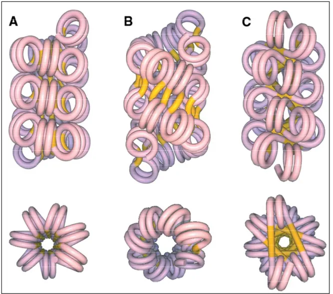
..
...47FIGURE 13: TWO WIDELY ACCEPTED MODELS OF 30NM CHROMATIN FIBER….48 FIGURE 14: ELECTRON - MICROGRAPH OF A HISTONE-DEPLETED METAPHASE CHROMOSOME FROM HELA……….………...………...50
15
FIGURE 15: THE DISTRIBUTION OF EUCHROMATIN (E) AND HETEROCHROMATIN (H) IN A NORMAL THYMUS LYMPHOCYTE………51 FIGURE 16: PROPERTIES OF EUCHROMATIC AND HETEROCHROMATIC REGIONS……….….53 FIGURE 17: REMODELER FAMILIES, DEFINED BY THEIR ATPASE………...58 FIGURE 18: MODEL OF DNA MOVEMENT DURING A REMODELING EVENT…….65 FIGURE 19: SEQUENCE ALIGNMENT OF THE MAIN MAMMALIAN HISTONE H3 VARIANTS………72
CHAPTER II
FIGURE 1: NAP-1 ALLOWS THE PROPER INCORPORATION OF LINKER HISTONE H1 AND ITS TRUNCATED MUTANTS IN 601 DINUCLEOSOMAL TEMPLATES…....85 FIGURE 2: REPRESENTATIVE ELECTRON CRYO-MICROSCOPY IMAGES OF RECONSTITUTED 601 TRINUCLEOSOMES………...87 FIGURE 3: HYDROXYL RADICAL FOOTPRINTING OF CONTROL AND H1 CONTAINING DINUCLEOSOMAL SUBSTRATES………89 FIGURE 4: HYDROXYL RADICAL FOOTPRINTING OF CONTROL AND ASSEMBLED WITH FULL LENGTH H1 AND ITS TRUNCATED MUTANTS DINUCLEOSOMES…..91 FIGURE 5: HYDROXYL RADICAL FOOTPRINTING OF TRINUCLEOSOMES……...93 FIGURE 6: MOLECULAR MODELS FOR THE NUCLEOSOME PARTICLE…………...96 SUPPLEMENTARY FIGURE 1: ELECTRON CRYO-MICROSCOPY IMAGES OF RECONSTITUTED 601 TRINUCLEOSOMES ASSEMBLED WITH HISTONE H1
TRUNCATED MUTANT 1-177………..…
…..
………...99SUPPLEMENTARY FIGURE 2: DNASE I FOOTPRINTING OF 601 DINUCLEOSOMES CONTAINING EITHER FULL LENGTH HISTONE H1 OR THE TRUNCATED MUTANTS………...100 SUPPLEMENTARY FIGURE 3: HYDROXYL RADICAL FOOTPRINTING OF M ONONU CLEOSO M ES CONTAI NI NG EI THER H1 OR THE HI STONE H1
TRUNCATED MUTANTS…………
………..
……….…...101FIGURE SM-1: RAW INTENSITY TRACE OBTAINED WITH A MONONUCLEOSOME WITH H1………...103 FIGURE SM-2: RAW INTENSITY TRACE, DE-TRENDED TRACE AND CONVERGED SINUSOIDAL FITTING FUNCTION,…………... ………….……….104
16
FIGURE SM-3: BAND POSITION X(N) IN PIXELS AS A FUNCTION OF BAND NUMBER……….104 FIGURE SM-4: PLACEMENT OF ANALYZED GELS ON THE NUCLEOSOME……105 FIGURE SM-5: COMPARISON OF MONONUCLEOSOME AND DINUCLEOSOME ACCESSIBILITIES IN THE PRESENCE OF H1………….………106 FIGURE SM-6: ACCESSIBILITY ON THE MONONUCLEOSOME CORE, WITHOUT H1, WITH THE GLOBULAR DOMAIN GH1 AND WITH FULL-LENGTH H1………..107 FIGURE SM-7: ACCESSIBILITY ON THE MONONUCLEOSOME LINKER, WITHOUT
H1 WITH THE GLOBULAR DOMAIN GH1 AND WITH FULL-LENGTH H1……....…107
FIGURE SM-8: EXPERIMENTAL ACCESSIBILITY PROFILE FOR NUCLEOSOMES WITHOUT HISTONE H1……….……….109 FIGURE SM-9: THREE-CONTACT NUCLEOSOME CONFIGURATION………...110 FIGURE SM-10: TWO-CONTACT NUCLEOSOME CONFIGURATION A.……...…….110 FIGURE SM-11: TWO-CONTACT NUCLEOSOME CONFIGURATION B……….111 FIGURE SM-12: EXPERIMENTAL PROTECTION PATTERN SHOWN COLOR CODED ON A NUCLEOSOMAL DNA LOOP………...………...113 FIGURE SM-13: STRUCTURE-DERIVED AND EXPERIMENTAL RELATIVE ACCESSIBILITY FOR MONONUCLEOSOMES WITH GH1…………...114 FIGURE SM-14: EXPERIMENTAL RELATIVE ACCESSIBILITY PATTERN FOR FULL-LENGTH H1 SHOWN ON A MONONUCLEOSOMAL LOOP………...115
CHAPTER III
F I G U R E 1 : T H E H I S T O N E V A R I A N T H 2 A L 2 C A N S U B S T I T U T E F O R CONVENTIONAL H2A IN THE NUCLEOSOME……….……….126 FIGURE 2: DNASE I AND HYDROXYL RADICAL FOOTPRINTING SHOW ALTERATIONS IN THE STRUCTURE OF THE HISTONE VARIANT H2AL2 NUCLEOSOME……….……….127 FIGURE 3: HIGHER ACCESSIBILITY OF H2AL2 MONO-NUCLEOSOMES AND NUCLEOSOMAL ARRAYS TO MICROCOCCAL NUCLEASE AND EXONUCLEASE III DIGESTION………..………..….………...130 FIGURE 4: “ONE POT ASSAY” OF CONVENTIONAL AND H2AL2 NUCLEOSOME.132 FIGURE 5: AFM IMAGING SHOWS THAT THE H2AL2 HISTONE OCTAMER IS
17
FIGURE 6: ELECTRON CRYO-MICROSCOPY VISUALIZATION OF CONVENTIONAL H2A AND HISTONE VARIANT H2AL2 TRI-NUCLEOSOMES………...134 FIGURE 7: SWI/SNF AND RSC REMODELING OF CONVENTIONAL AND H2AL2 NUCLEOSOMES………..………….………136 FIGURE 8: THE PRESENCE OF H2AL2 INTERFERES WITH BOTH SWI/SNF AND RSC NUCLEOSOME REMODELING AND MOBILIZATION………..138 FIGURE 9: XBA 1 NUCLEASE RESTRICTION AND AFM ANALYSES OF THE RSC-INDUCED RELOCATION OF CONVENTIONAL AND HISTONE VARIANT H2AL2 NUCLEOSOMES………...…...139 SUPPLEMENTARY FIGURE 1: NORTHERN BLOT ANALYSIS SHOWS THAT H2AL2 IS SPECIFICALLY EXPRESSED IN TESTIS……….……….142
CHAPTER IV
FIGURE 1: NAP1 FACILITATED DEPOSITION OF LINKER HISTONE H1 ON DIFFERENT MONONUCLEOSOMAL SUBSTRATES………..147 FIGURE 2: HYDROXYL RADICAL FOOTPRINTING ANALYSIS OF CONVENTIONAL AND H2AL2 VARIANT DINUCLEOSOMAL SUBSTRATES IN PRESENCE AND ABSENCE OF LINKER HISTONE H1………...…..…148 FIGURE 3: HYDROXYL RADICAL FOOTPRINTING SHOW NO BINDING OF LINKER HI ST ON E H1 TO T HE H2 A. BB D AN D S WA P P ED M UT AN T H 2 A . DD BB D DINUCLEOSOMAL SUBSTRATE………...………150 FIGURE 4: ELECTRON CRYO-MICROGRAPHS OF VARIOUS CONTROL AND H1 DEPOSITED H2A HISTONE VARIANT TRI-NUCLEOSOMES………...152
18
RESUME DE LA THESE
(Interaction entre H1 et le nucléosome: cartographie à haute
résolution et organisation tri-dimentionnelle du complexe)
La composition et l’organisation de base du nucléosome ont été établies depuis déjà quelques décades (1). La structure de la particule du cœur nucléosomal (NCP) a été résolue à l’échelle atomique par diffraction aux rayons X (2, 3). Cependant, le même type d’information concernant la structure complète du nucléosome, c'est-à-dire la particule du cœur (NCP) contenant de l’ADN de liaison (linker) associée avec l’histone de liaison, n’est pas disponible. Les nombreux efforts investis depuis plus de trente ans pour comprendre le mode d’association de l’histone de liaison avec l’ADN nucléosomal n’ont pas abouti et les données reportées restent controversées.
Dans ce travail, nous avons étudié en détails l’interaction de l’histone H1 avec l’ADN nucléosomal afin de comprendre comment cette interaction conduit à l’organisation en fibre nucléosomale. Nous avons pu résoudre ce problème ancien par l’utilisation de : (i) l’incorporation de H1 par une chaperonne d’histone physiologique, NAP-1, (ii) la reconstitution de nucléosomes parfaitement homogènes sur une matrice d’ADN contenant la séquence 601 fortement positionnante, (iii) une combinaison de cryo-microscopie électronique (EC-M) et de technique d’empreinte aux radicaux OH°, (iv) une modélisation mécanique du polymère ADN de type « coarse-grain ». Notre « cartographie » par empreinte OH° de résolution d’un nucléotide montre que le domaine globulaire de H1 (GH1) interagit à travers le petit sillon avec des « patch » d’ADN de 10 pb de part et d’autre de la dyade du nucléosome. De plus, GH1 organise environ un tour d’hélice d’ADN de chaque ADN de liaison du nucléosome. En même temps, une suite de 7 acides aminés (120-127) de la partie COOH-terminale est requise pour la formation de la structure en tige de l’ADN de liaison.
En utilisant les données expérimentales, nous avons construit un modèle 3D qui explique comment les différents domaines de H1 interagissent avec l’DN nucleosomal et qui prédit la structuration spécifique en tige de l’ADN de liaison. Dans ce modèle, la structure du GH1 est assez large pour occuper l’espace entre l’ADN d’entré-sortie et d’interagir avec environs 10 pb de chaque linker et avec l’ADN nucleosomal à la dyade. L’association de la partie C-terminale de H1, en même temps que du GH1, « pince » efficacement les linkers entrant-sortants de manière à les assembler en structure de tige. Environ 20-30 pb des deux linkers s’associent entre eux à l’extérieure du NCP dans une tige superhélicale à 2 débuts avec
19
une légère courbure de 100-120 pb. Il est à noter que ce modèle exige l’existence de trois points de contact de GH1 avec l’ADN nucléosomal, donc d’une orientation spécifique des linkers. Selon ce modèle, si l’orientation des ADNs de liaison dans la particule de cœur du nucléosome est perturbée ceci aurait affecté l’association de H1 avec le nucleosome et abolie la possibilité de formation de la structure en tige.
L’incorporation de l’histone variant H2A.Bbd au sein du nucléosome conduit à l’altération de sa structure. Nos images d’AFM (Microscopie à Force Atomique) et de EC-M montrent une grande flexibilité de la structure de la particule variante H2A.Bbd, ou l’angle d’entré-sortie de l’ADN est d’environ 180° (conformation parallèle) par opposition à la forme « V » des nucléosomes conventionnels. La raison de cette déformation de structure réside dans l’existence du domaine d’accrochage (docking) « défectueux ».
Dans ce travail, nous avons caractérisé les propriétés structurales et fonctionnelles du nucléosome contenant un autre variant d’histone de la famille de H2A, H2AL2, qui par analogie à H2A.Bbd présente un domaine d’accrochage « défectueux ». Les trinucléosomes, reconstitués avec H2AL2, présentent une structure de type « collier de perles » très similaires à celle des trinucleosomes H2A.Bbd. De plus, nos données biochimiques et microscopiques démontrent, en accord avec le modèle, que l’histone H1 est incapable de s’associer correctement avec ces deux nucléosomes variant et de former la structure en tige. Des expériences avec des histones chimères contenant les domaines d’accrochage variants et conventionnels intervertis ont montré que la cause est à nouveau le domaine d’accrochage « défectueux » qui contribue à l’ouverture du nucléosome et l’impossibilité d’accommoder et retenir l’histone H1.
20
SUMMARY OF THE THESIS
The composition and the basic organization of the nucleosomes were established since few decades (1). The structure of the nucleosomal core particle (NCP) was solved with nearly atomic precision by X-ray diffraction (2, 3). However, the same type of information for the structure of a complete nucleosome, i.e. the NCP containing linker DNA with associated linker histone, is not available. The numerous efforts (invested since more than 30 years) to understand the mode of association of the linker histone with the nucleosomal DNA have not led to a successful outcome and the reported data have a controversial character.
In this work we have been able to dissect how histone H1 interacts with the nucleosomal DNA and to understand how this interaction leads to the spatial organization of the nucleosomal templates. We have solved this long-stayed problem in the field thanks to the use of: (i) physiologically relevant linker histone chaperone NAP-1 assisted deposition of histone H1, (ii) 601 DNA sequence for reconstitution of strongly positioned nucleosomes, (iii) a combination of electron cryo-microscopy with OH° footprinting techniques and, (iv) Coarse-grain DNA mechanics. The one base pair resolution mapping by OH° footprinting showed that the globular domain of histone H1 (GH1) interacts, through the minor grove of DNA, with 10 bp localized symmetrically to the nucleosomal dyad. In addition, GH1 organizes ∼ one helical turn of DNA from each linker of the nucleosome. A row of seven aminoacids (120-127) of the COOH-terminus of histone H1 was required for the formation of the stem structure of the linker.
A 3D molecular model, based on these data and coarse-grain DNA mechanics, was constructed. The model explains how the different domains of H1 interact with nucleosomal DNA and predicts a specific H1-mediated stem structure of the linker DNA. In this model, the GH1 structure was large enough to fill the space between the exiting and entering DNA and to simultaneously interact with ∼10 bp of each linker DNA as well as with the nucleosomal dyad. The binding C-terminus of H1 together with the binding of GH1 efficiently "clamped" the exiting and entering linkers and resulted in the formation of the stem structure. Within the stem, the linkers come together along ∼20-30 bases outside the core particle, slightly curving into a two-start superhelical stem with a large pitch of around 100-120 bp. Note that the model requires three contacts of GH1 with nucleosomal DNA and thus, a specific orientation of the linkers. According to the model, if the orientation of the linkers in the core particle is perturbed, this should affect the binding of H1 to the nucleosome and would not allow the formation of a stem.
21
The incorporation of the histone variant H2A.Bbd within the nucleosome resulted in alteration of its structure. AFM and EC-M images show a highly flexible structure of the H2A.Bbd variant particles where the entry/exit DNA form ~ 180° angle (the linkers are close to parallel) in contrast to the V shape in the conventional nucleosomes. The defective docking domain of H2A.Bbd appeared to be responsible for the altered structure of the H2A.Bbd particles. In this work we have characterized the structural and functional properties of a nucleosome, containing H2AL2, another variant of the H2A family, which, as H2A.Bbd, exhibited a defective docking domain. H2AL2 reconstituted trinucleosomes had a type of a “beads on a string” structure very similar to that of H2A.Bbd ones. Our biochemical and EC-M data demonstrate, in agreement with the model, that histone H1 was unable to bind properly to the variant nucleosomes and to generate a stem structure. Experiments with swapped docking domains of conventional histone H2A and the variant histone H2A.Bbd showed that this is determined by the "defective" docking domain of the these H2A histone variants.
22
INFORMATION OF THE LABORATORIES IN WHICH THE THESIS WAS PREPARED
a)
Main Laboratory
Unité INSERM U823
Institut Albert Bonniot
Domaine de la Merci
38706 La Tronche cedex
France
b)
Affiliated laboratory
Laboratoire Joliot Curie
École Normale Supérieure
46 allée d'Italie
69364 Lyon cedex 07
23
LIST OF ABBREVIATIONS
AA: Aminoacid INCENP: Inner centromere protein
ACF: ATP-utilizing chromatin
assembly and remodeling factor
IPTG:
isopropyl-beta-D-thiogalactopyranoside
ADP: Adenosine diphosphate ISWI: Imitation SWI
AFM: Atomic force microscopy LSD1: Lysine-specific demethylase 1
ARP: Actin-related proteins MBD: Methyl CpG-binding domain
ATM: Ataxia telangiectasia mutated MeCP2: Methyl CpG binding protein 2
ATP: Adenosine triphosphate Msx1: msh homeobox 1
BSA: Bovine serum Albumin NAP-1: Nucleosome assembly protein
CBP: CREB Binding Protein NCP: Nucleosome core particle
CENP-A: Centromeric protein A NHR: Non- Histone Region
CCD: Charge coupled device NMR: Nuclear magnetic resonance
CH: Carboxy terminal helix NP40: Nonyl
phenoxylpolyethoxylethanol 40
CHD: Chromodomain NRL: Nucleosome repeat length
CHRAC: Chromatin accessibility
complex
NURF: Nucleosome remodeling factor
CL: Carboxy loop PAD4: Peptidylargenine deiminase 4
CTD: Carboxy terminal domain PAGE: Polyacrylamide gel
electrophoresis
DBD: DNA binding domain PARP-1: Poly-ADP ribose polymerase1
DNA: Deoxy ribonucleic acid PCR: Polymerase chain reaction
DNase I: Deoxy ribonuclease I PHD: Plant Homeo Domain
DSB: Double strand break PRMT: Protein arginine
methytransferases
DTT: Dithiothreitol PTM: Post translational modification
EC-M: Electron cryomicroscopy RNA: Ribonucleic acid
EDTA: Ethylenediaminetetraacetic acid RNAPII: RNA polymerase II
FPLC: Fast protein liquid
chromatography
RSC: Chromatin Structure remodeling
FRAP: Fluorescence recovery after
photo bleaching
RT-PCR: Reverse transcription polymerase
chain reaction
FTIR: Fourier transform infra-red SAXS: Small-angle X-ray scattering
GD: Globular domain
SDS-PAGE:
Sodium dodecyl sulfate PAGE
GNAT: Gcn5-related
N-acetyltransferase
SWI/SNF: SWItch/Sucrose NonFermentable
HAT: Histone acetyl transferase TAF: Tata associating factor
HDAC: Histone deacetylases TCA: Trichloroacetic acid
HILS1: H1-like protein in spermatids TFE: 2, 2, 2-trifluoroethanol
HMG: High Mobility Group UV: Ultraviolet
HP1: Heterochromatin protein 1 Xist: X-inactive-specific trascript
HSP: Heat shock protein 3D: Three dimension
24
CHAPITRE I: CHAPITRE D’INTRODUCTION
Le chapitre est dédié à la présentation de la littérature sur les propriétés structurales et biologiques de la chromatine. Y sont inclus dans l’ordre chronologique les découvertes essentielles dans le domaine. Une attention particulière est portée aux aspects structuraux de la chromatine, plus précisément, comment la molécule d’ADN, d’une longueur de quelques mètres, est repliée dans le noyau de la cellule de dimension de quelques microns. Les histones de liaison jouent un rôle essentiel dans la cascade de repliements de la chromatine, et déterminent la structure de l’ordre supérieur de sa structure. Les histones de liaison ont une structure particulière qui contienne un domaine globulaire central, très conservé, et une partie N-terminale courte et C-terminale longue de compositions d’acides aminés variables. Malgré le rôle clé de l’histone de liaison H1 dans la dynamique de la chromatine, son localisation et interaction avec le nucléosome sont encore des sujets de controverse. En effet, l’existence de plusieurs modèles alternatifs montre l’ambigüité du sujet. Les cellules ont développé trois stratégies majeures pour « ouvrir » la chromatine compactée pour que des processus vitaux pour la cellule puissent s’accomplir. Ce sont le remodelage de la chromatine par des complexes protéiques dépendants de l’ATP, l’incorporation d’histones variant et les modifications post-translationnelles des histones.
25
CHAPTER I: INTRODUCTION
I .1 Chromatin introduction
The basic thread of life in eukaryotes, containing the genetic information, is a complex of DNA and protein called chromatin housed inside the nucleus of the cell. The word has been derived from the Greek "khroma" meaning colored and “soma" meaning body, based on its stainability with basic dyes (4). Chromatin must be compact enough to accommodate two meters of DNA (6 x 10⁹ bp) in a micron sized (5-10µm) nucleus and at the same time must be rapidly accessible to permit its interaction with protein machineries that regulate the functions of chromatin: replication, transcription, repair and recombination. The dynamic organization of chromatin structure thereby influences, potentially, all functions of the genome.
I .2 Chromatin history
The history of chromatin (Figure 1) can be said to begin in 1880 with the W. Flemming (4, 5), who has suggested the name ‘chromatin’. In 1871, while developing the methods for the isolation of nuclei from pus leukocytes, Miescher reported a strong phosphorus rich acid, which he called nuclein (6). Later he performed additional experiments on sperm heads of the Rhine salmon and fractionated a basic component that he called protamine and an acidic component that was highly similar to the nuclein. In 1884 Albrecht Kossel (7), who continued the work of Miesher in E. Hoppe-Seyler laboratory, described the ‘histone’ in acidic extracts from avian erythrocyte nuclei. In the beginning, Miesher`s work was overshadowed by the more hyped discovery of genetic rules by Austrian monk Gregor Mendel and the theory of evolution by British scientist Charles Darwin. This period continued and more progress was done in the field of genetics. In 1900 Mendelian principles were rediscovered by H. de Vries (8), followed by the development of gene theory and principles of linkage in 1910 by T. H. Morgan (9). Another big achievement in the field was made by Franklin Griffith in 1928 (9) while describing the principle of transformation which lead Oswald Avery, Colin Macleod and Maclyn McCarty in 1944 (10) to demonstrate DNA as the molecule responsible for the process.
Discovery of polytene chromosomes in Drosophila and gene localization studies by E. Heitz and H. Bauer (1933), T. Painter (1933) and C. Bridges (1935) (9) inspired D. Mazia (11) to use proteases and nucleases to study salivary gland polytene chromosomes and plant chromosome. Nucleases particularly revolutionized the study in chromatin field. It was then
26
the April 1953 issue of Nature journal which published three papers describing the famous structure of DNA double helix by Watson & Crick (12), Wilkins, Stokes & Wilson (13) and Franklin and Gosling (14) .
Using biophysical approach to study the chromatin structure was made possible by the studies of G. Zubay`s laboratory in 1959 (15) when they were able to prepare the soluble chromatin. In the meanwhile, histone proteins were fractionated by the group of E. Johns (16-18).
Electron micrography was introduced in the chromatin field in 1970 by H. Davies (19) who has observed chromatin threads of 30nm in the chicken erythrocyte nuclei. Such fibers were seen subsequently in purified chromatin preparations by the group of Klug, A in 1976 (20). ‘Beads on the thread’ were visualized by two independent groups: Olins and Olins in 1974 (21), who named them as v (nu) bodies, and C.L.F. Woodcock 1976 (22).
In 1974 R. Kornberg, in collaboration with J. Thomas (23) postulated a model of chromatin structure describing it a repeat of ~200 base pairs of DNA in complex with core histone octamer, which in itself is made up of a Histone H3-H4 tetramer and two H2A-H2B dimers. This chromatin subunit was named as ‘Nucleosome’ by P. Chambon in 1975 (24). Linker histones were reported to link the nucleosome core particles in chromatin (23, 25). The organization of DNA and histones in nucleosome was borne out by X-ray crystallographic studies of the histone octamer and the core particle by the groups of Moudrianakis, E.N (1991) (26) and Richmond, T.J (1997) (2), respectively. However, there have been many controversial reports about the precise location of linker histones in chromatin (27, 28). This will be discussed in detail, in the later chapters of this thesis.
In vitro studies of chromatin fiber have led to two main models of the higher order
chromatin structure, namely zigzag model (29-32) and solenoid model (33, 34). Despite many refined and compelling studies, the structure of the 30-nm chromatin fiber remains open question.
27
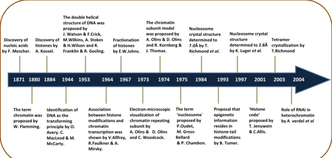
Figure 1: Diagram representing the hallmarks in the history of chromatin studies.
I .2.1 Nobel Prize related to chromatin
Owing to their commendable and unprecedented contribution to the field of chromatin, many scientists were conferred the prestigious Nobel Prize (Table 1).
Table 1: Showing Noble prizes awarded related to chromatin
Year Who Award
1910 Albrecht Kossel (University of Heidelberg)
Nobel Prize in Physiology or Medicine "in recognition of the contributions to our knowledge of cell chemistry made through his work on proteins, including the nucleic substances"
1933 Thomas Hunt Morgan (California Institute of Technology)
Nobel Prize in Physiology or Medicine "for his discoveries concerning the role played by the chromosome in heredity"
1962 Francis Crick, James Watson and Maurice Wilkins (MRC Laboratory of Molecular Biology, Harvard University and London University respectively)
Nobel Prize in Physiology or Medicine "for their discoveries concerning the molecular structure of nucleic acids and its
significance for information transfer in living material"
1982 Aaron Klug (MRC Laboratory of Molecular Biology)
Nobel Prize in Chemistry "for his
development of crystallographic electron microscopy and his structural elucidation of biologically important nucleic acid-protein complexes"
1993 Roberts and Sharp Nobel Prize in Physiology "for their
independent discoveries of split genes" 2006 Roger Kornberg (Stanford
University)
Nobel Prize in Chemistry "for his studies of the molecular basis of eukaryotic
transcription" Fractionation of histones by E.W.Johns. Discovery of histones by A. Kossel. Discovery of nucleic acids by F. Mescher. 1871 1880 1884 1944 1953 1964 1967 1973 1974 1975 1984 1993 1997 2001 2003 2004
The double helical structure of DNA was proposed by J. Watson & F.Crick, M.Wilkins, A. Stokes & H.Wilson and R. Franklin & R. Gosling.
The chromatin subunit model was proposed by A. Olins & D. Olins and R. Kornberg & J. Thomas. Nucleosome crystal structure determined to 7.0Å by T. Richmond et al. Nucleosome crystal structure determined to 2.8Å by K. Luger et al. The term chromatin was proposed by W. Flemming. Identification of DNA as the transforming principle by O. Avery. C. MacLeod & M. McCarty. Association between histone modifications and chromatin transcription was shown by V.Allfrey, R.Faulkner & A. Mirsky. The term ‘nucleosome` proposed by P.Oudet, M. Gross-Bellard & P. Chambon. Proposal that epigenetic information resides in histone-tail modifications by B. Tumer. ‘Histone code’ proposed by T. Jenuwein & C.Allis. Role of RNAi in heterochromatin by A verdel et al Tetramer crystallization by T.Richmond Electron-microscopic visualization of chromatin repeating subunit by A. Olins & D. Olins and C. Woodcock.
28
I .3 Chromatin structure
Chromatin exhibits a repeating structure. The basal repeating unit of chromatin, termed the nucleosome, is formed upon wrapping of two superhelical turns of DNA around an octamer of core histones (two of each H2A, H2B, H3 and H4). This structure provides the first level of compaction of DNA into the nucleus. Interconnected by linker DNA, nucleosomes form 10 nm "beads-on-a-string" filament. Upon addition of linker histones, the "beads-on-a-string" structure coils, in turn, into a 30 nm diameter structure known as the 30nm fiber. Chromatin is organized into functional territories (35) within an interphase nucleus. Long range fiber-fiber interactions finally compact the chromatin to highly condensed metaphase chromosome (Figure 2).
Figure 2: Organization of eukaryotic chromatin fibers. The fundamental unit of chromatin is
defined as nucleosome that forms the “beads-on-a-string” chromatin structure.
Internucleosomal interactions, linker histones and non-histone proteins mediate the further condensation of chromatin into 30nm fibers and higher order structures. Adapted from (36)
Structurally and functionally chromosome has been divided into two distinct domains. Chromosomal regions that do not undergo postmitotic decondensation were termed as heterochromatin by the German botanist Emil Heit in 1928, whereas fractions of the chromosome that decondense and spread out diffusely in the interphase nucleus were referred to as euchromatin (37).
29
I .3.1 DNA
In most living organisms (except for some viruses), genetic information is stored and transferred from a parent to its offspring by the molecule deoxyribose nucleic acid, called DNA for short. Regions of DNA which have the information encoded for giving rise to diffusible products (protein or RNA) are called the Genes. The process of transmitting the information from DNA to form protein via RNA is called central dogma of molecular biology. The proteins that are produced have functional roles in just about every aspect of a living cell. Some proteins play a structural role and others serve as enzymes that regulate many biochemical pathways (anabolic and catabolic) in living organisms.
DNA molecule has a right handed double-stranded helical structure in which the two strands run opposite and intertwined to each other (12). Each helix is a polymer of four basic nucleotides. The polymer backbone is composed of the alternating sugar (deoxyribose)-phosphate units, attached to which are four types of heterocyclic nitrogenous bases namely Adenine, Thymine, Guanine and Cytosine, which hold the two strands together. Adenine from one strand forms two hydrogen bonds with Thymine form the other stand, while as Guanine forms three hydrogen bonds with the Cytosine from the opposite strand. The DNA double helix also has two different-sized "grooves": a major groove and a minor groove (Figure 3). These grooves are binding sites for a wide variety of molecules that affect DNA at the molecular level such as proteins that control such functions as gene expression, regulation, replication and transcription. DNA double helical structure has been classified into three types namely B-DNA, A-DNA and Z-DNA. B-DNA is the most abundant form of DNA commonly found under physiological conditions in a cell. In this structure, the helix makes a turn every 3.4 nm, and the distance between two neighboring base pairs is 0.34 nm. Hence, there are about 10 pairs per turn. In a solution with higher salt concentrations or with alcohol added, the DNA structure may change to an A form, which is still right-handed, but every 2.3 nm makes a turn and there are 11 base pairs per turn. A-DNA forms are present under some biological conditions that are not yet well understood. Another DNA structure is called the Z form, because its bases seem to zigzag. Z-DNA is left-handed and also narrower than the other two types of DNA. One turn spans 4.6 nm, comprising 12 base pairs. The DNA molecule with alternating G-C sequences in alcohol or high salt solution tends to have such structure. Z-DNA has been found in synthetic short segments of Z-DNA.
30
Figure 3: A cartoon of DNA double helix showing DNA major and minor groove.
I .3.2 Nucleosome
Historically, the periodic nature of chromatin was identified by biochemical and electron microscopic studies. The partial digestion of chromatin, isolated from rat liver nuclei, generated fragments of 180-200 base pairs in length which were resolved by electrophoretic migration (38, 39). This regularity of chromatin structure was later confirmed by electron microscope analysis that revealed chromatin as regularly spaced particles or "beads on a string" (21, 24). In parallel, chemical cross-linking analysis permitted the precise determination of the stoichiometry of DNA and histones in the nucleosome to be 1/1 based on their mass (23). Together these observations led to the proposition that the nucleosome was the fundamental unit of chromatin. Pierre Chambon's laboratory was the first to use the term "nucleosome" (24).
Nucleosome is the basic repeating unit of the chromatin, composed of a core particle and a linker region (or inter-nucleosomal region) that joins adjacent core particles. The length of the linker region, however, varies between species and cell type. Therefore, the total length of DNA in the nucleosome can vary with species from 160 to 241 base pairs (40-44). Two copies of each of histone protein H2A, H2B, H3 and H4 are assembled into an octamer that has 146-147 bp of DNA wrapped in 1.65 left-handed super helical turns around it to form a nucleosome core particle (NCP) (26). A complete histone octamer is composed of a central (H3-H4)2 tetramer flanked by two H2A-H2B dimers. The four core histones are small basic
proteins (11 to 16 kDa), which are highly conserved through evolution. Histones induce the structural bending in the major and minor grooves of DNA in a way to compress and narrow
31
them down when they face the octamer and expand the ones facing outside (45). The nucleosome is stabilized by histone-DNA interactions which occur about once every 10 base pairs, resulting in impressive distortion of the DNA helix, and the final structure is stabilized by 116 direct histone-DNA and 358 water-bridged histone-DNA stabilizing interactions (46). The connections between DNA and histones are mainly non-specific and include non-polar interactions with the pentose groups in the DNA, hydrogen bonds to the phosphate groups of DNA, and electrostatic interactions between the positively charged aminogroups of the histones and the negatively charged DNA phosphate backbone (47).
The association of one molecule of linker histone H1 with the NCP yields 167 bp of DNA and thus protects approximately 20 bp of DNA from micrococcal digestion. This structure was originally termed the chromatosome (48). Nucleosomes are connected with one another to form what are called nucleosomal arrays which further fold into less understood 30nm fiber and higher order chromatin structures.
The function of the nucleosome is paradoxical, requiring it to play two opposite roles simultaneously. On one hand, nucleosomes must be stable, forming tight, sheltering structures that compact the DNA and protect it from harm. On the other hand, nucleosomes must be labile enough to allow the information in the DNA to be used. Chromatin modification enzymes must be allowed access to the DNA for functions like replication, repair and transcription. The method by which nucleosomes solve these opposed needs is not well understood, but may involve a partial unfolding of the DNA from around the nucleosome.
I .3.2.1 Nucleosome core particle
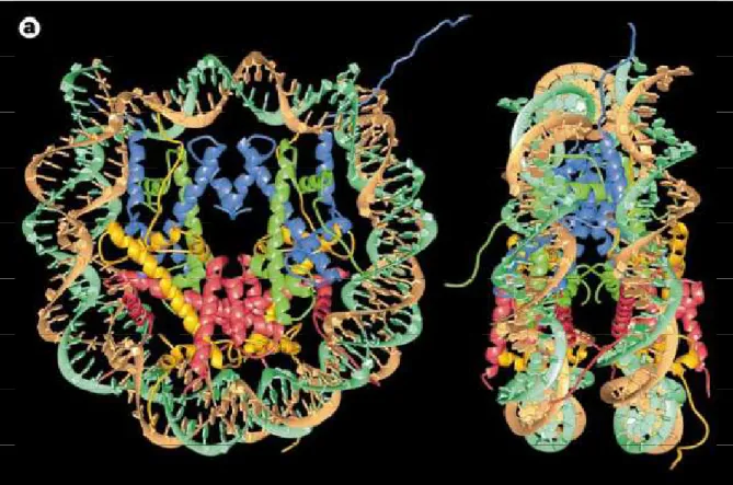
The nucleosome core particle (Figure 4) is the crystallizable substructure of the canonical nucleosome (49), defined by the DNA protection pattern of histone octamer in nuclease digestion of chromatin. The 205 kDa NCP contains two copies of each core histones H2A, H2B, H3 and H4 and 146 base pairs of DNA wrapped in about 1.65 superhelical turns around the histone core. Neutron scattering (50) and low resolution X-ray crystallographic studies (7Å) (51-53) demonstrated the disc shape to the NCP. However the more fine and high resolution structural details were possible only after the solution of 3.1Å structure of the histone core of the nucleosome (26) and 2.8 Å structure of NCP (2), using 146bp X chromosome α-satellite palindromic DNA and heavy atom labeled recombinant histone proteins. A refined structure of NCP using 146 bp α-satellite palindromic DNA and native
32
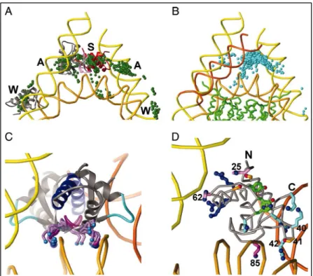
Figure 4: Nucleosome core particle: ribbon traces for the 146-bp DNA phosphodiester backbones (brown and turquoise) and eight histone protein main chains (blue: H3; green: H4; yellow: H2A; red: H2B. The views are down the DNA superhelix axis for the left particle and perpendicular to it for the right particle. For both particles, the pseudo-twofold axis is aligned vertically with the DNA centre at the top. Image adapted from (54)
chicken histone octamer cores demonstrated the asymmetries in the nucleosome core particle at 2.5 Å resolution (49).
The histone octamer looks like a tripartite assembly, when looking straight into the dyad axis, with a central V-shaped (H3-H4)2 tetramer flanked by two flattened balls, the
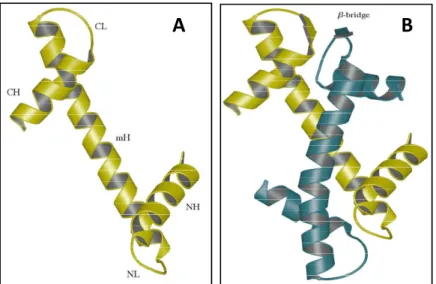
H2A-H2B dimers. The octamer surface has several grooves and ridges, which are set to incorporate the left handed superhelix of the DNA double helix. Individual core histones of the octamer share a common feature of having a symmetrically duplicated (evolutionary) helix-turn-helix motif called ‘histone fold’ motif (Figure 5A), consisting of three helices: a short helix on the N-terminal side of the symmetry center of the fold (NH), the long median helix (mH) and a short helix on the C-terminal side (CH). The helices are joined by loop NL between the NH helix and the mH helix, and loop CL between the median helix and the CH helix. Histone folds mediate histone-histone as well as histone-DNA interactions and account directly for the organization of 147 bp DNA, each fold pair associating with 27-28 bp DNA primarily binding to the phosphodiester backbone as they face the protein.
33
Histones assemble in pairs of heterodimers, in which the two monomers are intimately associated in a head-to-tail manner in a so called "handshake motif' (2, 26) and are stabilized largely through hydrophobic interactions. The short terminal helices are folded back and rotated over the median helix, which lead to interdigitation of the terminal helices and overlapping of the central helices (Figure 5B). This type of structure is stabilized by extensive hydrophobic interactions between the helices. Also there are some other interactions within the heterodimers, which lead to the formation of β bridges between the CL loop of H2A and the NL loop of H2B and between the CL loop of H3 and the NL loop of H4. These bridges form the primary DNA docking sites on the histone surface.
Figure 5: Showing the structure of the histone fold motif of H3 (A), H3-H4 heterodimer showing the two histones interdigitated in a handshake motif (B) Image adapted from (49)
Long median and C-terminal side helices of two H3 from two H3-H4 dimers interact to form a four-helix bundle, which assists the formation of heterotetramer (H3-H4)2. The
tetramer shape resembles a twisted open horseshoe (2, 26, 55) and determines the nucleosome positioning. Tetramer exists as soluble complex at physiological ionic strength solutions and interacts more strongly to the DNA than the H2A-H2B dimer. To complete the octamer two H2A-H2B dimers bind to the two opposite sides of the tetramer to form the tripartite structure. Histones also have a highly basic unstructured amino-terminal domain (‘tail’), which extends from the surface of the nucleosome (56, 57). These histone tails are targets for post-translational modifications, and are important for higher order chromatin structure. These tails, pass through DNA gyres, assume definite conformation once bound to DNA and are reported to be involved in interparticle and linker DNA interactions (58).
34
Histone octamer construction favors the placement of the arginine side chains of the core histones at the places which can fit in the minor grooves of the B-DNA to form a left-handed superhelical ramp with the minor grooves spaced more or less evenly along the ramp.
I .4 Linker histone
Linker histones are the arginine rich, highly diverse group of histones which lack the proper histone fold domain unlike the other core histones. They bind to linker DNA between the NCPs and help to compact and stabilize the higher order chromatin structure. They are constituted of a particular three domain structure: an unstructured N-terminus, relatively conserved central globular winged-helix domain and an unfolded lysine rich C-terminus (59). The linker histone family is highly diverse with at least 11 tissue, stage and species-specific
variants (60-62), which differ in molecular weight, amino acid sequence,
biochemical/biophysical and immunochemical properties (63).
The Linker histone function was studied by several groups. Fan and coworkers have systematically deleted linker-histone genes in mouse embryonic stem cells and generated mice null for H1°, H1a, H1c, H1d, H1e, or H1t, as well as several double mutants of H1 variants. Surprisingly, mice lacking any one of these subtypes develop normally (64-66), whereas the disruption of multiple H1 isoforms leads to embryonic lethality. These results suggest that the lack of a phenotype in single null mice is due to compensation by the remaining subtypes. Careful examination, however, revealed only limited functional redundancy of the variants, since the knockouts of histone H1d (H1.3) or H1e (H1.4) affected age dependent regulation of globin expression (67).
Tetrahymena thermophila has a single linker histone and the Knockout histone H1 strains grow at normal rates (68, 69) and reach near-normal cell densities, arguing that H1 in this organism is not essential for cell survival. However, it is believed that Tetrahymena histone H1 is not the real linker histone as it lacks a globular domain in its structure. On the contrary yeast Saccharomyces cerevisiae has a linker histone, Hho1p, (70) which contains two globular domains, without N and C terminal tails. The Hho1p knockout cell line is viable, although there are detectable alterations in gene regulation. A recent report has also shown Hho1p essential for chromatin compaction in stationary phase of yeast cell cycle (71). Other studies suggest that the linker histone subtypes play differential roles in the control of gene expression (67). Apart from being the main architectural protein of the chromatin and maintaining the chromatin structure, linker histone H1 has been reported to regulate core
35
histone acetylation in vivo (72) and some variants of H1 induce chromatin repression in a tissue specific manner (73). Recently, Konishi and his colleagues demonstrated a role for the linker histone H1c or H1.2 in triggering apoptosis in response to DNA damage (74).
I .4.1 Location of Globular domain on nucleosome
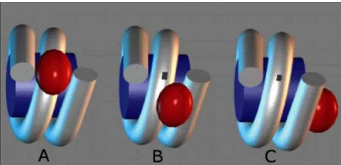
The central conserved winged-helix globular domain (GD) of the linker histone appeared to be internally located in the 30 nm chromatin fiber (75, 76), but its exact position within the nucleosome remains the major controversy in the available data (for review see (77-81). Several models have been postulated in the past 30 years (Figure 6) to explain exact location of globular domain on either native or reconstituted nucleosomal substrate. The very first model was postulated by Allan in 1980 (82), according to which GD binds 10bps
Figure 6: Different models of Globular domain localization on the nucleosome A. Symmetrical model (83), B. Bridging model (84) and C. Unsymmetrical model of (85).
entering and 10bps exiting DNA (linker DNA) of the nucleosome in such a way that it is placed at nearly dyad axis in a symmetrical manner. The model was validated by the GD specific DNase I footprint on nucleosomal dyad (86). This view was however challenged by the asymmetrical GD binding model of An W. and colleagues (87), which proposed that GD protects 20bp of either entering or exiting DNA. This pattern, in turn, probably can lead to directionality or polarity of the chromatin fiber. The polarity of H1 binding to nucleosome may be due to the presence of certain marker sequences found at one end of bulk chromatosomes (88) or due to a conformational change in the NCP (89).
36
Another study utilized the nucleosome positioning sequence 5S RNA from Xenopus
borealis and cross-linked the nucleosomal DNA to the GH5 and proposed a still much
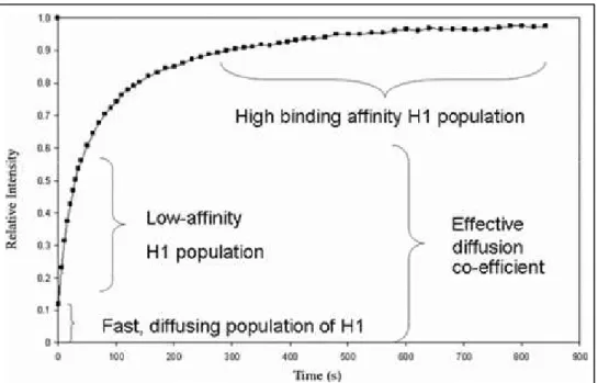
debated model for GH5 binding. According to the findings of this group the GD binds asymmetrically to the nucleosome inside the gyres of DNA at a distance of ∼ 65 bp from the dyad (90) and protects 15bp of DNA from one side and 5bp from other side of NCP (85). This model was objected by the “bridging model” of Zhou et al. (84), according to which GH5 interacts with the dyad and with only one (either the exiting or entering) of the free DNA arms. This model was supported by the findings of in vivo photobleacing experiments and subsequent modeling (28). Fluorescence recovery after photobleaching (FRAP) studies suggested the presence of only two DNA binding sites in globular domain. One of the two binding sites fit within major groove close to the dyad axis and the other within minor groove on the linker DNA in proximity of the NCP (28).
Recently Fan & Roberts (91) described the three binding site model for GH5 binding using the exhaustive rigid molecular docking programs (note that no experimental data were presented in (89)). This model favors the symmetrical binding model for GH5, in which one of the three binding sites contact the nucleosome at the dyad and two others bind symmetrically to the entering and exiting linker DNA (Figure 7).
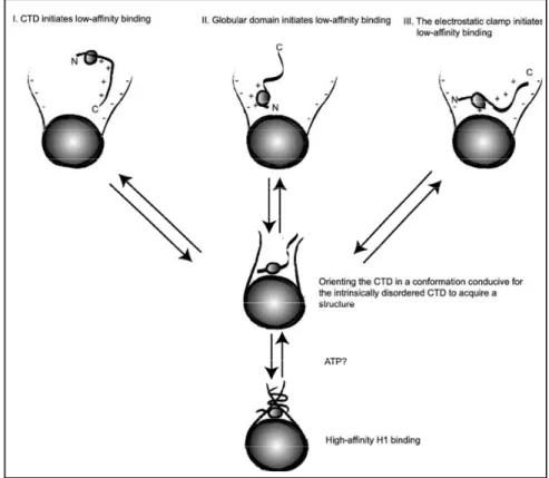
Several reasons could explain the controversial character of the reported data. The above in vitro studies used salt dialysis to deposit histone H1 to the nucleosomes. This would lead to improper assembly of histone H1 (92). In addition, the reconstitution on 5S DNA would result in the formation of nucleosomes exhibiting several translational positioning, which, in turn, would interfere with the mapping of histone H1:nucleosomal DNA contacts (93). The in vivo photobleaching studies could be viewed as indicative for the mapping of H1:nucleosomal DNA interactions, as they provide indirect information .
37
Figure 7: GH5 docked to the nucleosome. (A) Different possibilities to dock GH5 on the nucleosome, which include dockings far from the nucleosome dyad axis, near the dyad axis but contacting only one arm and over the dyad axis. (B) Nucleosome model with one DNA arm bent (orange). The 1,000 top-ranked GH5 solutions are concentrated over the dyad axis. (C) Lys-85 lies in the DNA minor groove at the dyad axis in 27 of the 30 top-ranked solutions or slightly to one side of the phosphate backbone at the dyad axis. (D) Interactions of GH5 side chains with the nucleosome. The top-ranked GH5 solution has Lys-69, Arg-73, and Arg-74 (blue) from helix H3, site I, contacting one arm, with His-25 and His-62 (lavender) and Ser-29 and Ser-71 (gold with red OG) nearby. Lys-85 (magenta) and its wing (site II) are centered in the DNA minor groove at the dyad axis; and Arg-40, Lys-42, Arg-94, and Lys-97 (light blue, right, site III) contact the other DNA arm. The Ser-41 side chain (gold, lower right) extends toward DNA at the dyad axis. The N and C termini of GH5 are indicated. Figure adapted from (91)
I .4.2 Role of the Carboxy terminus of Histone H1
Histone H1 is characterized by a long unstructured C-terminus (~100 aminoacid residues), which has been shown very important for the H1 induced chromatin condensation (82). The C terminus is highly rich in lysine, proline and alanine aminoacids (94). H1 N-terminal deletion protein can stabilize chromatin folding to the same extent as the full-length H1s. However, neither the globular domain alone nor the globular domain plus the N-terminus could facilitate chromatin folding (82, 83). These studies indicate that the ability of linker histones to stabilize chromatin folding resides in the C-terminal domain (CTD) of the protein, and the C-terminal performs its function by shielding negative charges on the DNA backbone. However, simple charge neutralization role of the CTD has been questioned in many studies where deletion of most of the C- terminal did not drastically interfere with the H1 induced fiber condensation (95). In fact specific sub domains have been identified in the CTD which
38
mainly control its functions (95). The main function of CTD, i.e. linker DNA conformation and stabilization and self association of nucleosomal array, has been assigned mainly to the first 25 aminoacids flanking the globular domain.
The sequence of CTD varies among various sub-types of H1 and exhibits different in
vitro DNA and chromatin (96, 97) condensation properties, which do not depend on their
length and charge distribution.
Unstructured in solution (98), CTD can adopt a segmental α-helical conformation upon DNA binding (99). Due to the intrinsic spectroscopic properties of DNA, the structure of the C-terminal bound to DNA cannot be resolved either by CD spectroscopy or Fourier transform infra-red (FTIR) spectroscopy. However organic solvent 2, 2, 2-trifluoroethanol (TFE) has been used (reported to be a genuine replacement for the DNA (100)) to study the secondary structure formation in the truncated C-terminal peptides. Such studies proved the induction of alpha helical structure in these unstructured peptides in TFE. Other studies also suggest CTD peptides acquire α-helical structure in both high salt solutions (101-103) and in presence of DNA (101, 102). CTD of some of the H1 variants also possesses one or more β-turn motif sequence S/TPKK (99-103) which has been reported to bind DNA minor groove and mediate condensation of naked DNA (104-106). The presence of three imperfect octapeptide repeats containing S/TPKK motif in H1d linker histone make it a better DNA condensing linker histone than H1t isotype which completely lack such motifs (97). The DNA compacting ability of H1d was found reduced by 1/3 upon deleting these three imperfect octapeptide repeats (107).
Interestingly the DNA compaction ability was reduced by 70% even if a stretch of 10 aminoacids between two of the repeats was deleted, suggesting that a specific secondary structure motifs in the C-termini is responsible for linker histone dependent DNA compaction (97, 107).
Studies (above described) suggest that there is strong circumstantial evidence that the CTD of H1 might assume a segmented α-helical conformation upon binding to DNA, which in turn will track the phosphate backbone, kinking around the linker DNA by virtue of proline-induced bends or breaks in the helix, following one or other of the grooves (possibly the major rather than the minor groove, since early studies revealed no protection of the minor groove from chemical modification (108)). Alternatively, helical segments of the CTD may lie on the face of the DNA, binding across rather than in the groove, but still kinking around the DNA (99).