HAL Id: inserm-00995588
https://www.hal.inserm.fr/inserm-00995588
Submitted on 23 May 2014
HAL is a multi-disciplinary open access
archive for the deposit and dissemination of
sci-entific research documents, whether they are
pub-lished or not. The documents may come from
teaching and research institutions in France or
abroad, or from public or private research centers.
L’archive ouverte pluridisciplinaire HAL, est
destinée au dépôt et à la diffusion de documents
scientifiques de niveau recherche, publiés ou non,
émanant des établissements d’enseignement et de
recherche français ou étrangers, des laboratoires
publics ou privés.
Severe and early quadriceps weakness in mechanically
ventilated patients
Isabelle Vivodtzev, Andrée-Anne Devost, Didier Saey, Sophie Villeneuve,
Geneviève Boilard, Philippe Gagnon, Steeve Provencher, Mathieu Simon,
Richard Baillot, François Maltais, et al.
To cite this version:
Isabelle Vivodtzev, Andrée-Anne Devost, Didier Saey, Sophie Villeneuve, Geneviève Boilard, et al..
Severe and early quadriceps weakness in mechanically ventilated patients. Critical Care, BioMed
Central, 2014, 18 (3), pp.431. �inserm-00995588�
L E T T E R
Severe and early quadriceps weakness in
mechanically ventilated patients
Isabelle Vivodtzev
1,2,3*, Andrée-Anne Devost
1, Didier Saey
1, Sophie Villeneuve
1, Geneviève Boilard
1,
Philippe Gagnon
1, Steeve Provencher
1, Mathieu Simon
1, Richard Baillot
1, François Maltais
1and François Lellouche
1ICU-acquired weakness has been reported in patients
with prolonged mechanical ventilation [1], leading to
prolonged weaning, poor quality of life after ICU
dis-charge, and high ICU-related cost [2]. Muscle weakness
is the primary manifestation of critical illness
polyneur-opathy or mypolyneur-opathy or both.
Although quadriceps strength has never been
object-ively quantified in the ICU, we previously evidenced
quadriceps muscle weakness by using magnetic
stimula-tion of the femoral nerve in patients with chronic
obstruct-ive pulmonary disease (COPD) and this non-invasobstruct-ive
technique allows a non-effort-dependent assessment of
quadriceps strength [3,4]. Thus, one objective of this
pilot study was to evaluate the feasibility of assessing
quadriceps strength by using this previously validated
technique in sedated patients on mechanical ventilation
at different stages after ICU admission using magnetic
stimulation of the femoral nerve. The study was
approved by the ethics committee of the Institut
Univer-sitaire de Cardiologie et de Pneumologie de Québec
(CER20392). Signed informed consent was obtained from
relatives for all patients.
Quadriceps twitch tension (Twq) assessment was
performed in 13 consecutive sedated and mechanically
ventilated patients with organ failure (Table 1). Twq
measurements were repeated after awakening in nine
patients. Mean Twq was 1.8 ± 1.3 kg for the whole group
of patients. As shown in Figure 1, Twq was two times
lower in ICU patients than in COPD patients (P <0.001)
and four times lower than in healthy subjects (P <0.001).
Furthermore, there was no significant difference in Twq
when patients were sedated or awake. The
reproducibil-ity between these two measurements was good (Figure 2).
* Correspondence:[email protected]
1
Centre de Recherche de l’Institut Universitaire de Cardiologie et de Pneumologie de Québec, Université Laval, 2725 chemin Sainte-Foy, Québec G1V 4G5, Canada
2Univ Grenoble Alpes, Grenoble HP2 38000, France
Full list of author information is available at the end of the article
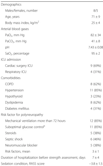
Table 1 Patient characteristics at baseline
Demographics
Males/females, number 8/5 Age, years 71 ± 9 Body mass index, kg/m2 25 ± 4
Arterial blood gases
PaO2, mm Hg 82 ± 34
PaCO2, mm Hg 41 ± 8
pH 7.43 ± 0.08
SaO2, percentage 95 ± 2
ICU admission
Cardiac surgery ICU 9 (69%) Respiratory ICU 4 (31%) Comorbidities COPD 8 (62%) Hypertension 11 (85%) Hypothyroid 3 (23%) Dyslipidemia 8 (62%) Diabetes mellitus 4 (31%) Risk factor for polyneuropathy
Mechanical ventilation more than 72 hours 12 (85%) Suboptimal glucose controla 11 (85%)
Steroids 5 (38%) Septic shock 6 (46%) Neuromuscular blocker 5 (38%) Risk factors, mean 3 ± 1 Duration of hospitalization before strength assessment, days 7 ± 4 Sedation condition, RASS score −3.8 ± 1.5
Data are presented as mean ± standard deviation or as number (percentage).
aSuboptimal glucose control is defined as repeated measurements of capillary
or venous glucose measurements above 10 mmol/L (at least two consecutives). COPD, chronic obstructive pulmonary disease; PaCO2, arterial
pressure in carbon dioxide; PaO2, arterial pressure in oxygen; RASS, Richmond
Agitation-Sedation Scale; SaO2, arterial saturation in oxygen.
© Vivodtzev et al.; licensee BioMed Central Ltd. The licensee has exclusive rights to distribute this article, in any medium, for 6 months following its publication. After this time, the article is available under the terms of the Creative Commons Attribution License (http://creativecommons.org/licenses/by/4.0), which permits unrestricted use, distribution, and reproduction in any medium, provided the original work is properly cited.
Vivodtzev et al. Critical Care
2014
2014, 18:431 http://ccforum.com/content/18/3/431
Strength measurements have been performed in patients
during septic shock (n = 2) or after a dialysis session (n = 2),
and a major reduction of muscle strength (Twq <1 kg) was
observed in these circumstances.
Our results confirm the evidence of early severe
muscle weakness in mechanically ventilated patients
and show that measurement of muscle strength by
magnetic stimulation of the femoral nerve may be
useful in ICU patients, particularly for assessing
re-covery or the effect of therapeutic interventions, as
previously suggested by Ginz and colleagues [5]. A
noteworthy result is that some events (such as dialysis
and sepsis) may modify the muscle strength and need
to be considered when interpreting muscle strength
data in this context. Our data showing that muscle
weakness is an early process in the ICU favor early
treatment to prevent rather than delay treatment to
treat this condition.
Abbreviations
COPD:Chronic obstructive pulmonary disease; Twq: Quadriceps twitch tension.
Competing interests
The authors declare that they have no competing interests. Authors’ contributions
IV and FL contributed to the study concept and design, data analysis, the interpretation of results, and the writing of the manuscript. A-AD, DS, SV, GB, and PG participated in the recruitment of patients, data acquisition, and the writing of the manuscript. SP, MS, and RB participated in the recruitment of patients. FM contributed to the study concept and design and the writing of the manuscript. All authors read and approved the final manuscript.
Acknowledgments
Funding was provided by the Canadian Foundation for Innovation (FRSQ). Author details
1
Centre de Recherche de l’Institut Universitaire de Cardiologie et de Pneumologie de Québec, Université Laval, 2725 chemin Sainte-Foy, Québec G1V 4G5, Canada.2Univ Grenoble Alpes, Grenoble HP2 38000, France. 3Inserm U 1042, Avenue des Maquis du Grésivaudan, Grenoble 38043,
France.
Published: References
1. De Jonghe B, Sharshar T, Lefaucheur JP, Authier FJ, Durand-Zaleski I, Boussarsar M, Cerf C, Renaud E, Mesrati F, Carlet J, Raphaël JC, Outin H, Bastuji-Garin S, Groupe de Réflexion et d’Etude des Neuromyopathies en Réanimation: Paresis acquired in the intensive care unit: a prospective multicenter study. JAMA 2002, 288:2859–2867.
2. Herridge MS, Cheung AM, Tansey CM, Matte-Martyn A, Diaz-Granados N, Al-Saidi F, Cooper AB, Guest CB, Mazer CD, Mehta S, Stewart TE, Barr A, Cook D, Slutsky AS, Canadian Critical Care Trials Group: One-year outcomes in survivors of the acute respiratory distress syndrome. N Engl J Med 2003, 348:683–693.
Figure 1Quadriceps twitch tension (Twq) in ICU patients. Stimulation was applied in ICU mechanical ventilation patients who were sedated (n = 13) or awake (n = 7) (grey), patients with age-related chronic obstructive pulmonary disease (COPD) (n = 18), and healthy subjects (n = 16) (shaded). The ends of the boxes define the 25th and 75th percentiles, and a line at the median and error bars define the 10th and 90th percentiles. *Previously measured in our laboratory [3]. ns, Not significant.
Figure 2Reproducibility of quadriceps twitch tension (Twq) measurements in sedated versus awake conditions. (A) Linear regression between Twq measured in sedated versus awake conditions in mechanical ventilation patients (Spearman coefficient correlation, r = 0.93, P= 0.02). (B) Bland-Altman comparison of sedated and awake Twq measurements. Limits of agreement (reference range of differences) were −1.18 and 0.98 kg. The means bias was −0.13 kg with a standard deviation of 0.47 kg.
Vivodtzev et al. Critical Care Page 2 of 3
23 May 2014 2014, 18:431
3. Saey D, Debigare R, LeBlanc P, Mador MJ, Cote CH, Jobin J, Maltais F: Contractile leg fatigue after cycle exercise: a factor limiting exercise in patients with chronic obstructive pulmonary disease. Am J Respir Crit Care Med2003, 168:425–430.
4. Vivodtzev I, Flore P, Levy P, Wuyam B: Voluntary activation during knee extensions in severely deconditioned patients with chronic obstructive pulmonary disease: benefit of endurance training. Muscle Nerve 2008, 37:27–35.
5. Ginz HF, Iaizzo PA, Urwyler A, Pargger H: Use of non-invasive-stimulated muscle force assessment in long-term critically ill patients: a future standard in the intensive care unit? Acta Anaesthesiol Scand 2008, 52:20–27.
Cite this article as: Vivodtzev et al.: Severe and early quadriceps weakness in mechanically ventilated patients. Critical Care
Vivodtzev et al. Critical Care Page 3 of 3
10.1186/cc13888
2014, 18:431 2014, 18:431