HAL Id: hal-00379714
https://hal.archives-ouvertes.fr/hal-00379714 Submitted on 29 Apr 2009
HAL is a multi-disciplinary open access
archive for the deposit and dissemination of sci-entific research documents, whether they are pub-lished or not. The documents may come from teaching and research institutions in France or abroad, or from public or private research centers.
L’archive ouverte pluridisciplinaire HAL, est destinée au dépôt et à la diffusion de documents scientifiques de niveau recherche, publiés ou non, émanant des établissements d’enseignement et de recherche français ou étrangers, des laboratoires publics ou privés.
Class II histone deacetylases are directly recruited by
BCL6 transcriptional repressor.
Claudie Lemercier, Marie-Paule Brocard, Francine Puvion-Dutilleul, Hung-Ying Kao, Olivier Albagli, Saadi Khochbin
To cite this version:
Claudie Lemercier, Marie-Paule Brocard, Francine Puvion-Dutilleul, Hung-Ying Kao, Olivier Albagli, et al.. Class II histone deacetylases are directly recruited by BCL6 transcriptional repressor.. Journal of Biological Chemistry, American Society for Biochemistry and Molecular Biology, 2002, 277 (24), pp.22045-52. �10.1074/jbc.M201736200�. �hal-00379714�
J. Biol. Chem., Vol. 277, Issue 24, 22045-22052, June 14, 2002
Class II Histone Deacetylases Are Directly Recruited by
BCL6 Transcriptional Repressor
*Claudie Lemercier , Marie-Paule Brocard , Francine Puvion-Dutilleul
§, Hung-Ying
Kao
¶, Olivier Albagli
§**, and Saadi Khochbin
**§§From the INSERM U309, Equipe Chromatine et Expression des Gènes, Institut Albert Bonniot, Domaine de la Merci, 38706 La Tronche Cedex, France, the § CNRS UPR 1983, 7 rue Guy Môquet, BP 8, 94801 Villejuif Cedex, France, and the ¶ Department of Biochemistry, School of Medicine, Case Western Reserve University, the Research Institute of University Hospitals of Cleveland and the Comprehensive Cancer Center of Case Western Reserve University and University Hospitals of Cleveland, Cleveland, Ohio 44106
Received for publication, February 20, 2002, and in revised form, March 28, 2002
Abstract
BCL6 is a member of the POZ/zinc finger (POK) family involved in survival and/or differentiation of a number of cell types and in B cell lymphoma upon chromosomal alteration. Transcriptionalrepression by BCL6 is thought to be achieved in part by recruitinga repressor complex containing two class I histone deacetylases (HDACs). In this study we investigated whether BCL6 could alsotarget members of class II HDACs. Our results indicate that three related class II deacetylases, HDAC4, HDAC5, and HDAC7 can associate with BCL6 in vivo and in vitro. Using electron microscopy, wefound that endogenous BCL6 and class II HDACs partially co-localizein the nucleus. Overexpression experiments showed that BCL6 andHDAC4, -5, or -7 are intermingled onto common nuclear substructures and form stable complexes. A highly conserved domain in the N-terminal region of HDAC5 and HDAC7 as well as the zinc finger region of BCL6 were found necessary for the complex formation in vivo andin vitro. Moreover, our data point to the zinc finger region ofBCL6 as a multifunctional domain which, beside its known capacity to bind DNA, is involved in the nuclear targeting of the protein and in the recruitment of the class II HDACs, and hence constitutesan autonomous repressor domain. Since PLZF, a BCL6 relative, couldalso interact with HDAC4, -5, and 7, we suggest that class IIHDACs are largely involved in the control of the POK transcriptionfactorsactivity.
Introduction
The presence of an N-terminal BTB/POZ (bric à brac, tramtrack, broad complex/pox virus, and zinc finger) domain and C-terminal C2-H2 zinc finger(s) defines the family of POK ((BTB/)POZ andkrüppel-like zinc finger) proteins found in both vertebratesand Drosophila. POK proteins are sequence-specific DNA-bindingtranscription factors, some of them being involved in human oncogenesis.For instance, non-Hodgkin lymphomas are often associated withthe structural alteration, and presumably mis-regulation, of theBCL6 (also named LAZ3) gene encoding a six-zinc finger POK protein (1, 2). Moreover, a few cases of acute promyelocytic leukemiaare associated with the t(11;17) reciprocal translocation thatfuses the retinoic acid receptor to PLZF, a nine-zinc fingerPOK protein. Finally, other POK proteins,
such as HIC-1 (3, 4) and APM-1 (5) have been proposed to act as tumor suppressors in different humanmalignancies.
BCL6 regulates at least lymphocyte (6, 7), myocyte (8, 9), male germ cells (10), and possibly keratinocyte survival and/or differentiation (11). Recently, a set of BCL6 potential target genes has been identified by DNA microarray screening inlymphocytes. BCL6 was found to repress a number of genes involved in B cell activation and terminal differentiation, inflammation, and cell cycle regulation (12). Earlier studies have shaded light on the molecular mechanisms by which BCL6 negatively regulates gene transcription. BCL6 recruits, through multiple contacts,a repressor complex containing both silencing mediator of retinoidand thyroid receptors or its close relative nuclear receptor co-repressor,two vertebrate homologues of the yeast repressor SIN3 (mSINA/B), and two related class I histone deacetylases, HDAC11 and HDAC2 (13-20). However, these co-repressors do not explainall the regulatory properties of the POK proteins. In fact, it appears that POK transcriptional repressors are capable of recruiting other co-repressors. Indeed, C-terminal binding protein mightinteract with Drosophila TTK (21), whereas another unrelatedco-repressor, termed B-CoR, specifically binds to BCL6 in a silencing mediator of retinoid and thyroid receptors/nuclear receptor co-repressormutually exclusive manner (22).
Whereas class I deacetylases (HDAC1,-2, -3, and -8) are closely related to the yeast rpd3
gene product, other HDACs (HDAC4, -5, -6, -7, -9, and -10) have been characterized in vertebrates,and collectively referred to as class II HDACs. HDAC4, -5, -7,and -9 share two regions, each encompassing half of the protein:a C-terminal catalytic domain resembling that of the yeast HDA1 deacetylase and a N-terminal "regulatory" domain (23-26). The organizationof HDAC6 and HDAC10 is atypical. Indeed, both HDAC6 and HDAC10harbor two regions of homology with the class II catalytic domain(23, 24, 27-29). However, whereas HDAC6 contains two activecatalytic domains (24), the second catalytic domain-like regionof HDAC10 does not appear to be active (29). Another featureof all of the class II members is their ability to shuttle ina regulated fashion between the nucleus and the cytoplasm (seeRef.
30 forreview).
The role of class II HDACs in the control of transcription is believed to be important but is only beginning to be analyzed.In particular, although class I and class II HDACs represent two structurally distinguishable families, their functional relationship,beyond their common ability to deacetylate histone in vitro, remainslargely unknown. In this study, we investigated whether POK proteins,such as BCL6, could also associate with class II histone deacetylases. Our data showed that indeed HDAC4, -5, and -7 are recruited byBCL6 in vivo and in vitro. Together with previously publisheddata, these findings indicate that BCL6, as well as other POKproteins, directly and indirectly recruit both class I and classIIHDACs.
Materials and Methods
DNA Constructs-- pcDNA-HA-HDAC5 and HA-HDAC4 (31) and pcDNA-HA-HDAC7 (25)
have been described before. HDAC5, HDAC4, and HDAC7 deletion fragments were constructed by PCR using the high Fidelity Taq/Pwopolymerases mixture (Roche Molecular Biochemcials) and cloned in-frame into pGEX5 plamids (Amersham Biosciences). Human BCL6(LAZ3) cDNA encoding the full-length protein (1-706) or deletionfragments (encoding amino acids 132-706, 501-706, 132-519, 1-626,1-573, and 1-519) were amplified by PCR from the pTL-LAZ3 plasmid (13) without the Flag tag, and subsequently cloned into pcDNA3.1 plasmid (Invitrogen). Gal4 DNA-binding fusion protein were obtained by
subcloning BCL6 fragments into pcDNA-Gal4 DB vector (32). L8G5-Luc and L8-Luc reporter plasmids containing 8 LexA operators with 5 Gal4-binding sites or without Gal4 sites, respectively,as well as the LexA-VP16 activator plasmid (31, 32), havebeen described before. The SV40-based PLZF expression vector (pSG5-PLZF) is a generous gift of Dr. Koken (33). For producing the proteinsin baculovirus, BCL6 cDNA encoding the zinc finger region (aminoacids 501-706) was cloned into the pBacPAK9 vector (CLONTECH),in-frame with a histidine tag at the Cterminus.
Antibodies-- The antibodies used were: anti-BCL6 N3 or C19 rabbit antibodies (Santa Cruz
Biotechnology) or anti-Flag M2 mouse antibody (Sigma-Aldrich) to detect BCL6; anti-HA Y11 rabbit antibody (Santa-Cruz Biotechnology) or 3F10 anti-HA rat antibody (Roche Molecular Biochemicals)to detect the HA-tagged transfected HDACs; anti-HDAC4 N18 and anti-HDAC5 P16 goat polyclonal antibodies (Santa Cruz Biotechnology) to detect endogenous class II HDACs; anti-PLZF mouse antibody (33) and anti-Gal4DB (RK5C1, Santa Cruz). For ultrastructural studies in UTA-L cells, secondary antibodies were gold-labeled goat anti-rabbit immunoglobulin (IgG) and goat anti-mouse IgG (British Biocell International Ltd., Cardiff, UnitedKingdom).
Cell Culture, Transfections, and Reporter Gene Assays-- Human U2OS osteosarcoma-derived
UTA-L (34) cells and human lung carcinoma A549 cells were grown in "growth medium," that is a 50/50 mixture of Dulbecco's modified Eagle's/Ham's F-12 medium (Invitrogen) containing 10% fetal calf serum (Sigma-Aldrich) and antibiotics. For UTA-L cells, growth medium was supplemented with tetracycline (Sigma-Aldrich, 2 µg/ml), G418 (Invitrogen, 500 µg/ml),and puromycine (Sigma-Aldrich, 1 µg/ml). To induce BCL6 expression,UTA-L cells were rinsed twice with growth medium, then trypsinized, centrifuged, and plated in growth medium. Transfections of bothUTA-L and C2 cells were performed using 1.5 µg of the appropriateexpression vector(s) and 3 µl of FuGENE 6 (Roche Molecular Biochemicals) according to the supplier's instructions. Induction of BCL6 expression in UTA-L cells was performed 24 h before transfection with any transiently transfected HDACs. Note that the expression of BCL6 continues during the transfection and the subsequent culture, as these steps are also performed in a tetracycline-free medium.HeLa cells were grown in a standard growth medium (Dulbecco'smodified Eagle's, 10% fetal calf serum, 2 mM L-glutamine, and
antibiotics) and transfected as described before (31). Typically, 20 ng of pcDNA-Gal4 DB plasmids, 400 ng of L8G5-Luc or L8-luc reporter plasmid, 100 ng of LexA-VP16 activator plasmid, and 100ng of pCMV- (CLONTECH) were used in each transfection. Cellsextracts were prepared 24 h post-transfection and processed forluciferase (Luciferase Assay System, Promega) and -galactosidase (Luminescent -gal detection kit, CLONTECH) assays. Luciferaseunits were normalized according to the -galactosidasevalues.
Immunofluorescence and Confocal Microscopy Analyses-- Immunofluorescence analyses
were performed as previously described (34). Briefly, 24 h after transfection, cells on a 3.5-cmdishes were rinsed with PBS, fixed in formaline (Sigma-Aldrich)for 10 min, washed in PBS, permeabilized in a 0.25% Triton, 50mM NH4Cl solution in PBS for 10 min, rinsed in PBS, and exposedfor 1 h at room temperature to the primary antibodies dilutedat 1/100 in a PBS, 0.2% gelatin solution (except for M2 anti-Flagwhich was diluted at 1/800). Cells were then rinsed in PBS, 0.2%gelatin and exposed to the appropriate secondary antibodies, allwere purchased from Molecular Probes: goat anti-rabbit Alexa 488plus goat anti-mouse Alexa 594 (Figs. 2B and 5C) and goat anti-ratAlexa 488 plus goat anti-rabbit Alexa 594 (Fig. 6E). The disheswere then rinsed with PBS and mounted in Mowiol (Merck) to beanalyzed by a
laser scanning microscope (Leica) using a ×63 objective.The images were finally processed with Adobephotoshop.
Electron Microscopy-- Non-induced and 24-h induced UTA-L cell cultures were transfected
with an expression vector encoding the desired HDAC andprepared for electron microscopy as previously described (35). Briefly, cell cultures were fixed 24 h after transfection with formaldehyde, dehydrated in methanol, and embedded in Lowicryl K4M. Ultrathin sections were collected on Formvar carbon-coatedgold grids. For the detection of the overexpressed HDACs (in UTA-Lcells), grids were incubated in the presence of the Y11 anti-HAantibody (at 1/10 dilution in PBS for 1 h) and anti-rabbit IgGconjugated to 10-nm gold particles (at 1/25 dilution in PBS for30 min), successively, prior to being stained with either uranylacetate alone or by the EDTA regressive staining method, which preferentially bleaches the condensed chromatin. BCL6 was detected either with the C19 anti-BCL6 or M2 anti-Flag antibodies. Forthe simultaneous detection of overexpressed BCL6 and HDACs (in UTA-L cells), grids were floated for 1 h on a mixture of the M2 anti-Flag and Y11 anti-HA antibodies, each at 1/10 dilution in PBS, then for 30 min over a mixture of anti-mouse IgG and anti-rabbitIgG conjugated to gold particles of different size (5 and 10 nm).Co-detection of the endogenous BCL6 and either HDAC4 or HDAC5was performed on human A549 cells and mouse C2 cells as follows.Grids were incubated successively with BCL6 C19 antibody (1/10in PBS for 1 h), HDAC4 (N18) antibody (1/10 in PBS), HDAC5 (P16)antibody (1/10 in PBS), and finally, a mixture of gold-labeledantibodies consisting of 6-nm donkey anti-rabbit antibody and12-nm donkey anti-goat antibody (Jackson Immunoresearch Laboratories,Inc., West Grove, PA), each diluted 1/20 in PBS with 1% bovineserum albumin and 1% Triton X-100.
Electrophoretic Mobility Shift Assays-- Electrophoretic mobility shift assays were performed
essentially as described before (32) except that the gels were preparedand run in 0.5 × TBE (44.5 mM Tris-HCl, pH 8.0, 44.5 mM boricacid, 1 mM EDTA). Oligonucleotides were end-labeled with [ -32P]ATP and T4 polynucleotide kinase. After removal of unincorporated nucleotides, the sense and antisense strands were annealed and used as a probe. The 20-bp BCL6-binding site used in this study(5'-GAAAATTCCTAGAAAGCATA-3') was described before (36).
Co-immunoprecipitation and Western Blotting-- Cell extracts were prepared from frozen
pellets of UTA-L cells co-transfected with pcDNA-HDAC5 (or HDAC4 or HA-HDAC7) and pcDNA-BCL6 or pcDNA3 empty vector. Proteins were extracted in 0.5% Nonidet P-40, 20 mM Tris-HCl, pH 8, 150 mM NaCl, 1 mM phenylmethylsulfonyl fluoride, 10% glycerol, 1 mM EDTA for 20min on ice. Cell debris was removed by centrifugation for 10 minat 12,000 × g and the soluble material was submitted to immunoprecipitationwith the C19 anti-BCL6 polyclonal antibody for 1 h at 4 °C. Theimmune complexes were precipitated with Sepharose-protein G (AmershamBiosciences) for an additional hour at 4 °C. After four washesin lysis buffer and two in PBS, the bound proteins were elutedin SDS-PAGE sample buffer and analyzed by Western blotting. Blots were subsequently probed with primary antibodies (C19 anti-BCL6or 3F10 anti-HA antibodies) and secondary antibodies conjugated to peroxidase, before being detected by chemiluminescence (ECL+,AmershamBiosciences).
In Vitro Protein-Protein Interaction Assays-- In vitro translated proteins were produced from
pcDNA3 plasmids using T7 RNA polymerase, [35S]methionine (ICN), and the TNT rabbit reticulocyte lysate system(Promega). GST fusion proteins were produced in Escherichia coli
glutathione-agarose beads according to the supplier's instructions (Amersham Bioscience). GST pull-down assays were performed essentially as described before (31). For direct protein interaction, the BCL6 zinc finger region (amino acids 501-706)fused to a histidine tag was produced in baculovirus using the BacPAK Baculovirus Expression System (CLONTECH). The protein was then purified from insect cell extracts by nickel affinity column (Ni-NTA-agarose, Qiagen), eluted with 250 mM imidazole and finallydialyzed against 20 mM Tris-HCl, pH 7.5, 10% glycerol, and 1 mM dithiothreitol. The purified protein was used in GST
pull-downexperiments as above and bound BCL6 was detected by Western blotwith C19 anti-BCL6antibody.
Results
Partial Association of Endogenous BCL6 and Class II HDACs-- In a first attempt to examine
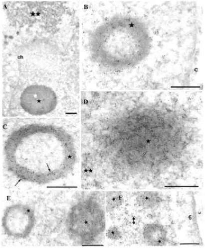
whether BCL6 and class II HDACs could associate in vivo, we used immunogold labeling and electronmicroscopy (EM) to co-detect these proteins in human A549 lungcells which express endogenous BCL6, HDAC4, and HDAC5 mRNA (datanot shown). EM analyses indicated that endogenous HDAC4 or endogenous HDAC5 (12 nm gold particles) as well as endogenous BCL6 (6 nm-gold particles) were detected as individual foci in the nucleus of these cells (Fig. 1, A, B, D, and E). Interestingly, some clusters showed an association or juxtaposition between the endogenousBCL6 and either endogenous HDAC4 (Fig. 1, A and B,
arrow) or endogenous HDAC5 (Fig. 1E, arrow). These data indicate that HDAC4 and
HDAC5molecules are entrapped within or juxtaposed to some foci of BCL6in A549 cells. Note, however, that this co-localization is partial as our results also showed that class II HDACs and BCL6 can alsobe independently engaged into distinct complexes in these cells (Fig. 1D). In mouse C2 myocytes, which also express BCL6 and classII HDACs (Ref. 8 and data not shown; see also, Refs. 37-39),a similar partial co-localization was observed between endogenousBCL6 and endogenous HDAC4 (Fig. 1C and data not shown). We concludethat, at physiological levels of expression, a fraction of theclass II HDAC molecules are associated
with BCL6 in at least two cell lines.
Co-localization of BCL6 with HDAC4, -5, and -7 in a BCL6-inducible Cell Line-- To search
for a potential interaction between BCL6 and class II HDACs and because of their low expression level in various cell lines, we next used a cell line stably transfected with an inducible BCL6 expression vector (UTA-L cells). In these cellsa flagged-version of BCL6 is under the control of a tetracycline-sensitivepromoter (34). Upon tetracycline removal, BCL6 protein expressionis induced within 24 h as shown on Fig. 2A, and is functional as revealed by electrophoretic mobility shift assays using a BCL6-target sequence (Fig. 2B). We next examined the subcellular distribution of BCL6 and class II HDACs. To this end, induced (BCL6 expressing) UTA-L cellswere transiently transfected with a vector encoding HA-taggedHDAC4, -5, or -7, and subcellular localization of these proteinswas analyzed by scanning confocal microscopy. In the three cases,we observed a nearly complete coincidence between the two stainings. In almost all the HDAC4-transfected cells, both proteins were found to co-localize onto typical BCL6 nuclear bodies (Fig. 2C,top), whereas a few cells also displayed co-staining onto cytoplasmicinclusions (data not shown). When HDAC5 and BCL6 were co-expressed, the two proteins were concentrated onto common nuclear subdomains (Fig. 2C, middle). Finally co-expression of HDAC7 and BCL6 alsoshowed a complete co-localization between the two proteins (Fig.2C, bottom).
We next confirmed and refined these findings upon EM. Indeed, HA-HDAC4 could be detected in cytoplasmic inclusions (Fig. 3A,double star) as well as in, and sometimes around, typical BCL6 nuclear bodies (Fig. 3, A and B, star). In the presence of BCL6 (Fig. 3C), HDAC5 (arrows) was also present, both in the BCL6 bodies(star) and in their interior. Upon higher HDAC5 expression (Fig.3D and data not shown), both HDAC5 and BCL6 stainings were co-distributed within nuclear inclusions (double star) indistinguishable from those formed when HDAC5 was expressed alone. In such nuclei, BCL6bodies (star) were found enclosed within HDAC5 nuclear inclusionsand both structures were intensely co-labeled with the anti-Flagand anti-HA antibodies (Fig. 3D). Finally, both HDAC7 and BCL6proteins also co-localized either onto "free" BCL6 bodies (star, Fig. 3E) or onto BCL6 bodies (stars) embedded within larger nuclearinclusions (double star) also containing both proteins (Fig. 3F
and data not shown).
In summary, when overexpressed with BCL6, the nuclear fraction of HDAC4, -5, and -7 was primarily targeted to BCL6 bodies. Upon higher expression levels, HDAC5 and HDAC7 formed nuclearinclusions, each presumably arising from a BCL6 body and fusingwith each other to form larger inclusions containing both BCL6 bodies as well as dispersed BCL6 molecules.
BCL6 and Class II HDACs Form Stable Complexes in Vivo-- The association between BCL6
and class II HDACs staining observed by both confocal and electron microscope analyzes suggestedthat they could form stable complexes in vivo. To directly testthis hypothesis, we transiently co-transfected HA-HDAC4 or -5or -7 with BCL6 in non-induced UTA-L cells. An anti-BCL6 antibodywas then used to immunoprecipitate BCL6-containing complexes,and the potential presence of associated HA-tagged HDACs was examined.Western blot analysis of the immunoprecipitated materials, using an anti-HA antibody, showed that the three HA-HDACs were co-immunoprecipitatedwith BCL6, indicating that they are capable of forming a stablecomplex with BCL6 in vivo (Fig. 4, HDAC5 panel, lane 4; HDAC4panel, lane 8, and
HDAC7 panel, lane 12). Under the same conditions, when expressed alone, neither of the
three HA-HDACs was immunoprecipitatedwith the anti-BCL6 antibody (Fig. 4, HDAC5, -4, and -7 panels,lanes 3, 7, and 11, respectively). Moreover, parallel co-immunoprecipitation with an irrelevant antibody (anti-Gal4) failed to show the presenceof any of the three HDACs (data not shown). We conclude that classII HDACs not only co-localize with BCL6, but are also capable of forming stable complexes with this transcription factor in vivo.
Delineation of the BCL6-binding Site on HDAC5 and HDAC7-- To precisely determine the
region involved in the interaction between the class II HDACs and BCL6, a GST pull-down approachwasundertaken.
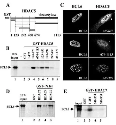
Different fragments of HDAC5 were fused to GST (Fig. 5A), and the respective purified proteins were used to monitor their ability to interact with in vitro translated 35S-labeled BCL6. A fragment of HDAC5 containing most of the N-terminal non-deacetylase region of the protein (amino acids 123-673) efficientlyinteracted with BCL6 (Fig. 5B, lane 4). The N-terminal end ofthe protein encompassing the first 122 amino acids as well asthe deacetylase domain alone (674-1113) did not efficiently interactwith BCL6 under the same conditions (Fig. 5B, lanes 3 and 5, respectively). We then precisely mapped the N-terminal BCL6-binding domain of HDAC5 using four smaller fragments covering this 123-673 region of interaction with BCL6 (Fig. 5A). This fine mapping allowedthe identification of the region
123-292 of HDAC5 as the minimal interaction domain with BCL6 (Fig. 5B, lanes 6-9). To evaluate the in vivo relevance of our in vitro interaction domain mapping, we examined the ability of different HDAC5 mutants to co-localize with BCL6 in cells by immunofluorescence analyses.Consistent with our mapping data, Fig. 5C shows that the N-terminal region of HDAC5 (123-673, top panel) almost systematically co-localized with BCL6, as did the full-length HDAC5. Under the same conditions, the isolated deacetylase domain (674-1113) showed no association with BCL6 in most of the cells (middle panel). Moreover, the in vitro defined "minimal" BCL6-binding site (HDAC5-(123-292), bottom
panel) clearly kept the ability to co-localize with BCL6, albeitperhaps less efficiently than the
HDAC5-(123-673)derivative.
Because both HDAC4 and HDAC7 share extensive sequence homology with HDAC5, and also associate with BCL6 in vivo, we investigatedthe possibility of an interaction between BCL6 and the N-terminal regions of HDAC4 and HDAC7. These two regions (HDAC4-(1-650) andHDAC7-(1-506)) were fused to GST and the purified proteins wereindeed shown to associate with 35S-labeled BCL6, whereas no binding was obtained with an equivalentamount of the GST protein alone (Fig. 5D). We concluded that BCL6 interacts with all the related class II HDACs in vitro.
Finally, a BCL6 minimal binding site in HDAC7 was also determined by GST pull-down. It showed that the most N-terminal domain of HDAC7-(1-254) efficiently interacted with the full-length BCL6(Fig. 5E), whereas neither the histone deacetylase region (500-938)nor the central part of the protein (241-533) retained BCL6. Adetailed analysis of the sequence of the minimal BCL6-interactingdomain defined above showed it to encompass the most conserved region in the N-terminal non-deacetylase domain of HDAC4, -5,and -7 (notshown).
Delineation of the BCL6 Regions Involved in the Interaction with HDAC5-- Reciprocally, we
next mapped the HDAC5-binding site of BCL6. To this end the ability of 35S-BCL6 deletion mutants (Fig. 6A) to interact with GST-HDAC5-(123-673) was tested in GST pull-down assays. As expected, full-length BCL6efficiently interacted with the HDAC5 N terminus in this assay (Fig. 6B, panel 1). Interestingly, BCL6 constructs lacking either the BTB/POZ domain (BCL6-(132-706), panel 2) or only containingthe ZF region of the protein (BCL6-(501-706), panel 3) were ableto associate with GST-HDAC5. Similar results were obtained withGST-HDAC7 (Fig. 6C, panels 1-3). These data pointed to an unexpectedrole for the ZF
region in the recruitment of class II HDACs.
The BCL6 ZF region is composed of six C2-H2 krüppel-like zinc fingers (Fig. 6A, fingers 1-6). To further refine our mapping,BCL6 deletion mutants were prepared lacking 2, 4, or all the 6ZFs. Removal of the two most C-terminal zinc fingers (fingers 5 and 6) did not significantly affect HDAC5 or HDAC7 binding (Fig. 6, B and C, panel 4). The additional deletion of the next twofingers (3 and 4) led to a drastic reduction in the capacity ofBCL6 to interact with both HDAC5 and HDAC7 (BCL6 1-573, Fig. 6,B and C, panel 5). Fingers 3 and 4 therefore appear essential for the binding of these two histone deacetylases by BCL6. Moreover, the residual interaction observed in the total absence of zinc fingers is totally eliminated when the BTB/POZ domain was deleted(Fig. 6B, compare BCL6(1519) and -(132-519)), suggesting thatthe BTB/POZ domain represents also a minor site of interaction withHDAC5.
Although the in vitro data described above suggested a direct interaction between BCL6 and the class II HDACs, an indirect interaction because of the activity of proteins present in the reticulocyte lysates was possible. To rule out this possibility,we set up a baculovirus-based expression and purification of the ZF region of BCL6 (Fig. 6D, left panel). The purified protein was then incubated with purified bacterially expressed GST-HDAC5 fusions, containing or not the BCL6-binding sites, and immobilizedon glutathione beads. The HDAC5 (123-292) region, shown to interact with BCL6 in reticulocyte lysate, efficiently interacted withthe purified BCL6 ZF fragment as well (Fig. 6D, right, upper panel,lane 3). Under the same conditions, the fragment 451-673 of HDAC5 did not bind the BCL6 ZF, although equivalent amounts of GST fusion proteins were used in this assay (Fig. 6D, right, lower
panel).These data clearly showed that the N-terminal region of HDAC5can directly bind to
the ZF region of BCL6.
To gain access to the in vivo relevance of these in vitro mapping, we tested the ability of different BCL6 constructs to recruit HDAC5 in cells. UTA-L cells were transiently co-transfected with HA-HDAC5 cDNA and plasmids encoding full-length BCL6 or mutants lacking either the two (BCL6-(1-626)) or the four C-terminal zincfingers (BCL6-(1-573)). As expected, full-length BCL6 co-localizedwith HDAC5 in nuclear bodies and inclusions upon co-transfection(Fig. 6E, top). Interestingly, BCL6-(1-626) mostly localized inthe cytoplasm, where it formed bodies retaining a large portion of the HDAC5 pool (Fig. 6E, middle). Surprisingly, the further removal of the two adjacent zinc fingers (BCL6-(1-573)) led to a diffuse distribution of the protein in the cytoplasm. In contrastto 626), the BCL6-(1-573) derivative did not efficientlyretain HDAC5 in the cytoplasm, as HDAC5 recovered its "usual" distribution, being predominantly observed in the nuclei of these cells (Fig. 6E,
bottom).
Taken together, these data confirmed the results obtained in vitro as they showed the role of the N-terminal regions of bothHDAC5 and -7, and of the ZF region of BCL6, as essential interactioninterfaces in cells, and suggested that zinc fingers 3 and 4 ofBCL6 are particularly involved in this interaction. The fact thatthe deletion of the regions necessary for the in vitro
interaction, both in BCL6 or HDAC5, impaired their co-localization, is consistent with the idea that direct contacts are indeed important in vivo for the two proteins to associate. In addition, these results revealed that the last two zinc fingers of BCL6 are required forthe nuclear targeting of thisprotein.
The Zinc Finger Region of BCL6 Mediates Transcriptional Repression-- Data presented
above showed that the zinc finger region of BCL6 is involved in the recruitment of HDAC5 and HDAC7. It is therefore expected that the BCL6 ZF region repress transcription, when targeted to a promoter. To test this hypothesis, we targeted the BCL6 ZF region into a promoter containing GAL4-binding sites. The reportersystem contained eight copies of the binding sites for LexA immediatelyadjacent to five copies of the binding site for GAL4, all clonedupstream from a luciferase gene (Fig. 7A). In the presence ofLexA-VP16 fusion co-activator and the GAL4 DNA-binding domainalone (GAL4-DB), this reporter was activated to high levels of expression (Fig. 7B, +LexA-VP16). An expression vector was prepared expressing a fusion protein containing the ZF region of BCL6 (sixfingers, amino acids 501-706) fused to the GAL4 DNA-binding domain. Co-expression of LexA-VP16 and GAL4-BCL6-(501-706) showed that the ZF region could efficiently inhibit the transcriptional activity of LexA-VP16 (Fig. 7B, DB-BCL6 501-706 construct). Interestingly,the removal of the zinc fingers involved in the interaction with class II HDACs almost abolished the repressive effect of the domain.In control experiments, the expression of these GAL fusion
proteins had no effect on a promoter lacking the GAL4-binding site (Fig. 7B, L8-Luc
reporter). Since the ZF region of BCL6 could efficientlyrecruit HDAC5 in vitro and in vivo
(Fig. 6), we wanted to investigate the role of this HDAC activity in the ZF-mediated transcriptional repression described above. The same experiments as above wereperformed but cells were treated with HDAC inhibitor TSA 12 hbefore measurement of the luciferase activity. Surprisingly, therepressive activity of the ZF region of BCL6 was not abolishedby TSA treatment, suggesting that, besides HDACs, other types of co-repressors could also participate in the repressive activity of this region of BCL6 (not shown).
Interaction of Class II HDACs with PLZF, Another POK Family Member-- Finally, we
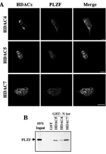
examined whether our observations could be extended to other (BTB/)POZ and krüppel-like zinc finger (POK) proteins. PLZF is another POK transcriptional repressor showing both functional similarities and association with BCL6 (Ref. 33 and references therein). Upon transient co-expression of both PLZF and classII HDACs in cells, an obvious co-localization of PLZF with HDAC4, -5, or -7 (Fig. 8A) was observed. Moreover, like BCL6, PLZF was able to interact directly with the N-terminal region of HDAC5,-4, and -7 in GST pull-down assays (Fig. 8B), suggesting thatthe class II HDACs interact with another POK factor at least and may therefore play a general role in regulating the function of these proteins.
Discussion
The data presented in this report show that BCL6 and PLZF harbor the capacity to recruit three related members of the class II HDACs, HDAC4, -5, and -7, and indicate that this recruitment relies, at least in part, on a direct interaction. A detailed investigation of these interactions revealed interesting propertiesof BCL6 and the studiedHDACs.
BCL6-Zinc Finger Region Is a Multifunctional Domain-- Two domains of BCL6 are involved
in the interactions with the class II HDACs: the N-terminal BTB/POZ domain and the C-terminal ZF region. A BCL6 derivative lacking the four C-terminal zinc fingers is severely impaired in its ability to bind HDAC5 and-7 in vitro and to co-localize with HDAC5 in vivo. Moreover, the deletion of both the entire ZF region and the BTB/POZ domain totally abrogates the interaction in vitro. Finally, the isolated ZF region of BCL6 is sufficient to directly interact with HDAC5 and -7 invitro, and exhibits an autonomous repressive activity when targeted to a promoter in vivo. In addition to its already characterized DNA binding capacity, all these results suggest a role for the ZF region as an important heteromerization interface with class II HDACs. The ZF region of POK proteins therefore appears as a multifunctional domain, mediating specific interaction with DNA as well as with various protein partners, including class II HDACs.Indeed, the recruitment of a co-repressor by the C2-H2 zinc fingerDNA-binding region of the CTCF transcription factor has been recently reported (40). Moreover, a deletion of the ZF region of theZF5 POK protein has been shown to eliminate its ability to self-interact(41), whereas the ZF region of both BCL6 and PLZF were foundto be involved in their heteromerization (33). Likewise, theZF region of PLZF mediates its association with PML, the major translocation partner of RAR in acute promyelocytic leukemia(42, 43).
A Limited Region of HDAC4, -5, and -7 Targets Transcription Factors-- Regarding the
BCL6-interacting region of class II HDACs, we identified their conserved non-catalytic N-terminal domain tobe necessary and sufficient for the interaction with BCL6 in vitroand for co-localization in cells. This region is present in HDAC4,-5, and -7, as well as in the recently
cloned HDAC9 (26) andin MITR (44, 45), suggesting that these last two proteinsprobably also interact with BCL6. These data emphasize the importance of this domain in protein/protein interactions. Indeed, this domain has already been shown to bind MEF2 transcription factors as wellas, at least for MITR, HDACs from both class I and class II and the CtBP co-repressor, indicating how the isolated HDAC5 N-terminal domain, or MITR, which lacks a catalytic deacetylase region, arecapable of transcriptional repression (31, 44,
46). Moreover, this domain contains three conserved serines, two of them being possibly phosphorylated by the calcium/calmodulin-dependent protein kinases (CaMK). A regulated phosphorylation of these serines seems to control the interaction of these HDACs with partners as wellas their intracellular localization (37, 47-52). Our data suggestthat BCL6 may interfere with the nuclear/cytoplasmic shuttling of class II HDACs. For instance, HDAC4 appeared cytoplasmic in most cells when overexpressed "alone," but it became almost systematicallydetectable in the BCL6 nuclear bodies when overexpressed in inducedUTA-L cells. Moreover, mapping experiments with either HDAC5 and-7 derivatives further indicated that the first half of theirN-terminal domain is necessary and sufficient to bind BCL6. This highly conserved subregion also encompasses the binding site for the MEF2 transcription factors (31, 48, 49, 52-54). These findings suggest a cross-talk between BCL6 and the CaMK/HDACs/MEF2 pathway. Interestingly, like MEF2 transcription factors and presumably class II HDACs (39), BCL6 positively controls myogenesis as BCL6-deficient myocytes are more prone to undergo apoptosis uponserum withdrawal, possibly because they are impaired in theircapacity to correctly arrest proliferation and/or to terminallydifferentiate (9).
BCL6 Recruits Multiple HDACs Directly or Indirectly-- HDAC4, -5, and -7 C-terminal
regions bind silencing mediator of retinoid and thyroid receptors/nuclear receptor co-repressor (25, 55) and, at least for HDAC7, mSIN3A (25). All these co-repressors were previously found to also bind BCL6 (13, 16,20). Moreover, B-CoR, a novel co-repressor interacting with the BCL6 BTB/POZ domain has been shown to associate with classII HDACs in vivo (22). Thus, BCL6 appears capable of interactingwith class II HDACs both directly through its C-terminal ZF region(and to a lesser extent, its BTB/POZ domain) and indirectly byrecruiting several co-repressors through its N-terminal half (16,22). The possibility of multiple, direct, and indirect, contacts between BCL6 and class II HDACs parallels the situation of BCL6-class I HDACs interaction (16, 20). It could both confer more stability to the DNA-bound repressor complex(es) and increase the regulatorypotential of the complex by broadening its linkage to distinctsignalingpathways.
What Could Be the Role of the BCL6-HDACs Interaction?-- An obvious possibility for the
role of BCL6-HDACs interaction is that BCL6 uses HDACs to exert transcriptional repression by local chromatin remodeling when targeted to specific promoters. It is also interesting to consider another hypothesis based on the ability of BCL6 and associated molecules to form specific "molecular reservoirs" allowing the assembly and reversible storageof class I and class II HDAC-containing regulatory complexes.In this respect, it is noteworthy that DNA replication has beenfound to progress on the periphery BCL6 nuclear core (35). BCL6-associated proteins could therefore be, at least in part, either depositedat specific promoters during DNA replication to maintain or alterthe local chromatin structure, or involved in a broader functionin chromatin maturation, especially the histone deacetylation that follows their association to the newly replicated DNA (56, 57). Finally, beyond the repression, another possibility would be that BCL6 could itself be a substrate of (at least some) itsassociated HDACs, which could thereby modulate its function. Thereport that the
acetyltransferase p300 may both acetylate BCL6 and regulate its repressive activity (58) provides support tothishypothesis.
Acknowledgements
We are grateful to Dr. J-J. Lawrence for encouraging this work, Dr. S. Rousseaux for the critical reading of the manuscript,Dr. M. Callanan for helpful discussion, and G. Géraud and C.Chamot (Service d'Imagerie of the Institut Jacques Monod, Universityof Paris VI/VII) for precious help in confocal microscopy analyses.We also thank S. Souquere-Besse, E. Pichard, and S. Curtet-Benitski for technical assistance, and M. Koken for the gift of the pSG5-PLZF expression vector and anti-PLZF antibody. Y. Kao wishes to thankDr. R. Evans for support and encouragement. Part of this workhas been carried out in R. Evan'slaboratory.
Footnotes
* This work is supported by grants from CNRS, INSERM, Association pour la Recherche contre le Cancer (ARC), Ligue Nationale contre le Cancer and Groupement des Entreprises Françaises etMonégasques dans la lutte contre le Cancer (GEFLUC).The costsof publication of this article were defrayed in part by the paymentof page charges. The article must therefore be hereby marked "advertisement" in accordance with 18 U.S.C. Section 1734 solely to indicate thisfact.
James T. Pardee-Carl A. Gerstacker Assistant Professor of CancerResearch. ** Both authors contributed equally to thiswork.
To whom correspondence may be addressed. Tel.: 71; Fax: 33-1-49-58-33-81; E-mail: albagli@vjf.cnrs.fr.
§§ To whom correspondence may be addressed. Tel.: 74; Fax: 33-4-76-54-95-95; E-mail: khochbin@ujf-grenoble.fr.
Abbreviations
The abbreviations used are: HDAC, histone deacetylase; BCL6, B cell lymphomas 6; BTB/POZ, bric à brac, tramtrack, broad complex/pox virus and zinc finger; PLZF, promyelocytic leukemia zinc finger; GST, glutathione S-transferase; EM, electron microscopy; ZF, zinc finger; PBS, phosphate-buffered saline; HA, hemagglutinin.
References
1. Kerckaert, J.-P., Deweindt, C., Tilly, H., Quief, S., Lecocq, G., and Bastard, C. (1993) Nat.
Genet.
5
, 66-702. Ye, B. H., Lista, F., Lo, Coco, F., Knowles, D. M., Offit, K., Chaganti, R. S., and Dalla-Favera, R. (1993) Science
262
, 747-7503. Wales, M. M., Biel, M. A., el Deiry, W., Nelkin, B. D., Issa, J. P., Cavenee, W. K., Kuerbitz, S. J., and Baylin, S. B. (1995) Nat. Med.
1
, 570-5774. Fujii, H., Biel, M. A., Zhou, W., Weitzman, S. A., Baylin, S. B., and Gabrielson, E. (1998)
Oncogene
16
, 2159-21645. Reuter, S., Bartelmann, M., Vogt, M., Geisen, C., Napierski, I., Kahn, T., Delius, H., Lichter, P., Weitz, S., Korn, B., and Schwarz, E. (1998) EMBO J.
17
, 215-2226. Dent, A. L., Shaffer, A. L., Yu, X., Allman, D., and Staudt, L. M. (1997) Science
276
, 589-5927. Ye, B. H., Cattoretti, G., Shen, Q., Zhang, J., Hawe, N., de Waard, R., Leung, C., Nouri-Shirazi, M., Orazi, A., Chaganti, R. S., Rothman, P., Stall, A. M., Pandolfi, P. P., and Dalla-Favera, R. (1997) Nat. Genet.
16
, 161-1708. Albagli, O., Dhordain, P., Lantoine, D., Auradé, F., Quief, S., Kerckaert, J.-P., Montarras, D., and Pinset, C. (1998) Differentiation
64
, 33-449. Kumagai, T., Miki, T., Kikuchi, M., Fukuda, T., Miyasaka, N., Kamiyama, R., and Hirosawa, S. (1999) Oncogene
18
, 467-47510. Kojima, S., Hatano, M., Okada, S., Fukuda, T., Toyama, Y., Yuasa, S., Ito, H., and Tokuhisa, T. (2001) Development
128
, 57-6511. Yoshida, T., Fukuda, T., Okabe, S., Hatano, M., Miki, T., Hirosawa, S., Miyasaka, N., Isono, K., and Tokuhisa, T. (1996) Biochem. Biophys. Res. Commun.
228
, 216-220 12. Shaeffer, A. L., Yu, X., He, Y., Boldrick, J., Chan, E. P., and Staudt, L. M. (2000)Immunity
13
, 199-21213. Dhordain, P., Albagli, O., Lin, R. J., Ansieau, S., Quief, S., Leutz, A., Kerckaert, J.-P., Evans, R. M., and Leprince, D. (1997) Proc. Natl. Acad. Sci. U. S. A.
94
, 10762-10767 14. Hong, S. H., David, G., Wong, C. W., Dejean, A., and Privalsky, M. L. (1997) Proc. Natl.Acad. Sci. U. S. A.
94
, 9028-903315. David, G., Alland, L., Hong, S. H., Wong, C. W., DePinho, R. A., and Dejean, A. (1998)
Oncogene
14
, 2549-255616. Dhordain, P., Lin, R., Quief, S., Kerckaert, J.-P., Evans, R. M., and Albagli, O. (1998)
Nucleic Acids Res.
26
, 4645-465117. Guidez, F., Ivins, S., Zhu, J., Soderstrom, M., Waxman, S., and Zelent, A. (1998) Blood
91
, 2634-264218. Huynh, K. D., and Bardwell, V. (1998) Oncogene
17
, 2473-248419. Lin, R. J., Nagy, L., Inoue, S., Shao, W., Miller, W. H., Jr., and Evans, R. M. (1998)
20. Wong, C. W., and Privalsky, M. L. (1998) J. Biol. Chem.
273
, 27695-27702 21. Wen, Y., Nguyen, D., Li, Y., and Lai, Z. C. (2000) Genetics156
, 195-20322. Huynh, K. D., Fischle, W., Verdin, E., and Bardwell, V. J. (2000) Genes Dev.
14
, 1810-182323. Verdel, A., and Khochbin, S. (1999) J. Biol. Chem.
274
, 2440-244524. Grozinger, C. M., Hassig, C. A., and Schreiber, S. L. (1999) Proc. Natl. Acad. Sci.
U. S. A.
96
, 4868-487325. Kao, H.-Y., Downes, M., Ordentlich, P., and Evans, R. M. (2000) Genes Dev.
14
, 55-66 26. Zhou, X., Marks, P. A., Rifkind, R. A., and Richon, V. M. (2001) Proc. Natl. Acad. Sci.U. S. A.
98
, 10572-1057727. Kao, H.-Y., Lee, C. H., Komarov, A., Han, C. C., and Evans, R. M. (2002) J. Biol. Chem.
277
, 187-19328. Fisher, D. D., Cai, R., Bhatia, U., Asselbergs, F. A. M., Song, C., Terry, R., Trogani, N., Widmer, R., Atadja, P., and Cohen, D. (2002) J. Biol. Chem.
277
, 6656-666629. Guardiola, A. R., and Yao, T.-P. (2002) J. Biol. Chem.
277
, 3350-335630. Khochbin, S., Verdel, A., Lemercier, C., and Seigneurin-Berny, D. (2001) Curr. Opin.
Genet. Dev.
11
, 162-16631. Lemercier, C., Verdel, A., Galloo, B., Curtet, S., Brocard, M. P., and Khochbin, S. (2000)
J. Biol. Chem.
275
, 15594-1559932. Lemercier, C., To, R. Q., Carrasco, R. A., and Konieczny, S. F. (1998) EMBO J.
17
, 1412-142233. Dhordain, P., Albagli, O., Honoré, N., Guidez, F., Lantoine, D., Schmid, M., de Thé, H., Zelent, A., and Koken, M. H. M. (2000) Oncogene
19
, 6240-625034. Albagli, O., Lantoine, D., Quief, S., Quignon, F., Englert, C., Kerckaert, J.-P., Montarras, D., Pinset, C., and Lindon, C. (1999) Oncogene
18
, 5063-507535. Albagli, O., Lindon, C., Lantoine, S., Quief, S., Puvion, E., Pinset, C., and Puvion-Dutilleul, F. (2000) Mol. Cell. Biol.
20
, 8560-857036. Chang, C. C., Ye, B. H., Chaganti, R. S., and Dalla-Favera, R. (1996) Proc. Natl. Acad.
Sci. U. S. A.
93
, 6947-695238. Miska, E. A., Langley, E., Wolf, D., Karlsson, K., Pines, J., and Kouzarides, T. (2001)
Nucleic Acids Res.
29
, 3439-344739. Zhao, X., Ito, A., Kane, C. D., Liao, T-S., Bolger, T. A., Lemrow, S. M., Means, A. R., and Yao, T-P. (2001) J. Biol. Chem.
276
, 35042-35048.40. Lutz, M., Burke, L. J., Barreto, G., Goeman, F., Greb, H., Arnold, R., Schultheiss, H., Brehm, A., Kouzarides, T., Lobanenkov, V., and Renkawitz, R. (2000) Nucleic Acids Res.
28
, 1707-171341. Numoto, M., Yokoro, K., and Koshi, J. (1999) Biochem. Biophys. Res. Commun.
256
, 573-57842. Koken, M. H., Reid, A., Quignon, F., Chelbi-Alix, M. K., Davies, J. M., Kabarowski, J. H., Zhu, J., Dong, S., Chen, S., Chen, Z., Tan, C. C., Licht, J., Waxman, S., de The, H., and Zelent, A. (1997) Proc. Natl. Acad. Sci. U. S. A.
94
, 10255-1026043. Melnick, A., and Licht, J. D. (1999) Blood
93
, 3167-321544. Sparrow, D. B., Miska, E. A., Langley, E., Reynaud-Deonauth, S., Kotecha, S., Towers, N., Spoh, G., Kouzarides, T., and Mohun, T. (1999) EMBO J.
18
, 5085-509845. Zhou, X., Richon, V. M., Rifkind, R. A., and Marks, P. A. (2000) Proc. Natl. Acad. Sci.
U. S. A.
97
, 1056-106146. Zhang, C. L., McKinsey, T. A., Lu, J., and Olson, E. N. (2001) J. Biol. Chem.
276
, 35-39 47. Wang, A. H., Kruhlak, M. J., Wu, J., Bertos, N. R., Vezmar, M., Posner, B. I., Bazett-Jones, D. P., and Yang, X. J. (2000) Mol. Cell. Biol.20
, 6904-691248. Lu, J., McKinsey, T. A., Nicol, R. L., and Olson, E. N. (2000) Proc. Natl. Acad. Sci.
U. S. A.
97
, 4070-407549. Lu, J., McKinsey, T. A., Zhang, C. L., and Olson, E. N. (2000) Mol. Cell.
6
, 233-244 50. McKinsey, T. A., Zhang, C. L., Lu, J., and Olson, E. N. (2000) Proc. Natl. Acad. Sci.U. S. A.
97
, 14400-1440551. Grozinger, C. M., and Schreiber, S. L. (2000) Proc. Natl. Acad. Sci. U. S. A.
97
, 7835-784052. Kao, H.-Y., Verdel, A., Tsai, C. C., Simon, C., Juguilon, H., and Khochbin, S. (2001) J.
Biol. Chem.
276
, 47469-4750753. Wang, A. H., Bertos, N. R., Vezmar, M., Pelletier, N., Crosato, M., Heng, H. H., Th'ng, J., Han, J., and Yang, X. J. (1999) Mol. Cell. Biol.
19
, 7816-782754. Miska, E. A., Karlsson, C., Langley, E., Nielsen, S. J., Pines, J., and Kouzarides, T. (1999)
55. Huang, E. Y., Zhang, J., Miska, E. A., Guenther, M. G., Kouzarides, T., and Lazar, M. A. (2000) Genes Dev.
14
, 45-5456. Krude, T. (1995) Exp. Cell Res.
220
, 304-31157. Tyler, J. K., Bulger, D. M., Kamakaka, R. T., Kobayashi, R., and Kadonaga, J. T. (1996)
Mol. Cell. Biol.
16
, 6149-6159Fig. 1.
Partial co-localization of endogenous BCL6 and endogenous class II HDACs in
human A549 lung cells and mouse C2 myocytes.
Immunogold labeling and electron microscopy were used to co-detect BCL6 and HDAC4 in A549 (A and B) and C2 (C) cells. The 6-nm gold particles, which localize BCL6, are scattered through the nucleus or constitute aggregates up to 100 nm in diameter. HDAC4, which is revealed by the 12-nm gold particles, mainly appeared as isolated or small groups of gold particles. The arrows point to BCL6 aggregates with enclosed HDAC4 molecules in A549 cells. These co-localization foci are located either in the interior of the nucleus (in A) or at the nuclear border (in B). Thearrowhead in C points to a cluster of 12-nm particles (HDAC4) juxtaposed to a cluster of
6-nm particles (BCL6) in C2 cells. D and E, co-detection of BCL6 and HDAC5 in A549 cells. In D, the 12-nm gold particles which localize HDAC5 are scattered through the cell or accumulated over a dot (arrow), about 150 nm in diameter, which is devoid of 6-nm gold particles, the latter constituting an independent aggregate (arrowhead). In E, the arrow points to juxtaposed clusters of 6- and 12-nm gold particles located at the nuclear border. c, cytoplasm; ch, perinuclear layer of condensed chromatin. Bars, 0.5 µm.
Fig. 2.
Co-localization of HDAC4, HDAC5, and HDAC7 with BCL6 in a
BCL6-inducible stably transfected cell line.
A, total cell extracts were prepared from BCL6 UTA-L stably transfected cells grown in the presence (+, non-induced) or absence ( , induced) of tetracycline for 24 h. BCL6 expression was detected by Western blotting using anti-BCL6 C19 antibody. B, overexpressed BCL6 binds to a BCL6-DNA target sequence: electrophoretic mobility shift assay analysis. Cell extracts were prepared from induced UTA-L cells and incubated with a 32P-labeled oligonucleotide containing a BCL6-binding site (lanes 2-5). A supershift (denoted by an asterisk) is observed in the presence of anti-BCL6 N3 (lane 3) or C19 (lane 4) antibodies but not in the presence of an irrelevant antibody (lane 5). Lane 1 contains only the labeled probe. C, co-localization of class II HDACs with BCL6 in UTA-L cells. HA-HDAC4 (top), -5 (middle), or -7 (bottom) were transfected in BCL6 expressing UTA-L cells. Cells were then subjected to a double staining using the polyclonal Y11 anti-HA(green) and mouse monoclonal M2 anti-Flag (red) antibodies to detect HDACs and BCL6,


