HAL Id: hal-02508014
https://hal.archives-ouvertes.fr/hal-02508014
Submitted on 13 Mar 2020HAL is a multi-disciplinary open access archive for the deposit and dissemination of sci-entific research documents, whether they are pub-lished or not. The documents may come from teaching and research institutions in France or abroad, or from public or private research centers.
L’archive ouverte pluridisciplinaire HAL, est destinée au dépôt et à la diffusion de documents scientifiques de niveau recherche, publiés ou non, émanant des établissements d’enseignement et de recherche français ou étrangers, des laboratoires publics ou privés.
Polyanionic Hydrogels as Reservoirs for Polycationic
Antibiotic Substitutes Providing Prolonged
Antibacterial Activity
Varvara Gribova, Fouzia Boulmedais, Agnès Dupret Bories, Cynthia
Calligaro, Bernard Senger, Nihal Engin Vrana, Philippe Lavalle
To cite this version:
Varvara Gribova, Fouzia Boulmedais, Agnès Dupret Bories, Cynthia Calligaro, Bernard Senger, et al.. Polyanionic Hydrogels as Reservoirs for Polycationic Antibiotic Substitutes Providing Prolonged Antibacterial Activity. ACS Applied Materials & Interfaces, Washington, D.C. : American Chemical Society, In press. �hal-02508014�
Polyanionic Hydrogels as Reservoirs for
Polycationic Antibiotic Substitutes Providing
Prolonged Antibacterial Activity
Varvara Gribova †,§, Fouzia Boulmedais‡, Agnès Dupret-Bories⊥, Cynthia Calligaro †,§, Bernard Senger†,§, Nihal Engin Vrana ∥, Philippe Lavalle* †,§,∥
† Institut National de la Santé et de la Recherche Médicale, INSERM Unité 1121 Biomaterials and
Bioengineering, 11 rue Humann, 67085 Strasbourg Cedex, France
§ Université de Strasbourg, Faculté de Chirurgie Dentaire, 8 rue Sainte Elisabeth, 67000
Strasbourg, France
‡ Institut Charles Sadron, CNRS UPR 22, 23 rue du Lœss, 67034 Strasbourg, France
⊥ Institut Claudius Regaud, Institut Universitaire de Toulouse Oncopole, 1 Avenue Irène Joliot
Curie, 31059 Toulouse Cedex 9, France
∥ SPARTHA Medical, 11 rue Humann, 67000 Strasbourg, France
* Corresponding author
ABSTRACT
Implantation of biomedical devices is often followed by bacterial infections that may seriously affect implant functionalities and lead to their failure. In the context of bacterial resistance to antibiotics, which is a growing problem worldwide, new strategies able to overcome these problems are needed. In this work, we introduce a new formulation of hyaluronic acid (HA)-based antimicrobial material: HA hydrogels loaded with polyarginine (PAR), a polycationic antibiotic substitute. The loading is possible through electrostatic interactions between negatively charged HA and positively charged PAR. Such hydrogels absorb high quantities of PAR, which is then gradually released from the hydrogel. This original system provides a long-lasting antibacterial effect in an in vitro model of repetitive infection, thus demonstrating a strong potential to fight multiple rounds of infections that are resistant to antibiotics treatment. In addition, HA-PAR hydrogels could be deposited onto/into medical devices such as wound dressings and mesh prostheses used in clinical applications. Finally, we performed first in vivo tests of hydrogel-coated mesh materials to verify their biocompatibility in a rat model, which show no difference between control HA hydrogel and PAR-loaded hydrogel in terms of inflammation.
INTRODUCTION
Implantation of medical devices is often followed by bacterial infections that may seriously affect implant functionalities and even lead to their failure.1 The rate of medical device-related infections vary from 5–8% for central venous catheters to 25–50% for heart assist devices.2 These infections are mostly induced by Staphylococcus epidermidis, Pseudomonas aeruginosa, Staphylococcus aureus and enterobacteria such as Escherichia coli.3 The bacteria
adhere to materials, that promote formation of biofilms, which are composed of bacteria in a hydrated polymeric matrix of their own synthesis.4,5
Conservative approaches to prevent or fight the infection include using of antibiotics (locally or systemically) and antiseptics (locally).6 New approaches such as materials containing antibiotics that are applied locally during the surgery are being developed. One of the approaches to fight the infection locally consists in using antibiotic-loaded collagen sponges, that are now widely used for different clinical applications.7-9 For instance, Musella et al. described insertion of a gentamicin-treated collagen tampon adjacent to a polypropylene mesh used for repairing groin hernias, which reduced by >6-fold the rate of wound infections.7 More recently, hyaluronic acid (HA) hydrogel coatings to protect implanted biomaterials in orthopedics, trauma and maxillofacial surgery were developed. Such hydrogels can be loaded with antibiotics and applied to the implant surface at the time of surgery.10 Currently, new antibiotic-releasing systems are being developed, for instance a pH-responsive hydrogel obtained by reacting oxidized dextran with aminoglycoside and an ornidazole has been recently described.11
However, bacteria resistance to antibiotics is a growing problem worldwide.12 Hence, antimicrobial agents able to overcome antibiotic resistance problem are needed. One of the most common antimicrobial agent family is based on metals such as silver, gold, copper and zinc.13 They can be used as surface coating or embedded into hydrogels as nanoparticles. However, there
remains uncertainties about safety of metallic nanoparticles such as silver as they have potential secondary effects and elevated cytotoxicity at high concentrations.14 Antimicrobial peptides (AMPs) are another category of emerging therapeutic agents.15 Despite recent progress, scale-up in their production remains a main challenge. Other molecules/substances recently used as antibiotic substitutes include pyrrolidinium ionic liquids,16 polyhexamethylene guanidine phosphate,17 hinokitiol,18 and curcumin.19 The exhaustive list of antibiotic substitutes used to functionalize the
hydrogels can be found in specialized reviews.20-22
Polyarginine (PAR) is a polycationic polypeptide presenting antimicrobial activity in solution, with minimum inhibitory concentration (MIC) depending on PAR length.23 It can be produced by polymerization, that makes PAR more profitable compared to AMPs. We have previously described thin films made of PAR and HA, constructed by layer-by-layer assembly,24 which demonstrated antimicrobial and anti-inflammatory properties.23,25,26 Interestingly, antimicrobial activity of PAR-containing layer-by-layer films was only observed for PAR of 30 arginine residues (PAR30). Moreover, this activity was demonstrated only when PAR30 was associated to HA, and did not work for other polyanions (e.g. alginate). This property is probably related to the strong diffusion capacity of PAR30 chains in (PAR30/HA) multilayers compared to their diffusion ability in the other films.26
HA is a polyanionic polysaccharide composed of a repeating disaccharide unit of (1,4)-glucuronic acid-β (1,3)-N-acetylglucosamine (GlcNAc) and it is naturally present in the extracellular matrix of vertebrate tissues.27,28 Synovial fluid, vitreous humor of the eye and hyaline cartilage are especially rich in HA. HA is known to promote wound healing, is biocompatible and already used for multiple biomedical applications such as dermal injections, osteoarthritis treatment, eye surgery, and wound regeneration.29 HA is also known to have antifouling properties which reduce bacterial adhesion.30-32 In one study, HA was grafted with antimicrobial peptide nisin in order to elaborate an antimicrobial biopolymer combining properties of nisin and HA.33
Another advantage of HA is the possibility to form hydrogels with tunable properties via cross-linking, which allows to adapt them to a given application and control HA degradation rate.34 Among different cross-linkers, BDDE (1,4-butanediol diglycidyl ether) is by far the most commonly employed nowadays, e.g. it is used in the production of majority of dermal fillers for esthetic purposes.35,36 The advantage of BDDE as a linking agent is that at high pH, it cross-links via hydroxyl groups,36 while leaving negatively charged carboxyl groups available for complexation with positively charged molecules, for instance with PAR.
In this work, we introduce a new formulation of HA-based antimicrobial material: HA hydrogels cross-linked with BDDE and loaded with PAR. We demonstrate that HA hydrogels can be loaded with PARs of different length, and some conditions provide a long-lasting antibacterial effect due to a gradual release of PARs from the hydrogel. Thus, they could be potentially used to fight persistent infections that are difficult to eradicate.37
The hydrogels were also deposited onto medical devices such as wound dressings and mesh prostheses used for hernia repair. Deposition of the HA-PAR hydrogels onto these materials is simple and shows efficient antimicrobial properties. This represents an important step because meshes used for hernia repair present a high risk of postoperative infection. For instance, up to 8% of ventral hernial repair by meshes become infected and may requireexcision.38
Antibacterial HA-PAR hydrogel, alone or deposited on medical devices, can be used in the future to treat infected wounds, prevent infections related to implantation and eradicate persistent infections due to its gradual release.
MATERIALS AND METHODS
Materials
Poly(L-arginine hydrochloride) (PAR) was purchased from Alamanda Polymers, USA. The different PAR polymers used differ by the numbers of arginine residues per chain: PAR10 (MW = 1.9 kDa), PAR30 (MW = 5.8 kDa), and PAR200 (MW = 38.5 kDa). Hyaluronic acid (HA, MW = 823 kDa) used as the polyanion was purchased from Lifecore Biomed (USA). Tris(hydroxymethyl)-aminomethane (Tris), butanediol diglycidyl ether (BDDE), thiazolyl blue tetrazolium bromide
(
MTT) and Mueller Hinton broth medium (MH) were purchased from Merck (Germany). DMEM (Dulbecco's Modified Eagle Medium) and 100x penicillin-streptomycin were purchased from Dominique Dutscher (France), fetal bovine serum (FBS) from Gibco/ThermoFicher Scientific (France). Gynecare Gynemesh® PS (Ethicon, USA) was kindly provided by Dr Nicolas Sananes.Preparation of HA hydrogels
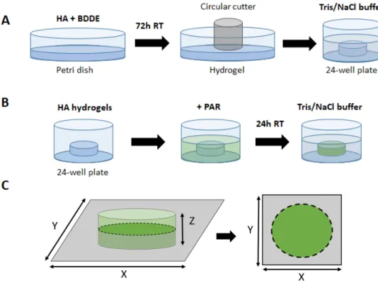
To prepare HA hydrogels, 1.5 mL of 2.5% HA (MW = 823 kDa) solution in NaOH (0.25 M) was mixed with 20% BDDE (v/v) and poured into a 35-mm diameter Petri dish. The mixture was allowed to cross-link at room temperature for 72 hours (Figure 1A). The hydrogel was further cut, most of the time, into 4-mm discs using a circle cutter, and rinsed in Tris 10 mM /NaCl 0.15 M buffer (further referred to as Tris/NaCl buffer). One fast 1 min rinsing and a longer 1-hour rinsing were performed. After this procedure, 4 mm diameter discs have swollen and became 6 mm diameter discs. Initially, HA of several molecular weights, as well as different concentrations of both HA and BDDE, were tested. Conditions allowing formation of a free-standing, handable optimal gel were finally selected and are now described in this study.
Rheological characterization of HA hydrogels
Rheological measurements were performed on HA-BDDE mixture using a Kinexus rheometer and d = 40 mm cone geometry. Briefly, 1.2 mL of freshly mixed HA-BDDE solution were deposited between the cone and the plate, and gelation was followed at 25°C for 72h at a frequency of 1 Hz. To avoid evaporation, solvent trap was used.
Loading and release of PAR
To load PAR into the hydrogels, the discs were immersed in PAR solution in Tris/NaCl buffer and incubated for 24h at room temperature on a moving plate (Figure 1B). For 6 mm discs in 24-well plate, 0.5 mL of PAR solution was used. After incubation, the discs were rinsed with Tris/NaCl buffer (one quick and one long rinsing, 5 min and 1 hour, respectively).
For PAR release experiments, discs loaded with fluorescein isothiocyanate-conjugated PARs (PAR-FITC, produced in the laboratory as described previously 23) were incubated at 37°C with respective solution (NaCl 1M, MH medium or DMEM supplemented with 10% FBS and 1% antibiotics). The release quantification of PAR-FITC in solution was performed by measuring the fluorescence of the supernatant with a spectrofluorimeter (SAFAS Genius XC spectrofluorimeter, Monaco) with excitation/emission wavelengths of 488/517 nm.
Confocal laser scanning microscopy (CSLM, LSM 710 microscope, Zeiss, Heidelberg, Germany) was also used to characterize PAR loading and release. To characterize PAR loading, the discs were incubated with PAR-FITC as described above, then disc X*Y cross-cut images were taken in the middle of the discs (Figure 1C).
Figure 1. Preparation of HA-PAR hydrogel discs. (A) Production of HA hydrogel discs: 1.5 mL
HA and BDDE well-mixed solution in is poured into a Petri dish and allowed to cross-link at room temperature for 72 hours. The hydrogel was further cut into the discs of required size using a circle cutter. (B) HA hydrogel discs loading with PAR for 24h, followed by rinsing in Tris/NaCl buffer. (C) Confocal microscopy imaging of hydrogel discs.
Fourier-transform infrared spectroscopy (FTIR)
FTIR experiments were performed on a Vertex 70 spectrometer (Bruker, Germany) using a DTGS detector. Spectra were recorded in the Attenuated Total Reflection (ATR) mode using single reflexion diamond ATR by averaging 128 interferograms between 600 and 4000 cm -1 at 2 cm-1 resolution using Blackman-Harris three-term apodization and Bruker OPUS/IR software
(version 7.5). HA gels were prepared in deuterated solution using the standard protocol, incubated in Tris-NaCl deuterared solution in the absence or the presence of 1 mg.mL-1 PAR and dried before
the measurement. The normalization was performed on the entire spectra recorded between 800 and 4 000 cm-1 by applying the min/max normalization of OPUS 7.5 software. This normalization
allows to compare spectra with each other when the optical layer thickness substantially varies. As the diffusion of PAR inside HA gel leads to its contraction, FTIR spectra of HA/PAR gels have higher intensities in comparison to HA gel because of the probing of more material on the surface. Thus, the normalization of FTIR spectra was applied by attributing to the highest peak of the HA the value of 1. This normalization allows the comparison of the spectra for the same quantity of HA probed by the IR source. As in each sample the polysaccharide peaks have the highest intensity, the normalization is obtained towards the quantity of HA probed by the IR source.
Fluorescence recovery after photobleaching (FRAP) experiments
FRAP experiments were performed on HA hydrogels incubated with 0.5 mg.mL-1 of fluorescently labelled PARs and immersed in Tris/NaCl buffer. One circular region (95 μm in radius in an image of 850 μm × 850 μm) was exposed for 64.4 s to the light of a laser set at its maximum power (λ = 488 nm). Then, the recovery of fluorescence in the bleached area was followed over time. Observations were carried out with the confocal microscope using a 10x objective. At the same time, four equally sized circular reference areas outside of the bleached area were monitored. The intensities in these areas were used to normalize the intensity in the bleached area so that bleaching due to image acquisition was accounted for.
Antibacterial assays
Staphylococcus aureus (ATCC 25923), Escherichia coli (ATCC 25922) and
Pseudomonas aeruginosa (ATCC 27853) strains were used to assess the antibacterial properties
of the test samples. Bacteria were pre-cultured aerobically at 37°C in a Mueller Hinton broth (MH) medium, pH 7.3. For this, one colony from previously prepared agar plate with streaked bacteria was transferred to 10 mL of MH medium and incubated at 37°C. Overnight culture was adjusted to OD620 nm = 0.001 (approximately 8x10⁵ CFU/mL) by diluting in MH, and added to the wells of a 24-well plate containing d = 6 mm HA hydrogels loaded with different concentrations of PAR
and sterilized with UV for 15 min. Antibacterial effect of HA-PAR hydrogels was quantified by measuring OD620 nm after 24h of incubation at 37°C on a moving plate (Figure S1). In repetitive culture experiments, bacterial suspension (OD620 nm = 0.001) was replaced every 24h.
In vitro cytotoxicity tests
Balb/3T3 (ATCC CCL163) mouse embryonic fibroblast cell line was cultured at 37°C in DMEM with 10% of FBS and 1% of penicillin-streptomycin (further referred to as “DMEM”). The cytotoxicity tests were performed according to ISO 10993-5. For direct cytotoxicity test, we used 4 mm diameter discs which swell and become ~6 mm diameter when placed in Tris/NaCl buffer. The discs, loaded or not with PAR, were sterilized with UV for 15 min and placed onto ~80% confluent layer of Balb/3T3 cells (passage 2 to 15) cultured in a 24-well plate (Figure S2). After 24h or 48h at 37°C, phase contrast images were taken around and under the discs to evaluate cell morphology. The images were captured using an Olympus CDX41 microscope (Olympus Corporation, Japan) with an Infinity 2 camera and Infinity Analyze software. Then, hydrogel discs were removed and MTT test was performed in order to measure cell metabolic activity. For MTT assay, the cells were incubated for 3h in 0.2 mg.mL-1 MTT-containing cell culture medium. The medium was then removed and formazan was dissolved in DMSO (0.5 mL per well of a 24-well plate). Absorbance of resulting solutions was measured at 570 nm using spectrophotometer.
Hydrogel deposition onto non-woven/mesh materials
To deposit the hydrogels onto materials (Medicomp®, a non-woven fabric used for wounds disinfection and GYNECARE GYNEMESH® PS Nonabsorbable PROLENE® Soft Mesh), HA-BDDE solution (50 µL) was deposited on 12 mm diameter fabric or mesh pieces and allowed to cross-link as previously described.
In vivo biocompatibility
Ten 8-week-old male Wistar rats (300-400g in weight), provided by a certified breeding centre (Charles River, France) were used for this study. The animals were received at the CREFRE (US 006/CREFRE - Inserm/UPS/ENVT) animal supplier (No. A31555010 issued December 17, 2015). Protocols were submitted to the CREFRE ethics committee with approval, in accordance with the European directive (DE 86/ 609/CEE; modified DE 2003/65/CE) for conducting animal experiments. One week of acclimatization was respected. The animals were housed in ventilated cages with a double level (two animals per cage according to European standards). The animals were carefully monitored (behavior and food intake) and were weighed weekly throughout the experiment. The 10 rats received each 2 round implants (diameter of 1 cm), one implant on left side and one implant on right side. In total, there were 5 implantations of dried and autoclaved hydrogels deposited onto mesh materials for each of the following conditions: i) HA-only hydrogels; ii) HA hydrogels loaded with PAR10 at 0.1 mg.mL-1; iii) HA hydrogels
loaded with PAR30 at 0.05 mg.mL-1; iv) HA hydrogels loaded with PAR30 at 0.1 mg.mL-1.
The rats were induced by isoflurane 4% and maintenance of 2%. Each rat was placed in a prone position on a heated pad. After shaving and scrubbing with betadine, two 20 mm dorsal incisions were made over the thoracolumbar area, one on the right side and one on the left side. One scaffold was inserted at both sides into subcutaneous pockets. All the incisions were closed with Vicryl® 3-0. All rats received buprenorphine (0.6 mg/kg) injected subcutaneously twice per day for 5 days. All animals survived the duration of the study with no adverse effects. Euthanasia were performed after 14 days. The animals were first anesthetized with isoflurane device and mask and then slowly injected with an overdose of pentobarbital (150 mg/kg) in intraperitoneal route. After the expiration of the animal death, the implants with surrounding tissue were explanted and collected to perform histology.
For histological analysis, the samples were fixed in 4% formalin. Macroscopic sections were embedded in paraffin. Five-µm thick sections were stained with hematoxylin-eosin-saffron (HES). For each sample, microscopic optical analysis was realized with the software NDP.view2 (Hamamatsu, Massy, France) after slides scanning (NanoZoomer, Hamamatsu) with the following criteria: semi-quantitative assessment of acute inflammation, chronic inflammation, fibroblastic reaction, edema, fibrosis, angiogenesis and periprosthetic histiocytic reaction.
Statistical Analysis
Rheology, loading/release, FRAP, antibacterial assays, cytotoxicity test experiments were performed at least three times. Either representative results or averages from three independent experiments are shown in the Figures. The data were processed by using SigmaPlot (Systat Software Inc., USA). One-way ANOVA on Ranks test was performed to evaluate statistical significance in the cytotoxicity assay.
RESULTS AND DISCUSSION
HA hydrogel formation and disc model
HA hydrogels gelation kinetics is followed in Figure 2A through macroscopic observations. At 24h, the gelation is starting, and after 48h, the hydrogel is in place. These qualitative observations were confirmed by rheological monitoring of changes in viscoelastic properties over time (Figure 2B). According to the graph, about 40h were required to reach a plateau. For commodity reasons, the hydrogels were cross-linked for 72h (over a week-end) before being cut to discs of different sizes (Figure 2C). Then, after rinsing and incubation in Tris/NaCl buffer, the gels swelled about 2 times (weight) (Figure 2D). For experiments in 24-well plates, 6 mm diameter and ~1 mm height discs were used.
PAR loading and release from HA hydrogels
To study loading of PAR in the hydrogels, polyarginines of three different chain lengths were used: PAR10, PAR30 and PAR200 (10, 30 and 200 arginine residues, respectively). For loading, hydrogels were incubated with PAR solutions prepared in Tris/NaCl buffer for 24h. Shorter loading time (3h) led to PAR loading on the disc periphery, while the center remained unloaded, as demonstrated with fluorescently labelled PAR (PAR-FITC) (Figure S3 A-B).
PAR loading into HA hydrogels was also characterized by FTIR. The spectra (Figure S3 C) show an amide I band at 1645 cm-1, due to the carbonyl group of amide bonds of both HA
and PAR, and saccharide peaks at 1040 and 1080 cm-1, attributed to C-O bond stretching.39,40 By
comparing HA gels before and after incubation with PAR30 solution, an increase of the amide I peak is observed with a shifting from 1614 cm-1 to 1626 cm-1 due to the contribution of the amide
I band of PAR. Moreover, an inversion of the intensities of the polysaccharide peaks was observed with PAR loaded hydrogels. This is explained by the influence of the carboxylic group state of HA on the hydrogen bond network, as reported by Rinaudo and co-workers.41 The presence of PAR
led to a higher contribution of 1080 cm-1 in comparison to 1040 cm-1 as reported for HA in acidic
form. This is probably related to the electrostatic interactions between positively charged arginine moieties of PAR and negatively charged carboxylic groups of HA. This suggests, together with fluorescent observations of PAR loading (Figure S3 A and B), that the loading of PAR in HA hydrogel was effective.
Figure 2. HA hydrogel preparation and characterization. (A) Monitoring of the HA gelation when
BDDE is added to produce hydrogel. (B) Rheological characterization of HA-BDDE hydrogel formation by monitoring elastic and viscous moduli as a function of time. Gelation was followed at 25°C for 72h at a frequency of 1 Hz. The experiment was repeated three times. (C) Production
of hydrogel discs of different sizes. (D) Hydrogel in NaOH 0.25 M (d = 18 mm after cutting) swelling in Tris/NaCl buffer.
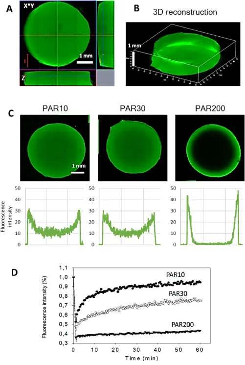
An example of a disc loaded with PAR30-FITC for 24h is shown in Figures 3A and 3B, where the disc morphology is visualized by CSLM. The disc is about 6 mm in diameter and 1 mm high, giving a volume of approximately 0.03 cm3 or 30 µL, and almost homogeneously loaded with PAR30-FITC although a higher fluorescence intensity on the disc’s periphery can be monitored.
Similar loading profile was observed for PAR10, while PAR200 chains mostly remained on the discs periphery and did not diffuse after 24h to the center of the hydrogel (Figure 3C), which is in line with our previous observations regarding the mobility of longer PAR chains in PAR/HA polyelectrolyte multilayer:23 PAR200 was the less mobile, while PAR30 showed intermediate mobility and PAR10 being the most mobile, as determined by FRAP experiments for multilayer films. Similar conclusions can be drawn here from fluorescence recovery curves determined from FRAP measurements and performed with loaded hydrogels (Figures 3D).
Figure 3. HA hydrogel loading with PAR. CLSM images of hydrogel discs incubated in 0.5
mg.mL-1 PAR30-FITC: cross-cuts in Z (A) and 3D reconstruction (B). (C) CLSM images and fluorescence intensity profiles of HA hydrogels incubated in 0.5 mg.mL-1 PAR10-FITC, PAR30-FITC and PAR200-PAR30-FITC. (D) Fluorescence recovery after photobleaching (FRAP) experiment: comparison of fluorescence recovery of the three PARs.
Total amounts of PAR contained in the hydrogels were estimated by incubation of hydrogels loaded with fluorescently labelled PAR in concentrated NaCl to promote PAR release. After 72h at 37°C in NaCl 1M, the release was almost complete for PAR10 and close to 80% for PAR30 and PAR200, according to the confocal microscopy images (Figure 4A). The percentage of PAR remaining in the hydrogel discs after 72h of incubation was measured with image processing; 100% corresponds to fluorescence intensity before release. Then, percentage of remaining PAR after NaCl 1M incubation was determined to obtain a value of released PAR: about 97%, 78% and 78% for PAR10, PAR30 and PAR200, respectively. Incomplete release of PAR30 and PAR200 correlates with lower mobility demonstrated by FRAP experiments (Figures 3D). Then, amounts of PAR-FITC released were quantified by measuring fluorescence intensity of the supernatant by spectrofluorimetry and referring to a calibration curve. The results show that the discs incubated in 0.5 mg.mL-1 PAR solutions released about 212 µg of PAR10-FITC, 157 µg of PAR30-FITC and 91 µg of PAR200-FITC after 72h in NaCl 1M (Figure 4B). When correcting the released quantities to 100%, it gives 218 µg, 201 µg and 117 µg of loaded PAR10-FITC, PAR30-FITC and PAR200-PAR30-FITC, respectively. Discs volume is approximately 30 µL, so the discs contain about 7.3 mg.mL-1 of PAR10, 6.7 mg.mL-1 of PAR30 and 3.9 mg.mL-1 of PAR200, respectively. Lower quantity of PAR200 in the hydrogels can be explained by its incomplete loading with most of PAR200 remaining on the discs periphery (Figure 3C).
PAR release from the hydrogels was then followed during 72 hours in microbiological growth medium (MH) or cell culture medium (DMEM). PAR-FITC loaded hydrogels were placed into these media and incubated at 37°C, and PAR release was observed by confocal microscopy (Figure 4C). The release was faster for PAR10 in MH, compared to PAR30 and PAR200, which were released more gradually (Figure 4D). In DMEM, all three PAR had more or less similar release profiles and were completely released after 48h (Figure 4E).
Figure 4. Release of PARs in NaCl and in culture media. HA discs were incubated with 0.5 mg.mL -1 of three different FITC-conjugated PAR for 24h. The discs were rinsed, then incubated for 72h in NaCl 1M, MH or DMEM. (A) Confocal microscopy observations, percentage of PAR remaining after 72h is indicated. (B) Quantification by spectrofluorimetry of PAR release after 72h in NaCl 1M. The graphs represent averages from 3 independent experiments, and error bars represent standard deviations. (C) Confocal microscopy images of the discs before and after incubation with MH and DMEM. (D) and (E) Percentage of released PARs in MH and DMEM, where 100% represent fluorescence intensity of the discs in Tris/NaCl.
Antibacterial activity of HA-PAR hydrogels
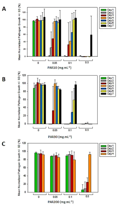
After showing that HA hydrogels can be loaded with PAR, which is released in microbiological growth medium and cell culture medium, we tested antibacterial activity of HA-PAR hydrogel towards Staphylococcus aureus (S. aureus) as our model and because its presence is widely reported in implant-related nosocomial infections. At high loading PAR concentrations (incubation in 1 mg.mL-1 PAR solutions), all three PARs inhibited S. aureus bacteria growth after 24h (Figure S4). However, high concentration of loaded PAR might be toxic to the mammalian cells. To evaluate antibacterial properties of HA-PAR hydrogels in more details, we performed repetitive bacterial culture in the presence of HA hydrogel discs incubated in different concentrations of PARs. HA-PAR discs were incubated with bacterial suspension for 24h, then bacteria suspension was replaced every 24h (Figure S1). This was done to mimic the worst conditions of repetitive bacterial infections, e.g. in case of urinary or venous catheter-associated infections.
Hydrogels incubated in 0.05 mg.mL-1 of PAR10 and PAR30 solutions showed 1 and 2 days of antibacterial activity, respectively. Hydrogels incubated in 0.1 mg.mL-1 of PAR10 and PAR30 solutions showed 2 and 4 days of antibacterial activity, respectively. For higher PAR10 and PAR30 concentrations (incubation in 0.5 mg.mL-1 of PAR solutions), antibacterial activity lasted for 6 and 7 days, respectively (Figure 5A and B).
Interestingly, PAR10 loaded into HA hydrogel showed antibacterial effect, which was not the case in PAR10/HA thin film, while in solution, all three PARs showed antibacterial activity 23. However, antibacterial effect of PAR30 was more prolonged, compared to PAR10, which is probably due to slower release of PAR30 from the hydrogels. This correlates with PAR10 and PAR30 mobility inside the hydrogels (PAR10 being more mobile) (Figure 3D), and with release profiles (Figure 4D).
For hydrogels incubated in 0.5 mg.mL-1 PAR200 solution, some bacterial growth was already observed after 24h, and the antimicrobial efficiency of the hydrogel decreased every following day (Figure 5C). Thus, PAR200 was much less efficient at equal w/v concentration when compared to PAR10 and PAR30. This can be due to its incomplete release from the hydrogels (Figure 4) in addition to its higher minimal inhibitory concentration (MIC) of 0.210 mg.mL-1, as described previously 23. MICs of PAR10 and PAR30 in solution (0.02 and 0.01 mg.mL-1, respectively) combined with their higher released amounts can explain the high antibacterial activity of PAR10 and PAR30 loaded hydrogels.
According to Figure 4, about 218 µg of PAR10 were released after 72h in NaCl. This quantity in a total volume of 0.4 mL of MH medium would correspond to a concentration of 0.545 mg.mL-1, which is more than 20 times higher than MIC10. For PAR30, 200 µg were released after 72h in NaCl, corresponding to a concentration of 0.5 mg.mL-1, which is almost 50 times higher than MIC30. Thus, hydrogels contain enough PAR to inhibit bacterial growth for several days. When PAR30 is released gradually, like in case of its release in MH (Figure 4C and D), it may provide, in theory, up to 40 days of antibacterial activity. In reality, after 72h, 80% of PAR30 is released (Figure 4D). However, the remaining 20% correspond to approximately 40 µg of PAR30. If released into the medium (400 µL), it would give a concentration of 100 µg/mL, which is 10 times higher than MIC. Thus, the remaining quantity is sufficient for several more days of antibacterial activity in MH medium. We have performed repetitive culture for up to 7 days, and no bacterial growth was observed for HA-PAR30 hydrogels incubated in 0.5 mg.mL-1 PAR30 solution.
Finally, we also tested if PAR30 loaded hydrogels can inhibit growth of other bacterial strains such as Escherichia coli. (E. coli) and (Pseudomonas aeruginosa) (P. aeruginosa), both
Gram-negative bacteria which are responsible, together with Gram-positive S. aureus, of the majority of nosocomial infections. The antibacterial effect towards Gram-negative bacteria was observed at PAR30 loading concentrations equal to 0.1 mg.mL-1 and higher (Figure S5). For loading
concentrations of 0.5 and 1 mg.mL-1, the antibacterial effect lasted for 3 days in repetitive in vitro infection model.
Figure 5. Antibacterial activity of HA hydrogels loaded with PAR10, PAR30, PAR200: repetitive
culture. Every 24h of bacterial culture, the samples were seeded with fresh bacteria. The graphs show bacterial growth in presence of hydrogel discs loaded with PAR10 (A), PAR30 (B) and PAR200 (C). Data were normalized, 100% corresponds to bacterial growth in the wells without hydrogels. The graphs represent averages from 3 independent experiments, and error bars represent standard deviations.
Biocompatibility of HA-PAR hydrogels
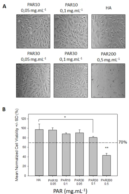
For use in clinical applications, antibacterial HA-PAR hydrogels have to be biocompatible. As a first step towards evaluation of HA-PAR hydrogel biocompatibility, we performed in vitro direct cytotoxicity test (Figure S2) according to the ISO 10993-5 norm. Microscopy observation of Balb/3T3 fibroblasts after 24h of incubation with hydrogel discs in DMEM medium, unloaded or loaded with 0.05 and 0.1 mg.mL-1 of PAR10 and PAR30, as well as 0.5 mg.mL-1 of PAR200. These concentrations correspond to the lowest antibacterial loading concentrations for each PAR (Figure 5). Confluent cell layers for the control, PAR10 and PAR30 (Figure 6A) were observed. However, in the condition where HA hydrogel was loaded with 0.5 mg.mL-1 PAR200, only rounded cells and cellular debris could be seen, suggesting cytotoxic effect of the aforementioned hydrogel.
These observations were confirmed by MTT test results: for unloaded hydrogels or those loaded with 0.05 and 0.1 mg.mL-1 of PAR10 and PAR30, cell viability was above 70% (100% corresponding to untreated cells), indicating absence of cytotoxicity according to ISO 10993-5 (Figure 6B). Condition in which the cells were incubated with 0.5 mg.mL-1 PAR200 loaded hydrogel showed decreased viability (43%) and was therefore considered as cytotoxic. PAR10 and PAR30 loading concentrations superior to 0.1 mg.mL-1 also showed cytotoxic response, however these concentrations are significantly higher than the necessary concentration needed to provide robust antimicrobial activities (Figure S6).
According to Figure 4E, the total release of PARs in DMEM takes 48h, therefore we checked cell viability after 24h and 48h of incubation with HA-PAR hydrogels. The results showed that cell viability for PAR10 and PAR30-loaded hydrogels was similar or even higher after 48h as compared to 24h, with some viability decrease for PAR200 (Figure S7).
Figure 6. Cytotoxicity assay. Balb/3T3 cells are seeded in 24-well plate and put in contact with
HA hydrogel discs loaded or not with PARs for 24h. “HA” correspond to BDDE-crosslinked HA hydrogel discs without PAR, other hydrogels were loaded with indicated PARs at different concentration. (A) Cell morphology observed by phase contrast microscopy. (B) Cell viability evaluation by MTT test. Dotted line corresponds to 70% viability (cytotoxicity limit according to ISA standard 10993-5). The graphs represent averages from 3 independent experiments, and error bars represent standard deviations. *p = 0.05, **p < 0.01.
HA-PAR hydrogel deposition on medical devices
After demonstrating the antimicrobial activity of PAR-loaded HA hydrogels, we evaluated the possibility to associate them to medical materials used for clinical applications. We selected a non-woven fabric used for wound cleaning and dressing (hydrophilic gauze made of 70% viscose and 30% polyester) and a polypropylene mesh used for treatment of urogynecological pathologies 42 (Figure 7A-C). HA hydrogels were successfully deposited onto both materials and showed antibacterial activity similar to HA hydrogel discs (Figure S8) after loading with PAR. Moreover, HA-PAR hydrogels deposited onto mesh prosthesis, dried and sterilized by autoclaving, showed similar antibacterial activity as compared to non-sterilized samples (Figure 7D). Thus, HA-PAR hydrogels can be easily deposited onto wound cleaning/dressing materials to prevent or fight infection on body surface, but also integrated into implantable mesh materials to reduce the risk of implantation-related infection.
In vivo biocompatibility assessment
Preliminary in vivo experiments were conducted on rats (10 animals). Each rat received 2 implants of hydrogel-coated meshes (d = 1 cm), one implant on left side and one implant on right side (Fig. 7E). All animals survived the duration of the study with no adverse effects and all animals gained weight in a normal way. After 14 days, the implants with surrounding tissue were explanted and collected to perform histological analysis. An example of the histological sample is shown in Figure 7F. The results of the analysis showed the presence of inflammation in the tissues surrounding the implants (Figure S9 and Table S1). However, there was no difference between HA-only hydrogels and HA-PAR hydrogels, suggesting that PAR addition does not promote or increase inflammatory response. As described before, BDDE-crosslinked HA hydrogel are regularly used as dermal filler and if such a limited inflammation occurs, it has never been reported
has being problematic. A future in vivo study confirming these results and including an infectious model should be planned to confirm the strong potential of this HA/PAR hydrogel for medical applications.
Figure 7. Deposition of HA hydrogels on medical devices. (A) Medicomp® was cut into 12-mm
diameter pieces and embedded with HA hydrogel. (B) Gynecare Gynemesh® was cut into 12-mm
diameter pieces and embedded with HA hydrogel. (C) Confocal microscopy image of PAR30-FITC loaded HA hydrogels deposited onto Gynecare Gynemesh®. (D) Effect of autoclaving on
antibacterial activity of HA-PAR hydrogels. Hydrogels were deposited onto polypropylene (PP) mesh prosthesis, loaded with 0.05 and 0.1 mg.mL-1 of PAR10 and PAR30 and autoclaved at 121°C
37°C; 100% corresponds to a positive control (bacteria grown in absence of HA-PAR hydrogels). (E) Rat implantation sites. (F) Example of histological cross-sections of the explants showing the implanted hydrogel and the surrounding tissues. The hydrogel is marked by an asterisk.
CONCLUSION
Because of the growing antibiotic resistance, new antibacterial agents are required, as well as new materials that can be deposited onto medical devices and allowing to control the release of antimicrobial agents. In this work, we introduced a unique and simple formulation of
hyaluronic acid (HA)-based antimicrobial material. We developed negatively-charged HA hydrogels which, via electrostatic interaction, can absorb high quantities of positively-charged antimicrobial agent polyarginine (PAR). Such formulation provides a prolonged antibacterial effect in vitro due to the gradual release of PAR. Among three different tested PAR lengths, PAR with 30 residues (PAR30) showed the most prolonged antibacterial effect in a repetitive infection
in vitro model, providing at least one-week of antibacterial activity against Gram-positive S.
aureus, the pathogen leading to multiple hospital-associated infections. HA-PAR30 hydrogels
were also efficient against Gram-negative bacteria E. coli and P. aeruginosa.
The hydrogels can be tuned in terms of their size, quantities of loaded PAR and their release kinetics. In addition, both PAR10 and PAR30-loaded antibacterial hydrogels with 2-4 days of antibacterial activity showed good in vitro biocompatibility. The hydrogels were successfully deposited on medical devices like fabrics or meshes used in clinical applications (Medicomp® and Gynecare Gynemesh®), and antibacterial activity of the resulting constructs was preserved after drying and autoclaving, which is important for its potential use in clinics.
Finally, we performed first in vivo tests of hydrogel-coated mesh materials biocompatibility in a rat model, which showed no difference between control HA hydrogel and PAR-loaded hydrogel. The next step of our work will be related to further in vivo studies of hydrogel biocompatibility, as well as their antibacterial activity in infected model.
Supporting Information
The following files are available free of charge: Supporting Information.pdf
Figure S1. Antibacterial activity of HA hydrogels loaded with PARs: repetitive culture. Figure S2. Schematic presentation of direct in vitro cytotoxicity test.
Figure S3. Loading of PAR30-FITC into HA hydrogel discs.
Figure S4. Antibacterial activity of HA hydrogel discs loaded with PARs: S. aureus.
Figure S5. Antibacterial activity of HA hydrogels loaded with PAR30: E. coli and P. aeruginosa. Figure S6. Cytotoxicity assay with higher concentrations of PAR10 and PAR30.
Figure S7. Cytotoxicity assay after 24 and 48 hours
Figure S8. Antibacterial activity of hydrogel discs and hydrogel-coated meshes loaded with PAR. Figure S9. In vivo biocompatibility assessement: histological analysis.
Table S1. Macroscopic outcomes of the implants.
Corresponding Author
Dr. Philippe Lavalle, PhD, Research Director INSERM INSERM / Université de Strasbourg, UMR_S 1121 11 rue Humann, 67000 Strasbourg, France
Phone: 33(0)368853061 philippe.lavalle@inserm.fr
Author Contributions
The manuscript was written through contributions of all authors. All authors have given approval to the final version of the manuscript.
ACKNOWLEDGMENT
This project has received funding from the European Union's Horizon 2020 PANBioRA research and innovation program under grant agreement no. 760921 and from the European Regional Development Fund (ERDF) in the framework of the INTERREG V Upper Rhine program “Transcending borders with every project”, project NANOTRANSMED. We would also like to thank Dr Nicolas Sananes who kindly provided us Gynecare Gynemesh®.
REFERENCES
(1) VanEpps, J. S.; Younger, J. G. Implantable Device-Related Infection. Shock 2016, 46, 597-608.
(2) Bayramov, D. F.; Neff, J. A. Beyond Conventional Antibiotics - New Directions for Combination Products to Combat Biofilm. Adv. Drug. Deliv. Rev. 2017, 112, 48-60.
(3) Romling, U.; Balsalobre, C. Biofilm Infections, Their Resilience to Therapy and Innovative Treatment Strategies. J. Intern. Med. 2012, 272, 541-561.
(4) Costerton, J. W.; Stewart, P. S.; Greenberg, E. P. Bacterial Biofilms: A Common Cause of Persistent Infections. Science 1999, 284, 1318-1322.
(5) von Eiff, C.; Jansen, B.; Kohnen, W.; Becker, K. Infections Associated with Medical Devices: Pathogenesis, Management and Prophylaxis. Drugs 2005, 65, 179-214.
(6) Darouiche, R. O. Antimicrobial Approaches for Preventing Infections Associated with Surgical Implants. Clin. Infect. Dis. 2003, 36, 1284-1289.
(7) Musella, M.; Guido, A.; Musella, S. Collagen Tampons as Aminoglycoside Carriers to Reduce Postoperative Infection Rate in Prosthetic Repair of Groin Hernias. Eur. J. Surg. 2001,
167, 130-132.
(8) van Vugt, T. A. G.; Walraven, J. M. B.; Geurts, J. A. P.; Arts, J. J. C. Antibiotic-Loaded Collagen Sponges in Clinical Treatment of Chronic Osteomyelitis. J. Bone Joint Surg. Am. 2018,
100, 2153-2161.
(9) Westberg, M.; Frihagen, F.; Brun, O. C.; Figved, W.; Grogaard, B.; Valland, H.; Wangen, H.; Snorrason, F. Effectiveness of Gentamicin-Containing Collagen Sponges for Prevention of Surgical Site Infection after Hip Arthroplasty: A Multicenter Randomized Trial. Clin. Infect. Dis.
2015, 60, 1752-1759.
(10) Pitarresi, G.; Palumbo, F. S.; Calascibetta, F.; Fiorica, C.; Di Stefano, M.; Giammona, G. Medicated Hydrogels of Hyaluronic Acid Derivatives for Use in Orthopedic Field. Int. J. Pharm.
2013, 449, 84-94.
(11) Hu, J.; Zheng, Z.; Liu, C.; Hu, Q.; Cai, X.; Xiao, J.; Cheng, Y. A Ph-Responsive Hydrogel with Potent Antibacterial Activity against Both Aerobic and Anaerobic Pathogens. Biomater. Sci.
2019, 7, 581-584.
(12) Li, B.; Webster, T. J. Bacteria Antibiotic Resistance: New Challenges and Opportunities for Implant-Associated Orthopedic Infections. J. Orthop. Res. 2018, 36, 22-32.
(13) Vasilev, K.; Cavallaro, A.; Zilm, P. Special Issue: Antibacterial Materials and Coatings.
Molecules 2018, 23, 585-588.
(14) AshaRani, P. V.; Low Kah Mun, G.; Hande, M. P.; Valiyaveettil, S. Cytotoxicity and Genotoxicity of Silver Nanoparticles in Human Cells. ACS Nano 2009, 3, 279-290.
(15) Mahlapuu, M.; Hakansson, J.; Ringstad, L.; Bjorn, C. Antimicrobial Peptides: An Emerging Category of Therapeutic Agents. Front. Cell Infect. Microbiol. 2016, 6, 194-205.
(16) Yu, Y.; Yang, Z.; Ren, S.; Gao, Y.; Zheng, L. Multifunctional Hydrogel Based on Ionic Liquid with Antibacterial Performance. J. Mol. Liq. 2020, 299, 112185-112192.
(17) Wu, D. Q.; Zhu, J.; Han, H.; Zhang, J. Z.; Wu, F. F.; Qin, X. H.; Yu, J. Y. Synthesis and Characterization of Arginine-Nipaam Hybrid Hydrogel as Wound Dressing: In Vitro and in Vivo Study. Acta Biomater. 2018, 65, 305-316.
(18) Chang, K. C.; Lin, D. J.; Wu, Y. R.; Chang, C. W.; Chen, C. H.; Ko, C. L.; Chen, W. C. Characterization of Genipin-Crosslinked Gelatin/Hyaluronic Acid-Based Hydrogel Membranes and Loaded with Hinokitiol: In Vitro Evaluation of Antibacterial Activity and Biocompatibility.
Mater. Sci. Eng. C Mater. Biol. Appl. 2019, 105, 110074-110082.
(19) Qu, J.; Zhao, X.; Liang, Y.; Zhang, T.; Ma, P. X.; Guo, B. Antibacterial Adhesive Injectable Hydrogels with Rapid Self-Healing, Extensibility and Compressibility as Wound Dressing for Joints Skin Wound Healing. Biomaterials 2018, 183, 185-199.
(20) Li, S.; Dong, S.; Xu, W.; Tu, S.; Yan, L.; Zhao, C.; Ding, J.; Chen, X. Antibacterial Hydrogels. Adv. Sci. (Weinh) 2018, 5, 1700527-1700543.
(21) Ng, V. W.; Chan, J. M.; Sardon, H.; Ono, R. J.; Garcia, J. M.; Yang, Y. Y.; Hedrick, J. L. Antimicrobial Hydrogels: A New Weapon in the Arsenal against Multidrug-Resistant Infections.
Adv. Drug. Deliv. Rev. 2014, 78, 46-62.
(22) Yang, K.; Han, Q.; Chen, B.; Zheng, Y.; Zhang, K.; Li, Q.; Wang, J. Antimicrobial Hydrogels: Promising Materials for Medical Application. Int. J. Nanomedicine 2018, 13, 2217-2263.
(23) Mutschler, A.; Tallet, L.; Rabineau, M.; Dollinger, C.; Metz-Boutigue, M. H.; Schneider, F.; Senger, B.; Vrana, N. E.; Schaaf, P.; Lavalle, P. Unexpected Bactericidal Activity of Poly(Arginine)/Hyaluronan Nanolayered Coatings. Chem. Mater. 2016, 28, 8700-8709.
(24) Decher, G. Fuzzy Nanoassemblies: Toward Layered Polymeric Multicomposites. Science
1997, 277, 1232-1237.
(25) Ozcelik, H.; Vrana, N. E.; Gudima, A.; Riabov, V.; Gratchev, A.; Haikel, Y.; Metz-Boutigue, M. H.; Carrado, A.; Faerber, J.; Roland, T.; Kluter, H.; Kzhyshkowska, J.; Schaaf, P.; Lavalle, P. Harnessing the Multifunctionality in Nature: A Bioactive Agent Release System with Self-Antimicrobial and Immunomodulatory Properties. Adv. Healthc. Mater. 2015, 4, 2026-2036.
(26) Mutschler, A.; Betscha, C.; Ball, V.; Senger, B.; Vrana, N. E.; Boulmedais, F.; Schroder, A.; Schaaf, P.; Lavalle, P. Nature of the Polyanion Governs the Antimicrobial Properties of Poly(Arginine)/Polyanion Multilayer Films. Chem. Mater. 2017, 29, 3195-3201.
(27) Almond, A. Hyaluronan. Cell Mol. Life Sci. 2007, 64, 1591-1596.
(28) Dicker, K. T.; Gurski, L. A.; Pradhan-Bhatt, S.; Witt, R. L.; Farach-Carson, M. C.; Jia, X. Hyaluronan: A Simple Polysaccharide with Diverse Biological Functions. Acta Biomater. 2014,
10, 1558-1570.
(29) Fakhari, A.; Berkland, C. Applications and Emerging Trends of Hyaluronic Acid in Tissue Engineering, as a Dermal Filler and in Osteoarthritis Treatment. Acta Biomater. 2013, 9, 7081-7092.
(30) Pavesio, A.; Renier, D.; Cassinelli, C.; Morra, M. Anti-Adhesive Surfaces through Hyaluronan Coatings. Med. Device Technol. 1997, 8, 20-21, 24-27.
(31) Ardizzoni, A.; Neglia, R. G.; Baschieri, M. C.; Cermelli, C.; Caratozzolo, M.; Righi, E.; Palmieri, B.; Blasi, E. Influence of Hyaluronic Acid on Bacterial and Fungal Species, Including Clinically Relevant Opportunistic Pathogens. J. Mater. Sci. Mater. Med. 2011, 22, 2329-2338. (32) Romano, C. L.; De Vecchi, E.; Bortolin, M.; Morelli, I.; Drago, L. Hyaluronic Acid and
Its Composites as a Local Antimicrobial/Antiadhesive Barrier. J. Bone Jt. Infect. 2017, 2, 63-72. (33) Lequeux, I.; Ducasse, E.; Jouenne, T.; Thebault, P. Addition of Antimicrobial Properties
to Hyaluronic Acid by Grafting of Antimicrobial Peptide. Eur. Polym. J. 2014, 51, 182-190. (34) Khunmanee, S.; Jeong, Y.; Park, H. Crosslinking Method of Hyaluronic-Based Hydrogel
for Biomedical Applications. J. Tissue Eng. 2017, 8, 1-16.
(35) Fidalgo, J.; Deglesne, P. A.; Arroyo, R.; Sepulveda, L.; Ranneva, E.; Deprez, P. Detection of a New Reaction by-Product in Bdde Cross-Linked Autoclaved Hyaluronic Acid Hydrogels by Lc-Ms Analysis. Med. Devices (Auckl.) 2018, 11, 367-376.
(36) De Boulle, K.; Glogau, R.; Kono, T.; Nathan, M.; Tezel, A.; Roca-Martinez, J. X.; Paliwal, S.; Stroumpoulis, D. A Review of the Metabolism of 1,4-Butanediol Diglycidyl Ether-Crosslinked Hyaluronic Acid Dermal Fillers. Dermatol. Surg. 2013, 39, 1758-1766.
(37) Grant, S. S.; Hung, D. T. Persistent Bacterial Infections, Antibiotic Tolerance, and the Oxidative Stress Response. Virulence 2013, 4, 273-283.
(38) Arnold, M.; Kao, A.; Gbozah, K.; Heniford, B.; Augenstein, V. Optimal Management of Mesh Infection: Evidence and Treatment Options. Int. J. Abdom. Wall Hernia Surg. 2018, 1, 42-49.
(39) Wang, X. H.; Li, D. P.; Wang, W. J.; Feng, Q. L.; Cui, F. Z.; Xu, Y. X.; Song, X. H.; van der Werf, M. Crosslinked Collagen/Chitosan Matrix for Artificial Livers. Biomaterials 2003, 24, 3213-3220.
(40) Duarte, M. L.; Ferreira, M. C.; Marvao, M. R.; Rocha, J. An Optimised Method to Determine the Degree of Acetylation of Chitin and Chitosan by Ftir Spectroscopy. Int. J. Biol.l
Macromol. 2002, 31, 1-8.
(41) Haxaire, K.; Maréchal, Y.; Milas, M.; Rinaudo, M. Hydration of Polysaccharide Hyaluronan Observed by Ir Spectrometry. I. Preliminary Experiments and Band Assignments.
Biopolymers 2003, 72, 10-20.
(42) De Maria, C.; Santoro, V.; Vozzi, G. Biomechanical, Topological and Chemical Features That Influence the Implant Success of an Urogynecological Mesh: A Review. Biomed. Res. Int.