HAL Id: hal-02351800
https://hal.archives-ouvertes.fr/hal-02351800
Submitted on 3 Jun 2021HAL is a multi-disciplinary open access archive for the deposit and dissemination of sci-entific research documents, whether they are pub-lished or not. The documents may come from teaching and research institutions in France or abroad, or from public or private research centers.
L’archive ouverte pluridisciplinaire HAL, est destinée au dépôt et à la diffusion de documents scientifiques de niveau recherche, publiés ou non, émanant des établissements d’enseignement et de recherche français ou étrangers, des laboratoires publics ou privés.
Manuel Campos, David Cisneros, Mangayarkarasi Nivaskumar, Olivera
Francetic
To cite this version:
Manuel Campos, David Cisneros, Mangayarkarasi Nivaskumar, Olivera Francetic. The type II secre-tion system – a dynamic fiber assembly nanomachine. Research in Microbiology, Elsevier, 2013, 164 (6), pp.545-555. �10.1016/j.resmic.2013.03.013�. �hal-02351800�
The type II secretion system - a dynamic fiber assembly nanomachine 1
2
Manuel Camposa,c, David A. Cisnerosb, Mangayarkarasi Nivaskumara and Olivera Francetic
3
Institut Pasteur, Molecular Genetics Unit, Department of Microbiology, 75015 Paris, France. 4
CNRS ERL3526, 75724 Paris, France. 5
aUniversity Paris VII, 25 rue du Dr Roux, 75724 Paris CEDEX 15
6
bPresent address: Research Institute of Molecular Pathology (IMP), Dr. Bohr-Gasse 7, A-1030
7
Vienna, Austria and Max F. Perutz Laboratories (MFPL), University of Vienna, Dr. Bohr-8
Gasse 9, A-1030 Vienna, Austria 9
cPresent address: Yale University School of Medicine, New Haven, CT, USA
10 11
Abstract (100 w)
12
Type II secretion systems (T2SSs) share common origins and structure with archaeal flagella 13
(archaella) and pili, bacterial competence systems and type IV pili. All of these systems use a 14
conserved ATP-powered machinery to assemble helical fibers that are anchored in the plasma 15
membrane. The T2SSs assemble pseudopili, periplasmic filaments that promote extracellular 16
secretion of folded periplasmic proteins. Comparative analysis of T2SSs and related fiber 17
assembly nanomachines might provide important clues on their functional specificities and 18
dynamics. This review focuses on recent developments in the study of pseudopilus structure 19
and biogenesis, and discusses mechanistic models of pseudopilus function in protein secretion. 20
22
Introduction 23
24
The type 2 secretion systems (T2SSs) translocate folded proteins from the periplasm of Gram-25
negative bacteria across the outer membrane via a large channel that remains closed in the 26
resting state (reviewed by P. Howard in this issue). Upon secretion, the exoprotein substrates 27
are released or exposed on the bacterial surface (Rondelet and Condemine, this issue) to 28
perform diverse functions in macromolecule degradation, adhesion, electron transport or 29
pathogenesis. The remarkable progress made in the study of T2SS structure and organization 30
during the past decade has been described in detail in several recent reviews (Douzi et al., 31
2012, Korotkov et al., 2012, McLaughlin et al., 2012). This review focuses on the core 32
function of T2SS - the assembly of periplasmic filaments called the pseudopili, which play an 33
essential but poorly understood function in promoting the protein transport. 34
The T2SSs belong to an ancient and widespread superfamily of membrane nano-35
machines that specialize in the assembly of fibers built with subunits localized in the inner 36
membrane. This highly successful and versatile molecular device is found in all prokaryotes, 37
suggesting its ancient evolutionary origin. In archaea these systems form the base of archaella 38
(formerly archaeal flagella) (Jarrel and Albers, 2012), type IV-like pili and bindosomes, the 39
carbohydrate "capture" systems. In bacteria, beside the natural competence systems and T2SSs, 40
they are also found in the form of type IV pili (T4P), long flexible fibers extending from the 41
bacterial surface. T4P are involved in bacterial motility over solid substrates called twitching 42
motility, and in other functions including adhesion, DNA uptake, aggregation, biofilm 43
formation, signaling and protein transport (Pelicic, 2008). 44
All these diverse machineries have a common basic function - the assembly of helical 45
fibers from the plasma membrane localized pilin subunits. The pilin transmembrane (TM) 46
segments are highly conserved and comprise at the N-terminus a positively charged signal 47
anchor followed by a Gly or Ala residue, the cleavage site for a specific processing enzyme, the 48
prepilin peptidase. The hydrophobic segment extends into a long a-helical stem that is shielded 49
on one side by a globular domain composed of 4 to 5 anti-parallel b-strands separated by 50
different conserved or variable loop regions. Globular pilin domains are exposed on the surface 51
of assembled fibers with their variable sequence providing the fibers with diverse surface and 52
binding properties. 53
The striking primary sequence conservation of TM a-helical regions is the signature 54
allowing unambiguous identification of fiber assembly systems in bacterial and archaeal 55
genomes (Albers and Pohlschroder, 2009). Such sequence conservation suggests that a strong 56
selective pressure shaped these segments, which play crucial roles in several steps of the fiber 57
biogenesis pathway (Fig. 1). Their hydrophobicity is essential for interactions with the signal 58
recognition particle (Fig. 1a) and for co-translational targeting to the Sec translocase that 59
promotes pilin insertion in the membrane in an N-in - C-out orientation (Fig. 1b) (Arts et al., 60
2007b, Francetic et al., 2007). The specific prepilin peptidase removes the positively charged 61
signal anchor after the conserved Gly residue (Fig. 1c), and, in T4a pili and T2SS transfers a 62
methyl group to the new N-terminal residue. In the following, still poorly understood steps of 63
fiber biogenesis, the TM segment might mediate interactions with the conserved fiber assembly 64
factors in the membrane, including the polytopic IM protein of the PulF/PilC family (Fig. 1d). 65
Finally, the TM segments make contacts with neighboring pilins during fiber assembly and 66
form the hydrophobic core of assembled filaments (Fig. 1e). 67
68
T2SS and fiber assembly nanomachines 69
70
In the T2SS, between 12 and 15 genes are required for protein secretion. Their 71
organization is similar in different bacterial groups (Douzi et al., 2012). In most species these 72
genes form a single operon gspCDEFGHIJKLMNO (for general secretion pathway) (Fig. 2a), 73
with different additional genes required for the assembly and targeting of the secretin channel 74
GspD subunits. Each gsp gene is essential for protein secretion, with the exception of gspN, 75
which is also absent from many bacteria. While this secretion-null phenotype did not favor the 76
genetic analysis of T2SSs, biochemical dissection of the system led to the current model of 77
T2SS organization in the bacterial envelope. Furthermore, the three-dimensional structure of 78
most soluble domains of T2SS components has been determined (McLaughlin et al., 2012, 79
Korotkov et al., 2012). 80
The functional studies of the T2SS have benefited from the comparative analysis with 81
type IV pili (T4P), the most prominent and best-studied members of the helical fiber assembly 82
nanomachine family. These thin and flexible filaments are found on the surface of many Gram-83
negative and some Gram-positive bacteria (Pelicic, 2008). In Pseudomonas aeruginosa, the 84
core genes essential for TP4 biogenesis are organized in an operon pilABCD, encoding the 85
major pilin subunit PilA, a hexameric ATPase PilB, a polytopic membrane protein PilC and the 86
prepilin peptidase PilD. This basic fiber assembly module is found in all related systems, 87
including the T2SS, suggesting that pilus assembly and protein secretion, as well as natural 88
competence, share a common molecular mechanism (Hobbs and Mattick, 1993). 89
Five proteins contain an N-terminal prepilin peptidase cleavage site - the highly 90
abundant (major) GspG and low-abundance (minor) pseudopilins GspH, I, J and K. The 91
presence of these components and their processing and N-methylation by the prepilin peptidase 92
GspO suggested that T2SSs assemble short periplasmic fibers (Strom and Lory, 1991, Pugsley, 93
1993). These initially hypothetical structures, the pseudopili, could be visualized upon 94
overexpression of the pul genes encoding the Klebsiella oxytoca T2SS (Sauvonnet et al., 95
2000b). Pseudopilus surface assembly allowed the biochemical and structural characterization 96
of these fibers and provided an additional phenotype for genetic analysis of the T2SS. Fiber 97
assembly is not specific to the Pul system: at least two others, the chitinase-specific Gsp T2SS 98
from Escherichia coli (Vignon et al., 2003) and the P. aeruginosa Xcp system (Durand et al., 99
2003) also show this property. In contrast, the Hxc system in P. aeruginosa does not assemble 100
surface pili under these conditions and seems to belong to a distinct T2SS subclass based on 101
phylogenetic studies (Durand et al., 2011). While protein secretion strictly requires the 102
presence of all T2SS components except GspB and GspN, pilus assembly, studied under 103
overexpression conditions, does not require minor pseudopilins GpsH, GspI, GspJ and GspK, 104
and the inner membrane component GspM (Fig. 2) (Durand et al., 2005). GspI has been 105
implicated in pseudopilus assembly initiation since reduced amount of pili is found in gspI 106
(xcpV) mutants (Sauvonnet et al., 2000b, Durand et al., 2005). Similar to T4P in Neisseria spp. 107
(Wolfgang et al., 2000, Carbonnelle et al., 2006), the secretin pore in the outer membrane is 108
not required for fiber assembly but rather for the surface presentation of assembled fibers. In 109
the absence of K. oxytoca PulD, PulG pili are assembled in the periplasm (Vignon et al., 2003, 110
Cisneros et al., 2011). 111
112
The major pseudopilins and T2SS pili 113
114
Only major pseudopilins can form long surface polymers, while minor pseudopilins fail to form 115
such fibers even when overproduced (Durand et al., 2005). Although surface pili are considered 116
as an overproduction artifact, they facilitated the pseudopilus analysis using biochemical 117
approaches, fluorescence and electron microscopy (EM). When observed by negative staining 118
EM, pseudopili are highly similar to T4a pili, thin (≈ 6 nm), flexible and with bundling 119
propensity, which might result from the EM sample preparation (Sauvonnet et al., 2000a, 120
Durand et al., 2003). EM analysis of PulG pili shows a helical subunit arrangement: each 121
subunit is displaced relative to its neighbor in the fiber by a vertical shift (axial rise) of 10.4 Å 122
and a rotational (twist) angle of 84.7° (Kohler et al., 2004), values similar to those found in T4a 123
pili (Craig et al., 2006). X-ray crystallography of major pseudopilins showed a typical pilin 124
fold, with a long alpha-helical stem and an antiparallel 3-strand beta-sheet forming the globular 125
domain (Kohler et al., 2004, Korotkov et al., 2009). Interestingly, the C-terminal pseudopilin 126
domain is folded around a Ca2+ ion, forming a stable and well-structured loop (Korotkov et al.,
127
2009), which is also apparent in the NMR structure of major pseudopilin XcpTGspG from P.
128
aeruginosa (Alphonse et al., 2010). In V. cholerae the Ca2+ coordinating residues are essential
129
for protein secretion (Korotkov et al., 2009), apparently without affecting the monomer 130
stability. In the Pul T2SS, equivalent mutations reduce dramatically the levels of PulG, 131
suggesting the involvement of Ca2+ in pseudopilin folding and stability (Campos et al., 2010).
132
Interestingly, this loop has its counterpart in the variable loop of major T4a pilins that is 133
stabilized by a disulfide bridge (Craig et al., 2006). 134
For T4a pili, a combination of cryo-EM and X-ray crystallography led to the first low-135
resolution models of these fibers (Craig et al., 2003). The first PulG pilus models, generated by 136
a similar approach revealed the presence of deep groves on the fiber surface (Kohler et al., 137
2004). A prominent left-handed grove had been taken as evidence that PulG pili were 138
organized as a left-handed helix (Kohler et al., 2004). However, the missing TM segments and 139
the overall lack of detail precluded the use of this model for further structure-function analysis. 140
The helical parameters (i.e. axial rise and rotation) from this study were used as constraints to 141
build another series of PulG pilus models using a flexible ab initio approach (Campos et al., 142
2011). The 1000 models generated independently converged into a cluster of 200 highly similar 143
right-handed helix models. The analysis of inter-protomer distances in this model cluster 144
showed that the vast majority showed multiple highly conserved contacts (Campos et al., 145
2010). Each PulG protomer (P) within the fiber is involved in intricate contacts with protomers 146
P ± 1, P ± 3, P ± 4 and possibly P ± 7 via different interfaces (Fig. 3). Biochemical analysis was 147
used to test these contacts and validate the models, but also to probe the function of different 148
interfaces. Site-directed mutagenesis and functional studies showed that the most extensive and 149
important contacts involve the two closest protomers (P and P ± 1). In the model of gonococcal 150
T4a pilus, the key role in pilus assembly was attributed to electrostatic interactions between the 151
absolutely conserved pilin residue E5 of protomer P and the N-terminal amine group of the 152
nearest protomer above (P+1) (Craig et al., 2006). In the PulG pilus, in addition to this probable 153
contact, which cannot be tested experimentally, two important salt bridge interactions have 154
been identified involving negatively charged residues of the periplasmic a-helical stem (E44 155
and D48) and positively charged residues (R87 and R88) in a loop region of the globular major 156
pilin domain (Fig. 3). Highly conserved among the major pseudopilin family, these residues 157
were essential for both T2SS functions: protein secretion and pseudopilus assembly (Campos et 158
al., 2010). In contrast, mutations disrupting other interfaces differentially affect the two 159
functions and appear to play different roles, currently under investigation (Nivaskumar, 160
Campos et al, unpublished data). 161
The inter-protomer distances at the level of the hydrophobic pseudopilin segment have 162
been tested by double cysteine cross-linking studies. As predicted by the pseudopilus structural 163
models, the shortest distance in assembled pili is found between residues 10 (P+1) and 16 (P). 164
Placing Cys residues at these positions leads to covalent cross-linking of pili resulting in 165
detergent-resistant PulG multimers. Interestingly, a lower degree of crosslinking is observed 166
when cysteine residues are placed in neighboring positions, 9 and 16 on one hand, and 11 and 167
16 on the other. These mutually exclusive interactions suggest an iris-like movement of pilin 168
monomers in assembled fibers (Campos et al., 2010). Similar structural heterogeneity is 169
observed in archaella (Yu et al., 2012) and archeal pili (Wang et al., 2008) suggesting that other 170
related fibers undergo similar conformational changes. External forces might promote these 171
changes during assembly/disassembly and might propagate along the fibers, providing the 172
structural basis for a potential signaling mechanism, one of the major functions observed in 173 T4P. 174 175 Minor pseudopilins 176 177
Four low-abundance (minor) pseudopilins are essential for protein secretion under 178
physiological expression conditions. In contrast, surface pili are produced, although less 179
efficiently, in their absence when the components of the system are overproduced, leading to 180
the idea that minor subunits provide determinants on the pseudopilus surface that are involved 181
in secretion-specific interactions. In the simplest model, the secreted substrates interact directly 182
with the minor pseudopilins. However, since complementation studies suggest that minor 183
pseudopilins are not substrate specificity determinants {Possot, 2000 #29}, interactions with 184
other T2SS factors and alternative functions such as the secretin channel gating, regulation of 185
fiber length were proposed (Forest, 2008). Although extensive efforts were made to detect 186
minor pseudopilins in surface fibers, there is still no biochemical or microscopy-based evidence 187
for their co-assembly into pili in T2SS (Vignon et al., 2003). Nevertheless, they might be 188
present in periplasmic pseudopili under physiological conditions, as suggested by studies of the 189
Xanthomonas campestris T2SS (Kuo et al., 2005, Hu et al., 2002). In T4P, the equivalent minor
190
pilins have been detected in pilus fractions and visualized by immuno-EM (Giltner et al., 2010, 191
Winther-Larsen et al., 2005). 192
The most compelling argument for minor pseudopilin co-assembly into fibers is 193
provided by the structural studies. The crystal structure of the complex formed by the 194
periplasmic domains of EpsI and EpsJ from Vibrio cholerae indicated a 1-nm shift between the 195
two proteins, identical to that in assembled pili (Yanez et al., 2008, Lam et al., 2009, Kohler et 196
al., 2004). Furthermore, structural analysis of the GspJ-GspI-GspK trimer from enterotoxigenic 197
E. coli showed that they formed a pilus-like complex (Korotkov and Hol, 2008) with a 1-nm
198
axial rise and a rotational angle of 100° between neighboring subunits. An alpha-helical 199
domain of GspK inserted in the pilin b-sheet fold caps this complex, such that additional 200
subunits cannot fit on top. Therefore, GspK is likely to be localized at the tip of a putative 201
pseudopilus. Consistent with this model, the absolutely conserved residue E5 that is predicted 202
to neutralize the N-terminal positive charge of the protomer above is lacking in GspK. The 203
fourth minor pseudopilin XcpUGspH in P. aeruginosa interacts with the trimeric tip complex in
204
vitro, suggesting that minor pseudopilins form a quaternary complex (Douzi et al., 2009).
205
Based on extensive analysis of binding affinities between their periplasmic domains, Douzi et 206
al proposed a pseduopilus assembly model in which GspIXcpV plays a central nucleating role
207
and GspHXcpU links this complex to the major pseudopilin ({Douzi, 2009 #7}In addition,
208
structural modeling allowed fitting of EpsH subunit below the GspJ-GspI-GspK complex 209
(Yanez et al., 2008), although direct binding of a GspG subunit also provided a satisfactory fit 210
(Korotkov and Hol, 2008). 211
Early in vivo studies had suggested the role of GspI in assembly initiation (Sauvonnet et 212
al., 2000b). Overproduction of PulK and its Pseudomonas homologue XcpX reduced pilus 213
assembly, while deletion of xcpX led to assembly of longer pili, leading to the idea that minor 214
pseudopilins control the fiber length (Vignon et al., 2003, Durand et al., 2005). Recent 215
systematic analysis of minor pseudopilin mutants by immunofluorescence microscopy revealed 216
that not only pulK mutants, but also pulI and pulJ mutants make longer and fewer pili (Cisneros 217
et al., 2011). Removing all minor pseudopilins led to complete loss of piliation, suggesting a 218
role of the tip complex in pseudopilus assembly initiation. However, once initiated, pseudopilus 219
assembly seems to continue unabated under plate culture conditions, as a function of the 220
available pilin pool in the inner membrane. This observation further confirms the lack of active 221
retraction and length control, consistent with the absence of retraction ATPase in the T2SS. 222
The gspH mutants were identical to wild type cells with respect to pilus number, suggesting 223
that GspH does not play a role in initiation but rather at a later, elongation step, or in the 224
transition between initiation and elongation (Cisneros et al., 2012). 225
Mutants lacking all minor pseudopilins occasionally produced long pili, suggesting that 226
the assembly machinery spontaneously activates in rare cases and that minor pseudopilins play 227
a catalytic role. The minor pseudopilin pair GspJ-GspI is sufficient to restore a significant level 228
of piliation in these mutants, consistent with the central position of these proteins in the 229
quaternary pseudopilin complex (Cisneros et al., 2011, Douzi et al., 2009). In vivo they form a 230
staggered complex, as evidenced by close contacts between TM segment residues 10 (GspI) 231
and 16 (GspJ) shown by cysteine cross-linking (Cisneros et al., 2011). These contacts are 232
determined by a specific fit between the globular domains of these proteins, which form an 233
extensive interacting surface (Korotkov and Hol, 2008). Molecular dynamics simulations, 234
mutational and cross-linking studies provide evidence for self-assembly of tip pseudopilins: the 235
staggered GspJ-GspI complex bends in the inner membrane to facilitate the binding of GspK 236
alpha domain to its upper surface; this in turn facilitates the extraction of PulK, exerting a 237
pulling force on the membrane. The strong correlation between GspJ-GspI-GspK binding and 238
their ability to promote assembly initiation, suggests that these proteins transduce a signal to 239
activate the assembly ATPase in the cytoplasm. How this is signal transduced and which 240
protein-protein interactions participate in activation, are some of the major unresolved 241
questions. Biochemical and structural studies from the Sandkvist and Hol labs in particular 242
provide some clues about the possible molecular mechanism of these events, discussed below. 243
244
The pseudopilus assembly machinery 245
246
The energy for the pseudopilus and T4P assembly is generated by conformational changes of 247
the assembly ATPase of the GspE/PilB family. These hexameric ATPases form a ring-like 248
structure with a central opening, leading initially to the idea that pilins are translocated to the 249
periplasm via this channel (Camberg and Sandkvist, 2005). X-ray structures of EpsE from V. 250
cholerae (Robien et al., 2003) and XpsE from X. campestris (Chen et al., 2005) show a bilobed
251
monomer structure, with the ATP binding site at the hinge region between the N-terminal and 252
C-terminal domains. In the recent EpsE hexamer model, the NTD and CTD of neighboring 253
subunits share extensive contacts (Patrick et al., 2011). In this arrangement, which was tested 254
by site-directed mutagenesis, several arginine residues around the active site belonging to both 255
protomers seem to be required for ATP hydrolysis. Variants with alanine substitutions at these 256
positions are specifically defective in ATP hydrolysis but not in ATP binding or GspE 257
oligomerization. The strongest defect was associated with mutation R225A, in close proximity 258
to the bound nucleotide both in V. cholerae EpsE and in X. campestris ATPase XspE, 259
suggesting a direct role in ATP hydrolysis (Shiue et al., 2007). 260
In the hexamer, two neighboring subunits contribute to ATP binding and hydrolysis, 261
resulting in three ATPase active sites (Patrick et al., 2011). Structural insight into their 262
conformations comes from the studies of the closely related retraction ATPase PilT (Satyshur 263
et al., 2007). Each PilT monomer pair shows a different active site conformation,
264
corresponding to the ATP-bound, hydrolysis and release state (Misic et al., 2010). Extensive 265
rotation of the N-terminal domain around the active site hinge is involved in the transition 266
between the extreme states. Although these conformational states might appear stochastically, a 267
sequential model would be consistent with the biological data, suggesting that the addition of 268
one pseudopilin at a time at the level of the inner membrane drives both fiber polymerization 269
and protein secretion (Campos et al., 2010). Interestingly, the subunit arrangement in the 270
initiation complex GspJ-GspI-GspK (Korotkov and Hol, 2008) mirrors the three-state ATPase 271
model and might use the self-assembly energy or fit or induce the three-state ATPase motor 272
(Cisneros et al., 2011), thus contradicting the three-start helix assembly model proposed for 273
T4P (Craig et al., 2006). 274
How are these conformational changes transduced to promote pseudopilus assembly? 275
One candidate for this role is GspF, the core fiber assembly component, whose interaction with 276
GspE has been demonstrated by two-hybrid and biochemical approaches (Py et al., 2001). 277
GspF has three TM segments and two large cytoplasmic domains structured as 6 -helix 278
bundles (Abendroth et al., 2009). Little is known about the overall organization and 279
oligomerization state of GspF. Its homologue in T4P, PilG, forms dimers and tetramers whose 280
cryo-EM structure looks like a conical ring (Collins et al., 2007). Interactions of GspF of P. 281
aeruginosa with GspL and GspE homologues are suggested by their mutual stabilization (Arts
282
et al., 2007a) and by the yeast two-hybrid studies (Py et al., 2001). Details of these interactions
283
and their functional significance are still a major black box. 284
The N-terminal domain of GspE forms a stable complex with the cytoplasmic domain 285
of GspL, characterized by X-ray crystallography (Abendroth et al., 2005). GspL is the 286
membrane anchor for the ATPase and favors its oligomerization. It also regulates the 287
interactions with membrane acidic phospholipids, phosphatidyl glycerol and cardiolipin, which 288
enhance GspE ATPase activity dramatically (Camberg et al., 2007). 289
Biochemical, two-hybrid and live imaging approaches provide ample evidence for 290
interactions between GspL and GspM (Py et al., 2001, Possot et al., 2000, Buddelmeijer et al., 291
2006). In addition to its stabilizing effect on GspL, the C-terminal domain of GspM plays a 292
specific role in secretion (Camberg et al., 2007). Recent studies on V. cholerae T2SS show that 293
EpsL and EpsG can be cross-linked in vivo (Gray et al., 2011), suggesting that EpsL transmits 294
the ATP-ase driven conformational changes to the major pseudopilin to promote fiber 295
assembly. The GspL-GspG interacting surface requires further investigation, since epsG 296
mutants defective in interaction with EpsL map at residues that form an intra-molecular 297
hydrogen bond and affect the stability of EpsG monomer. Other T2SS components might 298
interact with the major pseudopilin. In N. meningitidis, the major pilin subunit PilE interacts 299
with PilG (a GspF homologue) and PilO, which is homologous to GspM (Georgiadou et al., 300
2012). 301
302
Pseudopilus assembly and protein secretion 303
304
T2SSs and T4P assembly systems are remarkably similar. Recent structural studies revealed 305
that all T2SS components have their equivalents in T4aPS (Ayers et al., 2010). The minor 306
pseudopilins, which are specifically required for protein secretion, have their counterparts in 307
minor T4 pilins. Another "secretion specific" component GspM has a structural equivalent in 308
the T4P PilO protein (Ayers et al., 2009). 309
Some components, like the prepilin peptidase, are functionally interchangeable between 310
the T4P and T2SSs. The Pul T2SS can assemble the major T4 pilin PpdD encoded by the 311
chromosome of E. coli and many other enterobacteria (Sauvonnet et al., 2000b, Kohler et al., 312
2004). Furthermore, the E. coli minor pilins can promote the initiation of PpdD assembly in a 313
heterologous system, catalyzed by the T2SS assembly machinery (Cisneros et al., 2012), 314
suggesting a role of minor T4 pilins in pilus assembly initiation. Minor T4 pilins are required 315
for pilus assembly in the presence of retraction ATPase PilT. Mutants of pilT are piliated, due 316
to the spontaneous assembly initiation events, the high concentration of pilins in the membrane 317
and their extremely high stability. Indeed, unlike the T2SS pseudopili, which are unstable and 318
short under physiological conditions, T4 pili are so stable that an energy-dependent process is 319
required to undo the fibers and reset the system. While flexible modeling predicts a similar 320
electrostatic and hydrophobic interaction network in pseudopili and T4P, the covalent disulfide 321
bonds stabilizing the T4 pilin monomers might be responsible for this remarkable fiber stability 322
(Campos et al., 2011, Li et al., 2012). 323
Interestingly, the minor T4 pilins of E. coli can promote the assembly of PulG, albeit 324
less efficiently than the cognate minor pseudopilins (Cisneros et al., 2012). The absence of 325
homology and therefore of structural complementarity between the globular domains of T4 326
pilins and the major T2SS pseudopilin (and vice versa) suggests that their role in assembly 327
initiation does not rely on complex formation between these proteins. The only similarities 328
between T4 pilins and T2SS pseudopilins are found in their hydrophobic segments, suggesting 329
a role in crucial interactions with the assembly machinery. According to the activation model, 330
the minor T4 pilin or minor pseudopilin complex formation acts upon the assembly machinery 331
to start the first cycle of ATP binding and hydrolysis. However, pseudopilus assembly is more 332
efficient in the presence of cognate minor pseudopilins, suggesting that a scaffolding 333
mechanism (via direct binding of pseudopilin globular domains) contributes to assembly 334
initiation (Cisneros et al., 2012, Cisneros et al., 2011, Burrows, 2012). In support of this model, 335
full activation of pseudopilus assembly that leads to protein secretion is achieved in the 336
presence of cognate pseudopilins in the T2SS. 337
338
Models of protein secretion 339
340
How is the energy of pseudopilus assembly transduced to promote protein secretion? 341
Based on the similarities of T2SS and T4P, at least two models of protein secretion mechanism 342
have been proposed (Nunn, 1999) known as "the piston" and the "Archimedes' screw" models. 343
The idea of the piston was based on the dynamics of T4a pili, where cycles of pseudopilus 344
extension and retraction push the substrate through the secretin channel. According to this 345
model, pseudopilus assembly provides the driving force for secretion, acting as a piston, while 346
minor pseudopilins determine the binding specificity (Shevchik et al., 1997, Nunn, 1999, 347
Vignon et al., 2003). Major support for the piston model comes from recent findings that the 348
minor pseudopilin complex XcpU-V-W-X of P. aeruginosa binds in vitro to the specific 349
substrate of the Xcp system, LasB, but not to the substrate of the second Hxc system present in 350
the same species (Douzi et al., 2011). 351
The major problem with this model is that in T2SS there is no evidence of fiber 352
retraction. Certain T4P assembly systems also secrete periplasmic proteins. One example is the 353
toxin co-regulated pilus (TCP) of V. cholerae, which secretes a soluble colonization factor 354
TcpF (Kirn et al., 2003). Interestingly, the TCP system lacks the retraction ATPase, again 355
suggesting that pilus retraction is not required for protein secretion. A unidirectional assembly 356
might be required to ensure the vectorial transfer of proteins from the inner membrane towards 357
the exterior. The lack of retraction ATPase in T2SS led to the proposal that pseudopilus 358
"collapses" or is degraded, and that GspK might catalyze the degradation of major pseudopilin 359
(Durand et al., 2005). Indeed, pseudopilins might be more prone to degradation than T4 pilins. 360
Their Ca2+-coordinating C-terminal loop, structurally equivalent to the disulfide loop in T4
361
pilins, might provide a basis for the control of subunit structural integrity (Korotkov et al., 362
2009). 363
In T4P and all helical fibers of this superfamily, subunits are shifted by a 1-nm 364
translation and variable rotation angles, with a symmetry that does not match that of the GspE 365
ATPase. This implies that, during assembly, the fibers rotate relative to the assembly ATPase 366
during addition of each new subunit (Mattick, 2002). The T2SS might be regarded as a 367
modified reverse ATP synthase that uses the energy release upon ATP hydrolysis for rotation 368
instead of generating ATP using the proton motive force (Streif et al., 2008). The Archimedes' 369
screw model postulates exoprotein binding to the pseudopilus at the base, suggesting that major 370
pseudopilins provide the binding surface (Fig. 4). Addition of pseudopilin subunits would be 371
coupled to the rotary motion of the fiber and to a net upward transfer of subunits and bound 372
exoproteins from the pseudopilus base into the secretin channel. So far, there is less 373
biochemical support for this model that for the piston model. However, the exoprotein PulA of 374
the K. oxytoca T2SS interacts with the PulG pili (Kohler et al., 2004) (Francetic et al, 375
submitted). In addition, major pseudopilin mutations lead to a change in substrate specificity in 376
Xcp and Hxc, the two T2SS of P. aeruginosa (Durand et al., 2011). 377
Evidence for rotation during or upon assembly of helical fibers comes from archaella, 378
which rotate to promote swimming (Ghosh and Albers, 2011, Herzog and Wirth, 2012); S.V. 379
Albers, personal communication). While bacterial flagella require proton motive force for 380
rotation, archaella use the energy of ATP hydrolysis and do not seem to require motor proteins 381
other than the assembly machinery itself (Streif et al., 2008). Given their conservation, it is 382
plausible that all helical fiber assembly nanomachines of this superfamily display the same 383
dynamics. While rotation of rigid archaella allows them to "carry the weight" of the archaeal 384
cell and support swimming, rotation of flexible pseudopili or T4P might not be easily observed. 385
The ATPase GspE in T2SS is anchored to the membrane via GspL, but GspF might be free to 386
rotate in the membrane. Components of the fiber assembly platform anchored in the cell wall 387
might play a role of the stator, and the link with GspC and GspD might be important in this 388
regard (Korotkov et al., 2011). 389
Clearly, the interactions between the membrane components of fiber assembly 390
nanomachines and fiber dynamics are a vast unexplored area that awaits further research. 391
Comparative analysis of T2SSs, T4P, archaeal pili and archaella should provide important 392
clues on the common mechanisms and significant differences that underlie the specialized 393
functions of these remarkable systems. 394
395
Acknowledgments 396
The study of protein secretion in our team has been funded by the Institut Pasteur grant 397
PTR339. MC was a scholar of the French Ministry of Science and Education, MN is the 398
recipient of Pasteur-Paris University fellowship and DC was funded by EMBO long term and 399
Roux fellowships. We are grateful to our collaborators Michaël Nilges, Guillaume Bouvier, 400
Peter J. Bond, Gérard Péhau-Arnaudet, Nadia Izadi-Pruneyre, Daniel Ladant and Gouzel 401
Karimova for their contributions to the pseudopilus project. We are grateful to all members of 402
the Molecular Genetics Unit for many stimulating discussions. We thank Tony Pugsley for 403
generous support and for the critical reading of the manuscript. 404 405 References 406 407
Abendroth, J., D. Mitchell, K. Korotkov, T. Johnson, A. Kreger, M. Sankvist and W. Hol, 408
2009. The three-dimensional structure of the cytoplasmic domains of EpsF from the 409
type 2 secretion system of Vibrio cholerae. J Struct Biol 166, 303-315. 410
Abendroth, J., P. Murphy, M. Sandkvist, M. Bagdasarian and W. G. Hol, 2005. The X-ray 411
structure of the type II secretion system complex formed by the N-terminal domain of 412
EpsE and the cytoplasmic domain of EpsL of Vibrio cholerae. J Mol Biol 348, 845-855. 413
Albers, S. V. and M. Pohlschroder, 2009. Diversity of archaeal type IV pilin-like structures. 414
Extremophiles 13, 403-410. 415
Alphonse, S., E. Durand, B. Douzi, B. Waegele, H. Darbon, A. Filluox, R. Voulhoux and C. 416
Bernard, 2010. Structure of the Pseudomonas aeruginosa XcpT pseudopilin, a major 417
component of the type II secretion system. J. Struct. Biol. 169, 75-80. 418
Arts, J., A. de Groot, G. Ball, E. Durand, M. El Khattabi, A. Filloux, J. Tommassen and M. 419
Koster, 2007a. Interaction domains in the Pseudomonas aeruginosa type II secretory 420
apparatus component XcpS (GspF). Microbiol 153, 1582-1592. 421
Arts, J., R. van Boxtel, A. Filloux, J. Tommassen and M. Koster, 2007b. Export of the 422
pseudopilin XcpT of the Pseudomonas aeruginosa type II secretion system via the 423
signal recognition particle-Sec pathway. J Bacteriol. 189, 2069-2076. 424
Ayers, M., P. L. Howell and L. L. Burrows, 2010. Architecture of the type II secretion and type 425
IV pilus machineries. Future Microbiol 5, 1203-1218. 426
Ayers, M., L. Sampaleanu, S. Tammam, J. Koo, H. Harvey, P. Howell and L. Burrows, 2009. 427
PilM/N/O/P proteins form an inner membrane compex that affects the stability of the 428
Pseudomonas aeruginosa type IV pilus secretin. J Mol. Biol. 394, 128-142.
429
Buddelmeijer, N., O. Francetic and A. P. Pugsley, 2006. Green fluorescent chimeras indicate 430
nonpolar localization of pullulanase secreton components PulL and PulM. J. 431
Bacteriol.188, 2928-2935. 432
Burrows, L. L., 2012. Prime time for minor subunits in type II secretion and type IV pilus 433
systems. Mol Microbiol 86. 434
Camberg, J. L., T. L. Johnson, M. Patrick, J. Abendroth, W. G. Hol and M. Sandkvist, 2007. 435
Synergistic stimulation of EpsE ATP hydrolysis by EpsL and acidic phospholipids. 436
EMBO J 26, 19-27. 437
Camberg, J. L. and M. Sandkvist, 2005. Molecular analysis of the Vibrio cholerae type II 438
secretion ATPase EpsE. J Bacteriol 187, 249-256. 439
Campos, M., O. Francetic and M. Nilges, 2011. Modeling pilus strcutures from sparse data. J 440
J Struct Biol. 173, 436-444. 441
Campos, M., M. Nilges, D. A. Cisneros and O. Francetic, 2010. Detailed structural and 442
assembly model of the type II secretion pilus from sparse data. Proc Natl Acad Sci 443
USA 107, 13081-13086. 444
Carbonnelle, E., S. Helaine, X. Nassif and V. Pelicic, 2006. A systematic genetic analysis in 445
Neisseria meningitidis defines the Pil proteins required for assembly, functionality,
446
stabilization and export of type IV pili. Mol Microbiol 61, 1510-1522. 447
Chen, Y., S. J. Shiue, C. W. Huang, J. L. Chang, Y. L. Chien, N. T. Hu and N. L. Chan, 2005. 448
Structure and function of the XpsE N-terminal domain, an essential component of the 449
Xanthomonas campestris type II secretion system. J Biol Chem 280, 42356-42363.
450
Cisneros, D., G. Péhau-Arnaudet and O. Francetic, 2012. Heterologous assembly of type IV 451
pili by a type II secretion system reveals the role of minor pilins in assembly 452
initiation. Mol Microbiol 86. 453
Cisneros, D. A., P. J. Bond, A. P. Pugsley, M. Campos and O. Francetic, 2011. Minor 454
pseudopilin self-assembly primes type II secretion pseudopilus elongation. 455
EMBO J 31, 1041-1053. 456
Collins, R. F., M. Saleem and J. P. Derrick, 2007. Purification and three-dimensional electron 457
microscopy structure of the Neisseria meningitidis Type IV pilus biogenesis protein 458
PilG. J Bacteriol 189, 6389–6396. 459
Craig, L., R. K. Taylor, M. E. Pique, B. D. Adair, A. S. Arvai, M. Singh, S. J. Lloyd, D. S. 460
Shin, E. D. Getzoff, M. Yeager, F. K.T. and J. A. Tainer, 2003. Type IV pilin structure 461
and assembly: X-ray and EM analyses of Vibrio cholerae toxin-coregulated pilus and 462
Pseudomonas aeruginosa PAK pilin. Molec Cell 11, 1139-1150.
463
Craig, L., N. Volkmann, A. S. Arvai, M. E. Pique, M. Yeager, E. H. Egelman and J. A. Tainer, 464
2006. Type IV pilus structure by cryo-electron microscopy and crystallography: 465
implications for pilus assembly and functions. Mol Cell 23, 651-662. 466
Douzi, B., G. Ball, C. Cambillau, M. Tegoni and R. Voulhoux, 2011. Deciphering the Xcp 467
Pseudomonas aeruginosa type II secretion machinery through multiple interactions
468
with substrates. J Bio Chem 286, 40792-40801. 469
Douzi, B., E. Durand, C. Bernard, S. Alphonse, C. Cambillau, A. Filloux, M. Tegoni and R. 470
Voulhoux, 2009. The XcpV/GspI pseudopilin has a central role in the assembly of a 471
quaternary complex within the T2SS pseudopilus. J Biol Chem 284, 34580-34589. 472
Douzi, B., A. Filloux and R. Voulhoux, 2012. On the path to uncover the bacterial type II 473
secretion system. Phil Trans R Soc B 367, 1059-1072. 474
Durand, E., S. Alphonse, C. Brochier-Armanet, G. Ball, B. Douzi, A. Filloux, C. Bernard and 475
R. Voulhoux, 2011. The assembly mode of the pseudopilus. A hallmark to distinguish a 476
novel secretion system subtype. J. Biol. Chem. 286, 24407-24416. 477
Durand, E., A. Bernadac, G. Ball, A. Lazdunski, J. N. Sturgis and A. Filloux, 2003. Type II 478
protein secretion in Pseudomonas aeruginosa: the pseudopilus is a multifibrillar and 479
adhesive structure. J Bacteriol 185, 2749-2758. 480
Durand, E., G. Michel, R. Voulhoux, J. Kurner, A. Bernadac and A. Filloux, 2005. XcpX 481
controls biogenesis of the Pseudomonas aeruginosa XcpT-containing pseudopilus. The 482
J Biol Chem 280, 31378-31389. 483
Forest, K. T., 2008. The type II secretion arrowhead: the structure of GspI-GspJ-GspK. Nat 484
Struct Mol Biol 15, 428-430. 485
Francetic, O., N. Buddelmeijer, S. Lewenza, C. A. Kumamoto and A. P. Pugsley, 2007. Signal 486
recognition particle-dependent inner membrane targeting of the PulG pseudopilin 487
component of a type II secretion system. J Bacteriol 189, 1783-1793. 488
Georgiadou, M., M. Castagnini, G. Karimova, D. Ladant and V. Pelicic, 2012. Large-scale 489
study of the interactions between proteins involved in type IV pilus biology in 490
Neisseria meningitidis: characterization of a sub-complex involved in pilus assembly.
491
Mol Microbiol 84, 857-873. 492
Ghosh, A. and S. V. Albers, 2011. Assembly and function of the archaeal flagellum. Biiochem 493
Soc Trans 39, 64-69. 494
Giltner, C. L., M. Habash and L. L. Burrows, 2010. Pseudomonas aeruginosa minor pilins are 495
incorporated into type IV pili. J Mol Biol 398, 444-461. 496
Gray, M. D., M. Bagdasarian, W. G. Hol and M. Sandkvist, 2011. In vivo cross-linking of 497
EpsG to EpsL suggests a role for EpsL as an ATPase-pseudopilin coupling protein in 498
the Type II secretion system of Vibrio cholerae. Mol Microbiol 79, 786-798. 499
Herzog, B. and R. Wirth, 2012. Swimming behaviour of selected species of archea. Appl 500
Environ Micribiol. 78, 1670-1674. 501
Hobbs, M. and J. S. Mattick, 1993. Common components in the assembly of type 4 fimbriae, 502
DNA transfer systems, filamentous phage and protein-secretion apparatus: a general 503
system for the formation of surface-associated protein complexes. Mol. Microbiol. 10, 504
233-243. 505
Hu, N. T., W. M. Leu, M. S. Lee, A. Chen, S. C. Chen, Y. L. Song and L. Y. Chen, 2002. 506
XpsG, the major pseudopilin in Xanthomonas campestris pv. campestris, forms a pilus-507
like structure between cytoplasmic and outer membranes. Biochem. J 365, 205-211. 508
Jarrel, K. F. and S. V. Albers, 2012. The archeallum: an old motility structure with a new name. 509
Trends Microbiol 20, 307-312. 510
Kirn, T. J., N. Bose and R. K. Taylor, 2003. Secretion of a soluble colonization factor by the 511
TCP type 4 pilus biogenesis pathway in Vibrio cholerae. Mol. Microbiol. 49, 81-92. 512
Kohler, R., K. Schafer, S. Muller, G. Vignon, K. Diederichs, A. Philippsen, P. Ringler, A. P. 513
Pugsley, A. Engel and W. Welte, 2004. Structure and assembly of the pseudopilin PulG. 514
Mol Microbiol 54, 647-664. 515
Korotkov, K., T. L. Johnson, M. Jobling, J. Pruneda, E. Pardon, A. Héroux, S. Turley, J. 516
Steayert, R. Holmes, S. M and H. WG, 2011. Structural and functional studies on the 517
inetractions of GspC and GspD in type II secretion system. PLOS Pathog. 518
Korotkov, K., M. Sandkvist and W. Hol, 2012. The type II secretion system: biogenesis, 519
molecular archtecture and mechanism. Nat Rev Microbiol 10, 336-351. 520
Korotkov, K. V., M. Gray, A. Kreger, S. Turley, M. Sandkvist and W. Hol, 2009. Calcium is 521
essential for the major pseudopilin in the type 2 secretion system. J Biol Chem 284, 522
25466-25470. 523
Korotkov, K. V. and W. G. Hol, 2008. Structure of the GspK-GspI-GspJ complex from the 524
enterotoxigenic Escherichia coli type 2 secretion system. Nat Struct Mol Biol 15, 462-525
468. 526
Kuo, W. W., H. W. Kuo, C. C. Cheng, H. L. Lai and L. Y. Chen, 2005. Roles of the minor 527
pseudopilins, XpsH, XpsI and XpsJ, in the formation of XpsG-containing pseudopilus 528
in Xanthomonas campestris pv. campestris. J Biomed Sci 12, 587-599. 529
Lam, A. Y., E. Pardon, K. V. Korotkov, W. G. Hol and J. Steyaert, 2009. Nanobody-aided 530
structure determination of the EpsI:EpsJ pseudopilin heterodimer from Vibrio 531
vulnificus. J Struct Biol 166, 8-15.
532
Li, J., E. Egelman and L. Craig, 2012. Structure of the VIbrio choleare type IV b pilus and 533
stability comparison with Neisseria gonorrhoeae type IV a pilus. J Mol Biol 418, 47-64. 534
Mattick, J. S., 2002. Type IV pili and twitching motility. Annu Rev Microbiol 56, 289-314. 535
McLaughlin, L., R. Haft and F. KT, 2012. Structural insights into the Type II secretion 536
nanomachine. Cu Op Struct Biol 22, 208-216. 537
Misic, A. M., K. A. Satyshur and K. T. Forest, 2010. P. aeruginosa PilT structure with and 538
without nucleotide reveal a dynamic type IV pilus retraction motor. J Mol Biol 400, 539
1011-1021. 540
Nunn, D., 1999. Bacterial type II protein export and pilus biogenesis:more than just 541
homologies. Trends Cell Biol 9, 402-408. 542
Patrick, M., K. V. Korotkov, W. G. Hol and M. Sandkvist, 2011. Oligomerization of EpsE 543
coordinates residues from multiple subunits. J BIol. Chem 286, 10378-10386. 544
Pelicic, V., 2008. Type IV pili: e pluribus unum? Mol Microbiol 68, 827-837. 545
Possot, O. M., G. Vignon, N. Bomchil, F. Ebel and A. P. Pugsley, 2000. Multiple 546
interactions between pullulanase secreton components involved in stabilization and 547
cytoplasmic membrane association of PulE. J Bacteriol 182, 2142-2152. 548
Pugsley, A. P., 1993. Processing and methylation of PulG, a pilin-like component of the 549
general secretory pathway of Klebsiella oxytoca. Mol Microbiol 9, 295-308. 550
Py, B., L. Loiseau and F. Barras, 2001. An inner membrane platform in the type II secretion 551
machinery of Gram-negative bacteria. EMBO Rep 2, 244-248. 552
Robien, M. A., B. E. Krumm, M. Sandkvist and W. G. Hol, 2003. Crystal structure of the 553
extracellular protein secretion NTPase EpsE of Vibrio cholerae. J Mol. Biol 333, 657-554
674. 555
Satyshur, K. A., G. A. Worzalla, L. S. Meyer, E. K. Heiniger, K. G. Aukema, A. M. Misic and 556
K. T. Forest, 2007. Crystal structures of the pilus retraction motor PilT suggest large 557
domain movements and subunit cooperation drive motility. Structure 15, 363-376 558
Sauvonnet, N., P. Gounon and A. P. Pugsley, 2000a. PpdD type IV pilin of Escherichia coli K-559
12 can be assembled into pili in Pseudomonas aeruginosa. J Bacetriol. 182, 848-854. 560
Sauvonnet, N., G. Vignon, A. P. Pugsley and P. Gounon, 2000b. Pilus formation and protein 561
secretion by the same machinery in Escherichia coli. EMBO J 19, 2221- 2228. 562
Shevchik, V., J. Robert-Baudouy and G. Condemine, 1997. Specific interaction between OutD, 563
an Erwinia chrysanthemi outer membrane protein of the general secretory pathway, and 564
secreted proteins. EMBO J 16, 3007-3016. 565
Shiue, S. J., I.-L. Chien, N. L. Chan, W. M. Leu and N. T. Hu, 2007. Mutation of a key residue 566
in the type II secretion system ATPse uncouples ATP hydrolysis from protein 567
translocation. Mol Microbiol 65, 401-412. 568
Streif, S., W. F. Staudinger, W. Marwan and D. Oesterhelt, 2008. Flagella rotation in the 569
archaeon Halobacterium salinarum depends on ATP. J Mol Biol 384, 1-8. 570
Strom, M. S. and S. Lory, 1991. Amino acid substitutions in pilin of Pseudomonas aeruginosa. 571
Effect on leader peptide cleavage, amino-terminal methylation, and pilus assembly. J. 572
Biol. Chem. 266, 1656-1664. 573
Vignon, G., R. Kohler, E. Larquet, S. Giroux, M. C. Prevost, P. Roux and A. P. Pugsley, 2003. 574
Type IV-like pili formed by the type II secreton: specificity, composition, bundling, 575
polar localization, and surface presentation of peptides. J Bacteriol 185, 3416-3428. 576
Wang, Y. A., X. Yu, S. Y. M. Ng, K. F. Jarrell and E. H. Egelman, 2008. The structure of an 577
archaeal pilus. J Mol Biol 381, 456-466. 578
Winther-Larsen, H. C., M. Wolfgang, S. Dunham, J. P. van Putten, D. Dorward, C. Lovold, F. 579
E. Aas and M. Koomey, 2005. A conserved set of pilin-like molecules controls type IV 580
pilus dynamics and organelle-associated functions in Neisseria gonorrhoeae. Mol 581
Microbiol 56, 903-917. 582
Wolfgang, M., J. P. M. van Putten, S. F. Hayes, D. Dorward and M. Koomey, 2000. 583
Components and dynamics of fiber formation define a ubiquitous biogenesis pathway 584
for bacterial pili. EMBO. J. 19, 6408-6418. 585
Yanez , M., K. Korotkov , J. Abendroth and W. G. J. Hol, 2008. The Crystal Structure of a B 586
inary Complex of two Pseudopilins: EpsI and EpsJ from the Type 2 Secretion System of 587
Vibrio vulnificus. J. Mol. Biol. 375, 471–486.
588
Yanez, M. E., K. V. Korotkov, J. Abendroth and W. G. Hol, 2008. Structure of the minor 589
pseudopilin EpsH from the Type 2 secretion system of Vibrio cholerae. J. Mol Biol 590
377, 91-103. 591
Yu, X., C. Goforth, C. Meyer, R. Reinhard, R. Wirth, G. F. Shchröder and E. H. Egelman, 592
2012. Filaments from Ignicoccis hospitalis show diversity of packing in proteins 593
containing N-terminal type IV pilin helices. J Mol Biol 422, 264-281. 594 595 596 597 598 599 600 601
602 Figures 603 604 605
Fig. 1. Pseudopilus biogenesis steps. The transmembrane segment of pseudopilins (in grey) 606
interacts with cellular and type II secretion system partners. The hydrophobicity of the segment 607
is crucial for the targeting via the SRP complex (A) to the Sec translocase (B) for correct 608
membrane insertion. Upon insertion, the prepilin peptidase cleaves the positively charged pre-609
peptide after the conserved Gly residue (C); the cleaved peptide interacts with at least one 610
component of the assembly platform (D) to finally become buried in the core of the assembled 611
fiber, while the globular hydrophilic domains are exposed on the pseudopilus surface (E). 612 613 614 615 616 617 618
619
Fig. 2. Involvement of T2SS components in pilus formation. (A) Top: Organization of the 620
Pul T2SS-encoding genes on the chromosome of Klebsiella oxytoca. Divergent operons pulAB 621
and pulCDEFGHIJKLMNO, and a monocystronic pulS gene encode the T2SS. The substrate 622
gene A is shown in yellow. Genes dispensable for the substrate secretion are shown in grey, 623
genes essential for surface exposure of the pseudopilus and the substrate are in blue, genes 624
essential for pseudopilus assembly and protein secretion are in pale orange, and genes essential 625
for protein secretion only are in green. Below: PulG pilus assembly in mutants lacking 626
individual T2SS components. Bacteria of strain MDS42 (Posfai et al., 2006) transformed with 627
different derivatives of plasmid pCHAP231 carrying the complete set of pul genes or single 628
nonpolar mutations as indicated (Possot et al., 2000) were grown for two days on rich solid 629
media. Bacterial and sheared pili fractions were analyzed by immunodetection using anti-PulG 630
antibodies, as described (Cisneros et al., 2011). The equivalent of 0.05 OD600nm of each fraction
631
was loaded. The asterisk indicates a cross-reacting band. (B) The schematic representation of 632
the Pul T2SS components in the bacterial envelope with the same color- code as above. OM, 633
outer membrane; PG, peptidoglycan; IM, inner membrane. Images of T2SS components were 634
prepared with PyMol (http://www.pymol.org). The model of GspD is adapted from (Reichow
635 et al., 2010). 636 637 638 639 640
641
Fig. 3. The structure of the PulG pseudopilus (left) and PulG residues involved in inter-642
protomer interactions (right). The central protomer P (orange) interacts with protomer P+1 643
(green), P+3 (blue) and P+4 (red). Arrows indicate predicted interactions between residues of 644
the central protomer P and its partners at each interface (indicated by the residue position, the 645
color code and the protomer number in index). Adapted from (Campos et al., 2010). 646 647 648 649 650 651 652
653
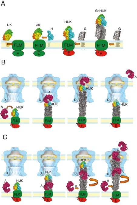
Fig. 4. Models of pseudopilus assembly and function in protein secretion. (A) The minor 654
pseudopilins (I-J-K) self-assemble in the inner membrane (IM) and bind to the fourth minor 655
pseudopilin GspH (H). This tetrameric complex transfers the activation signal to the assembly 656
ATPase via an unknown component of the assembly platform, possibly GspL (LMF). The 657
activation of the ATPase (E) leads to the elongation of the pseudopilin by successive addition 658
of major pseudopilin subunits (G), coupled to the fiber rotation relative to the assembly 659
ATPase. (B) According to the piston model, the substrate (A in pink) binds to the minor 660
pseudopilin complex on the tip (HIJK). Pseudopilus elongation adding GspG subunits (in grey) 661
pushes the substrate through the secretin channel. The system is reset by an unknown 662
mechanism to allow binding of the next substrate molecule. (C)In the Archimedes’ screw 663
model, minor pseudopilin complex initiates pseudopilus assembly and the substrate binds to the 664
major pseudopilins at the fiber base in the IM. Pseudopilus growth from the base, coupled with 665
rotation drives the substrate into the secretin channel (D) vestibule. Pseudopilus assembly force 666
pushes the secretin gate open and the substrate is expelled across the outer membrane (OM) 667
channel. A single pseudopilus initiation event leads to secretion of multiple substrate molecules 668
via continuous assembly of the major pseudopilins. 669