HAL Id: cea-01938121
https://hal-cea.archives-ouvertes.fr/cea-01938121
Submitted on 28 Nov 2018
HAL is a multi-disciplinary open access
archive for the deposit and dissemination of
sci-entific research documents, whether they are
pub-lished or not. The documents may come from
teaching and research institutions in France or
abroad, or from public or private research centers.
L’archive ouverte pluridisciplinaire HAL, est
destinée au dépôt et à la diffusion de documents
scientifiques de niveau recherche, publiés ou non,
émanant des établissements d’enseignement et de
recherche français ou étrangers, des laboratoires
publics ou privés.
A novel role for the Bcl-2 protein family: specific
suppression of the RAD51 recombination pathway
Yannick Saintigny, Anne Dumay, Sarah Lambert, Bernard Lopez
To cite this version:
Yannick Saintigny, Anne Dumay, Sarah Lambert, Bernard Lopez. A novel role for the Bcl-2 protein
family: specific suppression of the RAD51 recombination pathway. EMBO Journal, EMBO Press,
2001, 20 (10), pp.2596-2607. �10.1093/emboj/20.10.2596�. �cea-01938121�
Yannick Saintigny, Anne Dumay,
Sarah Lambert and Bernard S.Lopez
1UMR217 CNRS-CEA, CEA, Direction des Sciences du Vivant, DeÂpartement de Radiobiologie et Radiopathologie, 60±68 Avenue du GeÂneÂral Leclerc, 92 265 Fontenay aux Roses, Cedex, France 1Corresponding author
e-mail: lopez@dsvidf.cea.fr
Y.Saintigny and A.Dumay contributed equally to this work
The oncogenic role of Bcl-2 is generally attributed to its protective effect against apoptosis. Here, we show a novel role for Bcl-2: the speci®c inhibition of the con-servative RAD51 recombination pathway. Bcl-2 or Bcl-XL overexpression inhibits UV-C-, g-ray- or
mutant p53-induced homologous recombination (HR). Moreover, Bcl-2 recombination inhibition is independ-ent of the role of p53 in G1arrest. At an acute
double-strand break in the recombination substrate, Bcl-2 speci®cally inhibits RAD51-dependent gene conversion without affecting non-conservative recombination. Bcl-2 consistently thwarts recombination stimulated by RAD51 overexpression and alters Rad51 protein by post-translation modi®cation. Moreover, a mutant
G145ABcl-2, which is defective in Bax interaction and in
apoptosis repression, also inhibits recombination, showing that the death and recombination repression functions of Bcl-2 are separable. Inhibition of error-free repair pathways by Bcl-2 results in elevated fre-quencies of mutagenesis. The Bcl-2 gene therefore combines two separable cancer-prone phenotypes: apoptosis repression and a genetic instability/mutator phenotype. This dual phenotype could represent a mammalian version of the bacterial SOS repair system.
Keywords: apoptosis/DNA repair/homologous recombination/mutagenesis/RAD51
Introduction
Genome integrity and cell proliferation/viability are commonly regulated by a network of pathways including cell cycle checkpoints, DNA repair/recombination and programmed cell death. In response to genotoxic attacks, proliferating cells temporarily stall in their cycle, allowing the repair of the injured DNA (Hartwell, 1992, Hartwell et al., 1994). Alternatively, cells activate their pro-grammed cell death (Rich et al., 1999; Wyllie et al., 1999). p53 is the gene most frequently found to be mutated in human tumours (Hollstein et al., 1991; Levine et al., 1991). The p53 protein mediates cell cycle checkpoints and apoptosis (for review see Donehower and Bradley, 1993; Hainaut, 1995; Smith and Fornace, 1995; Ko and Prives, 1996). In addition, the status of p53 can affect
homologous recombination frequency (Wiesmuller et al., 1996; Bertrand et al., 1997; Mekeel et al., 1997; Dudenhoffer et al., 1999; Saintigny et al., 1999). How-ever, the effect of p53 on homologous recombination can be independent of G1 checkpoint alteration, suggesting
that p53 acts on recombination via a pathway other than the G1 checkpoint control (Dudenhoffer et al., 1999;
Saintigny et al., 1999). Since p53 protein also controls apoptosis, this raises the question, what effect does apoptosis regulation have on homologous recombination. In line with this, the mammalian recombination protein RAD51 is a target for caspase degradation during programmed cell death (Flygare et al., 1998; Huang et al., 1999). However, the relationship between apoptosis and regulation of homologous recombination is poorly understood.
One ef®cient and classic way to repress apoptosis is to overexpress Bcl-2 oncogene family members. Indeed, DNA damage can induce two pathways to apoptosis, one p53 dependent and one p53 independent, and both pathways can be inhibited by Bcl-2 (Strasser et al., 1994). Bcl-2 becomes oncogenic when overexpressed, such as in follicular B-cell lymphomas resulting from a t(14:18) translocation (Bakhshi et al., 1985; Cleary and Sklar, 1985; Tsujimoto et al., 1985). Bcl-2, as well as other members of its family such as Bcl-XL, has anti-apoptotic
activity (for review see Adams and Cory, 1998). Since cells become more resistant to genotoxic agents, this raised the question of how DNA repair pathways are regulated when protecting the cells against apoptosis. Here we address the question of whether the expression of Bcl-2 affects homologous recombination ef®ciency. Using a combination of different substrates and strategies to monitor recombination, the present paper focuses on the detailed characterization of the recombination pathways affected by Bcl-2 (or Bcl-XL) overexpression. We
over-expressed Bcl-2 in mammalian cell lines containing the tandem repeat recombination substrates depicted in Figure 1. These substrates allow the monitoring of different intrachromosomal recombination pathways. Using a substrate containing a cleavage site for the rare-cutting endonuclease I-SceI (Figure 1B), it is possible to target a unique double-strand break (DSB) in the recombination substrate (Liang et al., 1998). This strategy has permitted the de®nition of two homology-directed DSB repair pathways: (i) a non-conservative recombina-tion mechanism arising by single-strand annealing (SSA) and leading to deletion of the intervening sequence (NeoR/
HygS); (ii) a conservative recombination mechanism
initiated by strand invasion and leading to gene conversion with or without associated crossing over (double-resistant NeoR/HygR). In contrast to SSA, conservative
recombin-ation is RAD51 dependent in both yeast (Ivanov et al., 1996) and mammalian cells (Lambert and Lopez, 2000).
A novel role for the Bcl-2 protein family: speci®c
suppression of the RAD51 recombination pathway
Here we show that Bcl-2 expression speci®cally inhibits the RAD51-dependent conservative recombination path-way, independently of programmed cell death repression. We also show that the expression of Bcl-2 results in modi®cation of post-translation regulation of Rad51 protein. Taken together, the results suggest that RAD51-dependent conservative recombination and apoptosis are controlled by two separable functions of Bcl-2 (or Bcl-XL). In addition to its anti-apoptotic phenotype,
Bcl-2 also shows a mutator phenotype, both of which could contribute to a pre-cancerous predisposition.
Results
Expression of Bcl-2 in cell lines carrying intrachromosomal recombination substrates We used the recipient lines and strategies described in Figure 1. The pJS3-10 line derives from mouse Ltk±cells,
which are sensitive to HAT medium, and contains a copy of a duplication of inactive herpes simplex type I TK gene. Recombination between the two TK sequences restores a functional TK gene and thus HAT resistance. The frequency of recombination can be calculated from the number of HAT-resistant clones relative to the total number of viable plated cells. CHO-DRA10 contains a direct repeat of two inactive neomycin (NEO) resistance genes, separated by a hygromycin-resistant gene. Re-combination restores a functional NEO gene and thus resistance to G418 (NeoR). One NEO cassette contains one
I-SceI cleavage site. Expression of the I-SceI rare-cutting
endonuclease produces a DSB targeted into the recombin-ation NEO cassette (Liang et al., 1998). Repair of this DSB via a conservative gene conversion event produces a double-resistant NeoR/HygRclone, and a non-conservative
event produces a NeoRbut hygromycin-sensitive (HygS)
clone. A few double-resistant NeoR/HygR (2 out of 11
double-resistant clones analysed) recombinant clones result from intrachromatid crossing over then random re-integration of the excised product (Lambert and Lopez, 2000). These events are also initiated by strand exchange and are RAD51 dependent.
The different lines were transfected with mammalian expression vectors, to obtain derivative lines stably expressing Bcl-2 or mutant Bcl-2 proteins (Figure 2A). The wild-type Bcl-2 protein inhibits apoptosis and affects G0±G1cell cycle entry. These two functions of Bcl-2 can
be separated and the mutantY28ABcl-2 has been shown to
maintain its anti-apoptosis function but to be de®cient in its role in G0±G1 cell cycle entry (Huang et al., 1997).
Mutant G145ABcl-2 has been shown to be de®cient in its
interaction with Bax and in apoptosis repression (Yin et al., 1994). The different derivative lines are listed in Table I. Analysis by immuno¯uorescence shows that overexpres-sion of Bcl-2 in our lines does not seem to modify its classical localization (data not shown).
We ®rst veri®ed the apoptotic cell death repression activity resulting fromY28ABcl-2 expression. The
protect-ive effect of Y28ABcl-2 in the CHO-DRA10 line, after
treatment with ionizing radiation, was checked by differ-ent methods: (i) the frequency of cells with a sub-G1DNA
Fig. 1. Recombination substrates. (A) Parental pJS3-10 is a mouse Ltk±cell line containing a tandem repeat of two inactive TK genes (black boxes)
from herpes simplex virus type I (the grey square corresponds to the inactivating mutations). The cells are de®cient in thymidine kinase activity (tk±)
and, thus, sensitive to the HAT selective medium. Recombination between the two TK sequences restores a functional TK gene and resistance to HAT. The frequency of recombination is estimated by the frequency of HAT-resistant clones (Liskay et al., 1984). (B) Parental CHO-DRA10 is a hamster CHO K1 cell line containing a tandem repeat of two inactive neomycin (Neo) resistance genes (black boxes). In between the two Neo sequences is the hygromycin-resistant (Hyg) sequence (grey box). The parental lines are sensitive to G418 (NeoS) and resistant to hygromycin (HygR). All the
recombinants become G418 resistant (NeoR). One Neo cassette contains a cleavage site (grey arrow) for the yeast rare-cutting enzyme I-SceI.
Expression of I-SceI results in a DSB targeted in the recombination substrate. Conservative recombination leads to NeoR/HygRdouble-resistant clones.
Non-conservative recombination leads to NeoRsingle-resistant clones (Liang et al., 1998). Non-conservative recombination mainly corresponds to
single-strand annealing (SSA), an RAD51-independent process. Conservative recombination corresponds to gene conversion or intrachromatid crossing over (followed by re-integration of the excised product), two RAD51-dependent processes (Ivanov et al., 1996; Lambert and Lopez, 2000).
content, measured by ¯uorescence-activated cell sorting (FACS); and (ii) the nuclear fragmentation, visualized by means of Hoechst 33342, a ¯uorescent DNA intercalating agent. Forty-eight hours after irradiation (6 Gy), the percentage of apoptotic cells (sub-G1cell population) was
16.5% in the control cells and 5.3 and 4.3% in two independent clones expressingY28ABcl-2 (Figure 2B). The
frequency of fragmented nuclei (monitored by Hoechst ¯uorescence) consistently decreased 3- and 3.5-fold in two independent irradiated lines expressing Y28ABcl-2
(Figure 2C). Thus, both methods con®rmed in our cell line the well-established cell death repression activity of
Y28ABcl-2. These results are in agreement with a 2- to
3-fold increased viability after g-rays (6 Gy) of both mouse ®broblasts (pJS3-10) and CHO-DRA10 lines expressing Bcl-2 orY28ABcl-2 (Figure 2D).
Bcl-2 expression suppresses the induction of recombination byg-rays or UV-C
Both g-rays and UV-C stimulated homologous recombin-ation in the parental CHO-DRA10 lines (Figure 3).
Y28ABcl-2 expression inhibited induction of recombination
by g-rays (Figure 3A) as well as by UV-C (Figure 3B). Since these two different genotoxic stresses produce different types of DNA damage, Bcl-2 recombination inhibition (BRI) is not speci®c to the type of genotoxic stress.
Anti-recombination and anti-apoptosis are separable functions of Bcl-2
One important question is whether BRI requires the interaction between Bcl-2 and Bax proteins, and whether it is associated with or separable from the apoptosis repres-sion activity. More generally, BH3-only members of the Bcl-2 family are critical initiators of apoptosis that can be repressed by Bcl-2. However, mutants in the BH1 domain of Bcl-2 are unable to inhibit the pro-apoptotic activity of such BH3-only proteins (O'Connor et al., 1998).
To address this question, we expressed the mutant
G145ABcl-2 (mutated in the BH1 domain of Bcl-2) in the
CHO-DRA10 cell line.G145ABcl-2 is defective in the Bax
interaction and the protection against apoptosis (Yin et al., 1994). We ®rst veri®ed the effect on apoptosis by Table I. Cell lines
Cell lines Origin Expression of an exogenous human protein
p53 mutant protein Bcl-2 protein pJS 3.10 mouse L cell nonea none
pJS DR II.4 pJS 3.10 none Bcl-2 H175 DR 211 pJS 3.10 175 (Arg®His) none H175 DR II.3 pJS 3.10 175 (Arg®His) Bcl-2 H175 DR II.5 pJS 3.10 175 (Arg®His) Bcl-2 H273 DR11 pJS 3.10 273 (Arg®His) P273 DR 4 pJS 3.10 273 (Arg®Pro)
CHO-DRA10 hamster CHO-K1 noneb none
Cm3c CHO-DRA10 none none
A3c CHO-DRA10 none none
ADRA 8 CHO-DRA10 none Y28ABcl-2
ADRA14 CHO-DRA10 none Y28ABcl-2
ADRA17 CHO-DRA10 none Y28ABcl-2
BDRA1 CHO-DRA10 none G145ABcl-2
BDRA2 CHO-DRA10 none G145ABcl-2 aThe pJS 3.10 parental cell line expresses an endogenous wild-type p53
protein (see Bertrand et al., 1997; Saintigny et al., 1999).
bThe CHO-DRA10 parental cell line expresses an endogenous mutant
p53 protein.
cThe Cm3 and A3 cell lines are independent clones from the
CHO-DRA10 parental cell line transfected with the empty expression vector.
Fig. 2. Bcl-2 expression and apoptosis repression. (A) Detection by western blotting of the expression of Bcl-2. pJS3-10 is the mouse parental cell line; pJSDRII.4 is a pJS3-10 line overexpressing Bcl-2. CHO-DRA10 is the hamster parental line; ADRA14 and ADRA17 are two independent clones overexpressingY28ABcl-2. (B) Apoptotic sub-G1population, measured by FACS, 48 h after 6 Gy irradiation. Upper panel, an example of a typical
histogram (M1 corresponds to the sub-G1population); lower panel, frequency of sub-G1cells 48 h after irradiation. (C) Measurement of apoptosis by means of Hoechst ¯uorescence; the percentages correspond to the percentage of cells with fragmented nuclei. ADRA14 and ADRA17 correspond to two independent clones expressingY28ABcl-2. (D) Effect of Bcl-2 expression on survival of CHO-DRA10, after g-rays.
measuring the frequency of fragmented nuclei after exposure to radiation. Y28ABcl-2 exhibited a protective
effect, whereasG145ABcl-2 showed no anti-apoptotic
pheno-type in two independent clones (Figure 4A). We then measured the effect ofG145ABcl-2 expression on
radiation-induced recombination. Expression ofG145ABcl-2 as well as
ofY28ABcl-2 strongly impaired the induction of
recombin-ation by ionizing radirecombin-ation (Figure 4B). This result shows that recombination inhibition and apoptosis repres-sion are separable activities of Bcl-2. In addition, this result suggests that the Bcl-2±Bax interaction is not required for BRI.
Transient expression of Bcl-2 or Bcl-XLinhibits
radiation-induced recombination
Bcl-2 belongs to a family of anti-apoptosis genes (Adams and Cory, 1998). In order to determine whether BRI is speci®c to Bcl-2, we repeated these tests in cells express-ing another member of the Bcl-2 family: Bcl-XL(Adams
and Cory, 1998). We measured the impact of transient
overexpression of Bcl-2, Y28ABcl-2 or Bcl-XL on
radi-ation-induced recombination, tested 24 h after transfection in the pJS3-10 cell line and its derivatives (Figure 5).
Transient expression of Bcl-2 as well as of Bcl-XL
completely impaired radiation-induced recombination (Figure 5A). This result shows that BRI is not speci®c to Bcl-2 since another member of the family, such as Bcl-XL,
can also suppress the induction of recombination. Furthermore, the fact that transient expression of Bcl-2 or Bcl-XLsuppresses the recombination induction shows
that BRI results directly from Bcl-2 or Bcl-XLexpression
and not from a secondary associated phenotype selected during the long-term isolation of stable transfectants (2±3 weeks of selection).
Bcl-2 and Bcl-XLinhibit recombination
independent of p53 status
To investigate whether BRI is affected by p53 status, we used either the parental pJS3-10 (wild-type p53) or pJS3-10 expressing different mutant p53 proteins with Fig. 4. Effect of theG145ABcl-2 mutant on apoptosis (A) and radiation-induced recombination (B), 48 h after irradiation (6 Gy). (A) Frequency
of fragmented nuclei measured by Hoechst ¯uorescence. (B) Induction of recombination by g-rays. CHO-DRA10, parental line (control); A3, CHO-DRA10 transfected with an empty expression vector (control); ADRA8, CHO-DRA10 expressingY28ABcl-2; BDRA1 and BDRA2, two
independent clones expressingG145ABcl-2.
Fig. 3. Bcl-2 expression inhibits radiation-induced recombination. (A) g-rays (6 Gy). (B) UV-C; the doses are indicated on the ®gure. Black bar, parental CHO-DRA10 line (control line); grey bar (ADRA14, ADRA17), two independent clones expressingY28ABcl-2.
various effects on cell cycle control and/or on recombina-tion ef®ciency. His175 p53 affects the G1±S checkpoint
after radiation, whereas neither His273 nor Pro273 p53 modi®es the G1block after irradiation. Moreover, His175
or Pro273 mutant p53 proteins stimulate radiation-induced recombination whereas, the His273 mutant p53 protein does not (Saintigny et al., 1999).
Irradiation moderately stimulated recombination in the parental pJS3-10 and H273DR11 cell lines (expressing the His273 p53 protein) and strongly stimulated recombina-tion in lines expressing either His175 or Pro273 p53 (Figure 5B), as previously described (Saintigny et al., 1999). Transient expression of either Bcl-2,Y28ABcl-2 or
Bcl-XL inhibited radiation-induced recombination in all
cell lines tested (Figure 5B). Thus, the status of p53 (for recombination as well as for the G1checkpoint) did not
affect BRI. Whatever the extent of recombination stimu-lation, Bcl-2 suppressed radiation-induced recombination. Effect of Bcl-2 overexpression on spontaneous recombination
Expression of Bcl-2 inhibits recombination induced by profound genotoxic stresses such as UV or g-radiation. We
checked whether spontaneous recombination, i.e. without a drastic exogenous genotoxic stress, is also affected by Bcl-2 expression. Spontaneous recombination was meas-ured by ¯uctuation analysis using two assays: the Luria and DelbruÈck or Lea and Coulson test (Luria and DelbruÈck, 1943; Lea and Coulson, 1948; Capizzi and Jameson, 1973).
In mouse L-cells, expression of Bcl-2 has no effect on the spontaneous recombination rate in the parental pJS3-10 line (Table II). Expression of the mutant His175 p53 protein led to a 6-fold increase in spontaneous recombination in the pJS3-10 line (Table II; Bertrand et al., 1997; Saintigny et al., 1999). In these lines, expression of Bcl-2 abolished the p53 stimulation of the spontaneous recombination rate to the level of the parental wild-type p53 pJS3-10 line (Table II). The hamster CHO-DRA10 line contains an endogenous mutant (Lys211) p53 protein (Hu et al., 1999). In two independent CHO-DRA10 derivative lines, expression of Y28ABcl-2 resulted in a
4-fold decrease in the spontaneous recombination rate (Table II).
Thus, Bcl-2 does not inhibit the basal level of spontan-eous recombination, but suppresses the stimulation of Table II. Spontaneous homologous recombination between direct repeat sequences
Cell lines Expression of exogenous Bcl-2
or Y28ABcl-2 protein Number of independentcultures Recombination rate (3 10
±6/cell/generation)
Luria and DelbruÈck Lea and Coulson
pJS 3.10 none 18 1.5 6 0.8 1.9 pJS DR II.4 Bcl-2 12 2 6 0.7 2.3 H175 DR 211 none 18 9.7 6 0.7 11.7 H175 DR II.3 Bcl-2 6 1.8 6 0.6 1.7 H175 DR II.5 Bcl-2 12 2.7 6 0.6 3.4 (3 10±7/cell/generation) CHO-DRA10 none 6 5.5 6 0.4 6.6 ADRA14 Y28ABcl-2 6 1.3 6 0.6 1.5 ADRA17 Y28ABcl-2 6 1.4 6 0.6 1.6
Fig. 5. Transient expression of Bcl-2 family members inhibits radiation-induced recombination, independently of p53 status. Cells were irradiated at a dose of 6 Gy. The expressed transgenes and their respective symbols are indicated on the ®gure. (A) Parental (wild-type p53) pJS3-10. (B) pJS3-10 derivative cell lines expressing different mutant p53 with various effects on recombination (Saintigny et al., 1999). The expressed mutant p53 are reported in Table I.
recombination resulting here from the expression of mutant p53 proteins, even in the absence of profound exogenous genotoxic stress.
Bcl-2 speci®cally inhibits conservative
recombination events induced by a unique DSB Taken together, these results show that Bcl-2 expression inhibits recombination stimulation by UV-C and g-rays as well as by mutant p53 proteins, independently of the toxicity of the treatment. In order to further the molecular characterization of the recombination pathway affected, we checked the effect of Bcl-2 expression on a unique and acute DSB, targeted to the recombination substrate. We used the I-SceI strategy in the CHO-DRA10 lines (Figure 1). Expression of I-SceI, which produces the DSB, is not toxic for the cells but strongly stimulates homology-directed recombination (Liang et al., 1998). More importantly, this strategy distinguishes between two recombination pathways: conservative recombination (double-resistant NeoR/HygR) and non-conservative
re-combination (single NeoR). Conservative events
(double-resistant NeoR/HygR) are initiated by strand invasion and
are thus RAD51-dependent, whereas non-conservative events (single NeoR) mainly correspond to SSA, an
RAD51-independent process (Ivanov et al., 1996; Lambert and Lopez, 2000). SSA systematically results in the deletion of the sequence between the tandem recombination markers; it is thus an error-prone pathway. As previously shown, transfection by I-SceI stimulated the total number of recombinant (NeoR) by 100- to
1000-fold (Liang et al., 1998). These recombinants correspond to the sum of conservative and non-conserv-ative events. The expression of Bcl-2 did not modify the overall frequency of NeoR recombinants (Figure 6A).
However, the expression of Bcl-2 did modify the relative proportion of the different classes of events. Bcl-2
expression resulted in a signi®cant 3-fold decrease in the frequency of double-resistant (NeoR/HygR) clones
(Figure 6B). This result shows that the expression of Bcl-2 does not affect the non-conservative recombination events, but speci®cally inhibits conservative recombination events (double-resistant NeoR/HygR). The percentage of
conserv-ative events (double-resistant NeoR/HygR colonies)
rela-tive to all the recombinant colonies (NeoR alone) was
examined (Figure 6C). Since this value is normalized to the frequency of NeoR colonies (representing the whole
recombinant population), the calculation is based on an internal standard and is independent of transfection and cleavage ef®ciencies. In the control cell line, conservative events (NeoR/HygRcolonies) comprised 25% of the total
recombinant colonies. In the two independent lines overexpressing Bcl-2, the percentage of double-resistant NeoR/HygR colonies fell to 5.2 and 7.6%, respectively
(Figure 6C).
Taken together, these results show that global DSB healing is unaffected and that Bcl-2 does not act on all recombination pathways, but speci®cally on the conserv-ative homologous recombination pathway. The fact that the frequency of NeoR is unaffected suggests that
non-homologous end-joining (NHEJ) and SSA pathways are not inhibited by Bcl-2 expression.
Finally, this result shows that Bcl-2 is able to exercise its effect on a unique DSB, in the absence of an extreme deleterious genotoxic stress.
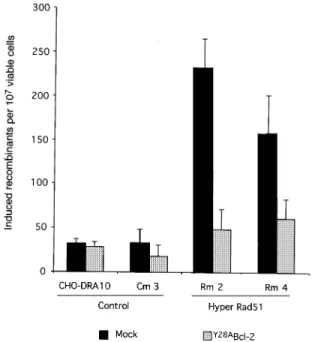
Bcl-2 suppresses RAD51-induced recombination Conservative recombination is RAD51 dependent, whereas NHEJ and SSA are RAD51 independent. To con®rm further that Bcl-2 overexpression inhibits recom-bination events promoted by RAD51, we used a CHO-DRA10 derivative cell line overexpressing mammalian RAD51. We have previously shown that overexpression Fig. 6. Effect of Bcl-2 on recombination induced by I-SceI. (A) Number of total recombinants (NeoR). (B) Number of conservative events
(NeoR/HygR). (C) Distribution (percentage) of class of events: non-conservative events (grey bars); conservative events (black bars). The numbers
on top of the histograms indicate the exact value of the percentage. Cm3, control corresponding to the parental CHO-DRA10 cell line transfected with the empty expression vector. ADRA14 and ADRA17 correspond to two independent clones expressingY28ABcl-2.
of the mouse MmRAD51 cDNA strongly stimulates homo-logous recombination after ionizing radiation (Lambert and Lopez, 2000). We tested here whether Bcl-2 expres-sion is able to suppress the stimulation of radiation-induced recombination resulting from the expression of MmRAD51.
At a dose of 6 Gy, overexpression of MmRAD51 led to a 10-fold stimulation of radiation-induced recombination compared with the control lines. In the lines transfected with the Bcl-2 expression vector, RAD51-stimulated recombination was reduced to the level of the control lines (Figure 7). This result indicates that the recombin-ation stimulrecombin-ation provoked by the overexpression of MmRAD51 was thwarted by the expression of Bcl-2, and is consistent with the data showing a speci®c inhibition of conservative recombination events.
Bcl-2 expression affects post-translation modi®cation of Rad51 protein
The data presented above show a speci®c inhibition of the RAD51 recombination pathway. In order to gain some clues regarding the molecular mechanisms involved, we focused on the Rad51 protein. Rad51 protein acts in a huge protein complex, but we have previously shown that it plays a pivotal role in gene conversion regulation. Overexpression of only Rad51 protein or of a dominant-negative form is suf®cient to stimulate or to inhibit gene conversion (Lambert and Lopez, 2000). These results indicate that acting on the regulation of Rad51 protein alone could be suf®cient to regulate the whole gene conversion pathway. We thus checked the status of Rad51 protein in cell lines overexpressing Bcl-2. First, the amount of Rad51 protein was measured by western
blotting (Figure 8A). The fact that the amount of Rad51 protein was identical in lines expressing Bcl-2 and in control lines showed that Bcl-2 did not affect transcription, RNA maturation or translation ef®ciencies of RAD51. Thus, if Rad51 is a target, the consequences of Bcl-2 expression should act post-translationally.
After genotoxic stress, Rad51 protein re-localizes in nuclear foci (Haaf et al., 1995). Since Bcl-2 has been reported to inhibit the nuclear transport of proteins such as p53 (Ryan et al., 1994), we checked whether Bcl-2 expression affects the formation of Rad51 nuclear foci (Figure 8B). No differences in the percentage or kinetics of Rad51 focus formation were observed between the Bcl-2-overexpressing and control cell lines. This result excludes the down-regulation of gene conversion via inhibition of Rad51 protein nuclear transport.
We analysed Rad51 protein by two-dimensional gel electrophoresis (TDGE). In the ®rst dimension, proteins migrate according to their isoelectric point; the second dimension corresponds to a classical SDS±polyacrylamide gel and the proteins migrate essentially according to their molecular weight. A post-translation modi®cation can affect either the molecular weight and/or the charge of the protein. Subtle modi®cations can thus be detected by TDGE. In the control line, Rad51 protein, detected with an anti-Rad51 antibody, corresponded to a faint spot plus two main spots, with the same molecular weight. This showed that Rad51 was present in three forms, which differ according to their charge. In the extract from cell lines expressing Bcl-2, the total amount of Rad51 protein is the same, but the distribution between the three spots changed. Indeed, most of the Rad51 protein is present in the central spot and the most basic spot almost disappeared. This result suggests that a large fraction of Rad51 protein became more acid.
Taken together, the western blot and TDGE results show that expression of Bcl-2 affects the post-translation modi®cation of Rad51 protein.
Bcl-2 leads to a mutator phenotype
Although the RAD51-dependent repair pathway is af-fected, these cells have increased resistance to ionizing radiation, implying that other repair pathways should be pro®cient. Our results show that Bcl-2 speci®cally inhibits the free RAD51 pathway, but that the alternate error-prone repair pathways are pro®cient. This hypothesis is attested to by the fact that SSA, an error-prone recombination process that systematically leads to sequence deletion, is not decreased by Bcl-2 expression (see above). Consequently, one prediction would be that mutagenesis should be increased in cells expressing the ectopic Bcl-2.
We ®rst measured spontaneous mutagenesis in the Na+/
K+-ATPase membrane pump gene. Mutation in this
gene leads to ouabain resistance of the mutant cells. Spontaneous mutagenesis was calculated by ¯uctuation analysis using the Luria and DelbruÈck or the Lea and Coulson assay (Luria and DelbruÈck, 1943; Lea and Coulson, 1948; Capizzi and Jameson, 1973). In two independent clones, expression of Bcl-2 led to a 2.5-fold increase in the spontaneous rate of mutagenesis per cell per generation (Table III).
Fig. 7. Bcl-2 inhibits recombination stimulated by RAD51. Cells were irradiated at 6 Gy. Cm3, control cell line corresponding to the parental CHO-DRA10 transfected with the empty expression vector. Rm2 and Rm4 are two independent clones overexpressing mouse MmRAD51, leading to a stimulation of radiation-induced recombination (Lambert and Lopez, 2000). Black bars, transfection with an empty vector; grey bars, transfection with aY28ABcl-2 expression vector.
We then measured mutagenesis induced either by UV-C or by ionizing radiation. Expression of Bcl-2 led to a strong stimulation of UV-induced mutagenesis (Figure 9A). At a dose of 30 J/m2, UV-induced
mutagen-esis of the Na+/K+-ATPase membrane gene locus was
stimulated from 18- to 20-fold in two independent clones expressing Bcl-2, compared with the control cell line.
We also measured the g-ray-induced mutagenesis of two different loci: the Na+/K+-ATPase membrane gene and
the HPRT gene. Mutation of the latter gene leads to
6-thioguanine (6-TG) medium resistance. In two inde-pendent clones, Bcl-2 expression stimulated g-ray-induced mutagenesis of both loci (Figure 9B). At a dose of 6 Gy, mutagenesis was stimulated 10-fold at the ouabain resist-ance locus and 80- to 100-fold at the HPRT locus.
Discussion
The oncogenic role of Bcl-2 is generally attributed to the inhibition of a variety of apoptotic deaths. Thus, Bcl-2 is Fig. 8. Status of Rad51 protein in lines expressing Bcl-2. (A) Western blot. Arrows indicate Rad51 protein, actin (internal standard) and
overexpression of the exogenous human Bcl-2. Cell lines are described in Table I. CDR1 is a clone from pJS3-10 transfected with an empty expression vector. (B) Rad51 foci measured by immuno¯uorescence. Top: an example of Rad51 foci (left panel) and nucleus coloration by DAPI (right panel). Bottom: the kinetics of Rad51 foci formation after irradiation (6 Gy). (C) TDGE revealed with an anti-Rad51 antibody. Rad51 is indicated by an arrow. Left panel, extract from the control cell line (A3); right panel, extract from a cell line expressing an ectopic Bcl-2 (ADRA8).
Table III. Spontaneous mutagenesis rate Cell lines Expression of exogenous
Y28ABcl-2 protein Number of independentcultures Mutagenesis rate (3 10
±8/cell/generation)
Luria and DelbruÈck Lea and Coulson
CHO-DRA10 none 6 6.1 6 0.56 5.8
ADRA14 Bcl-2 6 14 6 0.49 14.9
ADRA17 Bcl-2 6 16 6 0.48 12.7
an oncogene when it is overexpressed, such as in follicular B-cell lymphomas, in which Bcl-2 overexpression results from a t(14;18) translocation (Bakhshi et al., 1985; Cleary and Sklar, 1985; Tsujimoto et al., 1985). Consequently, Bcl-2 studies classically use overexpression of Bcl-2 to mimic the pathological level that occurs in some cancers. In the present paper we show that overexpression of Bcl-2 protects the cells against apoptosis after genotoxic stress such as ionizing or UV radiation, but suppresses the associated stimulation of homologous recombination. This effect is not speci®c to Bcl-2 since Bcl-XLshows the same
behaviour. Moreover, it is not affected by the impact of p53 on the G1 checkpoint and on recombination. One
hypothesis could be that, after radiation, cells bearing improperly repaired DNA and accumulating mutations are protected from apoptosis by Bcl-2. This hypothesis may be true in part, but several lines of evidence do not totally ®t with it. Indeed, for a unique and non-toxic DSB, we show here that Bcl-2 does not affect all DSB repair systems, but very speci®cally the conservative RAD51-dependent recombination pathway. Moreover, the effect on recom-bination does not require activation by a profound genotoxic stress, and Bcl-2 decreases the recombination frequency to the basal level of the non-treated control cell line independently of the extent of recombination stimu-lation by different stresses (g, UV, mutant p53 protein expression). In addition, the functions of Bcl-2 in recombination and apoptosis can be separated in the mutantG145ABcl-2. We show that Bcl-2 expression affects
post-translational regulation of Rad51 protein, even in the absence of genotoxic stress. This suggests that Bcl-2 acts on the recombination pathway itself. This conclusion is consistent with the fact that theG145ABcl-2 mutant exhibits
separation of functions for the death repression activity and BRI.
The use of the rare-cutting endonuclease I-SceI allowed us to determine the precise recombinational DSB repair pathway affected by Bcl-2. I-SceI produces a DSB targeted in the recombination substrate with no detectable associ-ated effect on cell survival. Bcl-2 expression does not signi®cantly modify the total frequency of I-SceI-induced recombination (NeoR clones), but speci®cally decreases
the frequency of conservative recombination events (double NeoR/HygR resistant clones). Since
non-vative recombination is RAD51 independent and conser-vative recombination is RAD51 dependent (Ivanov et al., 1996; Lambert and Lopez, 2000), the present data show that Bcl-2 expression speci®cally affects the RAD51 pathway even in the absence of stress able to induce apoptosis. These results were con®rmed when using cell lines overexpressing the mouse MmRAD51 protein. In the present paper we studied more speci®cally the effect of Bcl-2 expression on Rad51 protein. Overexpression of only Rad51 protein or of a dominant-negative form is suf®cient to stimulate or to inhibit gene conversion (Lambert and Lopez, 2000). Thus, the regulation of Rad51 protein would be suf®cient to control the whole gene conversion pathway. We have shown that the amount of Rad51 protein was not modi®ed by Bcl-2 expression. This shows that Bcl-2 does not act on the ef®ciency of RAD51 gene transcription or translation of Rad51 protein. We also show here that the expression of Bcl-2 does not affect the nuclear transport of Rad51 or foci formation. Nevertheless, since gene conversion is inhibited, if Rad51 foci actually represent repair foci, this result suggests that either the foci are formed but are inactive or that the late steps of the recombination process are inhibited. One hypothesis could be that Bcl-2 affects the post-translation regulation of Rad51 protein. This hypothesis is consistent with the fact that the amount of Rad51 protein measured Fig. 9. Bcl-2 stimulates induced mutagenesis. In black is the control line CHO-DRA10. In gray are ADRA14 and ADRA17, two independent clones expressingY28ABcl-2. The values correspond to the number of induced mutants: the number of resistant clones (ouabain or 6-TG) for 107viable
treated cells minus the number of resistant clones (ouabain or 6-TG) in 107non-treated cells. (A) UV-C-induced mutagenesis. Mutagenesis was
measured at the ouabain locus. (B) g-ray-induced mutagenesis at two loci: ouabain resistance (in black) and HPRT locus (6-TG resistance, in grey). Cells were irradiated at 6 Gy.
by western blotting remains unchanged by the expression of Bcl-2. We also show here that Bcl-2 affects the distribution of the different Rad51 protein forms, as measured by TDGE analysis. Indeed, the basic fraction of Rad51 became more acid in the Bcl-2-overexpressing cell line. Phosphorylation is a good candidate mechanism, compatible with our TDGE data. Phosphorylation modi-®es the charge of the protein, resulting in a change in the migration pattern in TDGE. Rad51 was shown to undergo phosphorylation. For instance, phosphorylation by cAbl inactivates Rad51 strand-exchange activity (Yuan et al., 1998), but favours the formation of Rad51 foci (Chen et al., 1999). Other kinases could act on Rad51 protein, and the characterization of the transduction signal pathway involved in BRI is under investigation.
It has recently been reported that targeted cytoplasmic radiation induces mutation of the nuclear genome, sug-gesting, as is the case here, that there is a tight connection between cytoplasm metabolism and the maintenance of genome stability (Wu et al., 1999).
Paradoxically, although expression of Bcl-2 inhibits one DSB repair pathway, these cells show increased resistance to irradiation. Among the different DSB repair pathways, Bcl-2 selectively affects the error-free DSB repair path-way (the RAD51-conservative recombination pathpath-way), but is without effect on the mutagenic pathways (SSA, NHEJ). Since Bcl-2-overexpressing cells remain more resistant to genotoxic stress, despite its effects on error-free DNA repair systems, error-prone DNA repair systems must compensate. We show here that SSA remains pro®cient. Conservative recombination is not the only error-free DNA repair pathway affected by Bcl-2. Expression of Bcl-2 has also been shown to result in an attenuation of the nucleotide excision repair (NER) system in UV-irradiated cells (Liu et al., 1997). The fact that different repair pathways are affected suggests that distinct patterns of mutagenesis should occur. In line with this, Bcl-2 overexpression results in increased mutagenesis induced by different genotoxic stresses such as benzene metabolite-induced oxidative stress (Kuo et al., 1999), UV or g-rays (this work), but also in increased spontaneous mutagenesis in the mutant p53 lines (this work). We have measured mutagenesis at two different loci, HPRT and the ouabain resistance gene, that do not exactly monitor the same kind of mutagenesis (Friedrich and Cof®no, 1977). We have also measured mutagenesis induced by different types of genotoxic stress (UV-C, g-rays), which produce different kinds of damage. We show that, in all cases, Bcl-2 overexpression results in an increase in mutagenesis. High levels of chromosome aberrations after ionizing radiation have also been noted in human lymphoblast lines expressing Bcl-2 (Cherbonnel-Lasserre et al., 1996). Taken together, these results suggest a wide mutagenesis spectrum. Since NER, base excision repair as well as conservative recombination are error-free repair pathways, this indicates that Bcl-2 (or Bcl-XL) expression results in a
concerted and speci®c inhibition of the error-free path-ways, resulting in a strong mutator phenotype in response to a broad spectrum of genotoxic stresses.
The data presented here show that Bcl-2 does not affect all the DSB repair pathways, but acts on the regulation of the balance between conservative versus non-conservative recombination. Control of this pathway is of particular
importance in cancer predisposition. It is generally assumed that the oncogenic role of Bcl-2 overexpression (as in some lymphomas) results from its death-repression activity. The present data reveal another separate onco-genic role of Bcl-2 overexpression: the mutator and genetic instability phenotypes due to the inhibition of error-free DNA repair pathways, such as conservative homologous recombination.
More generally, expression of Bcl-2 confers increased resistance to genotoxic stress associated with an increase in mutagenesis. This dual phenotype is reminiscent of the general consequences of the SOS system in bacteria, despite differences in the exact molecular control.
Materials and methods
DNA manipulations
All DNA manipulations were performed as described (Sambrook et al., 1989; Ausubel et al., 1999).
pSFFv-Bcl-2 expression vector was kindly provided by C.Cherbonnel-Lasserre and is described elsewhere (Cherbonnel-C.Cherbonnel-Lasserre et al., 1996). pEF-Y28ABcl-2 expression vector was kindly provided by Drs J.Adams
and S.Cory, and is described elsewhere (Huang et al., 1997). The
G145ABcl-2 mutant expression vector was kindly provided by Dr
Korsmeyer (Yin et al., 1994). Cells and Rad51 foci
Mouse L, pJS3-10 (Liskay et al., 1984) and CHO-K1 DRA10 cells (Liang et al., 1998) and their derivative lines were cultured at 37°C with 5% CO2in Dulbecco's modi®ed Eagle's medium (DMEM) supplemented with 10% fetal bovine serum. TK+(recombinant) clones were selected in
HAT medium (100 mM hypoxanthine, 2 mM aminopterin, 15 mM thymidine) as described (Liskay et al., 1984). Neo+(recombinant) clones
were selected with 500 mg/ml G418 and hygromycin-resistant clones were selected with 500 mg/ml hygromycin. Puromycin selection was performed at 5 mg/ml. Single transfections were performed using Transfast (Promega, Madison, WI). Co-transfections were performed using the calcium phosphate precipitate technique (Sambrook et al., 1989). The Rad51 foci were analysed as described (Haaf et al., 1995) using an anti-Rad51 antibody (Oncogene Research Products, Cambridge, MA).
Western blot analysis
All extract preparation steps were performed at 4°C. After washing with phosphate-buffered saline (PBS), cells were suspended in lysis buffer A (25 mM Tris±HCl pH 7.5, 1 mM EDTA, 600 mM NaCl, 0.5% NP-40, 2 mg/ml leupeptin, 2 mM pepstatin, 1 mM phenylmethylsulfonyl ¯uoride) and incubated for 40 min on ice. Extracts were centrifuged for 30 min at 15 000 g, supernatant was retrieved and protein concentration was determined using the Bio-Rad Protein Assay (Bio-Rad, Hercules, CA). A total of 40 mg/well of the boiled samples was loaded onto a 10% SDS±PAGE gel. After migration, the proteins were electrotransferred onto a nitrocellulose membrane and probed with speci®c antibodies: anti-human Bcl-2 (SC-509; Santa Cruz Biotechnology, Santa Cruz, CA) or anti-Rad51 (Ab-1; Oncogene Research Products). Standard procedures were used for the electrophoresis, transfer and western blotting. Antibodies were visualized using the ECL detection kit (Amersham Pharmacia Biotech, Orsay, France).
Measurement of apoptosis
Two different methods for the measurement of apoptosis were used (Celis, 1994).
Pro®le of DNA content (FACS analysis). For each point, 106cells were
plated in DMEM and incubated for 48 h at 37°C. Cells were then irradiated at 6 Gy using a137Cs irradiator (2 Gy/min). After 24 or 48 h
incubation at 37°C in DMEM, cells were trypsinized, collected by centrifugation (5 min at 2000 g), resuspended in 500 ml of PBS and ®xed by adding 1.5 ml of cold ethanol. The DNA content was estimated by propidium iodide ¯uorescence and DNA ¯ow cytometry (Becton FACScalibur).
During the subsequent rinse, apoptotic cells permeabilized by ethanol ®xation leak low-molecular-weight DNA into the cytoplasm. The lower Bcl-2 inhibition of homologous recombination
DNA content of these cells means that they contain less DNA stained by the ¯uorochrome. Thus, cells with lower DNA staining than that of G1 cells (the so-called `sub-G1peaks') are considered apoptotic.
Analysis of the morphological changes (Hoechst ¯uorescence). For each point, 5 3 104cells were plated in DMEM and incubated for 48 h at
37°C. Cells were then irradiated at 6 Gy using a137Cs irradiator (2 Gy/
min). Twenty-four or 48 h after irradiation, cells were washed and stained for 30 min at 37°C with Hoechst 33342 in culture medium. Hoechst 33342 penetrates the plasma membrane without permeabilization and inter-calates into the DNA. The cells were visualized by ¯uorescence microscopy. In contrast to normal cells, the nuclei of apoptotic cells appear to be one or more featureless, bright spherical beads. For each point, >500 cells (normal and apoptotic cells) were counted.
Recombination and mutagenesis measurements
Fluctuation analysis was used to measure spontaneous recombination and spontaneous mutagenesis. For each line analysed, several independent cultures were plated and cultured to con¯uence. Cells were then trypsinized, counted, and one portion was used for plating ef®ciency estimation. The remaining cells were plated under selection medium. Recombinant (TK+) L cells (pJS3-10 and pJS4-7-1 and derivatives) were
selected on HAT selective medium. Recombinants (Neo+) from the
CHO-DRA10 cell line (and derivatives) were selected on G418 or G418 + hygromycin. The resulting number of TK+or Neo+clones allowed us to
calculate the recombination frequency. The mutant colonies were selected on 2 mM ouabain or 20 mM 6-TG. The resulting number of ouabain- or 6-TG-resistant clones allowed us to calculate the mutagenesis frequency. The rate of recombination or mutagenesis per cell per generation was calculated by using the ¯uctuation analysis of Luria and DelbruÈck (Luria and DelbruÈck, 1943; Capizzi and Jameson, 1973) or Lea and Coulson (1948).
Recombination frequency after treatment with g-rays (in PBS, using a
137Cs irradiator 2 Gy/min) or UV-C (254 nm at 0.7 J/m2/s) was measured
at the dose indicated. After irradiation, the cells were incubated in DMEM at 37°C for 24 h. The cells were then trypsinized and divided into two fractions. The ®rst fraction was used to calculate the viability by measuring the plating ef®ciency. The second fraction was plated under HAT or G418 selection to measure the frequency of recombinant clones. Recombination after induction of a DSB was measured: 3 3 105cells
(for the control lines DRA10 and Cm3) or 1.8 3 106cells (for ADRA14
and ADRA17) were plated and transfected with 2 and 12 mg, respectively, of an expression vector for the I-SceI endonuclease (pCMV I-SceI). Twenty-four hours after transfection, G418 or G418 + hygromycin selection was initiated. The NeoRclone frequency was estimated by the
ratio of (number of Neo+clones)/(total number of cells plated). The NeoR/
HygRclone frequency was estimated by the ratio of (number of
double-resistant NeoR/HygRclones)/(total number of cells plated). The
percent-age of SI recombination events was calculated from the ratio of (frequency of double-resistant NeoR/HygRclones)/(frequency of
single-resistant NeoRclones).
Two-dimensional gel electrophoresis
Extract preparation. CHO cells were pelleted at 1200 r.p.m. for 5 min and washed twice in PBS. The pellet was resuspended quickly in extraction buffer [8 M urea, 1 M thiourea, 0.5% CHAPS, 50 mM dithiothreitol (DTT), 24 mM spermine] and incubated for 1 h at room temperature. After ultracentrifugation at 200 000 g for 1 h at 20°C, the concentration of supernatant was measured using Bradford reagent (Bio-Rad).
Two-dimensional electrophoresis. IPG strips (pH 4±7) were rehydrated overnight with 150 ml of extraction buffer containing 1% IPG buffer and 200 mg protein sample and traces of bromophenol blue. Proteins were isoelectrofocused using a Multiphor II Electrophoresis Unit cooled at 20°C with successive 30 min steps at 200, 1000 and 2000 V, and a ®nal step at 3500 V for 2.5 h.
Focused IPG strips were incubated for 15 min in equilibration solution (50 mM Tris±HCl pH 6.8, 6 M urea, 30% glycerol, 1% SDS, bromophenol blue) containing 50 mM DTT and then for 15 min in equilibration solution containing 200 mM iodoacetamide. For the second dimension, 10% SDS±PAGE was performed.
Acknowledgements
We would like to thank Drs S.Cory and J.Adams for providing the Y28ABcl-2 expression vector, Dr Korsmeyer for G145ABcl-2,
Dr C.Cherbonnel-Lasserre for Bcl-2 and Bcl-XL, Dr M.Liskay for the
pJS3-10 line, and Dr M.Jasin for the CHO-DRA10 line and I-SceI expression vector. We thank Drs D.Marsh and C.White for critical reading of the manuscript. We are grateful to Drs P.Bertrand and S.Roche for TDGE expertise. Y.S. is supported by an FRM fellowship, A.D. is supported by an INSTN/EDF fellowship and S.L. is supported by an INSTN fellowship. This work was supported by Electricite de France, ARC and ANRS.
References
Adams,J.M. and Cory,S. (1998) The Bcl-2 protein family: arbiters of cell survival. Science, 281, 1322±1326.
Ausubel,F., Brent,R., Kingston,R., Moore,D., Seidman,J., Smith,J. and Struhl,K. (1999) Current Protocols in Molecular Biology. John Wiley, Boston, MA.
Bakhshi,A., Jensen,J.P., Goldman,P., Wright,J.J., McBride,O.W., Epstein,A.L. and Korsmeyer,S.J. (1985) Cloning the chromosomal breakpoint of t(14;18) human lymphomas: clustering around JH on chromosome 14 and near a transcriptional unit on 18. Cell, 41, 899±906.
Bertrand,P., Rouillard,D., Boulet,A., Levalois,C., Soussi,T. and Lopez,B.S. (1997) Increase of spontaneous intrachromosomal homologous recombination in mammalian cells expressing a mutant p53 protein. Oncogene, 14, 1117±1122.
Capizzi,R.L. and Jameson,J.W. (1973) A table for the estimation of the spontaneous mutation rate of cells in culture. Mutat. Res., 17, 147±148.
Celis,J. (1994) Cell Biology: A Laboratory Handbook. Academic Press, San Diego, CA.
Chen,G. et al. (1999) Radiation-induced assembly of Rad51 and Rad52 recombination complex requires ATM and c-Abl. J. Biol. Chem., 274, 12748±12752.
Cherbonnel-Lasserre,C., Gauny,S. and Kronenberg,A. (1996) Suppression of apoptosis by Bcl-2 or Bcl-xLpromotes susceptibility to mutagenesis. Oncogene, 13, 1489±1497.
Cleary,M.L. and Sklar,J. (1985) Nucleotide sequence of a t(14;18) chromosomal breakpoint in follicular lymphoma and demonstration of a breakpoint-cluster region near a transcriptionally active locus on chromosome 18. Proc. Natl Acad. Sci. USA, 82, 7439±7443. Donehower,L.A. and Bradley,A. (1993) The tumor suppressor p53.
Biochim. Biophys. Acta, 1155, 181±205.
Dudenhoffer,C., Kurth,M., Janus,F., Deppert,W. and Wiesmuller,L. (1999) Dissociation of the recombination control and the sequence-speci®c transactivation function of p53. Oncogene, 18, 5773±5784. Flygare,J., Armstrong,R.C., Wennborg,A., Orsan,S. and Hellgren,D.
(1998) Proteolytic cleavage of HsRad51 during apoptosis. FEBS Lett., 427, 247±251.
Friedrich,U. and Cof®no,P. (1977) Mutagenesis in S49 mouse lymphoma cells: induction of resistance to ouabain, 6-thioguanine and dibutyryl cyclic AMP. Proc. Natl Acad. Sci. USA, 74, 679±683.
Haaf,T., Golub,E.I., Reddy,G., Radding,C.M. and Ward,D.C. (1995) Nuclear foci of mammalian Rad51 recombination protein in somatic cells after DNA damage and its localization in synaptonemal complexes. Proc. Natl Acad. Sci. USA, 92, 2298±2302.
Hainaut,P. (1995) The tumor suppressor protein p53: a receptor to genotoxic stress that controls cell growth and survival. Curr. Opin. Oncol., 7, 76±82.
Hartwell,L. (1992) Defects in a cell cycle checkpoint may be responsible for the genomic instability of cancer cells. Cell, 71, 543±546. Hartwell,L., Weinert,T., Kadyk,L. and Garvik,B. (1994) Cell cycle
checkpoints, genomic integrity and cancer. Cold Spring Harb. Symp. Quant. Biol., 59, 259±263.
Hollstein,M., Sidransky,D., Vogelstein,B. and Harris,C.C. (1991) p53 mutations in human cancers. Science, 253, 49±53.
Hu,T., Miller,C.M., Ridder,G.M. and Aardema,M.J. (1999) Characterization of p53 in Chinese hamster cell lines CHO-K1, CHO-WBL and CHL: implications for genotoxicity testing. Mutat. Res., 426, 51±62.
Huang,D.C., O'Reilly,L.A., Strasser,A. and Cory,S. (1997) The anti-apoptosis function of Bcl-2 can be genetically separated from its inhibitory effect on cell cycle entry. EMBO J., 16, 4628±4638. Huang,Y. et al. (1999) Role for caspase-mediated cleavage of Rad51 in
induction of apoptosis by DNA damage. Mol. Cell. Biol., 19, 2986±2997.
Ivanov,E.L., Sugawara,N., Fishman-Lobell,J. and Haber,J.E. (1996) Genetic requirements for the single-strand annealing pathway of
double-strand break repair in Saccharomyces cerevisiae. Genetics, 142, 693±704.
Ko,L.J. and Prives,C. (1996) p53: puzzle and paradigm. Genes Dev., 10, 1054±1072.
Kuo,M.L., Shiah,S.G., Wang,C.J. and Chuang,S.E. (1999) Suppression of apoptosis by Bcl-2 to enhance benzene metabolite-induced oxidative DNA damage and mutagenesis: a possible mechanism of carcinogenesis. Mol. Pharmacol., 55, 894±901.
Lambert,S. and Lopez,B.S. (2000) Characterization of mammalian RAD51 double strand break repair using non-lethal dominant negative forms. EMBO J., 19, 3090±3099.
Lea,D.E. and Coulson,C.A. (1948) The distribution of the numbers of mutants in bacterial populations. J. Genet., 49, 264±248.
Levine,A.J., Momand,J. and Finlay,C.A. (1991) The p53 tumour suppressor gene. Nature, 351, 453±456.
Liang,F., Han,M., Romanienko,P.J. and Jasin,M. (1998) Homology-directed repair is a major double-strand break repair pathway in mammalian cells. Proc. Natl Acad. Sci. USA, 95, 5172±5177. Liskay,R.M., Stachelek,J.L. and Letsou,A. (1984) Homologous
recombination between repeated chromosomal sequences in mouse cells. Cold Spring Harb. Symp. Quant. Biol., 49, 183±189.
Liu,Y., Naumovski,L. and Hanawalt,P. (1997) Nucleotide excision repair capacity is attenuated in human promyelocytic HL60 cells that overexpress BCL-2. Cancer Res., 57, 1650±1653.
Luria,S.E. and DelbruÈck,M. (1943) Mutations of bacteria from virus sensitivity to virus resistance. Genetics, 28, 491±511.
Mekeel,K.L., Tang,W., Kachnic,L.A., Luo,C.M., DeFrank,J.S. and Powell,S.N. (1997) Inactivation of p53 results in high rates of homologous recombination. Oncogene, 14, 1847±1857.
O'Connor,L., Strasser,A., O'Reilly,L.A., Hausmann,G., Adams,J.M., Cory,S. and Huang,D.C. (1998) Bim: a novel member of the Bcl-2 family that promotes apoptosis. EMBO J., 17, 384±395.
Rich,T., Watson,C.J. and Wyllie,A. (1999) Apoptosis: the germs of death. Nature Cell Biol., 1, E69±E71.
Ryan,J.J., Prochownik,E., Gottlieb,C.A., Apel,I.J., Merino,R., Nunez,G. and Clarke,M.F. (1994) c-myc and bcl-2 modulate p53 function by altering p53 subcellular traf®cking during the cell cycle. Proc. Natl Acad. Sci. USA, 91, 5878±5882.
Saintigny,Y., Rouillard,D., Chaput,B., Soussi,T. and Lopez,B.S. (1999) Mutant p53 proteins stimulate spontaneous and radiation-induced intrachromosomal homologous recombination independently of the alteration of the transactivation activity and of the G1 checkpoint. Oncogene, 18, 3553±3563.
Sambrook,J., Fritsch,E.F. and Maniatis,T. (1989) Molecular Cloning: A Laboratory Manual, 2nd edn. Cold Spring Harbor Laboratory Press, Cold Spring Harbor, NY.
Smith,M.L. and Fornace,A.J.,Jr (1995) Genomic instability and the role of p53 mutations in cancer cells. Curr. Opin. Oncol., 7, 69±75. Strasser,A., Harris,A.W., Jacks,T. and Cory,S. (1994) DNA damage can
induce apoptosis in proliferating lymphoid cells via p53-independent mechanisms inhibitable by Bcl-2. Cell, 79, 329±339.
Tsujimoto,Y., Cossman,J., Jaffe,E. and Croce,C.M. (1985) Involvement of the bcl-2 gene in human follicular lymphoma. Science, 228, 1440±1443.
Wiesmuller,L., Cammenga,J. and Deppert,W.W. (1996) In vivo assay of p53 function in homologous recombination between simian virus 40 chromosomes. J. Virol., 70, 737±744.
Wu,L.J., Randers-Pehrson,G., Xu,A., Waldren,C.A., Geard,C.R., Yu,Z. and Hei,T.K. (1999) Targeted cytoplasmic irradiation with a particles induces mutations in mammalian cells. Proc. Natl Acad. Sci. USA, 96, 4959±4964.
Wyllie,A.H. et al. (1999) Apoptosis and carcinogenesis. Br. J. Cancer Suppl. 1, 80, 34±37.
Yin,X.M., Oltvai,Z.N. and Korsmeyer,S.J. (1994) BH1 and BH2 domains of Bcl-2 are required for inhibition of apoptosis and heterodimerization with Bax. Nature, 369, 321±323.
Yuan,Z.M. et al. (1998) Regulation of Rad51 function by c-Abl in response to DNA damage. J. Biol. Chem., 273, 3799±3802.
Received August 25, 2000; revised and accepted March 22, 2001