HAL Id: tel-00797125
https://tel.archives-ouvertes.fr/tel-00797125
Submitted on 5 Mar 2013
HAL is a multi-disciplinary open access
archive for the deposit and dissemination of sci-entific research documents, whether they are pub-lished or not. The documents may come from teaching and research institutions in France or abroad, or from public or private research centers.
L’archive ouverte pluridisciplinaire HAL, est destinée au dépôt et à la diffusion de documents scientifiques de niveau recherche, publiés ou non, émanant des établissements d’enseignement et de recherche français ou étrangers, des laboratoires publics ou privés.
Regulation of plastid transcription by sigma factors and
anti-sense RNAs in Arabidopsis thaliana.
Malik Ghulam Mustafa
To cite this version:
Malik Ghulam Mustafa. Regulation of plastid transcription by sigma factors and anti-sense RNAs in Arabidopsis thaliana.. Molecular biology. Université Joseph-Fourier - Grenoble I, 2010. English. �tel-00797125�
Université de Grenoble; France
Ecole Doctorale de Chimie et Science du Vivant
Regulation of plastid transcription by sigma factors
and anti-sense RNAs in Arabidopsis thaliana.
Thesis
To obtain the grade of
Doctor of University of Grenoble, France
Discipline: Biology
Presented and defended publically by
Malik Ghulam Mustafa
on
15 December 2010
Directors of thesis : Dr. Silva Lerbs-Mache
: Dr. Livia Merendino
Composition of Jury:
Laurence Maréchal-Drouard
ReporterFrançoise Monéger
ReporterMichel Goldschmidt-Clermont
ExaminatorSilva Lerbs-Mache
Director of thesisAbstract
Abstract :
Les chloroplastes, responsables de la photosynthèse chez les organismes autotrophes, possèdent un génome plastidial codant de 100 à 130 gènes dont environ 80 pour des protéines principalement impliquées dans la photosynthèse, la transcription et la traduction. L'expression de ces gènes, coordonnée entre le plaste et le noyau, implique deux types d'ARN polymérases, la NEP (Nucleus Encoded RNA Polymerase) et la PEP (plastid Encoded RNA Polymerase) laquelle s‟associe à l‟un des 6 facteurs sigma (SIG), codés dans le noyau pour la reconnaissance spécifique de promoteurs de transcription.
Nous avons tout d‟abord analysé le rôle de ces facteurs sigma dans la régulation transcriptionnelle des deux opérons codant des sous-unités de l‟ATP synthase, atpI/H/F/A et atpB/E, en précisant le rôle particulier de SIG3 dans la reconnaissance spécifique du promoteur (-418) de l‟atpH. Nous avons identifié les promoteurs des transcrits polycistronique et ceux situés en amont des gènes atpH et atpE, et avons montré (1) que les gènes des deux opérons sont co-régulés par SIG3 et SIG2 sauf atpI régulé par SIG2 seul et (2), que SIG3 jouerait un rôle essentiel dans la surexpression monocistronique d‟atpH par la reconnaissance d‟un promoteur (-418) en amont de atpH. L‟analyse systématique des transcrits plastidiaux accumulés en fonction de l‟éclairement des plantes nous a permis de corréler cette surexpression à un éclairement élevé (1300 µE) de plantes matures.
SIG3 reconnaît aussi spécifiquement le promoteur de psbN, gène localisé sur le brin opposé de l‟opéron psbB/T/H/petB/petD, produisant un ARN anti-sens de psbT et de la région intergénique psbT/psbH. Nos résultats montrent que l‟anti-sens de psbT couvre la région codante, le 5'UTR et la quasi-totalité 3' UTR du transcrit sens psbT, pouvant ainsi réguler la production de PSBT en interférant dans la traduction par la formation d‟un duplex ARN. L‟anti-sens pourrait aussi intervenir dans le processing dans la région 5‟ UTR de psbH.
Chloroplasts, responsible for photosynthesis in autotrophic organisms, have a genome containing 100-130 genes, 80 of which code for proteins mainly involved in photosynthesis, transcription and translation. Gene expression, involves two types of RNA polymerases, NEP (Nucleus Encoded RNA Polymerase) and PEP (Plastid Encoded RNA Polymerase). Six nucleus encoded sigma factors participate to PEP promoter specificity.
We first have analyzed the role of sigma factors in the transcriptional regulation of the two atp operons, atpI/H/F/A and atpB/E, with special emphasis on the specific contribution of SIG3 to atpH gene expression. We identified the promoters responsible for polycistronic
Abstract transcripts and the internal promoters upstream of the atpH and the atpE genes. All genes of both atp operons are SIG3 and SIG2 dependent except atpI that is regulated by SIG2 only. The monocistronic -418 initiated atpH mRNA might contribute to the higher stoichiometry of atpH. A systematic analysis of plastid gene expression under different light conditions showed that SIG3 plays an important role in the transcript accumulation of atpH in high light (1300 µE) in mature plants.
Similarly, SIG3 also recognizes specifically the promoter of psbN located between psbT and psbH but on the opposite DNA strand and producing an anti-sense RNA to psbT. We showed that the anti-sense RNA covers the coding region, the 5‟ UTR and almost the entire 3‟ UTR of the psbT sense transcript and thus might regulate the expression of the psbT gene by interfering in the translation of psbT mRNA via duplex formation. It could also be necessary for a processing event in the 5‟ UTR of psbH.
Acknowledgements:
Acknowledgements
Many may disagree but my simple answer to the question of the professional essence of ph.d period will be to “have confidence in yourself and to the fellow researchers”. That‟s why I am highly indebted to Dr. Silva Lerbs Mache, the director of my thesis.
During this period, Dr. Silva not only provided all the lab material necessary for carrying out day to day research but also spent a lot of time for the analysis of the results. I have greatly benefitted from her scientific competence and had a chance of scientific discussions with her. I am extremely indebted and thankful to you Dr. Silva for the patience you showed and for the time you spared and spent for the improvement of the manuscript. It was exceptional. Thanks again.
But if someone asks me what Dr. Silva did best for me during ph.d period, I would say: „The best thing she did for me was the appointment of Dr. Livia Merendino as my co-director of thesis”.
It is without exaggeration that I was in dire need of a “ Livia Merendino” to be what actually I am. She stunned me by telling Silva in the first meeting that Mustafa knows how to extract RNAs. When we came out of the office, I reminded Livia that my first three extractions resulted in completely degraded RNAs. She replied, „I know but I am sure you won‟t spend three years to learn RNA extraction”. Since that day I never looked back and don‟t remember if I extracted degraded RNAs. Her trust and confidence in me have always been a source of great motivation for me.
I am highly indebted to Dr. Livia Merendino for having always kept her office door open for answering to all my questions. Without doubt, she showed great patience to have scientific, religious and social discussions with me. She has always been a source of hope, courage and confidence for me during the ups and downs of my professional and personal life. She invested a lot of time for the explanation of the theoretical aspects of experiments. Above all it was a pure pleasure to work and discuss with her. I am extremely short of words to pay gratitude to you Livia for all you have done for me. I wish you a life of unlimited pleasures and success.
I am thankful to Silva and Livia for having spared a lot of time for the correction of this manuscript. I am thankful to Régis Mache for his kind help to discuss and introduce important corrections in this manuscript.
I am also grateful to Emiline Lamdert and Florence Courtois and Jean Piere Alcaraz for technical assistance in the beginning of thesis. I am grateful to Emelie Demarsy, Wafa Zghidi whose work became the base of my thesis. I am thankful to Denis Falconet, Guillaume, Frank Buhr, David Downy, Gabrielle Tichtinsky and Abder for nice working environment and company.
I am also thankful to Higher Education Commission Pakistan for providing me the scholarship of 57 months for getting higher education in France.
Along with the wonderful professional life, I have experienced five best years of my personal life during my stay in France. I dedicate all this to my friends especially „Grenobloise‟ (I would like to write the individual names but the list is very long) whose company or contact had been a source of enjoyment, pleasure and peace of mind. To be honest, in their company I never missed my family. Thanks to all of you. I am thankful to Muhammad Imran Ashiq and his parents for a wonderful time in Angers.
I am extremely grateful to „ ma petite soeur‟ Sumaira Kousar‟ to take a lot of care and to be a real sister in true sense. I am highly thankful to Muhammad Asam Riaz for having waited till late in the evening to share dinner with me during my three years stay in Grenoble. I am grateful to my parents, brothers and sisters and friends in Pakistan for their best wishes, prayers and love.
Above all I am thankful to Almighty Allah for providing me the wit and health without which nothing would have been possible.
Dedication
iv
Dedication
I dedicate this humble work
To
My sweet uncle and a friend : Prof. Allah Dad Malik My high school teacher and a friend : Prof. Ahmad Nawaz Virk My university teacher and a friend : Prof. Naveed Irshad My elder brother who took great care of
the family in my absence : Dr. Shahid Iqbal
And to the co-director of my thesis:
Dr. Livia Merendino
Table of contents v INTRODUCTION ... 1 1. PLASTIDS ... 2 1.1. Proplastids ... 4 1.2. Chloroplast ... 4 1.3. Chromoplast ... 5 1.4. Etioplasts ... 5 1.5. Leucoplasts ... 6 1.6. Amyloplasts ... 6 1.7. Elaioplasts... 6 1.8. Proteinoplasts ... 6 1.9. Gerontoplasts ... 6 2. ORIGIN OF PLASTIDS ... 8 3. PHOTOSYNTHESIS ... 9 3.1. Light cycle ... 10
3.1.1. Photosystem II and cytochrome b6-f complex ... 11
3.1.2. Photosystem I ... 12
3.2. Dark cycle ... 14
3.3. ATP synthase ... 15
3.3.1. CF1 ... 15
3.3.2. CFo ... 16
3.3.3. ATP synthase gene expression ... 17
4. PLASTID GENE EXPRESSION ... 19
4.1. Post transcriptional regulation in chloroplast ... 19
4.1.1. Chloroplast ribonucleases ... 20 4.1.1.1. Endonuclease ... 20 4.1.1.2. Exonucleases ... 20 4.1.2. Turn over ... 21 4.1.3. 5’ End maturation ... 21 4.1.4. 3’ End maturation ... 22 4.1.5. Splicing ... 23 4.1.6. Editing ... 25
4.2. Transcriptional regulation in chloroplast ... 28
4.2.1. Promoters ... 29
4.2.1.1. NEP promoter ... 30
Table of contents
vi
4.2.2. RNA polymerases ... 30
4.2.2.1. Nucleus Encoded RNA Polymerase (NEP)... 30
4.2.2.2. Plastid Encoded RNA Polymerase (PEP) ... 31
4.2.2.3. Expression of genes and division of labour between NEP and PEP in Plastids ... 32
4.2.3. The transcriptional apparatus of Chlamydomonas reinhardti chloroplast ... 32
4.2.4. Role of sigma factors in plastid transcription of higher plants ... 33
4.2.4.1. SIGMA 1 ... 33 4.2.4.2. SIGMA 2 ... 34 4.2.4.3. SIGMA 3 ... 35 4.2.4.4. SIGMA 4 ... 36 4.2.4.5. SIGMA 5 ... 37 4.2.4.6. SIGMA 6 ... 37
4.2.5. Trancription Active Complex (TAC)……… ………. ………40
PROJECT ... 41
RESULTS: CHAPTER 1 ... 46
1. EXPRESSION ANALYSIS OF THE TWO PLASTID ATP OPERONS: THE LARGE ATPI/H/F/A AND THE SMALL ATPB/E OPERON. ... 47
1.1. Transcriptional analysis of atpI transcripts. ... 49
1.2. Transcriptional analysis of atpH transcripts. ... 52
1.2.1. 3’ end mapping of -418 initiated atpH transcripts. ... 55
1.2.2. 3’ end mapping of -45 processed atpH transcripts. ... 56
1.2.3. Regulation of the higher stoichiometry of ATPH. ... 57
1.3. Transcriptional analysis of atpF transcripts ... 61
1.4. Transcriptional analysis of atpA transcripts. ... 64
1.5. Transcriptional analysis of atpB transcripts. ... 66
1.6. Expression analysis of the atpE gene. ... 68
Conclusion: ... 71
CHAPTER 2 ... 73
2. DO LIGHT CONDITIONS INFLUENCE THE EXPRESSION OF SIG3 DEPENDENT GENES? ... 74
2.1. Illumination of etiolated plantlets. ... 76
2.1.1. Macroarray analyses ... 77
2.1.2. Action of light on SIG3 dependent gene expression ... 83
Conclusion on the macroarray results: ... 86
2.1.3. Primer extension analyses ... 86
2.2. Light stress of green plants (photoinhibition of chloroplasts). ... 88
Table of contents
vii
CHAPTER 3 ... 95
3. EXPRESSION ANALYSIS OF THE PSBT SENSE/ANTISENSE RNAS. ... 96
3.1 Previous results obtained in the laboratory. ... 96
3.2 Mapping of psbT anti sense RNA extremities. ... 98
3.3 Mapping of psbT sense RNA extremities. ... 99
3.4 Putative role of psbN expression on processing of psbB operon. ... 102
3.5 Existence of an internal psbT promoter within psbB gene. ... 104
Conclusion: ... 106
GENERAL DISCUSSION ... 107
Expression analysis of the two plastid encoded ATPsynthase operons: the large ATPI/H/F/A and the small ATPB/E operon. ... 108
Influence of light on the expression of SIG3 dependent transcripts... 111
Expression analysis of the psbT sense/antisense RNAs. ... 114
MATERIAL AND METHODS : ... 117
Cultivation of plants in vitro: ... 118
Cultivation of plants in soil: ... 118
Extraction of RNA: ... 119
Treatment of RNAs with DNase: ... 119
Northern Blot analysis: ... 120
Principal ... 120 Probe preparation: ... 120 Gel electrophoresis: ... 121 RNA Transfer ... 121 Hybridisation ... 122 Primer Extension: ... 122
Preparation of polyacrylamide gel: ... 123
Sequencing: ... 123
Oligo labelling: ... 124
PCR Amplification: ... 124
Cloning of the DNA Fragment: ... 124
Principle: ... 124
Table of contents
viii
Miniprep; Plasmid DNA Extraction: ... 125
RNA Analysis by RT-PCR: ... 125
Analysis of plastidial transcript profile expression by cDNA macroarray: ... 126
cDNA synthesis: ... 126
Hybridisation of labelled cDNAs on membrane: ... 126
Analysis of Results: ... 127
TAP (Tobacco Acid Pyrophosphate) treatment and 5’ RACE: ... 127
Principle: ... 127
Protocol: ... 127
Circular RT- PCR: ... 129
Principle: ... 129
Protocol: ... 129
Western Blot Analysis: ... 129
Principal ... 129
Protein extraction ... 130
Protein quantification... 130
Gel preparation ... 131
Protein Transfer ... 131
Western Blot analysis for smaller proteins ... 132
Procedure... 133
Introduction
1
Introduction
Introduction
2
1. Plastids
Plastids are cellular organells which are responsible for photosynthesis in autotrophic organisms like plants and algae. In contrast to many algal cells which contain a single chloroplast, mature cells of a plant leaf can have 100-150 plastids. Lens-shaped chloroplasts are 1 to 3 μm on their short axis and 5 to 8 μm on their long axis (Staehelin, 2003). Several authors (Mullet, 1988; Pyke & Leech, 1992) found 10-14 proplastids (undifferentiated plastids) of 1 μm diameter in each meristematic cell. It has been observed that the number of chloroplasts per cell depends on the size of the cell (Pyke & Leech, 1992) and that chloroplasts divide by binary fission in mature cells. Like in the bacterial cells, cytoskeletal proteins such as FtsZ form constriction rings at the mid-section of the dividing chloroplast (Osteryoung & Nunnari, 2003). In the mutants defective in chloroplast division (Robertson & Leech, 1995), there is a decreased number of chloroplasts (2 or more) but they are of large size, occupy the same volume and show similar photosynthetic capacity.
Plastids are surrounded by one envelope composed of two membranes. This envelope not only gives a shape to the plastids but also posses specific transport systems for the exchange of proteins and other metabolites. The outer membrane having larger pores is less selective while the inner membrane has a more selective and specific transport system (Ferro et al., 2002; Soll & Schleiff, 2004). Transport complexes on the outer (TOC) and inner (TIC) membranes interact with each other as the envelope membranes are tightly appressed (Nishizawa & Mori, 1989; Park et al., 1999; Sluiman & Lokhorst, 1988).
Proplastids of the seedlings grown in the dark are arrested to a transition state called etioplasts. Etioplasts have tubular network of membrane material called the prolamellar body. When etioplasts are exposed to light, they are rapidly transformed into chloroplasts. At the same time prolemellar bodies are transformed to thylakoid membrane system. During chloroplast development there is an increase in size and number of plastids, appearance and expansion of the thylakoid membrane and assembly of the energy transduction complex/processes contained within this membrane. Thylakoid membranes were found to develop by invagination of the inner membrane (von Wettstein, 2001), in rapidly greening cells of the alga Chlamydomonas reinhardtii (Hoober et al., 1991) and in cryofixed developing chloroplasts in rice seedlings (Bourett et al., 1999). The inner membrane invaginations produce vesicles which fuse to form thylakoid system. The thylakoid membrane, separate from the inner envelope membrane, is differentiated into cylindrical stacks of “appressed” membranes, called “grana”, that are interconnected with “unappressed”,
Introduction
3 stromal membranes. The highly elaborated, folded membrane system encloses a single, continuous lumen, which is an important compartment for the process of photosynthesis. This arrangement seems to maximize efficiency of the overall process.
Plastids have their own genome of 75-200 kilo base pairs. They can have as many as 10,000 genomes per cell depending upon the physiological state of the cell. The plastid genome contains 100-130 genes which encode ribosomal and transfer ribonucleic acids (rRNAs and tRNAs) and about 80 proteins mainly involved in photosynthesis, plastid gene transcription and translation. For example, the plastid genome of arabiopsis is composed of 154,478 bp (154.5 kb). It contains 87 protein coding genes, 4 ribosomal RNA genes and 37 tRNA genes (Fig.1). Due to the gene loss/transfer from the early plastid genome, plastids lost their autonomy as they encode a rather small number of proteins (around 90) although their proteome consists of 2500 proteins.
Figure 1. A.thaliana chloroplast DNA (inner circle: clock wise, outer: counter-clockwise). Function:
transcription (red), translation (yellow), photosynthesis (green), tRNA (black), other (gray), unknown (orange). Sequence: AP000423. See (Sato et al., 1999).
Introduction
4 Evidently, the plastid-encoded proteins are not sufficient for the efficient performance of the functions attributed to them. The majority of plastid proteins are encoded in the nucleus that are translated in the cytoplasm and transported to the plastids. The expression of nuclear and plastid genes need to be highly co-regulated for proper plastids‟ functionality.
The different types of plastids will shortly be described now. The nomenclature for different types of plastids is mainly taken from book titled “ The Structure and Function of Plastids” edited by (Robert & Kenneth, 2007).
1.1. Proplastids
Proplastids are small (0.5 to 1 μm in diameter) and undifferentiated organelles. Internal membrane system of proplastids is not well defined as it has only a few tubules which are connected to the inner membrane of the proplastid envelope (Whatley, 1977). The proplastids found in meristematic tissues, embryonic tissues and tissue-cultured cells are called germinal proplastids. All other types of plastids develop as a result of differentiation of germinal plastids (Lancer et al., 1976; Pyke, 1999). Very little is known concerning the proplastid activity but a high level of gibberellic acid accumulation in meristematic tissue and developing seeds indicates that the proplastids might be involved in the biosynthesis of gibberellic acid. The discovery of colocalisation of ent Kaurene synthase (an important enzyme in gibberelic acid biosynthesis) with proplastids in developing wheat tissues further supported this idea (Aach et al., 1997).
A particular form of proplastids named „nodule plastids‟ is found in nodule cells where nitrogen fixation takes place. They play an important role in the incorporation of the fixed nitrogen into a large number of metabolites.
1.2. Chloroplasts
Chloroplasts are green plastids (the colour is due to the high content in chlorophyll), which are present in leaves and unripe fruits of plants. Chloroplasts are essential for photosynthesis but they also take part in many metabolic reactions like biosynthesis of pigments, vitamins, plastoquinone and phylloquinone (vitamin K), fatty acids and lipids, aromatic and non aromatic amino acids, nitrogenous bases (purines and pyrmidines), isoprenoids and tetrapyroles. They are also involved in carbon oxidation via photorespiration (Ogren, 1984), in the starch synthesis and in a number of other metabolisms.
Introduction
5
1.3. Chromoplasts
„Chromo‟ stands for colour. Chromoplasts are brightly coloured plastids found in fruits, flowers, and even in some root cells. They develop from chloroplasts in ripening fruits (Bouvier et al., 1998) while in other tissues they may arise from proplastids (Ljubesic, 1972). Deruere and collaborators found that chromoplasts contain fibrils, composed of supramolecular structures that contains a carotenoid core, a layer of lipid, and an outer layer composed of fibrillin (Deruere et al., 1994). Carotenoids can be of red, yellow or orange colour (Juneau et al., 2002). Chromoplasts are involved in pigment synthesis and storage. Chromoplasts lack both thylakoid membranes and photosynthetic apparatus. They help attracting the pollinators and fruit dispersing animals by their bright colour. Carotenoids such as β-carotene found in carrots and lycopene found in tomatoes act as antioxidants in the human diet (Yeum & Russell, 2002).
1.4. Etioplasts
Shoot proplastids differentiate in the presence of light. If there is no light or extremely low light, proplastids are developmentally arrested during the transition from proplastids to chloroplasts. These developmentally arrested plastids are called etioplasts. They are found in dark grown stem and leaf tissues but not in root tissues grown in dark (Newcomb, 1967). The structure of etioplasts is characterized by the presence of a complicated prolamellar body (PLB) composed of tetrahydrally branched tubules arranged in symmetry (B. E. S. Gunning, 2001; Kirk & Tilney-Bassett, 1967). Carotenoids are needed for the stabilisation of the PLBs. PLBs develop in the darkness (Park et al., 2002) and they have plastoglobuli and ribosomes trapped inside the PLBs (B. E. S. Gunning, 2004). PLBs also contain protochlorophyllide a which is a precursor molecule of chlorophyll a. When the exposed to light, PLBs are converted to thylakoid membranes and etioplasts are then converted into chloroplasts. Induction of etioplast formation in dark and the ability of etioplast to develop into chloroplast in light had made it an attractive system for the study of disruption of PLB structure, chlorophyll synthesis and thylakoid and chloroplast development (Baker & Butler, 1976; Krishna et al., 1999; Leech, 1984). Wellburn & Hampp (1979) reported that when the etioplasts are exposed to light, photosysem I (PSI) activity can be measured within 15 minutes, photosystem II (PSII) within 2 hours and water-splitting, proton pumping and ATP formation within 2 to 3 hr. As etioplast to chloroplast transition is a step-wise controlled process, the assembly of the various photochemical complexes is easy to dissect and understand Lebkuecher et al. (1999).
Introduction
6
1.5. Leucoplasts
„Leuco‟ stands for „white‟. Leucoplast is the term used to categorize colourless, non-pigment-containing plastids. They store a variety of large molecules like starch in nonphotosynthetic tissues. They are usually found in roots responsible for monoterpene synthesis. They also lack both thylakoid membranes and photosynthetic apparatus.
1.6. Amyloplasts
Amyloplasts participate in starch storage having one to many large starch grains. They have a minimal internal membrane system. The presence of starch grains make them capable of graviperception. Like nodule proplastids, amyloplasts of some species like that of alfalfa (Medicago sativa) also contain a key enzyme of the glutamate synthase (GS-GOGAT) cycle and participate in nitrogen assimilation (Trepp et al., 1999).
1.7. Elaioplasts
They are also called as “elioplasts.” They are usually small and round plastids. Presence of numerous oil droplets dominates their internal structure. Elaioplasts are oil-containing leucoplasts as they store fats. Their name is derived from “elaiov” which means olive. Among the proteins found in elaioplasts, plastoglobule associated proteins (PAP) such as fibrillin are predominating (Hernandez-Pinzon et al., 1999; Wu et al., 1997). They play an important role in pollen maturation (Hsieh & Huang, 2004; Pacini et al., 1992; Ross et al., 2000; Ting et al., 1998). The elaioplast sterol lipids coat the outside of the pollen grain, whereas the PAPs are degraded and do not appear in the coat (Hernandez-Pinzon et al., 1999).
1.8. Proteinoplasts
They are also called “proteoplasts”. They are specialised for storing and modifying proteins. They were identified as plastids containing especially large and visible protein inclusions (Bain, 1968; Esau, 1944; Hurkman & Kennedy, 1976; Newcomb, 1967; Thomson & Whatley, 1980).
1.9. Gerontoplasts
„Geronto‟ stands for “old man”. During the senescence of foliar tissue it develops from a chloroplast (Harris & Arnott, 1973; Parthier, 1988). Gerontoplasts develop as a result of unstacking of grana, loss of thylakoid membranes of chloroplasts and a massive accumulation of plastoglobuli (Harris & Schaefer, 1981). They play an important role in controlled dismantling of the photosynthetic apparatus during senescence.
Introduction
7 Differrent types of plastids and their interconversions are depicted in Figure 2.
Figure 2. Types of plastids and their inter conversion (redrawn and revised from Møller (2005) and Robert & Kenneth (2007).
Introduction
8
2. Origin of plastids
Plastids originated one billion (for some scientists 1.2-1.5 billion years) years ago as a result of an endosymbiotic event between an ancient cyanobacteria and a eukaryotic cell. (Martin & Russell, 2003; Palmer, 2003) proposed that the eukaryotic host in the endosymbiotic origin of plastid was a mitochondriate eukaryote. During the course of evolution, these endosymbionts lost most of their genome and from autonomous organisms they became semi autonomous cellular organelles. Due to the gene loss/transfer from the plastid to the nucleus, mechanisms were developed for organelle biogenesis and metabolite exchange (Dyall et al., 2004).
Like the endosymbiotic origin of plastids, mitochondria originated more than 1.5 billion years ago from -proteobacterium like ancestor as a result of a single ancient invasion of an Archea type host (Gray et al., 1999). Some scientists believe in concurrent origin of eukaryotes and mitochondria (Martin et al., 2001; Martin & Muller, 1998; Moreira & Lopez-Garcia, 1998). There are two theories of mitochondrial origin, an anaerobic and an aerobic origin. Concerning the anaerobic origin, the methanotrophic proteobacterium provides essential compounds like hydrogen to the methanogenic archaean host (Martin & Muller, 1998). Those who believe in aerobic origin of mitochondria say that an aerobic proteobacterium invaded an anaerobic host (Andersson et al., 2003).
Schimper (1883) proposed probably for the first time that chloroplasts evolved as a result of symbiosis between non-photosynthetic host and a photosynthetic endosymbiont. This hypothesis was further supported/developed by Mereschkowsky (1905) that described plastids as “ Little workers, green slaves” and reduced forms of cyanobacteria. The discovery of DNA in the chloroplast by Ris & Plaut (1962) further supported the endosymbiotic origin of plastids. This idea became generally accepted at the end of 1960s. On the basis of the different types of endosymbiosis, plastids were classified as primary, secondary and tertiary plastids which originated as a result of primary, secondary and tertiary endosymbiosis respectively. There has been a long standing debate regarding the monophylytic and polyphylytic origin of primary plastids. If we consider that a single mitochondriate eukaryote took up a single cyanobacterial host at a single moment of time and established a stable relationship with it which resulted in the gene transfer and integration of the endosymbiont as an organelle, this will be considered as monophylytic origin of primary plastid. If a single mitochondriate eukaryote established a stable relationship and integration of the endosymbiont as an organelle took place after taking up a single/multiple cyanobacterial host
Introduction
9 at more than one moment of time, this will be considered as polyphylytic origin of primary plastid. Recently plastid origin from a single ancestor at a single point in time was viewed as an oversimplification by Larkum et al. (2007b) and a “shopping bag model” has been proposed for the origin of primary plastids (Howe et al., 2008; Larkum et al., 2007a; Larkum et al., 2007b). According to this model, the first endosymbiont might have persisted during certain rounds of host cell divisions and there might have been gene transfer from the endosymbiont to the host cell nucleus but finally this endosymbiont was lost. The successful integration of the endosymbiont as an organelle resulted after multiple rounds of intake and loss of the endosymbiont over a long period time.
3. Photosynthesis
It is a metabolic pathway that takes place in autotrophic organisms like plants, algae, cyanobacteria and in some aquatic animals/plants which can have both autotrophic as well as heterotrophic mode of life. Heterotrophic organisms cannot directly utilize light energy for their physiological or metabolic needs. Only the autotrophic organisms can convert light energy into chemical energy. In plants, photosynthesis takes place in green parts having chloroplasts in them. The specialized light absorbing green pigment of chloroplast is the chlorophyll which is found in thylakoids of chloroplast. The mesophyll of the leaves of higher plants is the most active photosynthetic tissue as it has large number of chloroplasts.
Plant chloroplasts use solar energy to oxidize H2O, reduce CO2 to synthesize organic compounds (carbohydrates; primary sugars) and at the same time to produce O2 as a byproduct.
This complex reaction can be expressed in a simplified form in the form of following equation:
6 CO2 + 12 H2O + photons → C6H12O6 + 6 O2 + 6 H2O On the basis of utilization of light, photosynthesis could be separated in two phases:
1. Light dependent reactions: In these reactions sun light electromagnetic
radiations are captured by chlorophyll and used to make high energy molecules. Water is oxidized to oxygen, NADP+ is reduced to NADPH and ATP is produced.
2. Calvin Cycle: These reactions utilize the high energy products of light
Introduction
10
3.1. Light reaction
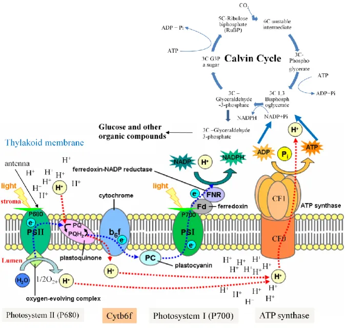
It takes place in the internal membranes, thylakoids, of the chloroplast. The light cycle is completed with the help of four major protein complexes: photosystem I (PSI), cytochrome b6-f complex, photosystem II (PSII) and the ATP synthase. These membrane complexes are the integral membrane complexes and are found vectorially in the thylakoid membranes. Orientation of these four complexes results in the oxidation of water to oxygen in the thylakoid lumen, reduction of NADP+ to NADPH on the stromal side of the membrane and the release of ATP molecules into the stroma as the protons (H+) move from the lumen to the stroma. The photosystems (PSI and PSII) are spatially separated, physically and chemically different and each one has its own antenna pigments and photochemical reaction center. Electron transport chain links the two photosystems. The PSI reaction center, its antenna pigments, its electron transfer proteins and coupling factor enzyme are found in stroma lamellae and at the edges of the grana lamellae while PSII reaction center, its antenna chlorophylls and its electron transfer proteins are predominantly found in stacked regions of grana lamellae. Stroma lamellae are the exposed thylakoid membranes which lack stacking while grana lamellae are the stacked thylakoid membranes. The ATP synthase is located in the less curved regions of the grana end membranes and stroma lamellae (Daum et al., 2010). The antenna pigments are the pigments which collect light and transfer the energy to the photochemical reaction center where chemical reaction leads to long term energy storage. The antenna pigments consist of carotenoids, also called accessory pigments, chlorophyll b and chlorophyll a. The absorption spectrum of carotenoids is 400-500 nm while that of chlorophyll a is ≈ 430 and chlorophyll b is ≈ 450 nm. The energy transfer from carotenoids to chlorophyll is less efficient than from chlorophyll to chlorophyll. Carotenoids also play the role of photoprotection and avoid the production of nascent/ single oxygen. If the excited state of the chlorophyll is not quenched rapidly, a reaction excites the molecular oxygen to produce nascent oxygen which is hyper active and can cause damage to cellular components.
PSII, cytochrome b6-f and PSI complexes are arranged in Z (zigzag) scheme. Transfer of excited electrons between a series of electron donors and acceptors is empowered by light energy. NADP+ is the final electron acceptor and is reduced to NADPH. Proton motive force produced across the thylakoid membranes by the light energy and the resulting proton gradient are used by ATP synthase to produce ATP molecules. So the end products of light cycle, high energy molecules ATP and NADPH, are then used in the dark cycle during the CO2 fixation.
Introduction
11
3.1.1. Photosystem II and cytochrome b6-f complex
Oxygenic photosynthesis in all photosynthetic organisms starts in a homodimeric multisubunit protein–cofactor complex which is embedded in the thylakoid membrane and is called PSII (Kern & Renger, 2007).
PSII is located in grana thylakoids and forms a supra molecular complex composed of polypeptides, pigments and co-factors. The protein subunits are either plastid or nucleus encoded. Water splitting complex oxidizes water and is also known as oxygen evolving complex (OEC). The reaction center is composed of several small polypeptides and homologous D1 and D2 proteins which are encoded by plastid psbA and psbD genes, respectively. The reaction center pigment (P680) receives the light energy directly or indirectly from the antenna pigments. The light energy causes an intramolecular rearrangement of the electrons and P680 changes from the ground state to the excited state (P680*). Then there is an intermolecular transfer of electrons from an electron donor (P680*) to an electron acceptor (pheophytine). Pheophytine is reduced by accepting electrons from P680*. The water splitting complex splits two molecules of H2O into 4 protons, 4 electrons and a molecule of oxygen. This reaction can be shown as under:
2H2O → 4H+ + 4e- + O2
The electrons produced as a result of water splitting are then transferred to to a redox-active tyrosine residue, also called Z or YZ molecule, which reduces photoxidized P680 by transferring electrons to it. High energy electrons enter into an „Electron Transport Chain” as pheophytine transfers these high energy electrons to the plastoquinone A (QA). Plastoquinone A transfers these electrons to the plastoquinone B (QB) which is a mobile electron carrier. The reduced QB transfers electron to the Cytochrome b6-f complex which consists of a rieske iron sulfur protein (FeSR), two b type cytochromes (cyt b) and a cytrochrome f with one covently bound heme c and subunit IV. Rieske iron sulfur protein has two iron atoms bridged by two sulfur atoms. Cytochrome b6-f complex is distributed equally between the stroma lamellae and grana lamellae. But it is not the mobile carrier of electrons. Oxidised FeSR is reduced by accepting electrons from the reduced plastoquinone B and transfers theses electrons to cytrochrome f (cyt f). Cytrochrome f (cyt f) then transfers electrons to plastocyanin (PC). The reduced PC oxidizes itself by transferring electrons to the oxidized pigment (P700) of PSI. Along with the transfer of electron to oxidised FeSR, plastoquinone B also transfers an electron to one of the cyt b and releases its 2H+ in the luminal side of the membrane. This reduced cyt b oxidizes by reducing the second cyt b which in turn transfers its electron to the
Introduction
12 oxidized plastoquinone. The plastoquinone gets a semi-quinone form by accepting one electron and is fully reduced by the similar flow of another electron. This reduced plastoquinone (quinol) oxidizes by picking protons from the stromal side of the thylakoid membranes and changes into plastohydroquinone.
The net result of activity of the Cytochrome b6-f complex is the transfer of two electrons to the P700 (PSI reaction center) and transfer of four proton from the stromal side of the membrane to the lumenal side. The electrochemical potential produced as a result of proton gradient is used by ATP synthase to produce high energy ATP molecules.
3.1.2. Photosystem I
PSI is located in thylakoids and consists of antenna chlorophylls, a reaction center (P700), phylloquinone, and iron-sulfur clusters. It is also a complex and highly organized membrane structure. The light energy in PSI is also absorbed by the same trans-membrane proteins as the one used by PSII. But here the maximum energy absorption is at 700 nm so the pigment of chlorophyll is called P700. The overall reaction can be shown in the following way:
plastocyanin → P700 → P700* → ferredoxin → NADPH ↑ ↓
b6f ← plastoquinone b6-f
When P700 absorbs energy, it obtains excited state (P700*). The electrons from P700* are transferred to ferrodoxin, a water soluble carrier, through a number of intermediate carriers. These electrons can have two different fates. The non cyclic electron transport and the cyclic electron transport. In non cyclic transport, the electrons are carried by ferredoxin to the enzyme ferredoxin NADP+ oxidoreductase which reduces NADP+ to NADPH. While in cyclic electron transport, electrons are transferred to P700 via a proton pump, cytochrome b6/f. The proton gradient is then used by the ATP synthase to produce ATP. NADPH and ATP are the end products of light phase which are used in the dark phase for carbon fixation and carbohydrate precursor synthesis. Photosystem I is composed of 19-21 protein subunits, 175 chlorophyll molecules, 2 phylloqinone and 3 Fe4D4 clusters (Ben-Shem et al., 2003; Jensen et al., 2007). Among the protein subunits, it consists of 15 core subunits (PsaA-PsaL and PsaN-PsaP) while the peripheral antenna, LHCI, consist of six LHCa proteins (LHCa1-6)
Introduction
13 but later on the existence of nine or ten LHCI proteins was also proposed but not yet confirmed (Dekker & Boekema, 2005). Most of the proteins of PSI are nucleus encoded and therefore should be transported from the cytosol to the plastid stroma and from stroma to be directed properly to the exact place in thylakoid membrane. This all renders it a great complexity. Some proteins (PsaA, PsaB, PsaC and PsaJ) are also encoded by chloroplast genes which add to the complexity of PSI subunit assembly process.
The electron flow from the PSII to cytochrome b6-f complex and PSI if schematically drawn in terms of it electric potential, forms a Z shape and hence called Z-scheme. It is depicted in Figure 3a.
Introduction
14
3.2. Calvin cycle
The Calvin cycle consists of a series of biochemical reactions that take place in the plastid stroma, are light-independent and cyclic. The substrate for carboxylation (ribulose-1,5-bisphosphate) is regenerated at the end of the cycle. The major enzyme involved in this cyclic reaction is RuBisCO (Ribulose-1,5-bisphosphate carboxylase oxygenase). It catalyses the carboxylation of a 5 carbon compound ribulose-1,5-bisphosphate by carbon dioxide in a two-step reaction. The ATP and NADPH molecules produced in the light cycle are used as energy source in this cycle.
Figure 3b: Schematic presentation of the components and mechanism of photosynthesis.
(I used power point to modify the figure of Tameeria who created the figure in April 2007 based on: Taiz and Zeiger, “Light-dependent reactions of photosynthesis at the thylakoid membrane” Plant Physiology, 4th edition, ISBN 0-87893-856-7. The original figure is available on http://en.wikipedia.org/wiki/Photosynthesis).
Introduction
15 The ADP and NADP+ produced during the dark cycle are taken up by light cycle to convert them again into ATP and NADPH molecules. The sum of reactions in the Calvin cycle is the following:
3 CO2 + 6 NADPH + 5 H2O + 9 ATP → glyceraldehyde-3-phosphate (G3P) + 2 H +
+ 6 NADP+ + 9 ADP + 8 Pi
In order to produce one mole of glyceraldehyde-3-phosphate (G3P), 3 moles of CO2 are needed which require three runs of Calvin cycle during which 9 ATP and 6 NADPH molecules are used. Glyceraldehyde-3-phosphate (G3P) is either converted to sucrose via triose phosphates or is used to regenerate ribulose-1,5-bisphosphate which again in calvin cycle. The localization of different components of photosynthesis, the flow of electrons from PSII to cyt b6-f and PSI and Calvin cycle are shown in figure 3b.
3.3. ATP synthase
It is an important enzyme involved in the ATP synthesis and hydrolysis in chloroplast thylakoid membranes during the process of photosynthesis by using proton motive force. It consists of two major portions, F1 and F0.
3.3.1. CF1
CF1 is a hydrophilic peripheral membrane protein complex attached to the thylakoid membranes and has five subunits , β, , , in a stoichiometry of 3:3:1:1:1 repectively. It is almost spherical and has a width of 11nm and its height is 9 nm (Richter et al., 2000). Its subunits ( , β, , , ) are encoded by atpA, atpB , atpC, atpD, atpE genes respectively where atpA, atpB and atpE are chloroplast genes while atpC and atpD are nuclear genes (Groth & Strotmann, 1999). Three s and three βs together form a heterohexamer ring (Figure 3c).
This heterohexamer ring consists of six structural domains of an N-terminal β barrel and in its centre of an -β domain containing the nucleotide binding site. The three catalytic sites of β subunits are βT, βD and βE. Two of them are occupied by Mg.ATP and Mg-ADP while the third one is an empty site (Noji & Yoshida, 2001). Interestingly they have always different nucleotide binding state at any given moment of time. Subunits and are present in the central cavity of , β heterohexamer ring and work as connectors between F1 and Fo (Figure 3c).
Introduction
16
3.3.2. CFo
CFo is a hydrophobic thylakoid membrane embedded protein complex and has four subunits “ a (IV), b (I), b‟ (II)‟, c (III)” in a stoichiometry of 1:1:1:14 respectively. The thickness of CFo was found to be 7.5 nm while it its diameter was estimated as 6.2 nm and 8.5 nm (Richter et al., 2000). Its subunits (I, II, III,IV) are encoded by atpF, atpG, atpH and atpI genes respectively (Figure 3c). AtpI, atpF, atpH are chloroplast genes while atpG is a nuclear gene.
Figure 3c: Schematic presentation of the arrangement of the subunits of ATP synthase and the genes encoding these subunits.
This figure is extracted from http://www.bio.miami.edu
All the subunits have transmembrane -helical structures. Subunits b and b‟ have only one transmembrane helix consisting of five helices. Subunit c has two anti-parallel transmembranes helices connected by two extramembrane polar loop (Groth and Strotmann 1999). Subunit c oligomer forms a ring outside where subunits b, b’ and a are located (Neff et al., 1997). Subunits b and b‟ are involved in binding with CF1. They form a stalk and are connected with subunit of CF1 while a and c subunits are involved in H+ translocation.
Introduction
17
3.3.3. ATP synthase gene expression
The genes which encode subunits of CF0 and CF1 belong to two transcriptionally active compartments i.e. nucleus and plastids. The mRNAs of nuclear genes atpC, atpD and atpG are translated in the cytoplasm. The resulting soluble precursor proteins have plastid specific transit peptides. These transit peptides are proteolytically cleaved after the proteins have been transported to the thylakoid membranes of chloroplast for the assembly into the ATP synthase CFoCF1 complex. The nuclear genes involved in ATP synthase synthesis are expressed at the same time in response to light, organ specific factors and plastid derived signals (Bolle et al., 1996). In chlamydomonas the biosynthesis of chloroplast encoded subunits of ATP synthase is controlled at the translational level. Drapier et al. (2007) proposed a CES (Control by Epistasy of Synthesis) regulation for uneven stoichiometry of both the ATP synthase complexes i.e. CF1 and CF0. CES is a process in which the presence of a subunit is required for the sustained synthesis of another plastid subunit belonging to the same complex (Choquet & Vallon, 2000; Choquet & Wollman, 2002; Wollman et al., 1999). Drapier et al. (2007) reported that nuclear encoded subunit is required for a sustained synthesis of chloroplast encoded subunit which in turn stimulates the translation of the chloroplast encoded subunit of theCF1 complex. An important feature of CES is the negative autoregulatory feedback of beta on its own translation.
In higher plants, (Sakai et al., 1998) tested the effect of tagetitoxin (a PEP inhibitor) on the transcript accumulation of a number of genes in isolated chloroplast (from mature tobacco leaves) and proplastid nuclei (cultured cells of tobacco) by northern blot hybridization. In tobacco, they observed 95-99 % reduction in transcript accumulation of atpA, atpB in developed chloroplasts and only 40-50 % reduction in transcript accumulation of these genes in proplastids. This suggest that atpA and atpB which encode subunits of ATP synthase are transcribed by two distinct RNA polymerases in proplastids and in chloroplasts. Spinach atpB gene codes for five different transcripts having their 5‟ ends at positions 455, 275, 180, -100 and +1 from the translation initiation codon of atpB (L. J. Chen et al., 1990; Mullet et al., 1985). Tobacco atpB transcripts map at positions -610, -500, -490, -290, -225, -90 (Orozco et al., 1990; Shinozaki & Sugiura, 1982) while -610, -490, 290 and -225 mRNAs were identified as primary transcripts (Kapoor et al., 1997; Orozco et al., 1990). Kapoor et al. (1997) also reported the existence of non-consensus type promoters (NCII) (which are NEP dependent) in atpB/E operon responsible for its expression in non photosynthetic plastids which was
Introduction
18 supported by the results of Sakai et al. (1998). Hirose & Sugiura (2004) reported that atpB mRNA needs an unstructured sequence encompassing the start codon for its translation as there is no Shine-Dalgarno (SD) sequence in the 5‟ UTR. atpB and atpE have overlapping stop and start codons in tobacco and arabidopsis. These overlapping codons result in the translational coupling of atpB/atpE transcripts in maize (Gatenby et al., 1989). In tobacco an atpE specific transcript of 1.3 kb was found by (Shinozaki et al., 1983), being issued from a promoter located within the coding region of the atpB gene (-430/-431 nt upstream of the atpE translation initiation codon) (Kapoor et al., 1994). Similarly an atpE transcript was found to be issued from a promoter located in the coding region of atpB (from -431 upstream of atpE translation initiation codon) in arabidopsis (thesis Wafa Zghidi, 2008). As atpE has a monocistronic transcript, the question is still open that whether in arabidopsis atpE is translationally uncoupled from atpB or not. The above mentioned results indicate that 1) majority of the transcripts for small ATP operon are issued from the promoters located in the 5‟ UTR of atpB but the promoter positions are not highly conserved among different species. 2) atpE is co-transcribed with atpB in higher plants but it is also transcriptionally uncoupled from atpB in tobacco and arabidopsis.
Early data indicated that spinach atpI/H/F/A contain almost 30 different transcripts which range from 0.5 kb to 6.0 kb in length (Cozens et al., 1986; Hudson et al., 1987; Stahl et al., 1993; Stollar & Hollingsworth, 1994). Miyagi carried out a detailed transcriptional analysis of the atpI/H/F/A operon in tobacco and found that rps2 is also co transcribed with atpI/H/F/A (Miyagi et al., 1998). Miyagi et al., (1998) found three transcription initiation sites and four processing sites in the non coding regions of this operon. They found that one of the primary transcripts being issued from position -208 from the translation initiation codon of atpI is dependent on a non consensus promoter (NCII) while the other two (131 atpI and -384 atpH) are synthesized from PEP dependent promoters.
The genes encoding for the subunits of ATP synthase complexes (CFO and CF1) in unicellular green alga chlamydomonas are dispersed throughout its plastid genome. Drapier and collaborators showed that atpA and atpH are co transcribed and are found in the same operon (atpA-psbI-cemA-atpH) (Drapier et al., 1998). They also showed a total of 8 transcripts for this transcriptional unit (atpA), three of them were described as primary transcripts and other transcripts were thought to be the result of processing events. Except these two genes none of the other atp genes are found in the same operon, rather minimum
Introduction
19 distance among them is 7 kb (Woessner et al., 1987). The atpE is neither located on the 3‟ end of atpB nor has overlapping stop and start codon with it, rather it is located 92 Kb away from it in a single copy region (Woessner et al., 1987).
4. Plastid gene expression
4.1. Post transcriptional regulation in chloroplast
The production of a functional protein from the information stored in the plastid DNA is not simply a two step process of transcription and translation. It consists of post transcriptional and post translational modifications. The main post transcriptional modifications are listed below:
RNA processing is a process of the cleavage of polycistronic transcripts and generation of the
5‟ and 3‟ ends of the cleaved transcripts.
Editing changes the amino acids specified by the DNA sequence by converting cytosine (C)
nucleotide to the uridine (U) nucleotide of an mRNA.
RNA splicing is a process in which (cis or trans) removal of group I and II introns is carried
out.
Even though the plastid genome is derived from an ancient cyanobacteria, its gene expression process consists of both eukaryotic and prokaryotic features. In addition, due to the transfer of plastid genes to the nuclear genome and the need of crosstalk between the two organellar genomes, the plastid gene expression machinery has become complex and dependent on nucleus encoded proteins. Interestingly, original mechanisms of gene expression regulation have been developed in the chloroplast to optimize its integration into the eukaryotic cell. Take the example of the RNA polymerases required for transcription. Cyanobacteria which are ancestors of plastids have only one RNA polymerase consisting of core enzyme and sigma factors all encoded by their single genome. But in higher plants chloroplasts have two RNA polymerases, PEP (Plastid Encoded RNA Polymerase) and NEP (Nucleus Encoded RNA Polymerase). PEP is plastid encoded but it requires nucleus encoded sigma factors for promoter specificity and the second type of RNA polymerase is encoded by the nucleus and hence called NEP. Similarly in contrast to cyanobacteria where the polycistronis transcripts are directly translated, plastid polycistronic transcripts need to be first processed and edited and then translated. In addition, plastid mRNA require the different
Introduction
20 trans factors for RNA stability (Stern et al., 2010), while bacterial mRNAs are stabilized by secondary structures.
4.1.1. Chloroplast ribonucleases
Ribonucleases are the enzymes which are responsible for maturation of precursor RNA and RNA degradation. Their activities depend on protein-protein interactions, protein modifications and RNA secondary or tertiary structures (Monde et al., 2000). There are two types of ribonucleases:
1- Endoribonucleases: They cleave the polynucleotide chain of RNA by breaking the phosphodiester band between two adjacent nucleotides.
2- Exoribonucleases: They cleave at the end (3‟) or the start (5‟) of a polynucleotide chain of RNA by breaking the phosphodiester band between two adjacent nucleotides.
4.1.1.1. Endoribonuclease
In chloroplast, endoribonucleases are thought to be responsible for 3‟ end formation (R. Hayes et al., 1996; Stern & Kindle, 1993) or initiation of RNA breakdown by breaking the stabilising stem-loop structures (H. C. Chen & Stern, 1991; Yang & Stern, 1997). The well known endoribonucleases in chloroplast are CSP41 (Chloroplast Stem-loop binding Protein, 41 kDa) (CSP41a and CSP41b), RAP 38 (Ribosome Associated Protein, 38 KDa) and RAP 41 (ribosome associated protein, 41 KDa), RNase E (Schein et al., 2008) , RNase J1(Zoschke et al., 2010), RNase P (Thomas et al., 2000), RNase Z (Canino et al., 2009) and CRR2 (Okuda et al., 2009).
4.1.1.2. Exoribonucleases
In chloroplast, exoribonucleases participate in the 3‟ and 5‟ end maturation and also in the 3‟-5‟ polyadenylation assisted degradation pathway of mRNAs. The well known exoribonucleases in the chloroplast are Polynucleotide phosphorylase (PNP) (Hayes et al., 2006; Perrin et al., 2004), RNase R (RNR1) (Kishine et al., 2004), RNase II RNB (Yehudai-Resheff et al., 2007) and RNase J1 that is also a 5‟-to -3‟ exoribonuclease.
Introduction
21
4.1.2. Turn over
The neo-synthesized polycistronic mRNA transcripts in plastids are spliced, processed, stabilised and edited to produce translation competent mRNAs. Both the neo-synthesized and translation competent mRNAs enter into a degradation pathway. The difference of the rate of synthesis and degradation determines the turnover of the mRNA. The decay or degradation of the mRNA takes place in three steps, 1) endonucleolytic cleavage, 2) polyadenylation and 3), exonucleolytic decay or turnover (Bollenbach et al., 2008; Stern et al., 2010). The translation competent mRNAs are protected at their 3‟ end by a stem loop structure of inverted repeat sequence (IRs) and by PPR proteins or other RNA binding proteins. The 5‟ end of such mRNAs are also protected by RNA binding protein like PPR proteins. When the transcripts have to be degraded, endonucleases cleave the secondary structures of the 3‟ end and in the 3‟ or 5‟ UTRs outside the PPR-protected regions. Endonuclease can also cleave the mRNAs internally where there are no attached ribosomes. (Stern et al., 2010) proposed endonucleolytic cleavage as the rate limiting step of the mRNA decay pathway. The endo-nucleolytically cleaved mRNAs are good substrates of polyadenylating enzymes. Polyadenylation can be catalysized either by PAP (poly (A) polymerases) or by PNPases (polynucleotide phosphorylase). PAP adds only homopolymeric poly (A) tails while PNPase adds heteropolymeric poly (A) tails. These polyadenylated transcripts are substrates for the exonuclease which degrades the RNAs in 3‟-5‟ direction. If the exonuclease encounters a secondary structure, an endonucleolytic cleavage and polyadenylation are required further degradation of the transcripts. For 5‟-3‟ degradation of transcripts, removal of the 5‟ end stability complex by an endonucleoytic activity is followed by a net 5‟-3‟ degradation. The existence of a 5‟-3‟ exonuclease dependent degradation activity has not yet been shown in chloroplast (Bollenbach et al., 2008; Stern et al., 2010). In brief mRNA turn over depends on the extent of transcriptional activity, mRNA stabilising structures or proteins (stem loops of IRs at the 3‟ end, mRNA binding proteins like PPR) and the activity of enzymes responsible for mRNA degradation/decay (endonuclease, polyadenylation enzymes, exonuclease).
4.1.3. 5’ End maturation
On the basis of their origin, transcripts found in chloroplasts of higher plants are divided in two types. The ones which originate directly from transcriptional events are called primary transcripts while the others which are produced as a result of processing of the
Introduction
22 primary transcripts are called processed transcripts. Processed transcripts are the predominant form of transcripts found in chloroplast of higher plants. Even in Chlamydomonas no primary transcripts can be detected which indicate that most if not all of the transcripts undergo processing and 5‟ end maturation and that this event is very fast (Stern et al., 2010). Primary transcripts have a tri-phosphate on their 5‟ ends while the processed transcripts have only one phosphate on their 5‟ end. It is possible to discriminate between primary and processed transcripts using a combination of TAP treatment and RT-PCR techniques.
Two pathways are proposed for the 5‟ end maturation of the transcripts. The first one is an endoribonuclease pathway while the second one is a 5‟-3‟ exoribonuclease pathway. Endoribonuclease starts maturation by site-specific cleavage of intercistronic transcripts or by dephosphorylation of primary transcripts. Once a free 5‟ end is available, the mRNA becomes a substrate for the 5‟-3‟ exonuclease actually. The extent of this 5‟ maturation is determined by the presence of sequence specific binding proteins like PPR proteins which bind to the specific elements/sequence or by secondary structures. In addition RNA binding proteins can also guide endoribonuclease for site specific cleavage.
RNAse J is thought to play a role in maize chloroplast mRNAs maturation (Pfalz et al., 2009). RNAse E which is another exoribonuclease was found to be involved in the 5‟ end maturation of plastid RNAs (Mudd et al., 2008). Arabidopsis thaliana possess both RNAse J and RNAse E while Chlamydomonas has RNAse J but lacks RNAse E. RNase J and E are thought to possess both the endoribonucleolytic and exoribonucleolytic activities and are the best candidates for 5‟ end maturation of plastid transcripts (de la Sierra-Gallay et al., 2008).
4.1.4. 3’ End maturation
Most of the mature 3‟ ends are produced as a result of processing and not by transcription termination which is inefficient in chloroplast. 3‟ end maturation is also carried out by exoribonucleases, endoribonucleases and RNA-binding proteins. In the most frequent situation, a newly synthesized mRNA is taken as a substrate by PNPase (polynucleotide phosphorylase) for a 3‟-5‟ exoribonuclease activity. PNPase continues its activity until it encounters an IR (inverted repeat) secondary structure. The same pathway is observed if a free 3‟ end is obtained upon endonucleolytic cleavage inside a polycistronic transcript. In this context, Pfalz and colleagues (Pfalz et al., 2009) proposed that endonucleolytic cleavages inside polycistronic transcripts are random and are followed by 5‟-to 3‟ and 3‟-to 5‟ exonucleolytic activity in order to define overlapping 5‟ and 3‟ ends respectively. Sequence specific binding proteins as PPR10 play their role in stabilising the 5‟ and 3‟ ends and limiting
Introduction
23 the 5‟ and 3‟ maturation. An example of this PPR10 hypothesis is a PPR protein HCF 152 that controls the intercistronic processing of psbH and petB in Arabidopsis (Meierhoff et al., 2003). In Chlamydomonas chloroplasts, the mature 3‟ end of atpB lies at the end of a stem loop forming inverted repeat (IR) sequence. The maturation of the 3‟ end of atpB pre-mRNA takes place by a two step process. As a first step, endonuclease cleaves 3‟ end of the pre-mRNA till 8-10 nt downstream of the mature 3‟ end. In the second step this intermediate product is trimmed by a 3′ → 5′ exonuclease to produce a mature 3‟ end (Hicks et al., 2002). It has also been proposed a synergetic relationship between the IR and ECS (endonuclease cleavage site).
4.1.5. Splicing
Splicing is the process that consists in removal of introns from the precursor mRNAs. There are two types of introns found in precursor mRNAs, group I and group II introns which are „self splicing‟ under non-physiological conditions in vitro (Saldanha et al., 1993) but not in land plants and in vivo need protein factors (Herrin et al., 1998).
Splicing of group I introns needs two times trans ester bond formation. The first bond is formed between 5‟ end of the intron and a nucleophilic guanine nucleotide and the second ester bond is formed between 3‟ OH of the upstream exon and 5‟ end of the downstream exon (Stern et al., 2010). Land plants have only one group I intron in trnL gene. Unicellular alga C. reinhardtii plastid gene rrnL has one intron while psbA has four introns (Turmel et al., 1993). All group I introns of chlamydomonas are autocatalytic in vitro but trnL of land plants has lost this ability (Goldschmidt-Clermont, 2008).
Six helical domains connected to a central core are conserved in group II introns (Fedorova & Zingler, 2007). Group II introns are divided into two subgroups on the basis of intron structure, group IIA and group IIB (Michel et al., 1989). Like group I introns, two steps of trans esterification are needed for splicing of this group of introns (Jarrell et al., 1988; Peebles et al., 1987). But here the 5‟ end of the intron is attacked by 2‟ OH (a nucleophile) of the bulged adenosine of the helix of the sixth domain (D6). The reaction results in the release of 5‟ exon and of the intron in the form of lariat. Then the 3‟ OH of the released 5‟ exon attacks the 3‟ splice site, reaction results in the union of 5‟ and 3‟ exons (spliced product) and release of the lariat intron (Fedorova & Zingler, 2007). The other mechanism of group II intron splicing is the hydrolytic splicing. This method is adopted in the absence of D6 free adenosine and OH- group of H2O is used as a nucleophile (Daniels et al., 1996; Jarrel et al.,
Introduction
24 1988; Vanderveen et al., 1987). The second step of trans esterification results in the release of ligated exons and lariat intron. Arabidopsis and tobacco have 20 group II introns in the plastid genomes while maize has 17 (Fedorova & Zingler, 2007).
Splicing of group I and II introns requires the involvement of nucleus-encoded factors. For example, 14 nuclear genes were found to be involved in trans-splicing of psaA precursor RNA (Goldschmidt-Clermont et al., 1990). The mature mRNA of psaA gene of chloroplast of C. reinhardtii is formed by three separate precursor RNAs in two steps of trans-splicing as it consists of three exons spread in the whole chloroplast genome (Herrin et al., 1998). Among the nucleus encoded factors four genes, Raa1, Raa2, Raa3 and Tr72 have been cloned. Raa2 encodes a protein which belongs to pseudouridine synthase family while the rest of three encode novel proteins. Interestingly, a small non-coding RNA, tscA, which is plastid encoded, is required for splicing of exon 1 and 2 to occur (Goldschmidt-Clermont et al., 1991). Among the land plants, products of nuclear genes crs1 and crs2 were found to be involved in splicing of group II introns of maize chloroplast (Jenkins et al., 1997). Crs1(chloroplast RNA splicing-1) is involved in splicing of atpF introns (Jenkins et al., 1997), while crs2 (chloroplast RNA splicing-2) is involved in splicing of many introns belonging to the subgroup B of the group II introns (Vogel et al., 1999).
Involvement of nuclear encoded proteins in splicing of group I and group II introns has been established. Using both genetic and biochemical approaches, 12 different nucleus-encoded proteins were found to be directly involved in chloroplast splicing (Kroeger et al., 2009; Schmitz-Linneweber & Barkan, 2007). Majority of these 12 nucleus encoded proteins which belong to a family of proteins containing four RNA-binding domains called CRMs (chloroplast RNA splicing and ribosome maturation) (Ostheimer et al., 2003). CFM2, a CRM domain protein, is required for trnL intron splicing. CFM3 is thought to participate in small ribosomal subunit biogenesis. PPR proteins also participate to RNA splicing. PPR4 contains 16 PPR repeats and one RRM domain and it is involved in the trans splicing of the rps12-1 in maize and Arabidopsis (Schmitz-Linneweber et al., 2006). De Longevialle and colleagues showed that OTP51, having 7 PPR repeats and two RRM domains, is involved specifically in intron splicing of ycF3-2 and in general for group IIA introns (de Longevialle et al., 2008). PPR 5 saves unspliced precursor of trnG from degradation (Beick et al., 2008; Kroeger et al., 2009). Kroeger et al., 2009 found the association of WTF 1 protein with the CAF1 (chromatin assembly factor 1) and CAF2 ribonucleoproteins (RNPs) in maize. WTF1 has DUF860 or PORR (plant organellar RNA recognition) conserved domain. Wtf1 mutant plants of maize showed defects in splicing of introns.