HAL Id: tel-03122693
https://tel.archives-ouvertes.fr/tel-03122693
Submitted on 27 Jan 2021HAL is a multi-disciplinary open access archive for the deposit and dissemination of sci-entific research documents, whether they are pub-lished or not. The documents may come from teaching and research institutions in France or abroad, or from public or private research centers.
L’archive ouverte pluridisciplinaire HAL, est destinée au dépôt et à la diffusion de documents scientifiques de niveau recherche, publiés ou non, émanant des établissements d’enseignement et de recherche français ou étrangers, des laboratoires publics ou privés.
Analyzing the role of SoxC genes in murine kidney
development
Vladimir Kozlov
To cite this version:
Vladimir Kozlov. Analyzing the role of SoxC genes in murine kidney development. Molecular biology. Université Côte d’Azur, 2020. English. �NNT : 2020COAZ6010�. �tel-03122693�
Etude du rôle des gènes SoxC au cours
du développement du rein de souris
Vladimir KOZLOV
Institut de Biologie Valrose (iBV)
Présentée en vue de l’obtention
du grade de docteur en Sciences de la vie et de la santé
d’Université Côte d’Azur Dirigée par : Andreas Schedl Soutenue le : 31 Août 2020
Devant le jury, composé de :
Thomas Lamonerie, professeur, Université Côte d’Azur
Andreas Schedl, directeur de recherche, Institut de Biologie Valrose
Muriel Umbhauer, professeur, Université Pierre-et-Marie-Curie
Seppo Vainio, professeur, Université d’Oulu
Etude du rôle des gènes SoxC au cours du développement du rein de souris
Jury :
Président du jury
Thomas Lamonerie, professeur, Université Côte d’Azur Rapporteurs
Muriel Umbhauer, professeur, Université Pierre-et-Marie-Curie Seppo Vainio, professeur, Université d’Oulu
Directeur de thèse
Etude du rôle des gènes SoxC au cours du développement du rein de souris
Résumé
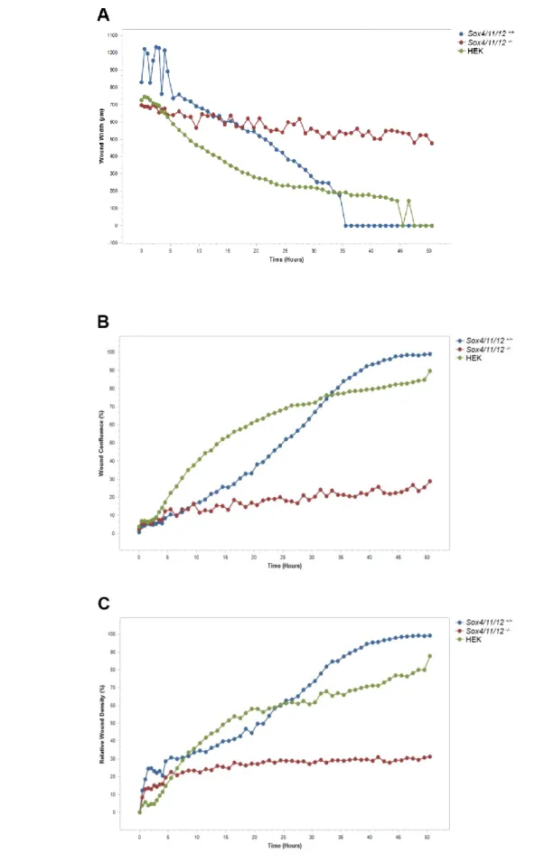
Les anomalies congénitales des reins et des voies urinaires (CAKUT) sont un groupe de malformations fréquemment trouvées chez les patients humains qui résultent de défauts dans le programme de développement de ces organes. Sox11 fait partie de la famille des facteurs de transcription SoxC, qui joue un rôle important dans le développement de divers organes chez les vertébrés. Les souris Sox11-/- meurent juste après la naissance et présentent une grande variété de CAKUT. Alors que les reins duplex sont une anomalie courante du développement rénal, le mécanisme moléculaire qui amène les mutants Sox11 à développer cette malformation reste incertain. Dans ce projet, j'ai analysé le phénotype rénal des souris Sox11-/- et montré que les reins duplex sont causés par l'expansion du mésenchyme métanéphrique (MM). D'autres expériences ont été réalisées pour déterminer si l'origine de cette expansion résidait dans une apoptose insuffisante ou dans l'absence de migration des cellules du MM. Une analyse tridimensionnelle par microscopie confocale des cellules apoptotiques a révélé que chez les embryons Sox11-/- le MM subissait une apoptose dans la région d'intérêt similaire à celles des embryons de type sauvage. Un test in vitro de cicatrisation des plaies a démontré que les gènes
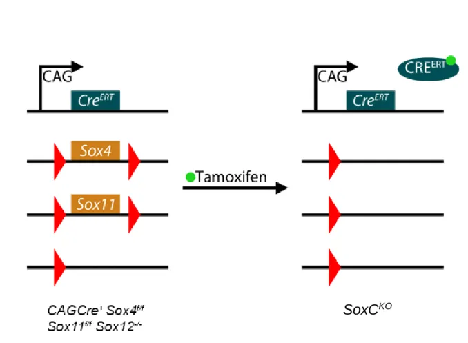
SoxC sont nécessaires pour la migration des cellules du MM. La délétion de ces gènes à un
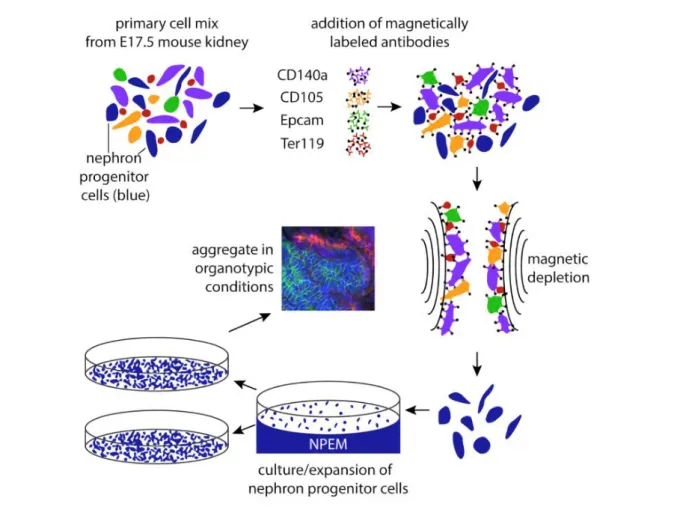
stade tardif du développement conduit à une hypoplasie rénale et à une diminution du nombre de néphrons due à l'arrêt de la néphrogenèse. Pour étudier le rôle des gènes SoxC au cours de la formation des reins, j'ai établi une culture de cellules progénitrices rénales permettant l’invalidation de ces gènes en présence de 4OH-Tamoxifène. L'analyse de l'expression génique a confirmé la suppression efficace des gènes SoxC, tout en maintenant l'identité des cellules progénitrices du néphron. Après induction de la délétion, ces cellules sont incapables de s'épithélialiser, du fait d’une activité accrue de la voie Wnt/β-caténine. Afin de valider ces résultats et d’identifier les cibles en aval des gènes SoxC, j'ai effectué une analyse ARN-seq d’organoïdes rénaux subissant une transition mésenchymateuse-épithéliale. Les données acquises par ces expériences révèlent que de nombreuses voies de signalisation sont affectées. En particulier, l’insuffisance du gène Ezh2 codant pour une protéine du groupe Polycomb, pourrait être à l’origine de changements transcriptomiques généralisés empêchant les cellules progénitrices déficientes en SoxC de se différencier. L’ensemble de ces données démontre l'importance des gènes de la famille SoxC dans le développement précoce et tardif du rein, dont l'analyse moléculaire conduira à des orientations possibles pour les recherches et les traitements futurs.
Mots clés : Développement rénal, duplication des voies urinaires, transition mésenchymateuse-épithéliale, cellules progénitrices
Analyzing the role of SoxC genes in murine kidney development
Abstract
Congenital Anomalies of Kidneys and Urinary Tract (CAKUT) are a group of birth defects that arise from defects in the developmental program of organ development and are
frequently found in human patients. Sox11 is a member of SoxC family of transcription factors, which plays an important role in the development of various organs in vertebrates.
Sox11 knockout mice die soon after birth and display a wide variety of CAKUT, including
duplex kidneys and nephrogenesis defects. While duplex kidneys are a common renal development anomaly, the molecular mechanism causing Sox11 mutants to develop duplex kidneys remains unclear. In this project, I analyzed the renal phenotype of Sox11 knockout mice and discovered that the duplex kidneys are caused by the expansion of metanephric mesenchyme (MM). Further experiments were performed to determine whether the origin of this expansion lies in insufficient apoptosis of MM cells or lack of MM cell migration. Three-dimensional confocal microscopy analysis with Lysotracker Red staining of apoptotic cells revealed that MM undergoes apoptosis in the region of interest in wildtype embryos, however the apoptosis seems to persist in Sox11-/- embryos as well. However, in vitro scratch wound
assay provided evidence that SoxC genes are necessary for migration of MM cells. In the late kidney development, SoxC deletion is leading to renal hypoplasia and reduced nephron number due to cessation of nephrogenesis. To study the role of SoxC in nephrogenesis, I set up an in vitro renal progenitor cell culture which allows for the timed deletion of SoxC genes by a 4OH-tamoxifen induced knockout. Gene expression analysis confirmed efficient
deletion of SoxC genes, while maintaining nephron progenitor cell identity. After deletion, renal progenitor cells were found to be unable to epithelialize, linked with increased activity of Wnt/β-catenin pathway. To validate these findings and discover the downstream targets of
SoxC genes, I developed a system for an RNA-seq analysis of renal organoids undergoing
mesenchymal-to-epithelial transition. Data acquired in these experiments reveal numerous regulatory pathways affected in SoxC-knockout renal progenitor cells, with the deficiency of Polycomb group gene Ezh2 as a likely origin of widespread transcription changes leading to the inability of SoxC-deficient progenitor cells to differentiate. Taken together these data demonstrate the importance of Sox11 and other SoxC genes in early and late kidney
development, with molecular analysis providing plausible directions for future research and interventions.
Keywords : Kidney development, duplicated collecting system, mesenchymal-epithelial transition, progenitor cells
Table of Contents
Acknowledgments ... 2
Introduction ... 3
The kidney ... 3
Renal anatomy and function in mammals... 3
Early renal differentiation ... 5
Nephrogenesis and nephron maturation ... 12
Sox genes ... 20
Sox gene structure and function ... 20
Sox genes functions in development ... 25
Congenital anomalies of kidney and urinary tract ... 29
Article 1: Duplex kidney formation: developmental mechanisms and genetic predisposition .... 31
Aims of the project ... 44
Results and Discussion ... 44
Role of Sox11 in CAKUT ... 44
Article 2: Sox11 gene disruption causes congenital anomalies of the kidney and urinary tract (CAKUT) ... 44
Role of SoxC in MET ... 80
Conclusions and perspectives ... 112
Sox11 role during ureteric bud emergence ... 112
SoxC genes in nephrogenesis ... 113
Materials and methods ... 115
2
Acknowledgments
First, I would like to thank Dr Seppo Vainio and Dr Muriel Umbhauer for having kindly accepted to participate in my thesis defence as reviewers, as well as Dr Thomas Lamonerie for taking the role of the president of the committee. It’s an honour to be a part of kidney research community.
Of course, none of this would be possible without Andreas accepting me as his student, directing my research and managing my stay in the lab, for which I am incredibly thankful. It’s immensely valuable to have a director who’s always available for discussion and advice, and who can turn my mistakes into valuable lessons about good research practices. The members of Schedl lab provided me with their invaluable help: Filippo with cloning and sequencing, Fariba with microscopy and cell culture, and Valerie with mouse work. I appreciate that Filippo, as my supervisor, has spent a colossal amount of his time and patience helping me with my experimental setup, evaluating my results and providing feedback on my manuscript. Fariba is very kind for hosting events at her private place, which is a valuable opportunity to socialize the French way for a foreign student like me. And I’m grateful to Valerie for bringing snacks to the office, making sure none of us students work while hungry.
I want to thank Yasmine and Haroun as my predecessors on this research project, for being available for discussion. Anaëlle, Fabio, Ioannis, Chamara and Rodanthi provided their support and suggestions to me, and I hope I was helpful in return. And though research work can be challenging, Amèlie, Clara and Aurelie were instrumental in keeping it not irritating, for which I’m grateful.
The institut de Biologie Valrose was a great place to work at, not just for the scenic location, but for wonderful people as well. I’m thankful to Collombat lab, Studer lab and especially Chaboissier lab for sharing their suggestions and their equipment. Thanks to PhD pizza club participants and organizers, it was a good place to practice giving presentations. And thanks to mouse facility group for their special attention to my mice.
I’m grateful to have a wonderful support network outside of the institute building, with 11 other students from the Renaltract group it was much easier to make our way in science together. I want to thank Aryuna and Artyom for providing an inspiring work environment while writing this manuscript. And finally, thank you my family for believing I can endure this journey.
3
Introduction
The kidney
Renal anatomy and function in mammals
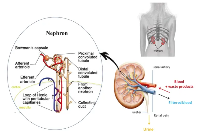
The kidneys are two bean-shaped organs situated in the back of the abdominal cavity in the retroperitoneal space between the dorsal wall and the peritoneum. They filter blood before sending it back to the heart and produce urine which is then excreted. Kidneys are complex organs that morphologically can be divided into three layers: the cortex that represent the outermost layer and contains the blood filtrating glomeruli, the medulla that consists of Henle’s loop and collecting ducts, and the renal pelvis that collects the concentrated urine and connects via the ureter to the bladder. Kidneys receive blood flow through the renal arteries, which direct blood into capillaries passing through the filtering apparatus of the kidney. Filtering is occurring inside the microscopical structures called nephrons, starting with a structure called glomerulus, continuing through a long epithelial tube passing from cortex to medulla and back, ending in a collecting duct.
The process of filtration occurs in three stages: filtration, secretion and reabsorption. Blood filtration involves a selective barrier that is compose of fenestrated endothelial cells, a negatively charged basement membrane and an ultrastructural molecular filter, the slit diaphragm. This glomerular filter allows fluids and small molecules such as waste products, nutrients and ions to pass from the blood into the Bowman’s space of the glomerulus, while larger molecules, such as proteins, are retained in the blood and exit through the peritubular capillaries called vasa recta. The fluid collected by the glomerulus passes into proximal tubules, where most of the water and nutrients are reabsorbed from the primary filtrate. Water and electrolytes are reabsorbed by the loop of Henle and the water content of urine is fine-tuned in the distal tubules and collecting ducts. The filtered blood is carried by the efferent arteriole, while the urine is transported via the ureter to the bladder ( Figure 1). (Scott and Quaggin, 2015)
4
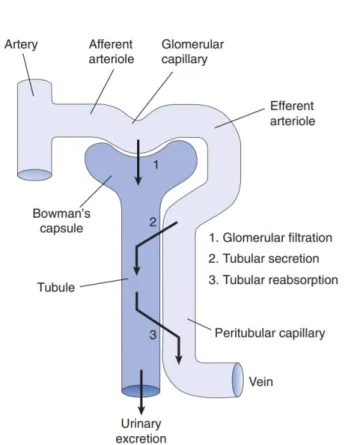
An adult human kidney contains approximately 1 million nephrons that filter around 180 litres of plasma per day. The initially produced pre-urine is processed all along the convoluted tubule and involves active tubular excretion and resorption. Concentration of the urine within the loop of Henle involves an osmotic gradient. It does this by actively transporting out salts from the ascending limb, which creates increased salt concentration in the interstitial fluid of the medulla. This results in the passive flow of water from the descending loop into the interstitial space. This water gets absorbed into blood, keeping the conditions inside the interstitial space constant (Figure 2).
5
Disposal of metabolic waste products while retaining electrolytes is one of the main functions of the kidney. This
way, the kidney maintains
homeostasis and water balance, and
regulates acid-base balance by
reabsorbing the filtered bicarbonate and excreting the fixed acids. Moreover, the kidney regulates the
blood pressure by controlling
electrolyte homeostasis, as well as by secreting renin, a crucial component of the renin-angiotensin-aldosterone system. The kidney also regulates the production of red blood cells in the bone marrow via the secretion of erythropoietin. Moreover, kidneys maintain bone density by converting
vitamin D into the biologically active calcitriol which in turn regulates calcium balance. The kidney also plays an important role in glucose balance by reabsorbing the filtered glucose using sodium-glucose cotransporters on proximal tubules (Vander’s Renal Physiology, 2013).
Early renal differentiation
In the early embryo, gastrulation leads to the development of the mesoderm that can be sub-divided into paraxial, intermediate and lateral mesoderm, according to their position. All kidney tissue in vertebrates arises from the intermediate mesoderm (IM) (Grobstein 1967; Fleming et al., 2013). Apart from kidneys, the IM also contributes to the development of the gonads and adrenal gland. Intermediate mesoderm forms paired elevations called urogenital ridges, which later on give rise to the nephrogenic cords (NC) and the the gonadal ridges that will form the kidneys and gonads, respectively.
Figure 2: Fundamental elements of renal function – glomerular filtration, tubular secretion and tubular reabsorption – and the association between the tubule and vasculature in the cortex. (From Vander’s Renal Physiology, 2013)
6
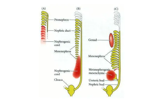
Along the anterior-posterior axis of the NC, three distinct types of kidney are developing in higher vertebrates: pronephros, mesonephros, and metanephros (Saxén 1987, Vize et al., 1997) (Figure 3). In mammals, pronephros and mesonephros are only transiently present during early embryonic life. The pronephros appears to be functional only in lower vertebrates. (Brändli 1999). The mesonephros develops caudally and degenerates or becomes part of the male reproductive system in mammals (Burns, 1955; Balinsky, 1972). The metanephros gives rise to the adult mammalian kidney.
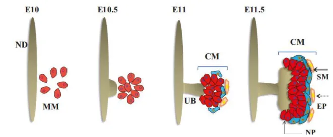
The nephric (or wolffian) duct (ND) is the first epithelial structure formed within the IM. Active migration and proliferation of ND cells results in caudal extension of ND and in induction of mesonephric tubules within the NC (Carroll et al., 2005). By embryonic day (E) 10.5 in the mouse, the ND reaches the Metanephric Mesenchyme (MM), a distinct zone of the NC (Figure 4). The MM cell population are nephron progenitor cells. Inductive signals produced by the MM cause ND to form an outgrowth, called the Ureteric Bud (UB), which makes contact with the adjacent MM and both the UB and the MM undergo extensive morphogenesis to form up to 20,000 nephrons and collecting ducts in mouse, forming a kidney (Murawski et al., 2010).
Figure 3: Three embryonic kidneys: pronephros (a), mesonephros (b) and metanephros (c). (Saxen, 1987)
7
The contact of renal progenitor cells with the UB is a critical event for metanephric development. Metanephric Mesenchyme (MM) gives rise to nephrons, stroma and potentially also some of the vasculature (Munro et al, 2017). Once the MM cells get in proximity of the UB, these cells are rapidly compartmentalized to form a region referred to as Cap Mesenchyme (CM). The first CM cells appear at E11 during MM condensation around the original UB tip (Figure 4). In the rest of the kidney development, CM cells make up a niche of nephron progenitor cells around each single UB tip of the branching ureteric tree. This cell population is exhausted by the second to third day after birth (Rumballe et al., 2011), after which nephrogenesis stops. No stem cell population which can form new nephrons in adult life has been identified.
Transcription factor ODD1, also known as OSR1, is the earliest known marker of the IM (James et al., 2006) (Mugford et al., 2008). The Osr1+ cell population gives rise to the ND, the MM and its epithelial derivatives (nephrons) (Grobstein, 1953, 1955). The ND is the first epithelial tubule to differentiate from the IM that is essential for all further urogenital development. PAX2, LIM1, and OSR1 are DNA-binding proteins that are required to specify the IM. (Patel et al., 2007; Dressler 1990). The Pax2 and Pax8 genes are redundant for the initiation of the specification of the ND lineage at E9.5. Pax2 is expressed during formation and differentiation of all epithelial structures derived from the IM. In Pax2 mutants, the mesonephric tubules fail to form. The ND appears normal but fails to extend caudally and degenerates. Mice Figure 4: Schematic representation of early kidney development. ND – nephric duct, MM – metanephric mesenchyme, UB – ureteric bud, CM – cap mesenchyme, NP - nephron progenitors, SM - stromal cells, EP - endothelial precursors. At the start of metanephric development, MM is specified adjacent to the ND. Later, at E10.5, ND stratification leads to UB outgrowth. This triggers proliferation of the MM. The growing UB invades MM niche, inducing condensation of MM around UB tips, forming CM. This niche is composed of several types of cells: NP, EP and SM. (Motamedi, 2015)
8
heterozygous for Pax2 mutations frequently exhibit a hypoplastic kidney (Dressler et al., 1990; Torres et al., 1995). Surprisingly, the Pax8 mutants do not display renal defects (Mansouri et al., 1998) suggesting that Pax2 might compensate for the loss of Pax8 in kidney development. (Bouchard et al, 2000; Grote et al., 2006). Pax2/Pax8 activate Lim1, also known as Lhx1, which is involved in the caudal extension and development of the ND (Pedersen et al. 2005; Tsang et al., 2000). Pax2/Pax8 also activate the expression of the transcription factor Gata3 and receptor
c-Ret. Gata3 expression commences in ND precursors at E8.5 and appears to control ND
extension (Grote et al., 2006). In contrast, Ret mutant mice develop a ND indicating that Gata3 function during ND extension is Ret independent. (Grote et al., 2008)
Wolffian duct and metanephric mesenchyme
Specification of the MM is the earliest step of the metanephric kidney development. This event occurs at around E10 in the caudal part of the ND (Figure 4). The induction of the MM is regulated by a variety of transcription factors including Osr1 (James et al., 2006; Mugford et al., 2008), Pax2/8 (Bouchard et al., 2000), Lim1 (Shawlot, and Behringer, 1995), Sall1 (Nishinakamura et al., 2001; Kiefer et al., 2010), Six1 (Xu et al., 2003), Six2 (Self et al., 2006),
Wt1 (Kreidberg et al., 1993), Hox11 paralogues (Wellik, 2002, Davis et al., 1994) or
transcriptional coactivator Eya1 (Xu et al., 1999; Sajithlal et al., 2005). Interestingly, specification of the MM doesn’t require the caudal extension of the ND, such as in the case of the conditional deletion of Gata3 in the ND (HoxB7-Cre; Gata3fl/fl). In these embryos, in the absence of the caudal part of the ND, the PAX2+ MM cells are still detected at E11.5 (Grote et al., 2008).
Osr1 is one of the upstream components in the network of transcription factors regulating the
initiation of kidney development. Osr1 orchestrates the specification of the mesonephric and metanephric mesenchyme. Mice lacking Osr1 do not form the MM and fail to express Lim1,
Eya1, Pax2, Six2, Sall1 or Gdnf (Mugford et al., 2006). Osr1 expression was found to be highly
restricted to the non-differentiated subpopulation of the CM (Xu et al. 2014).
Pax2 is expressed in both the UB and the MM. In Pax2 knockouts, a morphologically distinct
MM cell population forms but metanephric development does not occur due to the failure of UB outgrowth (Torres et al., 1995). The absence of UB induction is caused by the lack of the
9
bind to upstream regulatory elements within the GDNF promoter region and can transactivate expression of reporter genes (Brophy et al., 2011). The double Pax2/Pax8 mutants exhibit complete absence of the ND and the MM never differentiates. (Bouchard et al., 2002)
Lim1 (also known as Lhx1) is expressed in the tubule-forming tissue in pronephros,
mesonephros and metanephros (Kobayashi 2005). Deletion of Lim1 leads to a disorganization of the IM and a total absence of the MM (Tsang et al., 2000) suggesting that Lim1 is involved in the specification of the MM. In addition to its role in early kidney development, the conditional deletion of Lim1 at E11.5 in MM has demonstrated that its expression is also required for nephrogenesis and nephron patterning (Kobayashi et al., 2005). (Figure 5).
Eya1 is expressed early within the IM (Figure 5) becoming progressively restricted to the caudal
end where the UB grows out. At E11.5, Eya1 expression is localized to the MM. Eya1 is the transcriptional coactivator that has a specific role in determining metanephric cell fate within the nephrogenic mesoderm. The absence of MM specification in Eya1 mutants is not caused by a lack of signalling emanating from the UB, but due to a cell autonomous defect owing to the lack of Eya1. (Sajithlal et al., 2005)
Sall1 is expressed in multiple tissues including the brain, limbs, heart, ear, and kidney. Sall1
-/-mice display a lack of kidney development. Sall1 has a major role in UB invasion that appears to be independent of GDNF/RET signaling. At E10.5, Sall1 is expressed in the ND and MM. By E11.5, its expression is restricted to the CM surrounding the UB tip. In these mutants, the specification of the MM occurs, but the UB fails to invade the MM. MM cells isolated from Figure 5: A posterior cross-section of the mouse embryo at E8.5. The expression of transcription factor Osr1 is shown in blue, Lhx1, Pax2, Eya1 are shown in purple. Osr1 is expressed anterior compared to other genes. (Dressler, 2009; Sajithlal, 2005)
10
Sall1-/- embryos are capable of epithelialization. In Sall1 mutants, Gdnf is expressed normally at first, but its expression drops off at E11.5 due to the lack of a feedback loop with the UB (Nishinakamura et al., 2001).
Six1 is expressed in the MM starting from E10.5 and is maintained in CM until birth. Six1
-/-mice display smaller kidneys with a variable penetrance of phenotype. In Six1 mutants, the UB fails to invade the MM and the MM cells rapidly undergo apoptosis (Li et al., 2003). SIX1 was found to form a transcriptional complex with PAX2 and EYA1. (Xu et al. 2003). Six4, a gene in the same gene family, expresses in many of the same regions as Six1. Six4 and Six1, are necessary for development of the MM by regulating the level of Pax2-Gdnf expression.
Six1/Six4-deficient mice exhibit a more severe kidney phenotype than the Six1-deficient mice
with a complete absence of Pax2, Pax8, and Gdnf. (Brophy et al., 2001). (Kobayashi et al., 2007).
While it was found that ND forms by migration of the cells of the pronephric duct along an anterioposterior axis to give rise to the ND (Drawbridge et al., 2003; Kopan et al., 2014), the origin of MM remains uncertain. In the classical view the MM stems from the anterior IM. However, fate mapping and in vitro nephrogenesis experiments provided evidence that while the UB emerges from the descending pronephric duct, the metanephric anlagen arise from posteriorly located mesoderm (Taguchi et al., 2014)
Ureteric bud formation
By E10, the ND and MM have been specified at the caudal end of the NC. Later, at around E10.5, the induction of the UB occurs. The UB emerges from the ND and invades the adjacent MM. The formation of the UB is a crucial event in kidney development, with absence of UB leading to renal agenesis.
The initial outgrowth of the UB and its branching are both controlled by the GDNF/RET receptor-tyrosine kinase pathway (RTK). While c-Ret is located on the surface of the UB, GDNF is secreted by the MM (Pichel et al., 1996; Costantini, 2010). From E9 to E10 Gdnf expression is found along the ND before becoming restricted to the caudal end of the ND where the UB appears at E10.5.
Genetic deletion of Gdnf (Sanchez et al., 1996; Pichel et al., 1996; Sainio et al., 1997), its receptor c-Ret (Durbec et al., 1996), or its co-factor GdnfR-a1 (Jing et al., 1996) leads to UB outgrowth failure and subsequent kidney agenesis. However, in these mutants, the MM undergoes specification, which demonstrates that GDNF/RET signaling is not directly involved
11
in MM specification. The GDNF/RET signaling pathway acts through ETV4/ETV5 effectors and mice lacking both Etv4 and Etv5 fail to develop kidneys (Lu et al., 2009).
GDNF and c-Ret action is fine-tuned by many upstream genes such as Eya1, Gata3, Pax2, and
Sall1. Further description of molecular pathways regulating the UB formation can be found in
Article 1 below.
Branching
When the UB emerges from the ND and invades the MM, a repetitive dichotomous branching is initiated that is maintained until birth. This process includes the branching and elongation of the UB, which forms the ureteric tree of the kidney. Molecular analysis identifies at least two types of UB cells: 1) Tip cells that express Sox9, Wnt11 and Ret, and are highly proliferative forming new branches. 2) Stalk cells that have lost tip markers and show less proliferation that will form the collecting duct (CD). (Figure 6) Disruption in the morphogenetic program at this stage leads to renal malformation. Growth and branching of the ureteric tree is regulated through feedback loops primarily involving the signalling molecules WNT11 and GDNF by promoting cell migration and proliferation. (Majumdar et al., 2003) (Reginensi et al., 2011). The branching pattern is largely bifid, with occasional trifid and lateral branches (Blake, Rosenblum, 2014).
12
Nephrogenesis and nephron maturation
Cap mesenchyme
The cap mesenchyme of the
forming metanephros is
composed of nephron progenitor cells surrounding the UB tips on the periphery of the kidney. In the mouse, CM is emerging at E11.5 and persists until postnatal day 2 (Mugford et al., 2009). While individual CM cells undergo migration in accordance to their stage of differentiation, the CM as a whole stays just below the kidney capsule, under the layer of stromal cells. The CM can be
Figure 6: The process of ureteric tree branching is driven by a network of genetic and protein interactions principally controlled by Gdnf/Ret, Wnt and Fgf signalling networks. (Short, Smyth, 2016)
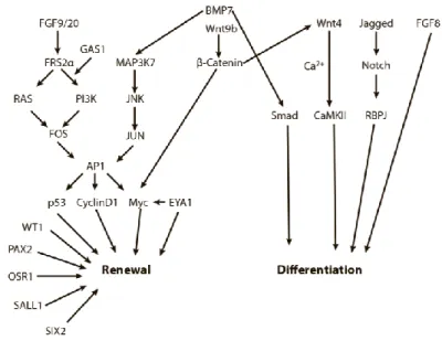
Figure 7: Schematic representation of important regulators of the balance between NPC renewal and differentiation. Arrows represent information flow between pathway nodes, and many of the complex molecular interactions have been simplified to allow for an overview of the major factors that balance the renewal-differentiation axis. (Oxburgh, 2018)
13
divided in two compartments corresponding to the expression of two expression factors: Cited1 (Boyle et al., 2007) and Six2 (Self et al., 2006). Cited1 expression is restricted to the unique subpopulation of CM cells. Fate-mapping approaches using Cited1-CreERT2 have shown that the CITED1+ cells develop exclusively into the epithelial structures of the nephron, apart from the CD (Boyle et al., 2008). Six2 is also expressed in CITED1+ cells, and the deletion of Six2 leads to the exhaustion of progenitor cell population due to premature differentiation (Self et al., 2006). Six2-/- mice develop smaller kidneys as a consequence of the decrease in the number of nephrons. In addition, cell fate analysis demonstrates the role of SIX2+ cells as the progenitors of the nephron lineage (Kobayashi et al., 2008). The importance of Six2 as a key transcription factor in nephron development leads to the conclusion that the CITED1+/SIX2+ subpopulation functions as a pool of non-committed progenitor cells during the nephrogenesis. As the differentiation starts, cells reach the CITED1-/SIX2+ subdomain of CM. These cells
undergo pSMAD activation which leads to higher levels of β-catenin signalling resulting in the formation of a fully-formed nephron. (Brown et al., 2013)
SIX2 was found to act synergistically with OSR1 to maintain progenitor identity of the CM population. SIX2 is required to maintain the expression of OSR1 to prevent premature differentiation of the CM (Xu et al., 2014). In Osr1 mutants, Six2 expression was not affected, however the embryos displayed reduced kidney size. On the other hand, in Six2-/- mutants a reduction of Osr1 expression was observed. Co-immunoprecipitation experiments confirmed the formation of a SIX2-OSR1 complex. OSR1 protects the progenitor pool identity by enhancing TCF interaction with the Groucho family transcription co-repressors, thereby repressing the Wnt/-catenin pathway. These results suggest that the early differentiation of CM starts with CITED1+/OSR1+/SIX2+ progenitor stage, progresses through CITED1 -/OSR1+/SIX2+ and finally to CITED1-/OSR1-/SIX2+.
WT1 is an essential transcription factor for the regulation of NPCs. In Wt1 knockout mice, the MM undergoes apoptosis and as a consequence the UB never forms. (Kreidberg et al., 1993). MM lacking Wt1 fails to undergo epithelialization when paired with wild-type UBs, despite preserved GDNF expression (Donovan et al., 1999). ChIP experiments demonstrate the binding of WT1 to genes which function as regulators of kidney development, such as Bmper, Bmp7,
Pax2, Sall1 and Fgf20, which is consistent with a role of WT1 as a crucial factor in kidney
development (Hartwig et al., 2010; Motamedi et al., 2014). (Figure 8) It was found that the elevation of BMP/SMAD pathway is directly responsible for the dysfunction of the WT1 mutant MM, as activation of this signalling pathway in organ cultures leads to apoptosis;
14
however, addition of FGF20 to the cultures rescues cell death, suggesting a deeper interplay of factors promoting MM survival. (Motamedi et al., 2014). WT1 was found to control expression of the membrane protein GAS1 in the CM; the inactivation of Gas1 leads to premature depletion of CM pool, suggesting a role in maintenance of progenitor population. GAS1 amplifies the PI3K signalling response in the CM, which results in overall function of WT1 promoting proliferation of CM through directing the response to FGF9/20 to the PI3K response (Kann et al., 2015).
PAX2 is a transcription factor that activates Gdnf, which orchestrates the recruitment of UB to MM (Brophy et al., 2001, Ranghini & Dressler, 2015) In the progenitor cell niche, PAX2 maintains the identity of the nephron lineage, preventing transdifferentiation to other cell types. Pax2 conditional inactivation in CM leads to the nephron progenitor cells differentiating to various stromal cell types (Naiman et al., 2017).
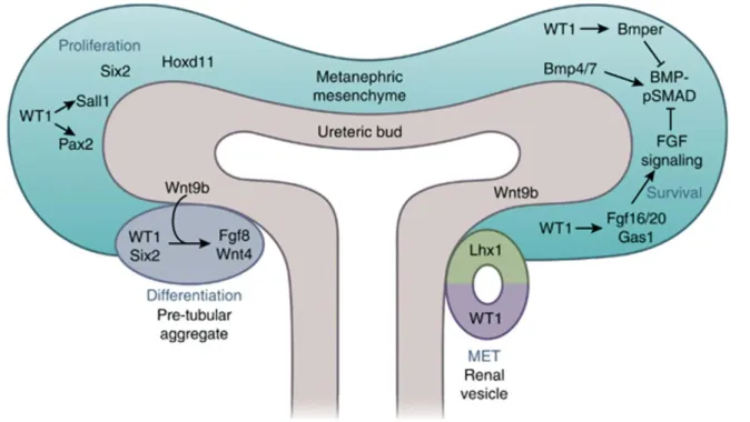
Figure 8: WT1, Six2, and Hoxd11 are individually required for survival and self-renewal of MM. SALL1 and PAX2 transcription factors lie downstream of WT1. BMP4 induces the apoptosis of MM; however, BMP7 promotes the MM differentiation. WT1 directly regulates BMPER, a BMP-pSMAD inhibitor, and activates the FGF signaling pathway via regulating Fgf20/Fgf16 and Gas1 expression to antagonize BMP-pSMAD signaling for the survival and self-renewal of MM. High expression level of Wnt9b in the ureteric tip increases the Wnt singling activity nearing the MM; there, the β-catenin-LEF/TCF complex works together with SIX2 and WT1 activating the critical differentiation factors, FGF8 and WNT4, to form PTA. Signals produced by PTA promote the MET to form renal vesicles. The proximal part of the renal vesicle expressing Wt1 will develop into the epithelial cells of the glomerulus and the distal part of the renal vesicle expressing Lhx1 will develop into the epithelial cells of the nephron tubule. (Dong, Pietsch, Englert, 2015)
15
SALL1 is expressed in the MM, with knockouts suffering from renal hypoplasia due to the defects in UB recruitment to MM. (Nishinakamura et al., 2001) Conditional inactivation of
Sall1 in CM leads to premature depletion of renal progenitors (Kanda et al., 2014). Sall1 null
CM exhibit decreased expression of genes important for nephron progenitor maintenance, such as Eya1, Myc, Six2, Fgf20, which is consistent with the role of SALL1 as a broad regulator of NPC self-renewal (Basta et al., 2017). However, that finding is unexpected in the context of SALL1 function as a transcriptional co-repressor by associating with NuRD complex, suggesting another explanation of gene expression decrease in Sall1 knockouts. One possible mechanism is synergy between SIX2 and SALL1, where the two act as transcriptional activators in the co-occupied loci. This was confirmed by observing co-occupation of self-renewal gene loci by SIX2 and SALL1 using ChIP (Kanda et al., 2014).
Eya1 mutants lack expression of Six2, with defects in early kidney development (Xu et al.,
1999) as well as premature differentiation of CM in conditional CM knockouts (Xu et al., 2014b). EYA1 is also interacting with SIX2 with the formation of the protein-protein complex, which increases the stability of MYC, promoting NPC proliferation on the cell cycle level, in addition to the transcriptional control (Xu et al., 2014b).
A multitude of signaling pathways play an important role in regulation of nephrogenesis. FGF2, a member of FGF signaling pathway, was revealed to promote mesenchyme survival without inducing MET (Perantoni et al., 1995; Dudley et al., 1999; Barasch, 1997). Inactivation of FGF receptors FGFR1 and FGFR2 in the MM leads to a decrease in MM growth and subsequent renal aplasia (Celli et al., 1998; Poladia et al., 2006). FGF2 and FGF9, generated by the UB, promote survival and proliferation of NPCs, and removal of the UB leads to death of MM (Brown et al., 2011). Fgf9 is also expressed in NPC themselves (Barak et al., 2012). However inactivation experiments demonstrate that this expression acts redundantly with Fgf20 (Ortega et al., 1998; Zhou et al., 1998; Dono et al., 1998; Colvin et al., 2001). Fgf20 is expressed solely in NPCs, and together with Fgf9 is essential for the maintenance of NPC proliferation. (Barak et al., 2012). Indeed, compound inactivation of Fgf9 and Fgf20 leads to apoptosis of NPC and loss of MM (Barak et al., 2012). Fgf expression in the MM is however not sufficient for the survival of NPCs in culture, indicating that additional factors from the UB or stroma are required.
WNT9B, a pro-differentiation factor expressed in the UB and promoting expression of Wnt4 and Fgf8 in PTA, is acting as a proliferation factor in CM, indicating a bimodal role of UB in nephrogenesis (Caroll et al., 2005; Karner et al., 2011) The switch between two functions of
16
WNT9B could be determined by the relative intensity of WNT9B signal to Six2 expression, and by activation of the BMP-SMAD signaling pathway (Brown et al., 2013; Karner et al., 2011). Cortical interstitium may play a role in modulating the response to WNT9B, keeping the balance between the proliferation and differentiation, by regulating nuclear versus cytoplasmic localization of the transcriptional regulator YAP. YAP cooperates with β-catenin to regulate expression of WNT9B targets in NPC such as Cited1.(Das et al., 2013) The Six2-mediated deletion of Yap in the CM (YapCM-/-) leads to an impaired transition of the renal vesicle to the S-shaped body structure resulting in hypoplastic kidneys. The conditional inactivation of Rho GTPase Cdc42 in CM compartment leads to a similar phenotype. In addition, lack of Cdc42 leads to reduced nuclear localization of YAP and also loss of Yap-dependent gene expression suggesting a role for Cdc42 to regulate the Yap activity (Reginensi et al., 2013).
BMP ligands are capable of activating both SMAD and MAPK signaling pathways. Bmp7 is expressed in the CM as well as in the CD (Dudley et al., 1997). The collecting duct provides a WNT9B signal which is required to maintain Bmp7 expression in the MM.(Park et al., 2012; Godin et al., 1998) NPC require BMP7 for proliferation and survival, and inactivation of Bmp7 leads to depletion and death of CM (Dudley et al., 1995; Luo et al., 1995). This requirement is present even at later stages of nephrogenesis, as conditional inactivation of Bmp7 at E12.5 after the onset of nephrogenesis recapitulated the Bmp7 null phenotype (Tomita et al., 2013) Downstream of BMP ligands and receptors, SMAD and MAPK signaling is activated. Activation of MAP3K7 promotes NPC proliferation, which explains the premature exhaustion of NPC in Bmp7 null mutant. The same lack of proliferation can be observed by inactivating JNK or activating Jun and ATF2, indicating that the proliferative signal is transduced through the BMP7-JNK-pJUN/pATF2 pathway (Blank et al., 2009).
Although Bmp7 null CM displays lack of proliferation, there are still CITED1+ cells retained within the compartment, though these NPC don’t progress to the epithelialized state, indicating additional functions of BMP7 in regulating MET (Brown et al., 2013; Dudley et al., 1997). Indeed, CM cells require BMP7-SMAD1/5 signaling to transform from CITED1-expressing state to the next compartment which is receptive to β-catenin activation (Brown et al., 2013). CITED1+ cells maintain low levels of activated SMAD1/5, and they increase as cells exit the CITED1+ compartment (Blank et al., 2008). This demonstrates a bimodal role of BMP7, a factor responsible both for maintenance and differentiation of CM.
17 Mesenchymal to epithelial transition in kidney
The process of the Mesenchyme to Epithelial Transition (MET) is a critical step during nephron progenitor differentiation. This process is initiated at E11.5 in murine kidneys, and is regulated through the crosstalk of multiple signalling pathways helping to maintain the proper balance between differentiation and self-renewal of the progenitor pool. The initiation of MET is mediated by the canonical WNT/β-catenin signalling, as demonstrated by a number of loss- and gain-of-function studies (Park et al., 2007; Dressler, 2009; Little, Mc Mahon 2012). The first inductive signal initiating the MET process is Wnt9b, expressed in the UB tips. (Caroll et al., 2005). A subpopulation of CITED1-/OSR1-/SIX2+ progenitor cells starts to aggregate below the UB tip, following the reduction in the expression of Bmp4 (Michos et al., 2007; Schedl 2007). These progenitor cells differentiate into pre-tubular aggregates (PTA) and start to express early epithelial markers after the loss of Six2 expression and under the influence of Wnt9b inductive signal. These markers include Lef1, Jag1, Cdh4/6, Wnt4, Fgf8, Pax8, Lim1 (Kobayashi et al., 2008). Inductive signal of Wnt9b may be substituted by sustained genetic activation of β-catenin in CM, however full epithelialization is not achieved in these conditions, indicating possible mechanisms of inhibition of β-catenin signal in the course of epithelialization (Park et al., 2007).
Positional identity of progenitor cells during aggregation was identified as an important factor in the process of nephrogenesis. The timing of NPC recruitment dictates the spatial positioning of each cell and the subsequent fate of cells along the proximal-distal axis of the nephron. This process of cell-streaming continues from PTA to late RV stage, when podocyte precursors become the last cells to be incorporated into the proximal RV. This finding forms a basis of Time-dependent Cell-fate Acquisition (TCA) model of nephron patterning. (Lindström et al., 2018)
Wnt4, expressed in the PTA, CSB and the distal part of SSB, is a central component of the
nephron induction pathway (Perantoni et al., 2005). Its expression is regulated by Wt1 (Essafi et al., 2011), Pax2 (Torban et al., 2006) and Fgf8 (Perantoni et al., 2005). Wnt4 expression in CM is supressed by the synergistic action of SIX2 and OSR1 proteins to prevent premature exhaustion of the progenitor cell population (Xu et al., 2014). Positive feedback loops increase the Wnt4 expression to reach the critical threshold when a PTA is transformed into a RV. Embryos lacking Wnt4 expression demonstrate inability to form any epithelial nephron structures and the lack of expression of Pax8, Fgf8 and Lim1 differentiation markers (Stark et
18
al., 1994). WNT4 is thought to induce nephrogenesis through its canonical direct target, β-catenin (Park, Valerius & McMahon, 2007). However, Wnt4 induction has been suggested to act through the non-canonical Wnt-Calcium-NFAT pathway as well (Tanigawa et al., 2011; Burn et al., 2011). Wnt4 induces calcium influx in primary MM, which causes epithelialization and can be mimicked by treatment with ionomycin (Tanigawa et al., 2011)
The role of WT1 in MET was studied using a conditional allele inactivated by Nestin-Cre. Nestin is expressed in CM cells in a WT1-dependent manner (Wagner et al., 2006), which allows to bypass the MM loss-of-function phenotype. Reduction in nephron numbers at later stages of development reveals the role of Wt1 throughout the renal differentiation process (Berry et al., 2015). The explanation for this is the role WT1 plays in controlling the transcription of Wnt4 (Essafi et al., 2011). Similar loss of CM differentiation was observed after inducible expression of a short hairpin RNA directed against Wt1 (Kann et al., 2015).
ChIP analysis was used to study the regulation of differentiation of NPCs, leading to a model where WNT/β-catenin and SIX2 regulate a number of genes, such as Bmp7, Wnt4, Fgf8, therefore influencing the balance between self-renewal and differentiation in the progenitor cell population. (Park et al., 2012) There is a remarkable overlap between regulatory targets of SIX2 and WT1, with SIX2-binding sites found on the Wt1 locus, and some of the SIX2 target genes containing WT1-binding motif (O’Brien et al., 2016) Notch was determined to be a negative regulator of Six2, with overactivation of Notch in the CM leading to the loss of SIX2 expression and further NPC differentiation, while inactivation causes loss of nephron differentiation (Boyle et al., 2011; Chung et al., 2017).
Anatomically, compartmentalization of cap mesenchyme reflects different stages of the differentiation process, as defined by sequential growth factor signaling. WNT9B induction mediated by UB activates the expression of Wnt4 and Fgf8 in the pre-tubular aggregate, which are required for the formation of the renal vesicle. (Park et al., 2012; Stark et al., 1994; Grieshammer et al., 2005). FGF8 is required for Wnt4 expression and for other components of the MET process, as evidenced by the lack of MET in the absence of Fgf8 even with Wnt4 ectopically induced (Perantoni et al., 2005).
19
Following MET, the renal vesicle forms a lumen, starts to pattern along the proximal-distal axis and undergoes complex transformation to form a “comma-shape” and then a “S- shape” body before developing into a mature nephron (Figure 9). This dynamic process is regulated by various signalling pathways, which give rise to multiple distinct cell sub-populations. These, in turn, form the distal tubule, Henle’s loop, proximal tubule and glomerulus. In the process of nephron formation, each cell sub-population expresses different segment-specific genes controlling their fate (Desgrange, Sereghini, 2015).
One of the major pathways involved in nephron segmentation is Notch signaling. NOTCH proteins are transmembrane molecules, which activate their downstream effectors after binding to Delta-like and Jaggedligands (Mukherjee et al., 2019). While Notch signaling is required for medial and proximal development (Cheng et al., 2007), Lim1 regulates the development of distal structures, which also express Jag1, Wnt4, Lef1 and Dkk1 (Perantoni et al., 2005; Dressler et al., 2006; Kopan et al., 2007; Schedl 2007; Little and McMahon 2012). Pax genes play a crucial role in nephron patterning, as evidenced by Pax2/Pax8 heterozygous embryos displaying abnormal segmentation in distal tubules (Narlis et al., 2007).
A gradient of ß-catenin activity along the proximal-distal axis controls the differentiation of segment-specific cell fates through interactions with the PTEN/PI3K, and Notch pathways. Conversely, BMP/pSmad and PI3K signalling are negatively modulating ß-catenin activity (Lindström et al., 2015) .
20
Wt1 is expressed in the proximal part of the comma-shaped body and its expression becomes
restricted to the presumptive podocyte layer of the S-shaped body. Wt1 is required for glomerulus podocyte layer specification, structure, function and maintenance (Kreidberg et al., 1993) (Hastie, 2017). Hnf1b was found to control proximal-intermediate nephron identity by regulating Irx1/2 and Notch pathway components Lfng, Dll1 and Jag1. HNF1B is recruited to regulatory regions of these genes. Hnf1b-/- mutants display lack of proliferation in CSB followed by apoptosis in late SSB. However, glomerulogenesis was weakly affected. (Heliot et al, 2013) (Massa et al., 2013)
Brn1, also known as Pou3f3, appears to be involved in the development of multiple nephron segments, including thick ascending limb of Henle’s loop, Macula Densa and distal tubule (Nakai et al., 2003). Brn1 is also important in nephron induction, the determination of nephron numbers, and in nephron size in the murine kidney (Rieger et al., 2016).
Sox genes
Sox is a family of 20 genes, that encode important transcriptional regulators that are involved
in a variety of developmental and pathological processes. The prototypical member of the group is Sry (Sex-determining region Y), located on the Y chromosome and responsible for sex determination in most mammals (Gubbay et al., 1990; Sinclair et al., 1990). Following the discovery, a number of genes were identified that share homology with the originally identified DNA-binding domain of Sry, dubbed Sry box or Sox genes. Sox genes are subdivided into 9 groups (A to H) on the basis of the homology of their DNA-binding domain. Genes in the same group share at least 80% homology inside their Sry box region, as well as substantial homology regions outside. (Wegner, 1999)
Sox gene structure and function
Multiple types of functional domains were identified in Sox genes. These include a DNA-binding domain of the HMG (High Mobility Group) type, a dimerization domain on the N-terminal side of the HMG box, and a trans-activation or trans-repression domain on the C-terminal side of the HMG box (Lefebvre et al., 2007)
21
22
Different Sox members expressed within the same cell type can target different genes, as well as the same Sox gene within different cell types. This points towards other factors that can determine targets of Sox genes, including DNA-binding motif, protein interactions, post-translational modifications and nucleus shuttling.
HMG box conformation is responsible for binding the minor groove of DNA and bending it in an L shape (Ferrari et al., 1992; Connor et al., 1994) with the consensus motif 5’-(A/T)(A/T)CAA(A/T)-3’ in vitro (Harley et al., 1994). Sox genes vary in their binding affinity depending on their subgroup, such as the preference of hSOX9 (human SOX9) to bind to the 5’-AACAAT-3’ motif, and hSOX4 to bind to the 5’-AACAAA-3’ motif (Mertin et al., 1999; Scharer et al., 2009). Nucleotides adjacent to the consensus motif are also responsible for the difference in specificity between Sox genes of different subgroups. For example, hSOX9 binds predominantly the 5’-AGAACAATGG-3’ sequence in vitro, while hSRY preferentially binds to 5’-(T/A)(T/A)AACAATAG-3’ (Mertin et al., 1999, Harley et al., 1994)
An identical consensus sequence can be bound by SOX proteins of different groups, and this interaction can regulate gene expression differently depending on the cellular context. For example, co-binding of SOX-D and SOX-E on the same enhancer sequence of the Col2a1 (Collagen, type II, Alpha I) gene promotes its expression and subsequent cartilage development, while in oligodendrocyte development, SOX-E and SOX-D compete to bind this enhancer and have opposite roles (Lefebvre et al., 1998; Stolt et al., 2006). The different action of different SOX factors, is thought to be brought about by their association with different interaction partners.
The HMG domain was also discovered to be responsible for the interaction with other transcription factors such as PAX proteins, POU factors and Zinc-finger proteins. A SOX protein can form heterodimers with different partners. For instance, SOX2 can synergize with BRN2 in neural primordium, with OCT2/3 in stem cells, and PAX6 in lens cells (Lefebvre et al., 2007).
TAD and TRD
TADs and TRDs were identified with the help of reporter assays in which isoforms of SOX proteins are truncated, but the HMG box is left intact. Truncated proteins lacking TAD/TRD domains are not able to activate or repress the reporter plasmid despite the HMG box retaining
23
its DNA-binding capability, indicating the critical role of TADs and TRDs in Sox transcriptional regulation function (Kamachi et al., 1999)..
TAD and TRD are located at the C-terminal side of the HMG box. Their protein sequences have a high variability between Sox members of different subgroups, providing specificity in regard to target gene regulation between Sox members. For example, chimeric proteins made of the chSOX9 (chicken SOX9) and chSOX1 revealed that the TAD of chSOX1 activates the DC5 enhancer of the Delta1 crystallin gene, although the 2 different HMG boxes can be interchangeable and display similar properties for DC5 enhancer’s binding and bending (Kamachi et al., 1999).
The SOX proteins contain 2 distinct TADs in SOX-E (SOX8/9/10) and SOX-F (SOX7/17/18) members. Which TAD displays the strongest functional relevance depends of the cellular context and can differ between SOX proteins of the same subgroup (Wegner, 2010), which means a gene’s target specificity exists even between the closest related Sox members. For example, Sox10 is an important regulator of neural crest lineage development. Early in development, it is required for the maintenance and survival of neural crest stem cells (Kim et al., 2003) and later for terminal differentiation of their derivatives (Stolt et al., 2002). Experiments using different Sox10 alleles lacking either one of the TADs showed that they were specifically required at 2 distinct stages of neural crest derivative differentiation, while early events were only slightly affected in both mutants (Schreiner et al., 2007)
Dimerization domains
Homodimerization domains, found only in SOX-D (SOX5/6/13) and SOX-E (SOX8/9/10), are responsible for the physical interaction with the same SOX protein or with a SOX member of the same subgroup. The existence for homodimerization implies the possibility of existence of two adjacent binding sites on DNA. Such a dimeric binding site is contained at the promoter of the Mpz (Myelin protein zero) gene, whose expression is controlled by SOX10 in neural crest derivatives. Reporter experiments demonstrated that removal of one of these binding sites severely decreased Mpz promoter activation by SOX10 (Schlierf et al., 2002). Therefore, protein dimerization ability is essential for this specific type of regulation.
Mutations of SOX9 in humans cause campomelic dysplasia syndrome, displaying skeletal malformations frequently associated with male sex reversal. Identification of mutation in hSox9
24
dimerization domain revealed the importance of this mechanism for gene regulation in cartilage development, but not for sex-determination genes. This was consistent with the patient’s phenotype, which didn’t show sex-reversal. (Bernard et al., 2003). This demonstrates that SOX homodimerization requirement for gene regulation is cell context-dependent.
Heterodimers can also form with other transcription factors through non-HMG domains. A short motif on N-terminal portion of SOX6 recruits the co-repressor CtBP2 (C-terminal binding protein 2) on the Fgf3 promoter to repress its transcription (Murakami et al., 2001)
Nuclear-cytoplasmic shuttling
The HMG box comprises 2 distinct nuclear localization signal motifs (NLS). These motifs are highly conserved among the different Sox members, but were functionally characterized only in SRY, SOX9 and SOX2. Nuclear import mediated by the N-terminal NLS motif was found to be dependent on the interaction with activated CALMODULIN, intracellular mediator of the Calcium pathway, and the transporting protein EXPORTIN4. The C-terminal NLS motif was shown to interact with IMPORTIN-B. In sex-reversal patients, mutations in both NLS motifs of hSox9 were described, pointing towards the functional relevance for the Sox nuclear import mechanism.
The HMG box also contains a Nuclear Export Signal motif (NES), described in SOX9 and SOX10, and requires EXPORTIN1 for the process of nuclear export. Although this NES motif is found among all Sox genes, there is a specific amino-acid consensus of the NES of Sox-E members, suggesting a specific nuclear export regulation for Sox8, Sox9 and Sox10. (Malki et al., 2010)
Post-translational modifications
Sox activity is influenced by phosphorylation, acetylation and sumoylation. Acetylation of the HMG domain of hSRY promotes NLS motif binding to Importin-β in vitro, and triggers the subsequent nuclear localization, while deacetylation triggers its cytoplasmic subcellular relocalization (Thevenet et al., 2004).
25
Phosphorylation of several SOX members was shown to promote their activity, such as phosphorylation of Sox2 in induced pluripotent stem cell reprogramming (Jeong et al., 2010), phosphorylation of Sox9 in chondrocytespecific gene activation (Huang et al., 2000), or phosphorylation of chSox9 in neural crest cell delamination in a and canonical Wnt- and BMP-dependent manner (Liu et al., 2013).
SOX3, SOX4, SOX6 and Sox-E were discovered to be modified by sumoylation, resulting in a decreased transactivation potential, decreased synergism with protein partners or increased nuclear localization and subsequent transactivation (Fernández-Lloris et al., 2006) (Girard et al., 2006) (Hattoriet al., 2006) (Savare et al., 2005).
Sox genes functions in development
Sox and WNT signalling
WNT growth factors facilitate essential embryogenesis processes through canonical and
non-canonical WNT pathways. As discussed above, canonical WNT/β-CATENIN,
WNT/CALCIUM and PCP pathways must be tightly controlled during kidney development to allow proper morphogenesis. In vitro studies showed that several SOX proteins could modulate WNT/β-CATENIN activity reporter through physical interaction with β-CATENIN, promoting its degradation or stabilization (Sinner et al., 2007). The in vivo functional relevance of this mechanism was demonstrated in the process of skeletogenesis, where WNT/βCATENIN is tightly controlled and its silencing is crucial for chondrocyte differentiation. This inhibition takes place with the binding of SOX9 to β-CATENIN leading to its proteosomal degradation. Gene targeting in mice revealed similar skeletal phenotypes between β-catenin stabilization and
Sox9 deletion, or β-catenin deletion and Sox9 overexpression (Akiyama et al., 2004). In
summary, WNT and SOX9 crosstalk is an essential mechanism of bone formation. Sox-WNT interaction was also found to be occurring during pancreas formation, T-cell development, sex determination and neural development (Kormish et al., 2010) (Tang et al., 2020). In addition, there is evidence for a WNT signalling role in regulation of the Sox genes through a feedback loop mechanism (Kormish et al., 2010)
26
The NOTCH signaling pathway is a pro-artery cell fate modulator during arterio-venous differentiation of endothelial cells (Lawson et al., 2001). Activation of the artery-specific gene
Dll4 requires binding of SOX-F (SOX7 and SOX18) to the NOTCH effectors NCID/RBPJ
(Notch intracellular domain/Recombination signal binding protein for immunoglobulin kappa J region), while the triple knock-down of Sox7, Sox18 and Rbjp abolishes arterial marker expression (Sacilotto et al., 2013). Thus, NOTCH and Sox-F cooperate in vascular development. In mouse retina development, the expression of Sox8 and Sox9, and subsequent Müller glial cells development, is induced by activated NOTCH signaling (Muto et al., 2009). In this context, NOTCH signalling is an upstream regulator for Sox function.
Sox and FGF-BMP signalling
Neural induction in Xenopus is triggered by endogenous BMP4 antagonists or by activation of FGF signalling. Sox2 is an early neural gene induced upon BMP inhibition, which stimulated responsiveness to FGF-neuralizing signal. Consequently, FGF treatment of Sox2-injected individuals triggered neural and neuronal differentiation in standard medium (Mizuseki et al., 1998). As a result, BMP signalling represses Sox-FGF interaction.
Sox and SMAD signalling
Adult epithelial cells of the mouse lung can be reprogrammed and re-specified into several airway epithelia cell types, in a reversible manner. Sox17 controls this plasticity at least at two levels: by direct binding and activation of the pro-proliferative CyclinD1 gene, and by repression of the anti-proliferative TGFβ/SMAD signalling pathway through physical interaction with SMAD3 (Lange et al., 2009). While adult lung cell plasticity relies on antagonistic effects driven by SOX17 and SMAD, SOX4-mediated inhibition of lymphocyte TH2 differentiation requires activated TGFβ signalling and subsequent binding of SMAD2/3 to the Sox4 promoter (Kuwahara et al., 2012). This way, SOX/SMAD crosstalk can be antagonistic or cooperative.
27 Sox genes in kidney development
Little is known about the roles of Sox genes during kidney formation. Campomelic dysplasia patients occasionally display renal malformations, such as renal hypoplasia, hydronephrosis, hydroureter, and rarely renal cysts (Houston et al., 1983). SOX9 is a gene causing campomelic dysplasia syndrome, thus it might also play important roles during kidney development. Expression analysis in mice supported this conclusion by discovering expression of several Sox members in embryonic kidneys: Sox8 and Sox9 which belong to the Sox-E subgroup, and the 3
Sox-C members Sox4, Sox11 and Sox12 (Kent et al., 1996)(Schepers et al., 2000) (Hoser et al.,
2008).
SoxE and ureter development
Sox9 was demonstrated to be transiently expressed in the ureteric mesenchyme where it is
required for its differentiation into ureter smooth muscle cells (SMC), and Sox9 removal in this compartment resulted in hydroureter (Airik et al., 2010). Another study further characterized the mechanism by which SOX9 regulates SMC differentiation (Martin et al., 2013). Martin et al., showed that SOX9 physically interacts with the transcription factor TSHZ3 (Teashirt3), also required for SMC differentiation. In contrast to Sox9, Tshz3 is expressed at every differentiation step in the differentiation of precursors into SMC. They proposed a model in which the SOX9/TSHZ3 complex inhibits the function of MYCOD (Myocardin), a master transcriptional activator of the smooth muscle program, and Sox9 downregulation removes this inhibition, concomitant with MYCOD-driven transcriptional program. Interestingly, Sox9 and
Tshz3 are both required independently for Mycod expression at the time of SMC induction.
Thus, Sox9 appears to act as a “buffering” factor of SMC differentiation program, by orchestrating the molecular steps driving progression through the myogenic program.
Both Sox8 and Sox9 were implicated in regulation of ureter branching and collecting duct development. Sox8/Sox9 double mutant mice maintained Ret expression but failed to initiate branching of the UB. SOX8/9 were found to be required, but not sufficient, for GDNF/RET signalling by controlling the expression of its major effector genes. In later development, Sox9 mutant mice displayed abnormal induction of nephrogenesis in the outermost layer of the
28
embryonic kidney. In this case, loss of ureteric tip identity leads to expansion of Wnt9b expression domain, causing the formation of ectopic aggregates (Reginensi et al., 2011).
Sox17 and human CAKUT
Sox17 has been shown to negatively regulate β-CATENIN transcriptional activity in vitro
(Sinner et al., 2007). To further analyze WNT/βCATENIN signalling during kidney development, a sequence analysis of the SOX17 gene in a human CAKUT cohort was performed and 3 mutations were identified in 8 patients (Gimelli et al., 2010). All of them displayed vesico-ureteral reflux with one patient suffering from a duplicated collecting system. Two of these mutations resulted in a single amino-acid change and one led to a two amino acid frame-shift insertion. Interestingly, the p.Y259N mutation was found in 2 unrelated sporadic cases and 4 familial cases derived from 2 families, in which the mutation segregated with the CAKUT phenotype. In vitro experiments demonstrated higher protein levels correlating with the stronger inhibition of the WNT/β-CATENIN reporter by the mutant protein compared to wildtype, despite having similar transcript levels. Therefore, the p.Y259N mutation renders SOX17 protein more stable with subsequent renal developmental defects due to inappropriate inhibition of WNT signalling, which makes Sox17 a CAKUT-causing gene
Cooperation between SOXC and WT1 in kidney development
Wt1 is a key gene for kidney formation. It is required for MM survival (Kreidberg et al., 1993)
(Motamedi et al., 2014), nephron formation (Essafi et al., 2011) (Murugan et al., 2012), and glomerulus maintenance (Hammes et al., 2001). Murugan et al. found that WT1 positively regulates Wtn4, a master gene for mesenchyme-to-epithelial transition of nephron precursors. Using the Xenopus pronephros as a model, the authors identified a 4 kb region upstream of the
Wtn4 translation start that was synergistically activated by WT1 and SOX11 in vitro, and
knockdown of each of these genes resulted in a downregulation of Wnt4 expression in vivo,. They further demonstrated that SOX11 physically interacts with WT1 in mouse embryonic kidneys (Murugan et al., 2012), suggesting that their cooperation might be a critical event for nephrogenesis in mammals. Likewise, interaction between WT1 and SOX4 was found to be important for renal development (Huang et al., 2013) Conditional ablation of Sox4 in nephron
29
progenitors results in significant reduction in nephron endowment, with loss of WT1+ podocytes and resulting renal failure. These observations are consistent with a specific role of SOX4 in podocyte lineage.
Congenital anomalies of kidney and urinary tract
Congenital anomalies of kidney and urinary tract (CAKUT) are developmental malformations that can have dramatic consequences in early and adult life. CAKUT are responsible for 30-50% cases of end-stage renal diseases in children (Seikaly et al., 2003). When asymptomatic, kidney malformations predispose to renal deficiency in adult life, promoting diseases such as hypertension or cardiovascular diseases. Kidney malformations in humans are found in 1 out of 500 newborns. They are often found in the prenatal development, representing approximately 20 to 30 % of all anomalies identified (Pichel et al., 1996). The in utero environment can be a contributing risk factor when issues such as maternal diabetes, diet or drug uptake are involved (Aisa et al., 2019) (Lee et al., 2018) (Boubred et al., 2006). In addition, familial clustering of kidney abnormalities is found only in 20% of patients, suggesting non-hereditary factors for predisposition to these diseases (Satko & Freedman, 2005)
Types of CAKUT include: renal agenesis (complete absence of kidney), renal hypoplasia (reduced size of the kidney), renal dysplasia (incomplete or abnormal development of the kidney), Wilms’ tumor (pediatric renal rumor), duplex kidney (duplication of kidney and collecting system), hydronephrosis and hydroureter (abnormal dilation of renal pelvis and ureter), vesicoureteral reflux (retrograde flow of urine), polycystic kidney disease (progressively expanding cystic structures in the kidney) and horseshoe kidney (two kidneys are fused at their lower pole). (Song & Yosypiv, 2011)
Since CAKUT were found to be inherited in some cases, a genetic basis for inheritance was proposed. The development of DNA sequencing capabilities that allowed human whole-genome analysis, coupled with genetic modification tools allowing targeted gene alteration in animal models, provided powerful tools to reproduce and follow CAKUT progression, allowing a better characterization of the mechanisms leading to human diseases. Based on the observation of human duplex kidneys samples, Mackie and Stephens proposed the ureteric bud (UB) theory (Mackie & Stephens, 1975). They found a strong correlation between the mispositioned ureter-bladder orifice and the hydronephrosis/hydroureter features displayed by
30
the corresponding kidney and ureter. They proposed a model in which the initial position of the UB (the future ureter) at the onset of kidney development dictates the final position of ureter orifice into the bladder, with an ectopic budding site leading to duplex kidneys. This hypothesis was confirmed using a mouse model (Foxc1 (Forkhead box C1) knock-out) (Kume et al., 2000). Later, a mutation of the FOXC1 gene was found in a cohort of CAKUT patients (Nakano et al., 2003). Currently, more than 180 monogenic causes of murine CAKUT have been described, with many of the 40 identified human CAKUT genes first described in a mouse model (van der Ven et al., 2018) including PAX2 (Paired box gene 2), EYA1 (Eyes absent homolog 1), SALL1 (Spalt-like transcription factor 1), HNF1B (HNF1 homeobox B) and SIX1 (SIX homeobox 1). While these genes were found mutated in a significant proportion of CAKUT patients (Yosypiv, 2012), their role as disease-causing factors is debated.
Treatments for CAKUT are limited in effectiveness. In the best cases renal failure can be treated with surgery and/or dialysis but the patients often fail recover full renal function. Kidney transplantation can cure the disease; however, patients face the obstacles of the low number of compatible donors and graft rejection issues. Regenerative medicine or cell therapy approaches that involve the in vitro production of renal structures or particular cell types could be the basis of future renal failure treatments. Extensive research has led to the identification of organ culture conditions that can mimic some of the renal development processes, however so far the
in vitro generation of a functional kidney has not been achieved. In addition, a high proportion
of human CAKUT cases escape genetic testing and remain without genetic or environmental explanation. To further the development of therapies and CAKUT diagnosis and etiology, it is important to understand how the kidneys develop in the normal situation and to characterize the molecular mechanisms controlling their morphogenesis.
31