Publisher’s version / Version de l'éditeur:
Vous avez des questions? Nous pouvons vous aider. Pour communiquer directement avec un auteur, consultez la première page de la revue dans laquelle son article a été publié afin de trouver ses coordonnées. Si vous n’arrivez pas à les repérer, communiquez avec nous à PublicationsArchive-ArchivesPublications@nrc-cnrc.gc.ca.
Questions? Contact the NRC Publications Archive team at
PublicationsArchive-ArchivesPublications@nrc-cnrc.gc.ca. If you wish to email the authors directly, please see the first page of the publication for their contact information.
https://publications-cnrc.canada.ca/fra/droits
L’accès à ce site Web et l’utilisation de son contenu sont assujettis aux conditions présentées dans le site LISEZ CES CONDITIONS ATTENTIVEMENT AVANT D’UTILISER CE SITE WEB.
The Journal of Biological Chemistry, 266, 17, pp. 10781-10786, 1991-06-15
READ THESE TERMS AND CONDITIONS CAREFULLY BEFORE USING THIS WEBSITE. https://nrc-publications.canada.ca/eng/copyright
NRC Publications Archive Record / Notice des Archives des publications du CNRC :
https://nrc-publications.canada.ca/eng/view/object/?id=6d318005-e122-4c01-b571-f41c639be9ed
https://publications-cnrc.canada.ca/fra/voir/objet/?id=6d318005-e122-4c01-b571-f41c639be9ed
NRC Publications Archive
Archives des publications du CNRC
This publication could be one of several versions: author’s original, accepted manuscript or the publisher’s version. / La version de cette publication peut être l’une des suivantes : la version prépublication de l’auteur, la version acceptée du manuscrit ou la version de l’éditeur.
For the publisher’s version, please access the DOI link below./ Pour consulter la version de l’éditeur, utilisez le lien DOI ci-dessous.
https://doi.org/10.1016/S0021-9258(18)99086-1
Access and use of this website and the material on it are subject to the Terms and Conditions set forth at
Structural and catalytic properties of a deletion derivative (Δ
₁₃₃₋₁₅₇) of
Escherichia coli adenylate kinase
Rose, Thierry; Brune, Martin; Wittinghofer, Alfred; Le Blay, Karine; Surewicz,
Witold K.; Mantsch, Henry H.; Bârzu, Octavian; Gilles, Anne-Marie
THE JOURNAL OF BIOLOGICAL CHEMISTRY
8 1991 by The American Society for Biochemistry and Molecular Biology, Inc.
Vol. 266, No. 17, Issue of June 15. PP. 10781-10786,1991
Printed in U.S.A.
Structural and Catalytic Properties
of
a
Deletion Derivative
(A133-15,)of
Escherichia coli Adenylate Kinase*
(Received for publication, February 19, 1991)
Thierry Rose$, Martin Brunes, Alfred Wittinghofer8, Karine Le Blay$, Witold K. Surewiczq, Henry H. Mantschll, Octavian BarzuS, and Anne-Marie Gilles$)I
From the $Unite de Biochimie des Regulations Cellulaires, Institut Pasteur, 75724 Paris Cedex 15, France, the §Max-Planck Institut fur Medizinische Forschung, Abteilung Biophysik, 6900 Heidelberg, Federal Republic of Germany, and the VSteacie Institute of Molecular Science, National Research Council of Canada, Ottawa K I A OR6
Escherichia coli adenylate kinase (AKe) as well as
the enzyme from yeast and mitochondria differs from the muscle cytosolic variant (AK1) by an insertion of 2 5 amino acid residues that are missing in AK1. The extra sequence, highly homologous in “large” size var- iants, is situated between residues 133 and 157 in AKe. Removal of 25 codons in the corresponding adk gene resulted in expression of a modified form of adenylate kinase (A133-157 AKe) which still conserved 7% of the maximal activity of the wild-type protein. The appar-
ent K,,, for nucleotide substrates was increased by a
factor of 4.6 (ADP), 23 (ATP) or 43 (AMP) in A133-157 AKe when compared with the wild-type enzyme. The secondary structure of
A133-157
AKe, as well as its ther- mal stability were very similar to the parent protein. However, the deleted protein was much more sensitive than the wild-type enzyme to inactivation by trypsin. Sodium dodecyl sulfate-polyacrylamide gel electropho- resis analysis of trypsin digested A133-157 AKe revealed accumulation of several well defined fragments which were not observed in the case of wild-type enzyme. We conclude that the additional sequence, although nec- essary for expression of full activity in AKe, is not critical for catalysis. It is perhaps responsible for in- teraction of enzyme with other cellular components although a different mechanism of water shielding for large and small size variants of AK can be also envis- aged.Adenylate kinase (AK, ATP:AMP phosphotransferase, EC
2.7.4.3),’ which catalyzes the reaction,
* This work was supported by grants from the Centre National de la Recherche Scientifique (URA 1129) and the Ministixe de la Re- cherche et de I’Enseignement Superieur and by the France-Canada Science and Technology Cooperation program. T he costs of puhlica- tion of this article were defrayed in part by the payment of page charges. This article must therefore be hereby marked “aduertise- rnent” in accordance with 18 U.S.C. Section 1734 solely to indicate this fact.
11 To whom correspondence should he addressed Unite de Bio- chimie des Regulations Cellulaires, Institut Pasteur, 28, rue du Doc- teur Roux, 75724 Paris Cedex 15, France. Tel.: 33-1-45-68-80-00 (ext. 7268); Fax: 33-1-43-06-98-35.
T he abbreviations used are: AK, adenylate kinase; ApBA, P 1 , P di(adenosine 5’)-pentaphosphate; A13:3-,57 AKe, deletion derivative of
E. coli AK lacking amino acid residues from 133 to 157; blue-Sepha- rose, Cibacron blue 3G-A-Sepharose CL-GB; HPLC, high perform-
ance liquid chromatography; SDS-PAGE, sodium dodecyl
sulfate-polyacrylamide gel electrophoresis; TPCK, L-l-tosylamido-2- phenylethylchloromethyl ketone; HEPES, N-2-hydroxyethylpipera- zine-N’-2-ethanesulfonic acid.
ATP.MgL‘
+
AMP ADP.M$’+
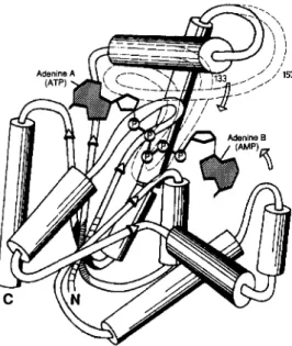
ADP,is an attractive model enzyme for studying structure-function relationships, as it is a small monomeric protein, easy to purify, handle, and detect (1, 2). The alignment of primary structures of different isoforms from different organisms (3) showed a striking difference between mammalian muscle cy- tosolic adenylate kinase (AKl) and the enzyme from Saccha- romyces cereuisiae (AKy), Escherichia coli (AKe) or mamma- lian mitochondria (AK2 and AK3). The latter four “long AKs” contain a continuous insertion of 25 amino acids that are missing in the short variant (AK1) of adenylate kinase. This insertion is bracketed by identical residues in all species of adenylate kinase: Ser-Gly-Arg at the N terminus and Asp- Asp at the C terminus (see Figs. 1 and 2). The three-dimen- sional structures of long and short adenylate kinases are remarkably similar (4-7). A schematic view of the structure of the long AKs as inferred from the three-dimensional analy- sis of the yeast and E. coli adenylate kinase complex with the bisubstrate analog ApsA (4, 6) is shown in Fig. 1. Although the extra sequence is not as well defined as the rest of the structure it was found t o be located close to the ATP binding site (adenine A of Ap5A). It seems to act as a lid that shields the active site from the surrounding medium. Additionally, NMR experiments suggest that of AKe (equivalent to His’43 in AKy) is located close to the ATP. M$+ binding site
(8, 9).
Since the extra sequence is highly conserved in long AKs (13 amino acid residues from 25 are identical in AK2, AK3, AKy, and AKe), we wanted to discover its possible functional role in this enzyme. We deleted the whole sequence situated between residues 133-157 in AKe and investigated the struc- tural and catalytic properties of the modified enzyme.
EXPERIMENTAL PROCEDURES
Chemicals-Adenine nucleotides, restriction enzymes, T4 DNA ligase, and coupling enzymes were from Boehringer Mannheim. DNA polymerase large fragment (Klenow) was from Du Pont-New England Nuclear. TPCK-treated trypsin and soybean trypsin inhibitor were from Sigma. Blue-Sepharose, polybuffer exchanger 94, and polybuffer 74 were from Pharmacia LKB Biotechnology Inc. Oligonucleotides were synthesized according to the phosphoamidinate method using a commercial DNA synthesizer (Cyclone’“ Biosearch).
Bacterial Strains, Plasmids, and Cloning Methods-Plasmid pEAK91, which contains the adk gene from E. coli on the high copy number plasmid pEMBL9 has been described previously (10). AKe is expressed from this plasmid in strain CK600 (SupE, hsdM’, hsdh), which contains a chromosomal copy of the adk wild-type gene. For the construction of the deleted gene involving BclI restriction enzyme we used strain GM119A which is dam- with unknown genetic back- ground. Thermosensitive strains CV2 and KG2 are plsA2 (=adk2) qlpD3, glpR2A phoA8 tonA22 T2-R rellA1 (X)pit-10 om pF627 fad-
10781
10782
E. coli Adenylate Kinase
FIG. 1. Schematic drawing of the three-dimensional struc-
ture of the ApsA complex of AKe based on the model of AK1,
a short version of adenylate kinase. The location of the extra sequence is tentative and was inferred from the structure of the AKy. AphA complex (4). The two arrows indicate the movement of the structural elements involved in the induced fit mechanism (see "Dis- cussion").
L701, and plsAI5 (=adkl5) recAl proC24 thyA25 nalAl2 argG34 metBl pyrC30 lac str-97 tsx-63 mlt-2 xyl-7 or 14, respectively. They were transformed with PEAK91 (A133-157) and plated a t 28 "C. After 48-h of growth, 100 colonies from each transformation were replated at 28 and 42 "C and scored for survival after 24 h (42 "C plates) and 48 h (28 "C plates). Mutagenesis was done using the thiophosphate method of Taylor et al. (11). If not indicated otherwise, the cloning techniques were performed as described by Maniatis et al. (12). Sequencing of the construction was performed with the T 7 polymer- ase sequencing kit from U.
s.
Biochemical Corp.Purification of Adenylate Kinase and Activity Assays-The wild- type enzyme from AKe-overproducting strain of E. coli was purified as described previously (13). A 1 3 3 . 1 ~ , AKe was purified essentially by
the same procedure involving chromatography on blue-Sepharose and Ultrogel AcA54. Enzyme fixed on a blue-Sepharose matrix a t p H 7.4 was eluted either with a salt gradient (between 0 and 1 M NaCl in 50 mM Tris-HC1, pH 7.4) or with a mixture of 3 mM ATP
+
3 mM AMP in the same buffer. AKe expressed by the chromosomal wild-type adk gene coeluted with A133-1s7 AKe both in blue-Sepharose and Ultrogel AcA54 chromatography. The two proteins were completely separated by chromatofocusing allowing a n accurate structural and kinetic analysis of the modified protein (see Fig. 4, under "Results"). Aden- ylate kinase activity was determined with the coupled spectrophoto- metric assay (13) a t 334 nm and 30 "C in 0.5-ml final volume on a Eppendorf PCP6121 photometer. One unit of enzyme activity corre- sponds to 1 pmol of product formed in 1 min a t 30 "C and pH 7.4.Trypsin Digestion and Peptide Separation of Deleted AKe-Wild- type and deleted forms of AKe were digested for 15 h at 37 "C with TPCK-trypsin (l%, w/w). Peptides were purified by reverse-phase HPLC on a Perkin-Elmer apparatus (series 410 equipped with a LC135 diode array detector) using a Nucleosil C-18 column (5 p M ,
4.6 X 25 mm) and an ammonium acetate, p H G.O/acetonitrile elution system at a flow rate of 1 ml/min and absorbance recording a t 230 and 280 nm.
Amino Acid and Sequence Analysis-Amino acid analyses were performed on a Beckman System 6300 high performance analyzer after 6 N HC1 hydrolysis for 22 h a t 110 "C. Manual sequencing of the isolated peptide was conducted using the 44dimethyla- mino)azobenzene 4'-isothiocyanate/phenylisothiocyanate double coupling technique (14).
Spectroscopic Studies-Infrared spectra were recorded with a Di- gilab FTS-60 spectrometer using a high sensitivity deuterated trygli- cine sulfate detector. Samples were prepared in 50 mM HEPES buffer in D,O, pH 7.4, at a protein concentration of 1 mM, and the spectra were obtained as described previously (15). Circular dichroism (CD)
spectra were recorded on a Jobin-Yvon CD6 or a Jasco-600 apparatus from 180 to 260 nm using quartz cylindrical cells of 0.2 or 0.5 mm and protein concentration ranging from 2 to 40 p ~ . Results are given as the mean residue molar ellipticity (0) expressed in degrees.cm2. dmol". For the estimation of secondary structure, CD curves in the 190-260 nm range were analyzed by the method of Chen et al. (16) using the program of Yang et al. (17). The equilibrium unfolding as a function of guanidine HCl was monitored at 20 "C and 222 nm. All samples were equilibrated for approximately 12 h before measure- ments were made. The fraction of folded protein, fN, was calculated as fN = (0 - 0 , ) / ( 0 ~
-
OU), where 0 is the observed ellipticity and ON and 0, are the values of ellipticity for the native and totally unfolded forms, respectively. Values for ON and 0, in the transition zone were determined by linear extrapolation.Thermal Denaturation-The thermal stability of the wild-type AKe and the A133.15v AKe was studied by high-sensitivity differential
scanning calorimetry using a Microcal MC-2D instrument a t a scan- ning rate of approximately 50 "C/h. Protein solutions used in these experiments were prepared in 50 mM HEPES buffer, pH 7.4, at a concentration of 2-2.5 mg/ml.
RESULTS
Expression and Purification of A133-157 AKe, a Deleted De- rivative of AKe-The E. coli adk gene on the high expression plasmid PEAK91 was mutated such that Asp-Ile substituted for T ~ r ' ~ ~ - H i s ' ~ ~ , thus creating an EcoRV restriction site (Fig.
2). Bacteria expressing the doubly-modified enzyme (Y133D,
H134I) exhibited an AK activity close to that of the same E.
coli strain without plasmid (about 1 unit/mg of protein). The modified PEAK91 plasmid was digested with EcoRV and BclI.
The BclI site was filled in with Klenow polymerase, the 75- base pair fragment removed and the plasmid religated. The resulting clones were screened for the presence of BclI site and the absence of the EcoRV site, and a positive clone was sequenced. The sequence of the resulting plasmid around the
+++ + +++++ ++ +++ + + ++ AKY PSGRSYHKIFNPPKEDMKDDVTGEALVQRSDDNAD ~ AK2 "-SGRSYHEEFNPPKEPMKDDITGEPLIRRSDDNKK- AK3 -SGRVYNIEFNPPKTMGIDDLTGEPLVQREDDRPE - AK1 --SGRV---DDNEE- SGRVYHVKFNPPKVEGICDDVTGEELTTRKDDQE sile directed mutagenesis GATATC i d
AKE' SGRVD~VKFNPPKVEGKDDVTGEELTTRKD~QEE ,eJl,lnlondiesl
'hA
&&
BCIIIECORV.lllinp __ EmRV Bcll "SGRVDDNEE - up wllh Klenow-Pol liallcn AK1 AKE A I S 1 5 7 ( 5 . 0 1 *b, PEAK91 l5.C "b,AKe. The sequence alignment of short and long AKs (3) shows the FIG. 2. Construction of the expression plasmid for A l ~ ~ - l s ,
conserved residues in and around the extra sequence. Three point mutations were introduced into single-stranded DNA from plasmid PEAK91 (10) to create an EcoRV site at the N-terminal site of the deletion. T h e BclI site was cleaved, filled in using Klenow polymerase, and after digestion with EcoRV the 75-base pair fragment was re- moved, the plasmid religated and analyzed, as described under "EX- perimental Procedures." The resulting amino acid sequence of A133.157 AKe is shown in comparison with the sequence of AK1 (pig muscle).
E. coli
AdenylateKinase
10783deletion site was now highly homologous to the sequence of mammalian cytosolic AK (Fig. 2). The soluble extract from
E. coli expressing plasmid PEAK91 (Al:<:\ 1 5 7 ) exhibited a 2-
fold higher specific activity under standard assay conditions (30 "C, p H 5.4, a n d 1 mM A D P as substrate) as compared with the same E. coli strain without plasmid. As several temperature sensitive mutants of the adh gene have been described showing that it is an essential gene, we wanted to know whether adk" strains could be complemented by the
deleted adk gene. Two types of temperature sensitive mutants (CV2 with a P87S mutation and KG2 with a S129F mutation) (3) were transformed with plasmid PEAK91 ( A 1 3 : l . 1 3 y ) . T h e
colonies were viable a t permissive and nonpermissive temper- atures, showing that the deletion derivative is not only active, but that the deleted region is not important for the viability of E. coli.
Al:\:x.157 AKe was expressed in a high yield in E. coli strain CK600 and purified by standard procedures involving blue- Sepharose and gel permeation chromatography (13). Under these conditions, the protein co-eluted with the wild-type
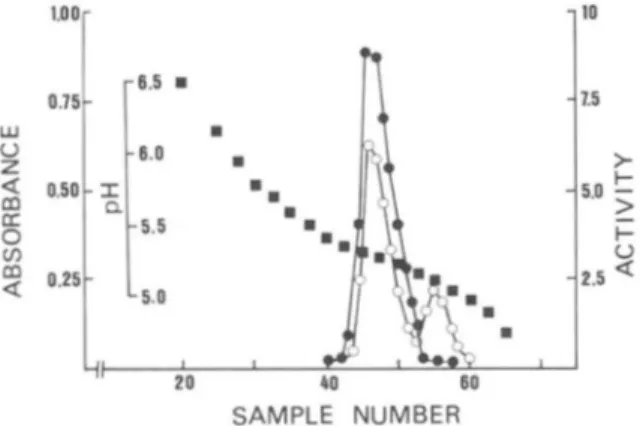
AKe expressed by the chromosomal gene (Fig. 3). T h e t w o proteins were subsequently separated by chromatofocusing (Fig. 4) as the pI of deleted enzyme (5.4) was higher than that of the wild-type protein (5.1).
Structural Analysis of Al:l:3.157 AKe-Amino acid analysis of
Al:3:3.157 AKe was in good agreement with what was expected from the removal of the sequence situated between Tyr"':' a n d
1 2 3 4
FIG. 3. SDS-PAGE (12.5%) of fractionsobtained during the purification of A l n n - t n i AKe. Lane 1, standard proteins, from top t o bottom: a, phosphorylase a (94,000); b, bovine serum albumin (67,000); c, ovalbumin (43,000); d , carbonic anhydrase (30,000); e,
soybean trypsin inhibitor (20,100); f, lvsozyme (14,400). Lane 2,
bacterial extract (35 p g of protein). Lane 3 , blue-Sepharose chroma- tography (35 p g of protein). Lane 4 , Ultrogel AcA54 chromatography
(18 pg of protein). h
1
lo w 0 z2
(r 0 v, m 6-
1.5>
SAMPLE NUMBERFIG. 4. Separation of A1:3:~-15i A K e from wild-type AKe by chromatofocusing. Proteins after Ultrogel AcA54 chromatography
(-5 mg) were loaded o n t o a polybuffer exchanger 94 column ( 0 3 X 15 r m l . 1.5 ml of Tris-HCI 50 mM, p H 7.4, were used to wash the column. Proteins were then eluted with 3.5 ml of 10-fold diluted polyhuffer 74 adjusted to pH 4 with 1 N HCI. Samples of 0 . 5 ml were collected at a
flow rate of 4 ml/h. H, pH; 0 , ahsorhance at 280 nm; 0, activity (units/ml).
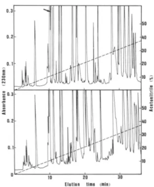
Lysl" in the wild-type protein (Table I). T o confirm the extent of deletion, digests with TPCK-trypsin of both wild- type and deleted AKe were analyzed by reverse-phase HPLC. The elution profile of tryptic peptides indicated that. the two peaks corresponding to peptides 132-136 and 137-156 in wild- type AKe were missing in the deleted enzyme (Fig. 5 ) . A new tryptic peptide in AKe arising from the fusion of Val':(' t o Asp""X (VDDQEETVR) had an elution time very similar to t h a t of peptide '"'KDDQEETVR"" in the wild-type enzyme.
The secondary structure of deleted AKe was analyzed using two different spectroscopic techniques. The Fourier-trans-
formed IR and CD spectra of AKe were similar to those of the parent enzyme (data not shown). In additional further experiments, we tested the stability of wild-t-ype and deleted protein against trypsin, guanidinium HCl, and tem- perature. Al:i:l I R i AKe was much more sensitive than the wild- type protein to proteolysis by trypsin. The first order rate
constant of inactivation by TPCK-trypsin at pH 7.4 and 30 "C was 2.1 x lo-" s" for wild-type protein and 3.9 x lo-:' s-' for its deleted variant. SDS-PAGE analysis of trypsin inactivated wild-type AKe did not reveal any well-defined tryptic frag- ment (18), whereas analysis of trypsin digested AKe
revealed accumulation of several well defined fragments (Fig. 6). Ap5A, a n d t o a lesser extent ATP, protected the deleted protein from inactivation by trypsin. When soluble extracts of bacteria harboring the plasmid overexpressing I ~ , ; AKe were incubated at 30 and 40 "C and submit.ted to SDS-PAGE analysis, we did not observe a significant proteolytic cleavage of t h e modified protein.
Figure 7 shows the excess heat capacity uersu.9 temperature curve for the wild-type AKe and the deleted protein. These curves were obtained by substracting the base lines from the raw calorimetric data using a cubic splines interpolation pro- cedure. The thermodynamic parameters derived from the excess heat capacity curves (Table 11) indicate relative close thermodynamic stabilities of both proteins. In particular, there is no significant difference between the denaturation temperatures of the wild-type AKe and its deletion derivative. Similar stabilities of the wild-type AK and the proteins
were also demonstrated by the results of equilibrium unfolding experiments in guanidine HCl (Fig. 8). For both proteins, t.he dependence of t h e folded fract.ion, f s , on the denaturant concentration indicates a two-state unfolding process. The midpoint transition concentrations of guanidine HCI were 0.93 a n d 0.90 M for the AKe and AI:<:{ AKe, respectively (Fig. 8).
Kinetic Properties of AI:?:, AKe-Table I11 shows the kinetic parameters of AKe compared with AKe and
rabbit muscle AK1. Deletion of the 25-amino acid extra sequence in E. coli protein decreased the catalytic activity by a factor of 62 (ATP formation) or 14 (ADP formation). The apparent K,,, for nucleotide substrates was increased by a factor of 4.6 (ADP), 23 (ATP), or 43 (AMP). Under identical experimental conditions rabbit muscle AK1 exhibited kinetic parameters close to those of wild-type AKe. The requirements for divalent cation were practically the same for all three
forms of adenylate kinase (not shown). However, excess AMP (4 mM) inhibited the wild-type AKe by about SO%, whereas a 2-fold higher concentration of nucleoside monophosphate did not inhibit the deleted bacterial variant.
DISCUSSION
The results of this study reveal numerous functional and structural similarities between AI:,.\ AKe and the wild-type protein. In particular both enzymes showed intrinsic trans- phosphorylating activities and similar resistance to tempera-
10784
E. coli Adenylate Kinase
TABLE I
Amino acid c ~ n ~ p < ~ . ~ i t i o n of A ~ , ~ , , adenylate kinase of E. coli compnrrd with that of wild-type protrin
Residues/molecule Amino
acid A,:,.< 157 Wild-type
Observed Calculated Observed Calculated"
c y s NDh 1 NDh 1 Asx 16.7 18 19.9 21 T h r 6.8" 8 9.6' 11 Ser 4.0 5 4.1' 5 Glx 22.1 23 25.0 26 Pro 8.0 8 10.3 10 Gly 17.2 18 19.6 20 Ala 19.0 19 19.0 19 Val 15.4 16 18.2 19 Met ND" 6 ND' 6 Ile 12.5 14 12.3 14 Leu 15.5 15 16.1 16 Tyr 4.9 6 5 . 9 7 Phe 3.9 4 4.8 5 His 2.2 2 3.1 3 Arz 11.6 12 12.6 13 Lys 12.7 14 17.1 18
" From the nucleotide sequence of the adk gene (10). ' Uncorrected values determined after 20 h of hydrolysis.
ND, not determined. a- b* C- d- e- #""" f-
"---
z - = a-
-
1 2 3 4 5 6 7FIG. 6. Proteolysis of Alns-157 AKe by trypsin and protection
by nucleotides. A1:l:l ls7A AKe at 1 mg/ml in 50 mM Tris-HCI, pH 7.4, and 50 mM KC1 was incubated a t 30 "C with TPCK-trypsin (2 pg/ml) in the absence (lanes 1 - 5 ) or presence of 4 mM ATP (lane 6 )
or 1 mM ApsA (lane 7). At different time intervals (0 min, lane I ; 1 min, lane 2; 2 min, lane 3 ; 5 min, lane 4 ; and 10 min, lanes 5-7), 20-
pl aliquots were withdrawn, boiled with electrophoresis buffer, and analyzed by SDS-PAGE (12.5%) and Coomassie Blue staining. The molecular weight standards are the same as indicated in Fig. 3.
1
IO 20 30
Elrllan tlns cmln)
FIG. 5 . Separation of tryptic peptides by reverse-phase
HPLC on a nucleosil C l n column. I'eptides resulting from 1 mg of digested protein were eluted with a linear gradient of 0 4 0 % aceto- nitrile ( - - - -) and detected by their ahsorhance at 230 nm (-).
The flow rate w a s 1 ml/min. Upper, digest of AI:,:, 1:); AKe. Lower,
digest of wild-type enzyme. Arrow indicates the peptide resulting from the lusion of \'all
''
to Asp"*.ture and guanidinium HCl denaturation. This suggests that the 25-amino acid residue insertion present in long variants of AKs is not essential for catalysis or for the maintenance of thermodynamically stable structures.
Despite these similarities, there are significant differences between AKe and Al:3:3.157 AKe. First, the deletion of the extra sequence reduced the catalytic activity of AKe drastically with a concomitant large increase of the K , values for nucleo- tide substrates. The crystal structure analysis of AK1 without substrate of AK3 with AMP and of AKe and AKy complexed
I
35 I 40 45 50 55 60
=I
65 7 0Temperature, 'C
FIG. 7. Differential scanning calorimetry curves of excess
specific heat uersus temperature for the wild-type AK (upper
trace) and the A1n3-157 enzyme (lower trace).
to the bisubstrate inhibitor ApsA has been completed (4, 6, 19). It revealed that the two adenine moieties of ApA are located in the AMP and ATP binding sites and that the
E. coli Adenylate Kinase
10785
uration, as shown in AK1, t o a closed more compact structure when both substrates are bound. This structural change leads to an induced fit of the substrates (20). In kinases it also served to shield the active site from the surrounding water
(21). The structural regions involved in this movement of two apparently rigid bodies, as indicated in Fig. 1, are the two helices to the right and the extra sequence to the left of the deep cleft. The extra sequence which in the structure of AK3 complexed to AMP points away from the substrate binding site is part of the active site in the AKe.Ap,A complex. Assuming the structures of all AKs are similar (7), this means that the extra sequence participates at least indirectly in catalysis. It might serve as a lid that closes off the ATP
l.OOr 0.75
-
fN 0.50-
0.25-
0.00-
1 1 I I I I 0.0 0.5 1.0 1.5 2.0 Guanidine-HCI (MIFIG. 8. Dependence of the fraction of folded protein, f,, on the concentration of guanidine HCl concentration.
a,
wild- type AKe; A, Alaa-la, AKe as determined by CD measurements.binding site such that no water can enter this site for hydrol- ysis. Since short adenylate kinases (AK1) have catalytic ac- tivities similar to or even higher than wild-type AKe (see Table 111), we have t o postulate a different way of shielding the substrate binding site from water.
Second, the trypsin digestion experiments also show a different behavior of the mutant enzyme as compared with AKe. The fact that no intermediates accumulated upon the trypsin digestion of wild-type protein but were observed in the case of mutant enzyme seems t o indicate that potential sites of tryptic attack in AKe become exposed upon removal of the 133-157 extra sequence. The high sensitivity of A133-157 AKe to inactivation by trypsin also raises the question of its resistance to endogenous E. coli proteolytic enzymes. The presence in high copy number of the plasmid PEAK1 (>lo0 at 37 "C) containing the truncated gene probably allows E.
coli strains temperature-sensitive in the aclk gene to survive
at the nonpermissive temperature with a less active enzyme. It had been shown earlier that an E . coli strain carrying a temperature-sensitive adk gene on the chromosome and on a plasmid survives at nonpermissive temperatures (3).
Third, although the denaturation temperature determined by differential scanning calorimetry and the denaturation characteristics in guanidinium HC1 are very similar for AKe and A133.157 AKe, calorimetric experiments indicate a higher
W H / A H c a l ratio for the wild-type AKe. In this context, it
should be noted that the van't Hoff enthalpy (WH) for the wild-type AKe denaturation was determined previously by CD and fluorescence spectroscopy. The A W H values obtained
in these earlier studies are very close to the calorimetric enthalpy
( e l )
shown here, indicating that protein denatur- ation is essentially a two-state process. The significantlyTABLE I1
Thermodynamic parameters for the unfolding of wild-type and A13:~.,~, AKe
T,,, is the temperature a t which the denaturation of the protein is half-completed. AH'"' is the calorimetric enthalpy of denaturation calculated from the area under the denaturation curve. AWH is the van't Hoff enthalpy
calculated according to the equation. A H " H = 4RT: C,,,,,,/AH'"', where R is the gas constant and C,,,,,. is the observed excess specific heat a t T,, (26).
Enzyme T," m a ' m v n o A H ' H / P 1
"C kcallmol
Wild-type AKe 51.8 95 180 1.9
A X - I S ~ AKe 51.5 79 87 1.1
T h e AWH value shown here for wild-type AK is higher than reported previously (27). The present value represents an average obtained from a larger number of experiments with different batches of protein.
TABLE 111
Kinetic parameters of wild-type and deleted AKe and AKI from rabbit muscle
The reaction medium (0.5 ml final volume) contained either 50 mM Tris-HC1, p H 7.4, 100 mM KCl, 1 mM glucose, 0.4 mM NADP', 2 mM MgC12, different concentrations of ADP, and 3 units of each of hexokinase and glucose-6-phosphate dehydrogenase or 50 mM Tris-HC1, pH 7.4, 0.2 mM NADH, 0.5 mM phosphoenolpyruvate, 80 mM KC1 2 mM MgCl,, and 5 units each of lactate dehydrogenase and pyruvate kinase. The reaction was started with enzymes diluted a t 2-50 Gg/ml in 50 mM Tris-HC1, p H 7.4, supplemented with 1 mg/ml of bovine serum albumin. &(AL)p) and V m a r l ~ ~ p ) were determined from plots of l / u uersus 1/ADP2, which assumes that the two
molecules of ADP bind to the enzyme with the same affinity. The apparent K,,, for ATP and for AMP was determined a t a single fixed concentration of cosubstrates (1 mM ATP and 0.3 mM (wild-type AKe), 1 mM (rabbit muscle A Kl ) , 1.8 mM (Al:I:r-lsi AKe) AMP, respectively). Th e V m a x ( A T P . A M P ) was obtained by extrapolating the
reaction rates for infinite concentrations of ATP and AMP and assuming that the concentration of one nucleotide substrate does not affect the apparent K,,, for the second nucleotide substrate.
Enzyme K m m D P , V m e r l A D P ) K m ~ ~ ~ ~ , K,"(AMPI VmaxIATP,AMP)
P M mg of protein pmol/min/ PM mg of protein pmol/min/ Wild-type AKe 92 605 51 38 1020 IliAKe 420 9.7 1180 1640 74
10786
E. coli Adenylate Kinase
higher A H H value derived from the calorimetric data (Table 6. Muller, C. W., and Schulz, G. E. (1988) J . Mol. Bid. 2 0 2 , 909-
11) is likely related to the use of considerably higher concen- trations of protein in differential scanning colorimetry exper- iments than those employed in CD or fluorescence measure- ments (3, 22). Indeed, the increase in AWH with increasing protein concentration has been noted for other proteins and is believed to reflect a concentration-dependent increase in intermolecular interactions among the protein molecules (23). In this study, the denaturation of wild-type and mutant AKe were compared at similar concentrations. The lower
AH""/
AH""' value for the A133-157 protein (Table 11) suggests that, at
least under the conditions of the calorimetric experiments, the degree of intermolecular interaction decreases upon dele- tion of the (133-157) fragment.
The observation made in this study may be crucial for understanding the role of AK in the metabolism of E. coli. Previous genetic and biochemical experiments have suggested that AKe may be directly involved in phospholipid synthesis through formation of a complex with sn-glycerol-3-phosphate acyl transferase, a membrane-bound enzyme that catalyzes the first step in phospholipid synthesis (24, 25). The hypoth- esis that the extra sequence of AKe may be involved in protein-protein and/or in protein-lipid interaction is being investigated in our laboratories.
Acknowledgments-We thank Agnes Ullmann and Ken Holmes for continuous support, Susan Michelson for critical comments, Philippe Glaser for helpful advice, Renate Schumann for expert technical assistance, and Mireille Ferrand for excellent secretarial help.
REFERENCES
1. Noda, L. (1973) in The Enzymes (Boyer, P. D.) 3rd Ed., Vol. 8, pp. 279-305, Academic Press, New York
2. Ray, B. D., Rosch, P., and Nageswara Rao, B. D. (1988) Biochem- istry 27,8669-8676
3. Haase, G. H. W., Brune, M., Reinstein, J., Pai, E. F., Pingoud, A., and Wittinghofer, A. (1989) J . Mol. Biol. 207, 151-162 4. Egner, U., Tomasselli, A. G., and Schulz, G. E. (1987) J . Mol. 5. Dreusicke, D., Karplus, P. A., and Schulz, G. E. (1988) J. Mol.
Biol. 195,649-658 Biol. 199,359-371
912
Biol. 213,627-630
7. Schulz, G. E., Muller, C. W., and Diederichs, K. (1990) J. Mol. 8. Vetter, I., Reinstein, J., and Rosch, P. (1990) Biochemistry 29, 9. Vetter, I., Konrad, M., and Rosch, P. (1991) Biochemistry, in 10. Brune, M., Schumann, R., and Wittinghofer, A. (1985) Nucleic 11. Taylor, J . W., Oh, J., and Eckstein, F. (1985) Nucleic Acids Res. 12. Maniatis, T., Fritsch, E. F., and Sambrook, J . (1982) Molecular Cloning: A Laboratory Manual, pp. A3 and B20, Cold Spring Harbor Laboratory Press, Cold Spring Harbor, NY
13. Saint Girons, I., Gilles, A.-M., Margarita, D., Michelson, S., Monnot, M., Fermandjian, S., Danchin, A., and Birzu, 0. (1987) J. Biol. Chem. 262,622-629
14. Chang, J . Y., Brauer, D., and Whittman-Liebold, B. (1978) FEBS Lett. 93,205-214
15. Yang, P. W., Mantsch, H. H., Arrondo, J. L. R., Saint Girons, I., Gaillou, Y., Cohen, G. N., and Birzu, 0. (1987) Biochemistry 16. Chen, Y. H., Yang, J. T., and Chan, C. H. (1974) Biochemistry
13,3350-3359
17. Yang, J . T., Chuen-Chang, C. W., and Martinez, H. M. (1986) Methods Enzymol. 1 3 0 , 208-267
18. Gilles, A,"., Saint Girons, I., Monnot, M., Fermandjian, S., Michelson, S., and Birzu, 0. (1986) Proc. Natl. Acad. Sci. U. S. A. 83,5798-5802
19. Diederichs, K., and Schulz, G. E. (1990) Biochemistry 2 9 , 8138- 8144
20. Koshland, D. E., Jr. (1958) Proc. Natl. Acad. Sci. U. S. A . 4 4 , 98-104
21. Jenks, W. P. (1975) Adu. Enzymol. 4 3 , 219-410
22. Monnot, M., Gilles, A,"., Saint Girons, I., Michelson, S., Birzu, O., and Fermandjian, S . (1987) J . Bid. Chem. 2 6 2 , 2502-2506
23. Hecht, M. H., Sturtevant, J. M., and Sauer, R. T. (1984) Proc. Natl. Acad. Sci. U. S. A . 8 1 , 5685-5689
24. Esmon, B. E., Kensil, C. R., Cheng, C.-H. C., and Glaser, M. (1980) J. Bacteriol. 141, 405-408
25. Goelz, S. E., and Cronan, J . E., Jr. (1982) Biochemistry 21, 189- 195
26. Sturtevant, J. M. (1987) Ann. Reu. Phys. Chem. 38,463-488 27. Reinstein, J., Gilles, A,"., Rose, T., Wittinghofer, A., Saint
Girons, I., Birzu, O., Surewicz, W., and Mantsch, H. H. (1989)
J. Biol. Chem. 264,8107-8112 7459-7467 press Acids Res. 1 3 , 7139-7151 13,8765-8785 26,2706-2711