Publisher’s version / Version de l'éditeur:
Chemical Reviews, 104, 4, pp. 1719-1758, 2004-04-14
READ THESE TERMS AND CONDITIONS CAREFULLY BEFORE USING THIS WEBSITE. https://nrc-publications.canada.ca/eng/copyright
Vous avez des questions? Nous pouvons vous aider. Pour communiquer directement avec un auteur, consultez la première page de la revue dans laquelle son article a été publié afin de trouver ses coordonnées. Si vous n’arrivez pas à les repérer, communiquez avec nous à [email protected].
Questions? Contact the NRC Publications Archive team at
[email protected]. If you wish to email the authors directly, please see the first page of the publication for their contact information.
NRC Publications Archive
Archives des publications du CNRC
This publication could be one of several versions: author’s original, accepted manuscript or the publisher’s version. / La version de cette publication peut être l’une des suivantes : la version prépublication de l’auteur, la version acceptée du manuscrit ou la version de l’éditeur.
For the publisher’s version, please access the DOI link below./ Pour consulter la version de l’éditeur, utilisez le lien DOI ci-dessous.
https://doi.org/10.1021/cr020683w
Access and use of this website and the material on it are subject to the Terms and Conditions set forth at
Femtosecond time-resolved photoelectron spectroscopy
Stolow, A.; Bragg, A.; Neumark, D.
https://publications-cnrc.canada.ca/fra/droits
L’accès à ce site Web et l’utilisation de son contenu sont assujettis aux conditions présentées dans le site LISEZ CES CONDITIONS ATTENTIVEMENT AVANT D’UTILISER CE SITE WEB.
NRC Publications Record / Notice d'Archives des publications de CNRC:
https://nrc-publications.canada.ca/eng/view/object/?id=ed454ec1-8c6b-4eec-add9-747558b6ea30 https://publications-cnrc.canada.ca/fra/voir/objet/?id=ed454ec1-8c6b-4eec-add9-747558b6ea30
Femtosecond Time-Resolved Photoelectron Spectroscopy
Albert Stolow*
Steacie Institute for Molecular Sciences, National Research Council of Canada, 100 Sussex Drive, Ottawa, Ontario, K1A 0R6 Canada
Arthur E. Bragg and Daniel M. Neumark*
Department of Chemistry, University of California, Berkeley, California 94720
Received August 6, 2003
Contents
1. Introduction 1719
2. General Aspects of Femtosecond TRPES 1721 2.1. Neutral and Anionic Systems 1722 2.2. Photoelectron Angular Distributions 1724
2.3. Intensity Effects 1725
3. Development of Time-Resolved Photoelectron
Spectroscopy: 1985−1998 1726 3.1. Time-Resolved Photoelectron Spectroscopy
Experiments 1726
3.2. Time-Resolved Photoelectron Angular
Distributions 1727
3.3. Theoretical Developments 1727
4. Experimental Methods 1728
4.1. Photoelectron Spectroscopy of Neutrals and
Anions 1728
4.2. Photoelectron−Photoion Coincidence Methods 1729 4.3. Femtosecond Laser Technology 1730
5. Applications 1730
5.1. Non-adiabatic Intramolecular Dynamics 1730 5.2. Excited-State Intramolecular Proton Transfer 1736 5.3. Photophysics of Model Molecular Switches 1737
5.4. Superexcited States 1738
5.5. Time-Resolved VUV/XUV/X-ray Photoelectron
Spectroscopy 1739
5.6. Neutral Photodissociation Dynamics 1740 5.7. Neutral Reaction Dynamics 1742 5.8. Anion TRPES: I2-Nuclear Wave Packet
Dynamics 1743
5.9. Dissociation Dynamics of Polyatomic Anions 1744 5.10. Photodissociation, Recombination, and
Reaction Dynamics in Anionic Clusters 1744 5.11. Anion Vibrational Wave Packet and
Relaxation Dynamics 1746
5.12. Charge-Transfer-to-Solvent and Solvated
Electron Dynamics 1746
5.13. Anion Intramolecular Electronic Relaxation
Dynamics 1747
5.14. Photoelectron Angular Distributions 1749 6. Conclusions and Future Directions 1753
7. Acknowledgments 1753
8. References 1754
1. Introduction
The development of femtosecond time-resolved methods for the study of gas-phase molecular dy-namics is founded upon the seminal studies of Zewail and co-workers, as recognized in 1999 by the Nobel Prize in Chemistry.1 This methodology has been
applied to chemical reactions ranging in complexity from bond-breaking in diatomic molecules to dynam-ics in larger organic and biological molecules, and has led to breakthroughs in our understanding of fun-damental chemical processes. Photoexcited poly-atomic molecules and anions often exhibit quite complex dynamics involving the redistribution of both charge and energy.2-6 These processes are the
pri-mary steps in the photochemistry of many polyatomic systems,7are important in photobiological processes
such as vision and photosynthesis,8 and underlie
many concepts in molecular electronics.9
Femtosecond time-resolved methods involve a pump-probe configuration in which an ultrafast pump pulse initiates a reaction or, more generally, creates a nonstationary state or wave packet, the evolution of which is monitored as a function of time by means of a suitable probe pulse. Time-resolved or wave packet methods offer a view complementary to the usual spectroscopic approach and often yield a physically intuitive picture. Wave packets can behave as zeroth-order or even classical-like states and are therefore very helpful in discerning underlying dy-namics. The information obtained from these experi-ments is very much dependent on the nature of the final state chosen in a given probe scheme. Transient absorption and nonlinear wave mixing are often the methods of choice in condensed-phase experiments because of their generality. In studies of molecules and clusters in the gas phase, the most popular methods, laser-induced fluorescence and resonant multiphoton ionization, usually require the probe laser to be resonant with an electronic transition in the species being monitored. However, as a chemical reaction initiated by the pump pulse evolves toward products, one expects that both the electronic and vibrational structures of the species under observa-tion will change. Hence, these probe methods can be * To whom corresondence should be addressed. A.S.: telephone
(613) 993-7388, fax (613) 991-3437, E-mail [email protected]. D.M.N.: telephone (510) 642-3505, fax (510) 642-3635, E-mail [email protected].
1719 Chem. Rev. 2004, 104, 1719−1757
10.1021/cr020683w CCC: $48.50 © 2004 American Chemical Society Published on Web 03/12/2004
restricted to observation of the dynamics within a small region of the reaction coordinate.
The present review article focuses upon gas-phase time-resolved photoelectron spectroscopy, a probe methodology that has been demonstrated to be able to follow dynamics along the entire reaction coordi-nate. In these experiments, the probe laser generates free electrons through photoionization or photode-tachment, and the electron kinetic energy (and/or angular) distribution is measured as a function of time. Time-resolved photoelectron spectroscopy
(TRP-ES) was first developed in the early 1980s and applied to electronic dynamics on semiconductor surfaces.10In this review of the literature from 1999
until the present, we focus only on applications of femtosecond TRPES to isolated molecules and clus-ters, both neutral and negatively charged, in the gas phase. The first reviews of TRPES were published in 2000-2001,11,12followed soon after by others.13-17
TRPES experiments have been performed on neu-tral molecules for several years using nanosecond (ns), picosecond (ps), and femtosecond (fs) lasers to generate the pump and probe pulses. The type of dynamics one can follow depends on the temporal resolution of the laser system. TRPES experiments with ns or ps resolution have been used to probe lifetimes and radiationless decay pathways of excited electronic states. With fs resolution, experiments of this type can be done on very short-lived electronic states. In addition, one can follow a host of vibra-tional dynamics including dissociation, vibravibra-tional relaxation, and coherent wave packet motion. The results of ns or ps TRPES experiments are often complementary to information obtained with other techniques such as laser-induced fluorescence and dispersed emission. Femtosecond TRPES experi-ments are differential versions of experiexperi-ments in which mass-selected ion yields are measured as a function of pump-probe delay. For example, in photodissociation problems, ion-yield experiments are very useful for identifying any transient species and monitoring the production of products,18 whereas
TRPES provides an additional level of detail concern-ing the energy content of and charge redistribution in these species as a function of time.
Albert Stolow was born in Ottawa. He began studies in biochemistry at Queen’s University, Kingston, but turned to chemistry and physics after his second year. He did his Ph.D. studies in physical chemistry with John C. Polanyi at the University of Toronto. Albert was an NSERC postdoctoral fellow with Yuan T. Lee at the University of California, Berkeley, where he became interested in the dynamics of polyatomic molecules. In 1992, he joined the Femtosecond Science Program of the Steacie Institute for Molecular Sciences (National Research Council of Canada) to begin a research program in femtosecond dynamics based upon time-resolved photoelectron spectroscopy. He is currently a Senior Research Officer at the NRC. His research interests include the development and applications of femtosecond time-resolved spectroscopies for the study of non-adiabatic molecular dynamics in both gas and condensed phases, the coherent quantum control of molecular processes using nonperturbative laser fields, and the behavior of polyatomic molecules and clusters in strong nonresonant laser fields.
Arthur Bragg was born in 1977 in Rochester, Michigan. He studied chemistry and physics at Albion College, Albion, Michigan, as a Dow Chemical and Barry M. Goldwater Scholar, graduating with a B.A. and honors in 1999. Art subsequently received a National Science Foundation predoctoral fellowship and began graduate studies at the University of California, Berkeley. He currently studies the dynamics of anions using time-resolved photoelectron imaging in the group of Professor Daniel M. Neumark, with particular interest in the intramolecular electronic relaxation and alignment dynamics of photoexcited molecular and cluster anions.
Daniel Neumark received his B.A. in chemistry and physics and his M.A. in chemistry from Harvard University in 1977, where he did undergraduate research with Dudley Herschbach. He spent a year at Cambridge University working with David Buckingham, and then attended graduate school at Berkeley, where he joined the research group of Yuan Lee. He received his Ph.D. in physical chemistry in 1984, and then worked as a postdoctoral researcher with Carl Lineberger at the University of Colorado, Boulder, from 1984 to 1986. In 1986, he returned to Berkeley as a faculty member in the Chemistry Department. His research interests focus on several areas in chemical dynamics and spectroscopy, including (i) studies of reaction dynamics using transition-state spectroscopy, time-resolved photoelectron spectroscopy, and state-resolved photodissociation experi-ments on stable molecules and reactive free radicals, (ii) size-dependent spectroscopy and dynamics of clusters ranging from semiconductor clusters to He nanodroplets, and (iii) the effects of clustering and solvation on fundamental chemical processes. These issues are addressed through a combination of negative ion photodetachment and neutral beam experi-ments.
TRPES experiments have also been performed on mass-selected negative ions. While the more complex sources and lower number densities in negative ion experiments present challenges that are absent in the neutral experiments, detachment energies are gener-ally significantly lower than ionization energies in neutral species and are typically overcome with easily generated probe laser wavelengths. Whereas in stud-ies of radiationless transitions most neutral TRPES experiments “lose track” of dynamics once internal conversion to the ground state occurs, this limitation does not hinder negative ion experiments. In addi-tion, studies of clusters are straightforward in nega-tive ion experiments because the ions can be mass-selected prior to their interaction with the laser pulses; analogous neutral studies require collecting photoions in coincidence with photoelectrons so that the identity of the ionized species can be ascertained. In a typical instrument for a gas-phase experiment, a skimmed neutral molecular beam or mass-selected negative ion beam interacts with pump and probe laser pulses in the interaction region of a photoelec-tron (PE) analyzer. Since these experiments involve pulsed lasers, time-of-flight electron energy analyzers have been used in most time-resolved PE experi-ments to date, although both 2D and 3D imaging methods are beginning to contribute. It is particularly useful to use high collection efficiency analyzers to increase the data acquisition rate; hence, parabolic reflectors19 or, more commonly, “magnetic bottle”
analyzers20-22with a collection efficiency of 50% or
better have been incorporated into many of the instruments currently in use. The recent develop-ment of high-power femtosecond lasers with repeti-tion rates of 1 kHz or higher has also greatly facilitated time-resolved PE experiments with fem-tosecond temporal resolution. The combination of photoelectron spectroscopy and femtosecond lasers is particularly appealing because the energy resolution of typical electron energy analyzers (10-50 meV) is comparable to the energy resolution of a typical femtosecond pump-probe experiment (∼25 meV for 100 fs pulses).
TRPES is particularly well-suited to the study of ultrafast non-adiabatic processes because photoelec-tron spectroscopy is sensitive to both elecphotoelec-tronic configurations (molecular orbitals) and vibrational dynamics. An elementary but useful picture is that emission of an independent outer electron occurs without simultaneous electronic reorganization of the “core” (be it cation or neutral)sthis is called the “molecular orbital” or Koopmans’ picture.23 These
simple correlation rules indicate the continuum state expected to be formed upon single photon, single active electron ionization or detachment of a given molecular orbital. The probabilities of partial ioniza-tion into specific continuum electronic states can differ drastically with respect to the molecular orbital nature of the probed electronic state. If a given probed electronic configuration correlatessupon re-moval of a single active outer electronsto the ground electronic configuration of the continuum, then the photoionization probability is generally higher than if it does not. This suggests the possibility of using
the electronic structure of the continuum as a probe of non-adiabatically evolving electronic configurations in the neutral excited state, as was first proposed theoretically by W. Domcke in 1991.24,25
2. General Aspects of Femtosecond TRPES
From a physical point of view, time-resolved ex-periments involve the creation and detection of wave packets, defined to be coherent superpositions of molecular eigenstates. Due to the differing energy phase factors of the eigenstates, this superposition is nonstationary. Extensive theoretical treatment of the propagation and probing of nonstationary states in a TRPES experiment may be found elsewhere;26-32only a descriptive summary of the theory follows. The three aspects of a femtosecond pump-probe wave packet experiment are (i) the preparation or pump, (ii) the dynamical evolution, and (iii) the probing of the nonstationary superposition state. The amplitudes and initial phases of the set of prepared excited eigenstates are determined by the amplitude (i.e., the “spectrum”) and phase (i.e., the “temporal shape”) of the pump laser field and the transition probabilities between the ground state |g〉 and the excited state of interest. Once the pump laser pulse is over, the wave packet χ(∆t), given by eq 1, evolves freely according to relative energy phase factors in the superposition. The a˜ncomplex coefficients contain
both the amplitudes and initial phases of the exact (non-Born-Oppenheimer) molecular eigenstates |Ψn〉, which are prepared by the pump laser. The En are the excited-state eigenenergies, given here in wave-numbers. The probe laser field interacts with the wave packet typically after the pump pulse is over, projecting it onto a specific final state |Ψf〉at some
time delay ∆t. This final state acts like a “movie screen” onto which the wave packet dynamics is projected. Knowing something about this “movie screen” and whether the projection is “faithful” will obviously be important. The time dependence of the differential signal, Si(∆t), for projection onto a single final state |Ψf〉can thus be written:
where
The complex coefficients b˜ncontain the a˜nfrom eq 1 as well as the probe transition dipole moment and generalized vibronic overlap factors to the final state |Ψf〉. The most detailed information in this final state
is the resolved differential signal Si(∆t). It arises from a coherent sum over all two-photon transition
am-|χ(∆t)〉)
∑
n a˜n|Ψn> e-i2πcEn∆t (1) Si(∆t) ) |〈Ψf|b(rb)‚Eµ BPROBE|χ(∆t)〉|2)|
∑
n b˜ne-i2πcEn∆t|
2 (2)b˜n) a˜n〈Ψf|µb(rb)‚EBPROBE(∆t) |Ψn〉
Si(∆t) )
∑
n men∑
plitudes consistent with the pump and probe laser bandwidths and therefore implicitly contains inter-ferences between degenerate two-photon transitions. It can be seen that the signal as a function of ∆t contains modulations at frequencies (En - Em), the set of level spacings in the superposition. This is the relationship between the wave packet dynamics and the observed pump-probe signal. It is the interfer-ence between individual two-photon transitions aris-ing from an initial state, through different excited eigenstates, and terminating in the same single final state, which leads to these modulations. It is, in essence, a multilevel quantum beat. The power spectrum of this time domain signal is comprised of frequencies and amplitudes (i.e., modulation depths) which give information about the set of level spacings in the problem and their respective overlaps with a specific, chosen final state |Ψf〉. Different final states
|Ψf〉will generally have differing overlaps with the
wave packet leading to differing amplitudes in the power spectrum (due to differing modulations depths in the time domain signal). By carefully choosing different final states, it is possible for the experimen-talist to emphasize particular aspects of the dynam-ics. In general, there is often a set of final states |Ψf〉
which may fall within the probe laser bandwidth. We must differentiate, therefore, between integral and differential detection techniques. With integral detec-tion techniques (e.g., total fluorescence, total ion yield), the experimentally measured total signal,
S(∆t), is proportional to the total population in the
set of all energetically allowed final states,∑Si(∆t), created at the end of the two-pulse sequence. Infor-mation is lost in carrying out the sum∑Si(∆t) since the individual final states |Ψf〉may each have
differ-ent overlaps with the wave packet. Therefore, dif-ferential techniques such as dispersed fluorescence, translational energy spectroscopy, or photoelectron spectroscopy, which can disperse the observed signal with respect to final state |Ψf〉, will be important.
Clearly, the choice of the final state is of great importance, as it determines the experimental tech-nique and significantly determines the information content of an experiment. In many studies of non-adiabatic processes in polyatomic molecules, the initially prepared “bright” state is often coupled to a lower-lying “dark” state. The fact that this latter state is “dark” means that observation of its dynamics is potentially obscured, depending greatly upon the nature of the final state.
The molecular ionization (or detachment) continu-um as the final state in polyatomic wave packet ex-periments has both practical and conceptual advan-tages:24,25,33,34(a) Charged particle detection is
extreme-ly sensitive. (b) Detection of the ion or preselection of the anion provides mass information on the carrier of the spectrum. (c) Ionization/detachment is always an “allowed” process, with relaxed selection rules due to the range of symmetries of the outgoing electron. Any molecular state can be ionized/detachedsthere are no “dark” states. (d) The final state in an ioni-zation/detachment process is often a stable cation/ neutral species providing a well-characterized tem-plate on which to project the evolving wave packet.
(e) Highly detailed, multiplexed information can be obtained by differentially analyzing the outgoing photoelectron as to its kinetic energy and angular distribution. (f) Higher order (multiphoton) processes, which can be difficult to avoid in femtosecond experi-ments, are readily revealed. (g) Photoelectron-pho-toion coincidence and anion photodetachment mea-surements allow for studies of cluster solvation effects as a function of cluster size and for studies of vector correlations in photodissociation dynamics.
2.1. Neutral and Anionic Systems
As discussed in the Introduction and illustrated by the work of Baumert35and Neumark,36,37TRPES has
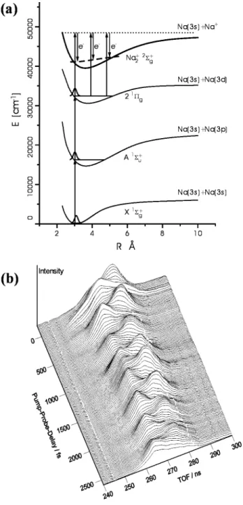
the multiplexed ability to follow detailed vibrational wave packet dynamics in excited molecular states, in some cases all the way to the products. In the work by Baumert and co-workers on Na2, two-photon
excitation was used to create a wave packet on the excited 21Π
gstate of Na2, and the PE spectrum was
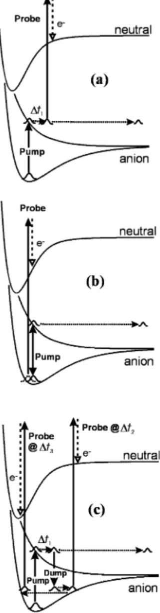
measured after delayed ionization with a probe pulse, as schematically depicted in Figure 1a. The photo-electron spectrum changes with the phase of the wave packet because of the difference between the potentials for the 21Πg state and the X2Σ
g +
state of Na2+, with higher (lower) electron kinetic energy at
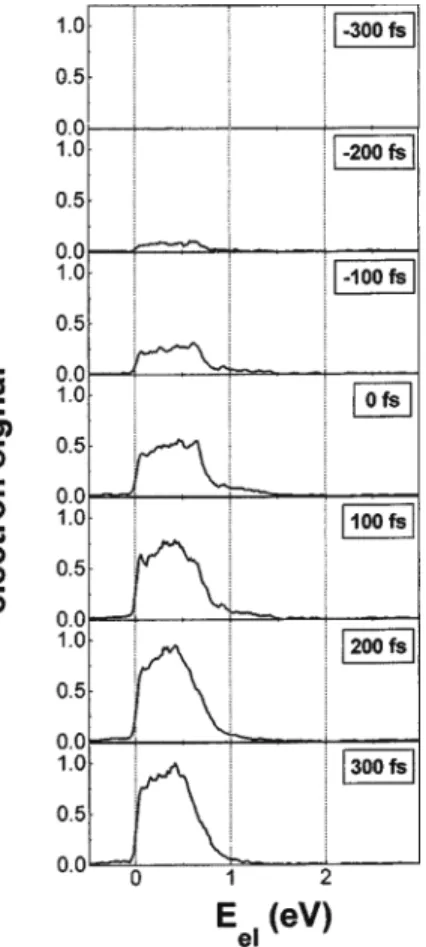
the inner (outer) turning point. As a result, a plot of the photoelectron spectra vs time (Figure 1b) shows the complete excited-state wave packet dynamics. The negative ion TRPES experiments on I2- by
Neumark and co-workers probed the time scale for direct dissociation to I(2P
3/2) + I-on the repulsive A′ 2
Σg,1/2 +
state, schematically drawn in Figure 15a; the PE spectrum evolves from a broad transient feature associated with dissociating I2-to very sharp features
associated with the atomic I-product in about 300
fs, as shown in Figure 2.
Hayden38first demonstrated that dynamics due to
non-adiabatic processes can also be monitored by TRPES, using internal conversion in hexatriene as an example. Non-adiabatic processes lead to changes in electronic symmetry, and, as proposed by Dom-cke,24the simple Koopmans’ picture suggests that one
might be able to directly monitor the evolving excited-state electronic configurations during non-adiabatic processes while simultaneously following the coupled nuclear dynamics via the vibrational structure within each photoelectron band. In Figure 3, we show a picture of excited-state polyatomic wave packet dy-namics probed via TRPES, shown here for the case of neutral molecule dynamics. (A similar picture can be envisaged for the case of anion excited-state dynamics.) A zeroth-order bright state, R, is coher-ently prepared with a femtosecond pump pulse. According to the Koopmans’ picture, it should ionize into the R+ continuum, the electronic state of the
cation obtained upon removal of the outermost va-lence electron (here chosen to be the ground electronic state of the ion). This process produces a photoelec-tron band ǫ1.
We now consider any non-adiabatic coupling pro-cess which transforms the zeroth-order bright state R into a lower lying zeroth-order “dark” state, β, as induced by promoting vibrational modes of
appropri-ate symmetry. Again, according to the Koopmans’ picture, the β state should ionize into the β+
ioniza-tion continuum (here assumed to be an electronically excited state of the ion), producing a photoelectron band ǫ2. Therefore, for a sufficiently energetic probe
photon (i.e., with both ionization channels open), we expect a switching of the electronic photoionization channel from ǫ1 to ǫ2 during the non-adiabatic
pro-cess. This simple picture suggests that one can directly monitor the evolving excited-state electronic configurations (i.e., the electronic population
dynam-ics) during non-adiabatic processes while simulta-neously following the coupled nuclear dynamics via the vibrational structure within each photoelectron band. In other words, the electronic structure of the molecular ionization (or detachment) continuum acts as a “template” for the disentangling of electronic from vibrational dynamics in the excited state, as was first experimentally demonstrated for fs non-adia-batic processes in 1998-1999.39,40
The two limiting cases for Koopmans’-type correla-tions in such experiments, as initially proposed by Domcke,24,25 have been demonstrated
experimen-tally.41,42The first case, Type I, is when the neutral
excited states R and β clearly correlate to different
Figure 1. Femtosecond PE spectroscopy of Na2. (a)
Potential energy curves of Na2/Na2+ and experimental
excitation schema: one- and two-photon excitation at 618 nm creates vibrational wave packets at the inner turning point of the A1Σu+ and 21Πg states, respectively, with
dynamics probed via nuclear-induced changes in the ion-ization potential. (b) Time-resolved photoelectron spectra showing coherent wave packet motion on the excited Na2
states. Reprinted with permission from ref 35. Copyright 1996 American Physical Society.
Figure 2. Experimental fs photoelectron spectra of I2-.
Spectra at various delays (-30 to 320 fs); A, B, and C correspond to the X1Σg+
, A′3Π2u, and A3Π1u states of I2
measured in a one-photon UV background spectrum, while dashed lines indicate the results of time-resolved wave packet simulations. Reprinted with permission from ref 37. Copyright 1999 American Institute of Physics.
Figure 3. A TRPES scheme for disentangling electronic
from vibrational dynamics in excited polyatomic molecules. A zeroth-order electronic state R is prepared by a fs pump pulse. Via a non-adiabatic process it converts to a vibra-tionally hot lower-lying electronic state, β. The Koopmans’-type ionization correlations suggest that these two states will ionize into different electronic continua: R f R+ +
e-(ǫ
1) and β f β+ + e-(ǫ2). When the wave packet has
zeroth-order R character, any vibrational dynamics in the R state will be reflected in the structure of the ǫ1
photo-electron band. After the non-adiabatic process, the wave packet has β zeroth-order electronic character; any vibra-tional dynamics in the β state will be reflected in the ǫ2
band. This allows for the simultaneous monitoring of both electronic and vibrational excited-state dynamics. Re-printed with permission from ref 91. Copyright 2000 Elsevier.
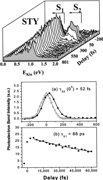
ion electronic continua, as suggested by Figure 3. Even if there are large geometry changes upon internal conversion and/or ionization, producing vi-brational progressions, the electronic correlations should favor a disentangling of the vibrational dy-namics from the electronic population dydy-namics. As an example, Stolow and co-workers studied ultrafast internal conversion in a linear polyene (decatetraene, DT). The DT optically bright S2 state internally
converts to S1on ultrafast time scales. As discussed
in more detail in a following section, the S2 state
electronically correlates with the D0 cation ground
state, and S1 correlates with the D1 cation first
excited state. In Figure 4 (top), we show the energy level scheme relevant to this experiment. A femto-second pump pulse at 287 nm prepared the excited S2 state at its origin. This then evolved into a
vibrationally hot S1state via internal conversion. The
coupled states were probed using single-photon 235 nm ionization. As suggested by Figure 3, the evolving
electronic and vibrational character of the wave packet alters the photoionization electronic channel, leading to large shifts in the time-resolved photo-electron spectrum. The experimental photophoto-electron kinetic energy spectra, shown in Figure 4 (bottom), indeed show a dramatic shift as a function of time. This shift is the direct (as opposed to inferred) signature of the changing electronic-state character induced by non-adiabatic coupling. This example demonstrates the selectivity of the molecular ioniza-tion (or detachment) continuum for specific excited-state configurations and shows that electronic popu-lation dynamics can be disentangled from vibrational dynamics during ultrafast non-adiabatic processes.40,42
The other limiting case, Type II, is when the neutral excited states R and β correlate equally strongly to the same ion electronic continua. This is expected to hinder the disentangling of electronic from vibra-tional dynamics42and is discussed in more detail in
a following section.
2.2. Photoelectron Angular Distributions
The other component of the molecular ionization or detachment continuum is that of the free electron. Within the molecular frame, the symmetries of the outgoing electron partial waves are likewise related to the symmetry of the electronic state undergoing ionization. This can be viewed most simply (eq 3) as being due to the requirement that the product of the symmetry species of the prepared excited state Γex,
the dipole operator Γµ, the ion state Γ+, and the free
electron wave function Γemust equal or contain the
totally symmetric irreducible representation ΓTS of
the molecular symmetry group, in order that the transition be allowed.
If, owing to a non-adiabatic process, the symmetry species of the initial zeroth-order state Γexchanges,
then the symmetry species of the outgoing electron Γe-must also change in order that the product remain
(or contain) the totally symmetric species ΓTS. It is
the symmetry species of the outgoing electron Γe-that
relates to the photoelectron angular distribution. Hence, measurement of time-resolved photoelectron angular distributions (PADs) also provides a probe of electronically non-adiabatic processes. We note, however, that a change in symmetry is not always required to produce a change in the PAD. Two excited-state wave functions of identical electronic symmetry may have quite different ionization matrix elements, leading to differences in the PADs. The photoemission is, in general, sensitive to orientation of the molecular frame with respect to the photoelec-tron recoil vector (however, unfavorable ionization dynamics may obscure this effect). In favorable cases, a laboratory-frame alignment of the molecular frame will lead to anisotropies in the PAD. A coherent superposition of rotational states leads to a time-evolving molecular axis distribution in the laboratory frame. In this situation, the PAD may evolve due to the evolution of the rotational superposition state and can directly reflect the axis distribution. The use of
Figure 4. (Top) Energy level scheme for TRPES of all-trans-decatetraene (DT), a molecule exhibiting Type I Koopmans’ correlations. The pump laser prepares the optically bright state S2. Due to ultrafast internal
conver-sion, this state converts to the lower-lying state S1with
∼0.7 eV of vibrational energy. The expected complementary Type I Koopmans’ correlations are shown: S2fD0+ e-(ǫ1)
and S1 f D1 + e-(ǫ2). (Bottom) TRPES spectra of DT
pumped at 287 nm and probed at 235 nm. There is a rapid shift (∼400 fs) from ǫ1, an energetic band at 2.5 eV due to
photoionization of S2into the D0cation ground electronic
state, to ǫ2, a broad, structured band at lower energies due
to photoionization of vibrationally hot S1into D1, the first
excited electronic state of the cation. The structure in the low-energy band reflects the vibrational dynamics in S1.
These results illustrate the disentangling of the vibrational from the electronic population dynamics. Reprinted with permission from Nature (http://www.nature.com), ref 40. Copyright 1999 Nature Publishing Group.
time-resolved PADs in excited-state molecular dy-namics has been recently reviewed, and we refer the reader to these reviews for detailed discussions.13,14,17
The measurement of time-resolved PADs is expected to be particularly valuable when dynamical processes are not discernible from a photoelectron kinetic energy analysis alone. In general, PAD measure-ments are more involved and their calculation more challenging in polyatomic systems than for the case of photoelectron energy distributions. A consideration of practical issues such as the nature of the labora-tory-frame alignment (i.e., how the pump pulse creates an anisotropic distribution of excited-state molecular axes in the laboratory frame) and the sensitivity of the ionization dynamics to the orienta-tion of the molecular frame is important. Unfavorable laboratory-frame alignment can potentially obscure the underlying photoionization dynamics and reduce the efficacy of the PAD as an experimental probe.
2.3. Intensity Effects
Amplified femtosecond laser pulses are inherently intense. The neglect of intensity effects can lead to misinterpretation of results. For example, even mod-erate (f/40) focusing of a 100 fs pulse (at λ ) 300 nm) yields an intensity of 1012W/cm2with a pulse energy
of only 1 µJ! It can be seen that amplified femtosec-ond pulse experiments will be very difficult to carry out in the perturbation theory (Golden Rule) limit. As the strong laser field physics of molecules is amply discussed elsewhere,43we summarize here only briefly
some potential manifestations of nonperturbative electric fields that can appear in almost any femto-second experiment using amplified pulses. A char-acteristic phenomenon observed in photoelectron spectroscopy via strong field ionization is above-threshold ionization (ATI), the absorption of ad-ditional photons by an electron already above the ionization potential (IP) that leads to a series of peaks in the photoelectron spectrum spaced by the photon energy.44
Other strong field phenomena may also directly affect femtosecond photoelectron spectra. As dis-cussed below, strong Rabi oscillations can lead to aligned states and to ground-state wave packets. Another important related consequence is the dy-namic (or AC) Stark effectsthe shifting and modifi-cation of potential energy surfaces during the strong electric field of the laser pulse. As discussed below, this leads to an alteration of the molecular dynamical evolution and to sweeping field-induced resonances that may introduce phase shifts in the probe laser signal. From a very practical point of view, it is often difficult to use one-photon preparation of an excited-state wave packet unless the absorption cross section for this step is large, as high-intensity femtosecond pulses generally favor nonlinear over linear pro-cesses. Since the ratio of one- to two-photon cross sections is fixed (for a given molecule) and the ratio of the one- to two-photon transition probabilities scales inversely with intensity, the only possibility is to reduce the laser intensity (W/cm2) until the
one-photon process dominates. The signal levels at this point, however, may be very small, and very sensitive
detection techniques such as single particle counting are usually required.
In the electronic continuum, the dynamic Stark effect manifests itself in terms of the ponderomotive potential Up, well-known in strong-field atomic
phys-ics.44Classically, the ponderomotive potential of an
electron in a laser field of strength E and frequency ω is given by the time-averaged “wiggling” energy:
A convenient rule of thumb is that the pondero-motive shift is ∼60 meV TW-1 cm-2 at 800 nm
(N.B.: 1 TW ) 1 × 10¹²W): Upscales linearly with
laser intensity and quadratically with wavelength. Due to the spatio-temporal averaging of intensities in the pulse, the ponderomotive effect is observed as a red shift in electron kinetic energy and broadening of the photoelectron spectrum.45
It is well-known in atomic physics that the dynamic Stark effect causes field-induced changes in the energies of the atomic levels and, hence, leads to field-induced multiphoton resonances known as “sweeping” or “Freeman” resonances and to fine structure in atomic ATI photoelectron spectra.46As
the dynamic Stark effect is general, similar argu-ments apply to molecular systems. Beginning in 1996, Baumert and Gerber and their co-workers made extensive studies on the effect of laser intensity on TRPES.47-51In particular, using the Na
2 system
as a model, they made the first studies of ATI photoelectron spectra as a function of time, using intense 40 fs pulses at 618 nm. This study (and following studies52) showed in detail how TRPES
maps the alteration of nuclear dynamics in intense laser fields, using sweeping resonances as a new tool for controlling molecular dynamics. Y. Chen and co-workers also reported the observation of intensity effects on time-resolved PE spectra, using 2 + 1 ionization of NO through its C2Π Rydberg state.53
They observed field-induced Freeman resonances attributed to the v ) 3 level of the NO A2Σ+state
being shifted about 200 meV into near-resonance with the NO C (v ) 0) level. A 2003 paper by S. L. Cong and co-workers showed how the dynamic Stark effect could be used constructively to tune or select specific ionization pathways in TRPES of the NO molecule, with resonances chosen between the F2∆
and E2Σ+states.54
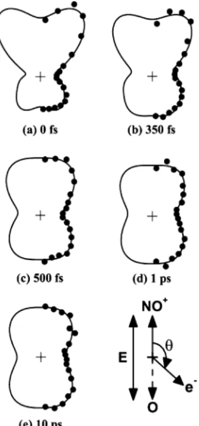
Nonperturbative intensities can have a particularly pronounced effect on the measurement of photoelec-tron angular distributions (PADs). The anisotropic nature of the photoexcitation pump process results in an alignment of both the angular momentum and molecular axis distributions of the excited state in the laboratory frame (defined by the polarization direction of the pump laser field). In the Golden Rule limit, for a single photon pump excitation, a cos2 θ
distribution of excited-state molecular axes in the laboratory-frame results from a parallel transition (sin2θ from a perpendicular transition).
Nonpertur-bative intensities will, due to rotational Rabi cycling between the initial and intermediate states, give rise
Up) e
2E2
to higher than expected alignment and can be used to align the molecular states in the laboratory frame.55-57Seideman has shown that, although the
pump laser intensity can have significant effects on PADs, the intensity effects of the probe laser are minimal.58This is due to the fact that the “lifetime”
of a free electron “near” the molecular core is usually much less than the Rabi period. The pump laser alignment of the molecular axis distribution in the laboratory frame directly affects the observed PADs since the relative contributions of parallel and per-pendicular ionization channels and the interferences between them depend on the degree of alignment. In cases of favorable ionization dynamics, this can be exploited to follow the evolution of molecular axis alignment as a probe of rotation-vibration coupling in both diatomic59and polyatomic60molecules.
3. Development of Time-Resolved Photoelectron
Spectroscopy: 1985
−
1998
3.1. Time-Resolved Photoelectron Spectroscopy
Experiments
TRPES experiments performed between 1985 and 1998 have been reviewed previously12and are
there-fore considered only briefly in the following discus-sion. The first gas-phase TRPES experiments using ns and ps lasers were demonstrated in the mid-1980s, by Pallix and Colson61on sym-triazine and by
Sekre-ta and Reilly62on benzene. In both cases, PES was
used to follow excited-state relaxation via intersystem crossing to a triplet state in real time.
In the early 1990s, experiments by Reilly,63Knee,64
and their co-workers showed that TRPES with ps resolution could be used to follow intramolecular vibrational relaxation (IVR) in excited electronic states. In IVR, the vibrational character of the initially excited zeroth-order vibrational state evolves (i.e., dephases) with time, but the molecule remains in the same electronic state. Reilly’s experiments were performed on p,n-alkylanilines, while Knee investigated IVR in fluorene, benzene, and aniline‚ CH4 clusters.
Syage65,66 demonstrated that picosecond TRPES
could be used to follow reaction and solvation dynam-ics within clusters, the first example of a particularly powerful application of the technique. This experi-ment was applied to excited-state proton transfer in phenol‚(NH3)n clusters. In a seminal study, Weber and co-workers used picosecond TRPES to follow spin-orbit coupling (intersystem crossing) from the S1 state in aniline and 2- and 3-aminopyridine,
making usesfor the first timesof the very important role of electronic symmetry correlations upon ioniza-tion.67 This work presaged the fs studies of
non-adiabatic processes that were to follow. C. A. de Lange and co-workers used picosecond TRPES to follow dissociation in the repulsive A band of CH3I,68
and to measure lifetimes of the predissociative B˜1E′′
and C˜′1A
1′Rydberg states of NH3.69Fischer, Schultz,
and co-workers used picosecond TRPES to measure lifetimes of vibrational levels of the B, C, and D excited electronic states of the allyl radical, all of which lie about 5 eV above the ground state.70-72
The first experiments at femtosecond time resolu-tion incorporating electron detecresolu-tion were reported in the mid-1990s. In 1993, Baumert, Gerber and co-workers,73 followed by Stolow and co-workers in
1995,33,34 applied femtosecond time-resolved ZEKE
spectroscopy to vibrational wave packet dynamics in Na3and I2, respectively. Gerber used a femtosecond
pump pulse to create a vibrational wave packet in the B state of Na3and measured both total ion and
ZEKE electron yield as a function of pump-probe delay. Stolow’s experiments, applied to the I2 B
state, were similar in principle but more extensive and presented an outline of the experimental pros-pects for fs TRPES as applied to excited-state dy-namics in polyatomic molecules, emphasizing the role of the molecular ionization continuum as the final state.
In 1996, Cyr and Hayden38 used femtosecond
TRPES in its first application to non-adiabatic dy-namics in polyatomic molecules, investigating the dynamics of internal conversion in 1,3,5-hexatriene. In this work, the S2 state of 1,3,5-hexatriene was
excited, and a combination of time-resolved ion yield and PE spectroscopy measurements was used to unravel the ensuing dynamics. The PE spectra for the cis isomer showed very rapid (∼20 fs) internal conversion from the S2 to the S1 state, followed by
IVR within the S1state on a time scale of 300 fs. This
experiment was the first example of how TRPES can be used to follow ultrafast non-adiabatic dynamics in polyatomic molecules.
The Stolow and Hayden experiments were soon followed by a flurry of activity in other laboratories, including the first negative ion experiments by Neu-mark36 on the photodissociation of I
2- and wave
packet dynamics experiments by Baumert35on Na 2.
Baumert also studied intensity effects on the coher-ent control of wave packet dynamics of threshold52
and above-threshold ionization (ATI)48
photoelec-trons, as well as the chirp- and pulse-length depen-dence of molecular photoionization dynamics.74
The first TRPES experiments on metal cluster anions were performed by Gantefo¨r et al.75in 1998
on Au3-. These negative ion experiments are
dis-cussed further in the context of more recent work in the following sections. Femtosecond TRPES was also applied to studies of dynamics in neutral clusters. In 1997, Soep and Syage studied the dynamics of the double proton-transfer reaction in the 7-azaindole dimer.76By comparing PE spectra obtained by (1 + 1)
ionization with 0.8 and 5.0 ps laser pulses, they found significantly higher PE yield with shorter pulse ionization, indicating a subpicosecond excited-state lifetime.
Stolow and Blanchet77 applied fs TRPES to the
photodissociation dynamics of polyatomic molecules, using the nitric oxide dimer (NO)2 as an example.
Nanosecond studies of (NO)2 photodissociation
dy-namics at 193 nm revealed that two product channels are open:78(NO)
2* f NO(A2Σ+v,J) + NO(X2Π v′,J′)
and (NO)2* f NO(B2Π v,J) + NO(X2Π v′′,J′′).
Fem-tosecond pump-probe measurement of the decaying (NO)2+parent ion signal as a function of delay time
TRPES spectra showed a prominent sharp peak at 0.52 eV due to the appearance of the NO(A2Σ+,v)
photodissociation product with a considerably slower appearance time of 0.7 ps. This difference in time scales between excited parent signal disappearance and product signal appearance was attributed to a two-step, likely non-adiabatic mechanism, rather than a direct photodissociation mechanism. This work showed that relying on parent ion yield mea-surements as a function of time in order to determine dissociation mechanisms and time scales may be potentially misleading, especially if non-adiabatic processes are involved.
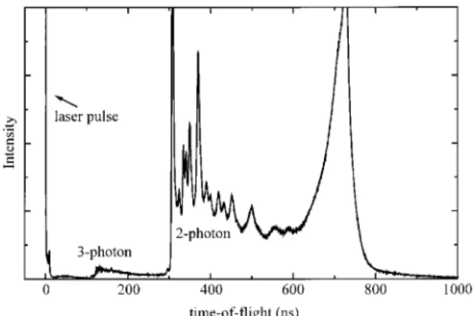
In 1997 Radloff, Hertel, Jouvet, and co-workers introduced an important extension of TRPES. They applied the photoelectron-photoion coincidence (PEP-ICO) detection technique to fs pump-probe spectros-copy,79,80allowing the dynamics of size-selected
neu-tral clusters to be studied via TRPES. As an illustrative example, shown in Figure 5, these au-thors studied internal conversion dynamics in ben-zene and benben-zene dimers. They found that the decay of the S2to S1states was very rapid for both species
(about 50 fs), whereas the S1 f S0 IC decay was
significantly faster in the monomer than in the dimer, 7.6 vs 100 ps. The same authors applied the PEPICO method to dynamics in ammonia clusters, investigating the dynamics of hydrogen transfer in electronically excited (NH3)2.81By monitoring the PE
signal associated with the (NH3)2+ and NH4+ ions,
they were able to sort out the rather complicated dynamics associated with this system, finding time constants of 170 fs for the decay of (NH3)2* to
NH4‚‚‚NH2, and 4 ps for dissociation of the latter
complex to NH4+ NH2.
3.2. Time-Resolved Photoelectron Angular
Distributions
In 1989, Y. Fujimura and co-workers presciently considered the effects of Coriolis coupling on ps time-resolved photoelectron angular distributions (PADs) in a model system.82 These authors showed that
b-axis Coriolis coupling leads to a k-coherence that
is directly observable in the time-resolved PADs. In 1993, K. L. Reid independently proposed an approach to the study of excited-state dynamics through the use of time-resolved PADs.83Reid showed that the
sensitivity of the PAD to the intermediate state alignment leads to evolution of the photoelectron angular distribution. Reid subsequently demon-strated this in a nanosecond-resolution experiment, using this time dependence to follow hyperfine de-polarization in the NO (A2Σ+) state.84These seminal
studies opened up the possibility of using the other component of the electronic continuumsthat of the free electronsas a probe of excited-state dynamics.
3.3. Theoretical Developments
In 1991, Seel and Domcke24,25 wrote pioneering
theoretical papers on femtosecond TRPES and pro-posed how it could be applied to non-adiabatic dynamics in polyatomic molecules, using S2 f S1
internal conversion (IC) dynamics in pyrazine as an example. These papers laid the foundation for the general application of fs TRPES to problems regard-ing excited-state non-adiabatic dynamics. Of particu-lar significance, Domcke showed how the electronic structure of the molecular ionization continuum and the ionization correlations of the neutral configura-tions involved can be used to help understand the
Figure 5. Time-resolved PEPICO spectra of benzene monomer (left) and dimer (right) with λpump) 200 nm and λprobe)
267 nm. The ultrafast internal conversion process can be seen to be very different in the dimer than in the monomer. The PEPICO technique allows complete separation of these channels. Reprinted with permission from ref 80. Copyright 1997 Elsevier.
non-adiabatic dynamics in the neutral excited state. In 1993, Meier and Engel85 applied a treatment
similar to Domcke’s to TRPES of diatomic Na2,
predicting many of the features associated with coherent vibrational wave packet motion that were later observed experimentally. These authors ex-panded these studies in 1994, looking at opportuni-ties for TRPES in the mapping of wave packet dynamics in double-welled potentials86and
develop-ing technically important approximation treatments of the photoionization dynamics, considerably sim-plifying the numerical task involved in computa-tions.30In 1996, Meier and Engel studied the
appli-cation of TRPES to non-adiabatic predissociation problems involving the coherent decay of sets of resonances,87 complementing Stolow’s 1996
experi-mental ion yield observation of these in the IBr molecule.88In 1997, Braun and Engel considered the
role of initial temperature of the ground state in TRPES, using the hot cesium dimer as an example.89
In 1998, Meier and Engel explicitly considered the important role of frequency chirp (i.e., the time ordering of frequencies in a broad bandwidth pulse) in fs TRPES experiments, relevant to the discussion of quantum coherent control in this area.90
In 1997, T. Seideman developed a formal nonper-turbative theory of time-resolved PADs.26 This
in-cluded exact (all orders) treatment of the matter-field interaction and included, in principle, the effects of both nuclear and electronic dynamics on PADs. This work laid the foundation for the development of a research program, beginning in 1999, by Seide-man and co-workers on several aspects of time-resolved PADs.14
4. Experimental Methods
4.1. Photoelectron Spectroscopy of Neutrals and
Anions
In any photoelectron spectroscopy measurement, the observables of interest are the electron kinetic energy (eKE) distribution and the photoelectron angular distribution (PAD). Spectrometers for fem-tosecond TRPES have modest energy resolution requirements as compared to modern standards for photoelectron spectrometers. The bandwidth (fwhm) of a Gaussian 100 fs pulse is ∼150 cm-1. A
pump-probe measurement involves the convolution of two such pulses, leading to an effective bandwidth of ∼25 meV. This limits the energy resolution required in measuring the energy of the photoelectrons. How-ever, TRPES experiments are very data-intensive, as they require the collection of many photoelectron spectra. As a result, most neutral and all anion TRPES experiments performed to date make use of high-efficiency electron energy analyzers in which a large fraction of the photoelectrons are collected.
The analyzer most commonly used in TRPES experiments has been the magnetic bottle time-of-flight spectrometer.22,91This technique uses a strong
inhomogeneous magnetic field (1 T) to rapidly par-allelize electron trajectories to a flight tube and a constant magnetic field (10 G) to guide the electrons to the detector.21,92With careful design, the collection
efficiency of magnetic bottle spectrometers can exceed 50%, while maintaining an energy resolution es-sentially equivalent to the fs laser bandwidth. The highest resolution is obtained for electrons created within a small volume (φ < 100 µm) at the very center of the interaction region. In contrast with ns laser experiments, however, it is not desirable to focus fs lasers to small spot sizes due to the inherent high intensity of such pulses, thus leading to mul-tiphoton ionization. Longer focal lengths mean that the Rayleigh range (the “length” of the focus) extends well beyond the favorable region, leading in general to an overall lower resolution.
Magnetic bottle analyzers have been used in many neutral and anion TRPES experiments. They are relatively simple, have high collection efficiency and rapid data readout, and, importantly, permit straight-forward photoelectron-photoion coincidence (PEPI-CO) measurements. Magnetic bottles suffer from the general disadvantage that they can only be used to determine eKE distributions; the complex electron trajectories in magnetic bottle analyzers make it impractical (if not impossible) to extract a PAD. There is an additional complication associated with the use of magnetic bottle analyzers in anion TRPES experiments. Mass-selected negative ion beams typi-cally have laboratory kinetic energies of 1 keV or higher; hence, the velocity of the ion beam is no longer negligible compared to that of the photoelec-trons. As a result, photoelectrons with the same speed in the center-of-mass frame of reference but scattered at different polar angles with respect to the ion beam will have different speeds in the laboratory frame.93This “Doppler broadening” significantly
de-grades the energy resolution of a high-efficiency analyzer with no angular resolution, such as a magnetic bottle, yielding eKE resolution as poor as several hundred millielectronvolts. This resolution is often sufficient for TRPES experiments but can be improved (with loss of signal) to several tens of millielectronvolts by pulsed deceleration of the ion beam, as has been demonstrated in several labora-tories,37,94-96or, as shown recently by Cheshnovsky,97
by impulsive deceleration of the detached electrons. These limitations of magnetic bottle analyzers may be overcome by the use of two-dimensional (2D) photoelectron imaging techniques, in which position-sensitive detection is used to measure the photoelec-tron kinetic energy and angular distributions simul-taneously. When used, as is common, with CCD camera systems for image collection, one gives up rapid data readout, as multi-kilohertz readout of CCD chips is very challenging. This precludes the possibility of simple PEPICO measurements. The most straightforward 2D imaging technique is pho-toelectron velocity-map imaging (VMI),98 a variant
of the elegant photofragment imaging method devel-oped by Chandler and Houston.99Photoelectron VMI
was first demonstrated by Eppink and Parker for neutrals98,100and by Bordas101and Sanov102for
nega-tive ions. Typically, a strong electric field projects nascent charged particles onto a microchannel plate (MCP) detector. The ensuing electron avalanche falls onto a phosphor screen, which is imaged by the CCD
camera. Analysis of the resultant image allows for the extraction of both energy- and angle-resolved information. In this case, a two-dimensional projec-tion of the three-dimensional distribuprojec-tion of recoil velocity vectors is measured; various image recon-struction techniques103-105 are then used to recover
the full three-dimensional distribution.
Photoelectron VMI thus yields close to the theo-retical limit for collection efficiency, along with simultaneous determination of the photoelectron eKE and angular distributions. In addition, and of prime importance to anion studies, VMI eliminates the Doppler broadening observed with magnetic bottle analyzers, obviating the need for deceleration meth-ods. In anion time-resolved photoelectron imaging (TRPEI) studies utilizing velocity-map imaging, the resolution limit closely approaches the laser band-width.
This 2D particle imaging approach may be used most straightforwardly when the image is a projec-tion of a cylindrically symmetric distribuprojec-tion whose symmetry axis lies parallel to the two-dimensional detector surface. This requirement usually precludes the use of non-coincident pump and probe laser reference frames (i.e., other than parallel or perpen-dicular laser polarizations), a situation which may provide detailed information on intramolecular dy-namics through the alignment dependence of the PAD. It may be preferable to adopt fully three-dimensional (3D) imaging techniques based upon “time-slicing”106,107or full time- and position-sensitive
detection,108where the full three-dimensional
distri-bution is obtained directly without the introduction of mathematical reconstruction. In fs pump-probe experiments where the intensities must be kept below a certain limit (often leading to single-particle counting methods), “time-slicing” (which requires significant signal levels) may not be practical, leaving only full time- and position-sensitive detection as the option.
Modern MCP detectors can measure both spatial position (x,y) on the detector face and time of arrival (z) at the detector face.108In this situation, a weak
electric field is used to extract nascent charged particles from the interaction region. Readout of the (x,y) position yields information about the velocity distributions parallel to the detector face, equivalent to the information obtained from 2D detectors. How-ever, the additional timing information allows mea-surement of the third (z) component of the laboratory-frame velocity, via the “turn-around” time of the particle in the weak extraction field. Thus, these detectors allow for full 3D velocity vector measure-ments, with no restrictions on the symmetry of the distribution or any requirement for image reconstruc-tion techniques. Very successful methods for full time- and position-sensitive detection are based upon interpolation (rather than pixellation) using either charge-division (such as “wedge-and-strip”)109,110 or
crossed delay-line anode timing MCP detectors.111,112
In the former case, the avalanche charge cloud is divided among three conductorssa wedge, a strip, and a zigzag. The (x,y) positions are obtained from the ratios of the wedge and strip charges to the zigzag
(total) charge. Timing information can be obtained from a capacitive pick-off at the back of the last MCP plate. In the latter case, the anode is formed by a pair of crossed, impedance-matched delay lines (i.e.,
x and y delay lines). The avalanche cloud that falls
on a delay line propagates in both directions toward two outputs. Measurement of the timing difference of the output pulses on a given delay line yields the
x(or y) positions on the anode. Measurement of the
sum of the two output pulses (relative to, say, a pickoff signal from the ionization laser or the MCP plate itself) yields the particle arrival time at the detector face. Thus, direct anode timing yields a full 3D velocity vector measurement. An advantage of delay line anodes over charge division anodes is that the latter can tolerate only a single hit per laser shot. This precludes the possibility of multiple coinci-dences, of interest in some experiments, and makes the experiment very sensitive to background, such as scattered UV light.
4.2. Photoelectron
−
Photoion Coincidence
Methods
Photoionization (photodetachment) always pro-duces two species available for analysissthe ion (neutral) and the electron. The extension of the photoelectron-photoion coincidence (PEPICO) tech-nique to the femtosecond time-resolved domain was first demonstrated by Stert and Radloff and was shown to be very important for studies of dynamics in clusters.79,81 In these experiments, a simple yet
efficient permanent magnet design “magnetic bottle” electron spectrometer was used for photoelectron flight measurements. A collinear time-of-flight mass spectrometer was used to determine the mass of the parent ion. Using coincidence electronics, the electron time-of-flight (yielding electron kinetic energy) is correlated with an ion time-of-flight (yield-ing the ion mass). In this manner, TRPES experi-ments may be performed on neutral clusters, yielding time-resolved spectra for each parent cluster ion (assuming cluster fragmentation plays no significant role). Signal levels must be kept low (much less than one ionization event per laser shot) in order to minimize false coincidences.
Time- and angle-resolved PEPICO measurements yielding photoion and photoelectron kinetic energy and angular correlations can shed new light on photodissociation dynamics in polyatomic molecules. As shown by Continetti and co-workers for the case of nanosecond laser photodetachment, correlated photofragment and photoelectron velocities can pro-vide a complete probe of the dissociation process.113,114
The photofragment recoil measurement defines the energetics of the dissociation process and the align-ment of the recoil axis in the laboratory frame, the photoelectron energy provides spectroscopic identi-fication of the products, and the photoelectron angu-lar distribution can be transformed to the recoil frame in order to extract vector correlations such as the photofragment angular momentum polarization. The integration of photoion-photoelectron timing imaging (energy and angular correlation) measure-ments with femtosecond time-resolved spectroscopy
was demonstrated, using timing imaging detectors, in 1999 by C. C. Hayden and co-workers115,116 at
Sandia National Laboratories. This coincidence-imaging spectroscopy (CIS) method allows the time evolution of complex dissociation processes to be studied with unprecedented detail.
4.3. Femtosecond Laser Technology
The progress in femtosecond TRPES over the past 10 years derives from prior developments in femto-second laser technology, since techniques for photo-electron spectroscopy have been highly developed for some time. There are several general requirements for such a femtosecond laser system. Most of the processes of interest are initiated by absorption of a photon in the wavelength range ∼200-350 nm, produced via nonlinear optical processes such as harmonic generation, frequency mixing, and para-metric generation. Thus, the output pulse energy of the laser system must be high enough for efficient use of nonlinear optical techniques and ideally should be tunable over a wide wavelength range. Another important consideration in a femtosecond laser sys-tem for time-resolved photoelectron spectroscopy is the repetition rate. To avoid domination of the signal by multiphoton processes, the laser pulse intensity must be limited, thus also limiting the available signal per laser pulse. As a result, for many experi-ments a high pulse repetition rate can be more beneficial than high energy per pulse. Finally, the signal level in photoelectron spectroscopy is often low in any case, and, for time-resolved experiments, spectra must be obtained at many time delays. This requires that any practical laser system must run very reliably for many hours at a time.
Modern Ti:sapphire-based femtosecond laser oscil-lators have been the most important technical ad-vance for performing almost all types of femtosecond time-resolved measurements.117Ti:sapphire
oscilla-tors are tunable over a 725-1000 nm wavelength range, have an average output power of several hundred milliwatts or greater, and can produce pulses as short as 8 fs, but more commonly 80-130 fs, at repetition rates of 80-100 MHz. Broadly tunable femtosecond pulses can be derived directly from amplification and frequency conversion of the fundamental laser frequency.
The development of chirped-pulse amplification and Ti:sapphire regenerative amplifier technology now provides millijoule pulse energies at repetition rates of >1 kHz with <100 fs pulse widths.118
Chirped-pulse amplification typically uses a grating stretcher to dispersively stretch fs pulses from a Ti: sapphire oscillator to several hundred picoseconds. This longer pulse can now be efficiently amplified in a Ti:sapphire amplifier to energies of several milli-joules while avoiding nonlinear propagation effects in the solid-state gain medium. The amplified pulse is typically recompressed in a grating compressor.
The most successful approach for generating tun-able output is optical parametric amplification of spontaneous parametric fluorescence or a white light continuum, using the Ti:sapphire fundamental or second harmonic as a pump source. Typically, an 800
nm pumped fs optical parametric amplifier (OPA) can provide a continuous tuning range of 1200-2600 nm.119Noncollinear OPAs (NOPAs)120pumped at 400
nm provide microjoule-level ∼10-20 fs pulses which are continuously tunable within a range of 480-750 nm, allowing for measurements with extremely high temporal resolution. A computer-controlled stepper motor is normally used to control the time delay between the pump and probe laser systems.
The development of femtosecond laser sources with photon energies in the vacuum ultraviolet (VUV, 100-200 nm), extreme ultraviolet (XUV, <100 nm), and beyond (soft X-ray) opens new possibilities for TRPES, including the preparation of high-lying mo-lecular states, the projection of excited states onto a broad set of cation electronic states, and, in the soft X-ray regime, time-resolved inner-shell photoelectron spectroscopy. High harmonic generation in rare gases is a well-established and important method for gen-erating fs VUV, XUV,121and soft X-ray radiation.122-124
Harmonics as high as the ∼300th order have been reported, corresponding to photon energies in excess of 500 eV. Both pulsed rare gas jets and hollow-core optical waveguides123,125 have been used for high
harmonic generation. Sorensen and co-workers126
used the sixth harmonic (9.42 eV) of a Ti:sapphire laser (generated from the third harmonic of the Ti: sapphire second harmonic) as the pump pulse in TRPES experiments. Even higher harmonics (17th) have been incorporated into TRPES experiments by Leone and co-workers and used for time-resolved inner-shell photoelectron spectroscopy.127-129As these
techniques become more commonplace, the range of applicability of TRPES will be increased significantly. Finally, it is worth noting that high harmonic generation provides the route to attosecond (10-18s)
pulse generation,130,131 generally producing
wave-lengths within the soft X-ray regime. Although in its infancy, the first TRPES experiments using attosec-ond pulses have been reported132 and were used to
study inner-shell electronic dynamics in atoms133
with sub-femtosecond time resolution. This provides a new and direct time domain approach to the study of electron correlation in atoms and molecules.
5. Applications
In the following sections, we review recent applica-tions (1999-2003) of TRPES to problems in the dynamics of isolated molecules, anions, and their clusters.