The MIT Faculty has made this article openly available.
Please share
how this access benefits you. Your story matters.
Citation
Seow, Kok et al. “C-Jun N-Terminal Kinase in Synergistic Neurite
Outgrowth in PC12 Cells Mediated through P90RSK.” BMC
Neuroscience 14.1 (2013): 153.
As Published
http://dx.doi.org/10.1186/1471-2202-14-153
Publisher
BioMed Central Ltd
Version
Final published version
Citable link
http://hdl.handle.net/1721.1/86393
Terms of Use
Creative Commons Attribution
R E S E A R C H A R T I C L E
Open Access
c-Jun N-terminal kinase in synergistic neurite
outgrowth in PC12 cells mediated through
P90RSK
Kok Huei Seow
1, Lihan Zhou
1,2, Gregory Stephanopoulos
1,3and Heng-Phon Too
1,2*Abstract
Background: Synergistic multi-ligand treatments that can induce neuronal differentiation offer valuable strategies to regulate and modulate neurite outgrowth. Whereas the signaling pathways mediating single ligand-induced neurite outgrowth, such as Akt, extracellular signal-regulated kinase (Erk), c-Jun N-terminal kinase (JNK), and p38 mitogen-activated protein kinase (P38), have been extensively studied, the mechanisms underlying multi-ligand synergistic neurite outgrowth are poorly understood. In an attempt to gain insight into synergistic neurite outgrowth, PC12 cells were treated with one of three combinations: pituitary adenylate cyclase-activating peptide (PACAP) with epidermal growth factor (EP), basic fibroblast growth factor (FP), or nerve growth factor (NP) and then challenged with the appropriate kinase inhibitors to assess the signaling pathways involved in the process.
Results: Response surface analyses indicated that synergistic neurite outgrowth was regulated by distinct pathways in these systems. Synergistic increases in the phosphorylation of Erk and JNK, but not Akt or P38, were observed with the three growth factor-PACAP combinations. Unexpectedly, we identified a synergistic increase in JNK phosphorylation, which was involved in neurite outgrowth in the NP and FP, but not EP, systems. Inhibition of JNK using the SP600125 inhibitor reduced phosphorylation of 90 kDa ribosomal S6 kinase (P90RSK) in the NP and FP, but not EP, systems. This suggested the involvement of P90RSK in mediating the differential effects of JNK in synergistic neurite outgrowth.
Conclusions: Taken together, these findings reveal the involvement of distinct signaling pathways in regulating neurite outgrowth in response to different synergistic growth factor-PACAP treatments. Our findings demonstrate a hitherto unrecognized mechanism of JNK-P90RSK in mediating synergistic neurite outgrowth induced by the co-treatment of growth factors and PACAP.
Keywords: PC12, Synergistic, NGF, FGFb, EGF, PACAP, Neurite outgrowth, JNK, P90RSK
Background
The effects of synergistic activity have been gaining atten-tion in the treatment of diseases such as cancer and AIDS [1,2]. Drug or ligand synergy is defined as the joint action of two or more agents for which the result is greater than the sum of the actions of the individual parts. Synergistic therapeutic strategies therefore have the potential to accel-erate the response to treatment, achieve higher efficacy,
and potentially reduce the side effects associated with sin-gle treatment approaches [3-5]. Indeed, several studies have demonstrated the benefits of the co-administration of neurotrophic factors [6] and the combinatorial treat-ment of nerve growth factor (NGF) with glial cell-derived neurotrophic factor (GDNF) [7] or insulin-like growth fac-tor (IGF)-1 [8] in promoting synergistic axonal or neurite elongation. Axonal and dendritic outgrowth from the neuronal cell body is a key cellular aspect of neuronal differentiation that is critically important not just in de-velopment, but also in recovery from injuries and neu-rodegenerative diseases [9]. Following nerve injury, the rate of nerve regeneration is critical, as a full functional recovery can be impeded by delayed regeneration [10].
* Correspondence:heng-phon_too@nuhs.edu.sg
1
Chemical & Pharmaceutical Engineering, Singapore-MIT Alliance, 4 Engineering Drive 3, Singapore, Singapore
2
Department of Biochemistry, National University of Singapore, 14 Medical Drive, Singapore, Singapore
Full list of author information is available at the end of the article
© 2013 Seow et al.; licensee BioMed Central Ltd. This is an open access article distributed under the terms of the Creative Commons Attribution License (http://creativecommons.org/licenses/by/2.0), which permits unrestricted use, distribution, and reproduction in any medium, provided the original work is properly cited.
Thus, accelerating or facilitating neurite outgrowth through a synergistic approach may provide a valuable treatment strategy for patients with nerve injury. How-ever, the mechanisms underlying such synergistic action are currently not well understood.
Rat pheochromocytoma PC12 cells have been widely used as a model to study neuronal differentiation. Upon treatment with ligands such as the NGF [11], basic-fibroblast growth factor (FGFb or FGF-2) [12], and pituit-ary adenylate cyclase-activating peptide (PACAP) [13], PC12 cells differentiate into sympathetic neuron-like cells characterized by long-term and stable neurite outgrowth. These ligands regulate neurite outgrowth through path-ways such as the extracellular signal-regulated kinase 1/2 (Erk) [14,15], p38 mitogen-activated protein kinase (P38) [16], c-Jun N-terminal kinase (JNK) [15,17], and the phosphatidylinositol 3-kinase (PI3K) [18]. In compari-son, epidermal growth factor (EGF) promotes prolifera-tion instead of differentiaprolifera-tion in these cells [19,20]. Studies have attributed this difference in cell fate to the kinetics of Erk activation, where transient or sustained Erk activation leads to proliferation or differentiation, respectively [14,21]. Although EGF alone does not in-duce neurite outgrowth in PC12 cells, it has been found to synergize with cyclic adenosine monophosphate (cAMP)-elevating agents such as PACAP and forskolin, thereby enhancing neurite outgrowth [22,23]. Consist-ent with the knowledge that Erk is important in regulat-ing differentiation, enhanced Erk activity has also been observed in the synergy model [23]. Similarly, cAMP-elevating agents have also been found to synergize with FGFb [24] and NGF [25,26] to enhance neurite out-growth, where both P38 and Erk have been found to regulate neurite outgrowth induced by NGF-cAMP [26,27]. Whereas NGF, FGFb and EGF can all cooperate with cAMP-elevating agents to enhance neurite out-growth, an interesting question is whether these three systems activate a common set of signaling pathways to mediate such synergy.
In this study, we investigated the activation and in-volvement of various signaling pathways in synergistic neurite outgrowth using three combinations of ligands: NGF-PACAP (NP), FGFb-PACAP (FP) and EGF-PACAP (EP). As expected, all three systems showed a synergistic phosphorylation of Erk concomitant with neurite out-growth. Interestingly, JNK, but not Akt or P38, was also synergistically activated in all three systems. Unexpect-edly, inhibition of JNK blocked neurite outgrowth in the NP and FP, but not EP, systems. This differential in-volvement of JNK was found to be dependent on the regulation of P90RSK activity. Thus, a JNK-P90RSK link was identified as a hitherto unrecognized mechanism mediating the synergistic effect in neurite outgrowth. Our results therefore demonstrate the involvement of
distinct signaling pathways in regulating neurite out-growth in response to different synergistic out-growth factor-PACAP stimulation.
Methods
Materials
Mouse recombinant NGF was purchased from Pepro-tech (Rocky Hill, NJ). Mouse recombinant EGF was pur-chased from Shenandoah Biotechnology (Warwick, PA). PACAP was purchased from American Peptide Company (Sunnyvale, CA). MEK inhibitor U0126, JNK inhibitor SP600125, PI3K inhibitor LY294002, and P38 inhibitor SB203580 were purchased from LC Laboratories (Woburn, MA). P90RSK inhibitor BRD7389 was purchased from Santa Cruz Biotechnology (Santa Cruz, CA). Primary anti-bodies against phospho-specific Erk1/2 (Thr202/Tyr204) (pErk), pan-Erk1/2, phospho-specific JNK (Thr183/ Tyr185) (pJNK), pan-JNK, phospho-specific P38 (Thr180/ Tyr182) (pP38), phospho-specific Akt (Ser473) (pAkt), phospho-specific P90RSK (Ser380) (pP90RSK), and pan-RSK were purchased from Cell Signaling Technologies (Danver, MA). An antibody against phospho-specific c-Jun (Ser73) (pc-Jun) was purchased from Abnova (Taipei, Taiwan). Human recombinant FGFb and an antibody against actin were purchased from EMD Millipore (Billerica, MA). Horseradish peroxidase-conjugated sec-ondary antibodies, Imperial Protein Stain and Hoechst were purchased from Thermo Scientific (Wilmington, DE).
Cell culture
Rat pheochromocytoma PC12 cells (American Type Cul-ture Collection, Manassas, VA) were culCul-tured in Dulbecco’s minimum essential medium (DMEM) supplemented with 10% heat inactivated fetal bovine serum (FBS, Sigma-Aldrich, St. Louis, MO) and 5% Horse Serum (HS, Hyclone, Thermo Scientific). Cells were cultured with 100U/ml peni-cillin and 100 mg/ml streptomycin, and maintained in a hu-midified incubator with 5% CO2at 37°C.
Western blot analyses
PC12 cells were seeded into the wells of 6-well plates pre-coated with poly-D-lysine at a density of 500,000 cells/well and cultured in growth medium for 48 hours. Following this, cells were incubated in serum-depleted medium (1% FBS, 0.5% HS) for an additional 16 hours. Cells were then simulated with individual or combinations of NGF, FGFb, EGF, and PACAP. For treatments with inhibitors, the cells were pre-incubated for 1 hour with the respective inhibi-tors prior to stimulations with the ligands. Cells were har-vested within 1 hour after ligand stimulation. Treated cells were washed once with PBS and subsequently lysed in 2% sodium dodecyl sulfate (SDS). Protein concentrations in the total cell lysates were quantified using the microBCA
assay (Pierce Biotechnology, Rockford, IL). The protein samples were then separated by SDS-polyacrylamide gel electrophoresis (SDS-PAGE), transferred onto nitrocellu-lose membranes, blocked with 5% milk and probed with antibodies against phosphorylated (p)Erk (1:5000 dilution), pJNK (1:1000 dilution), pAkt (1:1000 dilution), pP38 (1:1000 dilution), pP90RSK (1:1000 dilution), pc-Jun (1:1000 dilution), total Erk (1:5000 dilution), total JNK (1:1000 dilution), total P90RSK (1:1000 dilution), and actin (1:10,000 dilution). Blots were stripped with Re-store Western Stripping Buffer (Pierce Biotechnology) and re-probed for different proteins. The protein bands were developed with Immobilon Western Chemilumin-escent HRP Substrate (Millipore) on a ChemiDoc XRS system (Biorad, Hercules, CA). The band intensities were quantified using Quantity One 1-D Analysis software (Biorad). To enable comparisons of signals across different blots, lysates from NGF-PACAP–treated PC12 cells were used to generate a standard curve for each blot.
Measurement of neurite outgrowth
PC12 cells were seeded into the wells of 12-well plates at a density of 25,000 cells/well, and cultured as described for western blotting. After treatment with the respective ligands for 48 hours, the cells were fixed with 4% parafor-maldehyde for 20 minutes and permeabilized with ice-cold methanol for 15 minutes. The cell bodies were then stained with Imperial Protein Stain for 15 minutes and the nuclei with Hoechst stain for 5 minutes. The images of the cells were then captured using a Zeiss inverted fluor-escent microscope (Zeiss Oberkochen, Germany). The length of the neurites was quantified using HCA-Vision software (CSIRO, North Ryde, NSW, Australia). The neur-ite quantification procedure, which involved neuron body detection, neurite detection, and neurite analysis, was per-formed as previously described [28]. The neurite length obtained under control conditions (i.e., in the absence of NGF and PACAP) was subtracted from each treatment condition. Thereafter, the neurite length for each condi-tion was normalized against that obtained for cells grown under 50 ng/ml of NGF, assigned an arbitrary value of 1.
Statistical analyses
Statistical significance was determined using the Stu-dent’s t-test and the respective results are displayed as the mean ± standard deviation (S.D.). All experiments and measurements were replicated at least three times.
Results
Response surface analyses suggests that synergistic neurite outgrowth is regulated by discrete mechanisms in different systems
NGF [25,26], FGFb [24] and EGF [22,23] are known to synergize with cAMP-elevating agents to enhance neurite
outgrowth. NGF or FGFb cause considerably longer neur-ite outgrowth than EGF or PACAP (Figure 1). To better visualize the synergistic action between growth factors and PACAP on neurite length, we used a response surface model (RSM) [29] and examined the effect of NGF-PACAP (NP), FGFb-NGF-PACAP (FP) and EGF-NGF-PACAP (EP) treatments in these cells. The cells were treated with the ligands singly and in combination. In these plots, the neurite length obtained after 48 hours of combinatorial treatment was compared to that obtained by a summation of neurite length induced by the individual ligands (addi-tive effect). Surface plots of the three systems—NP (Figure 1a(i)), FP (Figure 1b(i)), and EP (Figure 1c(i))— clearly indicated that combinatorial treatments resulted in longer neurites than the additive effects of single ligand exposure, indicating synergism. These plots also showed that synergism (as indicated by the plateau regions) oc-curred over a wide range of doses of growth factors and PACAP. To further illustrate that synergistic neurite out-growth can occur even with low doses of PACAP, an iso-bologram [30] was plotted for each of the three systems (Figure 1a(ii), 1b(ii), & 1c(ii)). Significantly higher concen-trations of PACAP were required in the absence of any growth factors to obtain similar neurite lengths. In addition, in the NP and FP systems, the saturating neurite length for the combinatorial treatment was about twice that of the additive effect, whereas a difference of about 4-fold was observed for the EP system. This indicates a higher degree of synergism in the EP system, and suggests that synergistic neurite outgrowth in the EP system may differ mechanistically from those of the NP and FP sys-tems. Representative images of the neurite outgrowth in each system are shown in Figure 1d.
Synergistic phosphorylation of Erk & JNK upon combinatorial growth factor-PACAP treatment
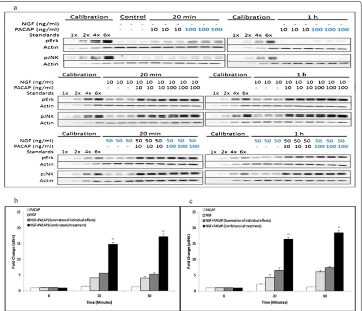
We hypothesized that there was likely to be synergistic activation of the various kinases that regulate synergistic neurite outgrowth. To examine the pathways involved in regulating synergistic neurite outgrowth in these sys-tems, we conducted a time-course to determine changes in the phosphorylation levels of four kinases—Akt, Erk, JNK, and P38—upon NGF, PACAP, and NP treatments. The kinases were activated throughout the entire 1 hour time-course (Additional file 1: Figure S1). Thus, for con-venience, subsequent analyses were performed only at 20 and 60 minutes time-points.
After treating the cells with multiple doses of NGF and PACAP, the phosphorylation levels of Erk, JNK (Figure 2a), P38 and Akt (Additional file 2: Figure S2a) were quantified and analyzed for synergism. Single lig-and treatment with NGF but not PACAP induced sus-tained Erk phosphorylation. To analyze for synergistic activation of Erk, effects upon combinatorial treatments
(See figure on previous page.)
Figure 1 Synergistic neurite outgrowth induced by combinatorial growth factor-PACAP treatments. (a(i)), (b(i)), (c(i)) Response surface plots for the NGF (0-50 ng/ml)-PACAP (0-500 ng/ml) (NP), FGFb (0-100 ng/ml)-PACAP (0-500 ng/ml) (FP), and EGF (0-50 ng/ml)-PACAP (0-500 ng/ ml) (EP) systems, respectively. Top panel: Experimentally obtained results of the growth factor-PACAP combinatorial treatment; Bottom panel: Additive effect calculated through the summation of the individual effects of the growth factors and PACAP. The x, y, and z axes denote concen-trations of PACAP (ng/ml), concenconcen-trations of growth factors (ng/ml), and neurite length, respectively. (a(ii)), (b(ii)), (c(ii)) Isobologram plots illus-trating the concentrations of growth factor and PACAP necessary to obtain a neurite length of 0.14 for the NP, FP, and EP systems, respectively. (d) Representative images of cells treated with each growth factor (50 ng/ml) with and without PACAP (100 ng/ml).
Figure 2 Synergistic and sustained phosphorylation of Erk and JNK upon combinatorial NGF and PACAP treatment. (a) Time-course of Erk and JNK phosphorylation at 20 and 60 minutes following NGF (0-50 ng/ml)-PACAP (0-100 ng/ml) (NP) treatment. Phosphorylation levels of the proteins were analyzed by western blotting, and normalized to the levels of actin. Fold-changes in (b) pErk and (c) pJNK were quantified by densitometry. Data for 50 ng/ml NGF and 100 ng/ml PACAP (highlighted in blue) were plotted and analyzed for synergism. Significant differences between combinatorial experimental treatment of NGF-PACAP and summation of their individual effects were calculated using the paired Student’s t-test. A value of p < 0.05 was considered significant (**p < 0.01). Graphs showing synergistic Erk and JNK phosphorylation at the other ligand concentrations were plotted in Additional file 3: Figure S3a & S3b. Western blot data and quantified results for pP38 and pAkt were plotted in Additional file 2: Figure S2a-S2c.
of NP was compared to the additive effect of the individ-ual ligands. In the presence of both ligands, Erk phos-phorylation was higher than the additive effects of NGF and PACAP separately (Figure 2b, & Additional file 3: Figure S3a). This is in congruence with the finding that NGF and NP treatment but not PACAP induced exten-sive neurite outgrowth, and is consistent with the idea that sustained Erk phosphorylation is involved in neurite outgrowth [21,27,31].
Similarly, sustained activation of JNK by NGF was ob-served (Figure 2c). In addition, we made the novel discov-ery that JNK was also synergistically phosphorylated upon combinatorial NP treatment (Figure 2c, & Additional file 3: Figure S3b) and it was sustained for up to 1 hour post-stimulation. On the contrary, using the same ana-lyses, synergistic phosphorylation of P38 (Additional file 2: Figure S2a, & S2b) and Akt (Additional file 2: Figure S2a, & S2c) were not observed in the NP system.
Having found that Erk and JNK were synergistically phosphorylated in the NP system, we next investigated if these trends were also common to the FP and EP sys-tems. Similar to the NP system, sustained and synergistic Erk (Figure 3a(i), & b(i)) and JNK (Figure 3a(ii), & b(ii)) phosphorylation were observed for the FP and EP treat-ments, respectively, within 1 hour of stimulation. Like-wise, neither P38 (Additional file 4: Figure S4a(i), & S4b (i)) nor Akt (Additional file 4: Figure S4a(ii), & S4b(ii)) were synergistically phosphorylated in the FP and EP systems. Thus, these results indicate that specific kinases were synergistically phosphorylated by growth factor-PACAP co-treatment, suggestive of their roles in mediat-ing synergistic neurite outgrowth.
The total protein levels of Erk, JNK, P38 and Akt upon treatment with single ligand or combinations of the growth factors and PACAP were unchanged across all conditions and time-points (Additional file 5: Figure S5).
Figure 3 Synergistic and sustained phosphorylation of Erk and JNK upon FP and EP treatments. Time-course of quantified Erk and JNK phosphorylation at 20 and 60 minutes upon (a) FGFb (50 ng/ml)-PACAP (100 ng/ml) (FP) or (b) EGF (50 ng/ml)-PACAP (100 ng/ml) (EP) treatment. Fold-change in (i) pErk, and (ii) pJNK were quantified by densitometry and normalized to the levels of actin. Significant differences between com-binatorial experimental treatment of growth factor-PACAP and the summation of their individual effects were calculated using the paired Student’s t-test. A value of p < 0.05 was considered significant (**p < 0.01; *p < 0.05).
Figure 4 Erk is required for neurite outgrowth in all 3 systems whereas JNK is required only for the NP and FP, but not EP, systems. Concentrations of growth factors and PACAP used were 50 ng/ml and 100 ng/ml, respectively. (a) Dose–response treatment of (i) MEK inhibitor (U0126), (ii) JNK inhibitor (SP600125), (iii) P38 inhibitor (SB203580), and (iv) PI3K inhibitor (LY294002) on neurite outgrowth in the NP system. (b), (c) Effect of U0126 (20μM), SP600125 (10 μM), SB203580 (20 μM), and LY294002 (20 μM) on neurite outgrowth in the FP, and EP systems, respectively. Significant differences between treatments with and without inhibitors were calculated using the paired Student’s t-test. A value of p < 0.05 was considered significant (**p < 0.01; *p < 0.05).
Erk is required for neurite outgrowth in all 3 systems whereas JNK is required only for the NP and FP, but not EP, systems
We next examined the role of these synergistically activated kinases in regulating neurite outgrowth using kinase inhibitors. As expected, treatment with the MEK inhibitor, U0126, inhibited neurite outgrowth in the NP system in a dose-dependent manner (Figure 4a(i), & Additional file 6: Figure S6). Similarly, inhibition of MEK also blocked neurite outgrowth in the FP and EP systems (Figure 4b, c, & Additional file 6: Figure S6), confirming the involvement of synergistic Erk phosphor-ylation in neurite outgrowth. Further supporting the in-volvement of synergistically phosphorylated kinases in regulating synergistic neurite outgrowth, the JNK inhibi-tor, SP600125, blocked neurite outgrowth in the NP (Figure 4a(ii), & Additional file 6: Figure S6) and FP sys-tems (Figure 4b, & Additional file 6: Figure S6). Surpris-ingly, SP600125 at the same concentration (10 μM) failed to inhibit neurite outgrowth in the EP system,
showing instead enhanced neurite outgrowth (Figure 4c, & Additional file 6: Figure S6). Higher concentrations of SP600125 were deemed to be cytotoxic (data not shown). Positive controls for the effects of U0126 and SP600125 are shown in Additional file 7: Figure S7a and S7b, respectively.
As expected, inhibition of the non-synergistically acti-vated nodes, P38 and Akt, by SB203580, and LY294002, respectively, did not block neurite outgrowth in all three systems (Figure 4a(iii)-(iv), b, c, & Additional file 6: Figure S6). Likewise, cells treated with doses of the in-hibitors at concentrations higher than 20μM resulted in high levels of cytotoxicity (data not shown). The positive controls for SB203580 and LY294002 are shown in Additional file 7: Figure S7c and S7d, respectively.
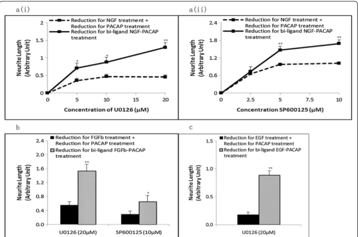
Next, the reduction in neurite outgrowth, after treat-ment with inhibitors, for the NP treattreat-ment was com-pared to the sum of reduction of neurite outgrowth in the single ligand treatments. With U0126 (Figure 5a(i)) and SP600125 (Figure 5a(ii)), the reduction in neurite
Figure 5 Net inhibitor-induced reduction in neurite length is greater in the synergistic systems than in the additive effect of the single ligand treatments. Net reduction in neurite outgrowth in the (a) NP system following treatment with various concentrations of (i) U0126 (MEK inhibitor) and (ii) SP600125 (JNK inhibitor). Reductions in neurite outgrowth in the (b) FP and (c) EP systems in the presence of specific kinase inhibitors. Significant differences between the effects of the combinatorial treatment of growth factor-PACAP (bi-ligand) versus the sum of the effects for each single ligand treatment were compared using the paired Student’s t-test. A value of p < 0.05 was considered significant (**p < 0.01; *p < 0.05).
outgrowth in the NP treatment was greater than the sum of reduction for the single ligand treatments. Simi-larly, for the FP (Figure 5b) and EP (Figure 5c) systems, inhibition of the kinases required for neurite outgrowth also resulted in a greater reduction in neurite outgrowth in the combinatorial growth factor-PACAP treatments than the sum of reduction for the respective single lig-and treatments. These results support the involvement of the various kinases in regulating synergistic neurite outgrowth in the respective synergistic systems.
Critically, these results also suggest that these systems utilize distinct pathways to regulate neurite outgrowth and that not all synergistically phosphorylated kinases are relevant to neurite outgrowth.
P90RSK is a downstream target of both Erk & JNK in the NP & FP systems but is only downstream of Erk in the EP system
Having found that JNK was involved in neurite out-growth in the NP and FP, but not EP, systems, we sought to identify the downstream targets that may be involved in mediating this differential requirement of JNK. Among the many downstream effectors of JNK, P90RSK has been recently shown to be involved in neurite outgrowth and PC12 cells differentiation [32-34]. Thus, we examined if P90RSK was synergistically phosphorylated and if it was involved in JNK-mediated neurite outgrowth.
As expected, P90RSK was synergistically phosphorylated in the NP (Figure 6a(i)-a(ii), & Additional file 8: Figure S8a), FP (Figure 6b(i)) and EP (Figure 6c(i)) systems from 20 minutes to 1 hour after stimulation. In all three systems, neurite outgrowth was inhibited in the presence of the P90RSK inhibitor, BRD7389 [35,36] (Figure 6a(iii), b(ii), c(ii), & Additional file 8: Figure S8b). In these systems, greater reductions in neurite outgrowth were also achieved in the combinatorial growth factor-PACAP treatments than for the sum of the reduction in neurite outgrowth in the respective single ligand treatments (Figure 6d), sup-porting the involvement of P90RSK in regulating synergis-tic neurite outgrowth in all three systems.
To validate the role of P90RSK as a downstream effector of synergistically activated JNK in the three
systems, the phosphorylation level of P90RSK was exam-ined after inhibition with SP600125. Surprisingly, treat-ment with SP600125 inhibited P90RSK phosphorylation in the NP (Figure 7a) and FP (Figure 7b), but not EP (Figure 7c), systems. These results strongly suggest that the regulation of P90RSK by the JNK pathway could be a critical determinant of JNK involvement in regulating synergistic neurite outgrowth.
In addition to JNK, P90RSK has also been reported to be a downstream target of Erk [37,38]. Unlike the case for JNK inhibition, inhibition of Erk activation with U0126 suppressed P90RSK phosphorylation in all three systems (Figure 7), providing further support for the role of P90RSK as an important mediator of neurite outgrowth. The total levels of Erk, JNK, and P90RSK were unchanged during the combinatorial growth factor-PACAP treat-ments both in the presence and absence of the inhibitors (Additional file 9: Figure S9).
Discussion
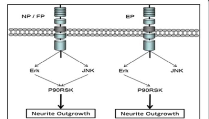
In this study, we demonstrated the involvement of dis-tinct combinations of signaling pathways in mediating synergistic neurite outgrowth induced by PACAP and different growth factors (Figure 8). In these systems, Erk, JNK, and P90RSK were all found to be synergistically phosphorylated. However, synergistic JNK phosphoryl-ation was not required for neurite outgrowth following stimulation with the combination of EGF and PACAP. Further investigations led to the crucial finding that the JNK-P90RSK link is critical to the involvement of JNK in regulating synergistic neurite outgrowth in some but not all growth factor-PACAP stimulation combinations.
cAMP-elevating agents have long been known to syner-gize with NGF [25,26], FGFb [24], and EGF [22,23] to en-hance neurite outgrowth. Although the pathways used by these individual ligands to regulate neurite outgrowth have been widely studied, little is known about the mecha-nisms underlying synergistic neurite outgrowth. RSM-based analyses provide a means to quantitatively compare the degree of synergism between different treatments [29]. By such analyses, the degree of synergism in the EP system was found to be higher than those in the NP and FP
(See figure on previous page.)
Figure 6 P90RSK is synergistically phosphorylated and is involved in neurite outgrowth in all three systems. (a(i)) Time-course of P90RSK phosphorylation at 20 and 60 minutes following NGF (0-50 ng/ml)-PACAP (0-100 ng/ml) treatment. Phosphorylation levels of the proteins were analyzed by Western blotting, and normalized to the levels of Actin. The blots used were the same as those used for pJNK in Figure 2a. (a(ii)), (b (i)), (c(i)) Time-course measurements of pP90RSK at 20 and 60 minutes following NGF (50 ng/ml)-PACAP (100 ng/ml) (NP), FGFb (50 ng/ml)-PACAP (100 ng/ml) (FP) or EGF (50 ng/ml)-PACAP (100 ng/ml) (EP) stimulations, respectively. The total protein levels of P90RSK upon treatment with single ligand or combinations of the growth factors and PACAP were unchanged across all conditions and time-points (Additional file 5: Figure S5). (a(iii)), (b(ii)), (c(ii)) Effect of P90RSK inhibitor, BRD7389 (0.2μM), on neurite outgrowth in the NP, FP, and EP systems, respectively. (d) Net reduc-tion in neurite outgrowth between combinatorial ligand treatment (bi-ligand) versus the sum of neurite outgrowth reducreduc-tion from treatment with each ligand separately in the presence of BRD7389 (0.2μM). Significant differences between the effects of combinatorial experimental treat-ment of growth factor-PACAP and summation of their individual effects, and that between the effects of treattreat-ments with and without inhibitors were calculated using the paired Student’s t-test. A value of p < 0.05 was considered significant (**p < 0.01; *p < 0.05).
systems, suggesting that different signaling pathways may regulate neurite outgrowth in these systems.
To determine the pathways involved in synergistic neurite outgrowth, four kinases were examined, each widely reported to be involved in PC12 cells differenti-ation: Erk [14,39,40], P38 [16,39], JNK [17,41], and Akt
[18,40,42]. Interestingly, our results showed that Akt and P38 were activated following ligand stimulation but not involved in neurite outgrowth in all three systems. In agreement with this, inhibition of these two kinases also failed to suppress NGF-induced PC12 cells neurite out-growth. These results were consistent with some of the earlier reports exploring neurite outgrowth [43-45] but not others [15-18,39-42]. A recent systems-based study revealed a two-dimensional Erk-Akt signaling code that was critical in governing PC12 cells proliferation and dif-ferentiation [46]. Thus, the controversy surrounding the involvement of P38 and Akt would be more adequately addressed using systems-based approaches in the future.
The sustained activation of Erk has been widely re-ported to be required for neurite outgrowth during dif-ferentiation [14,21,27,31]. Consistent with these reports, synergistic and sustained Erk phosphorylation was found to be involved in neurite outgrowth in all three growth factor-PACAP systems. This was especially evident in the EP system, where transient Erk activation was ob-served following treatment with EGF or PACAP alone. Similarly, synergistic and sustained JNK phosphorylation
Figure 7 P90RSK is regulated by Erk and JNK in the NP and FP systems, but only by Erk in the EP system. (a), (b), (c) Time-course meas-urement of pP90RSK at 20 and 60 minutes following NGF (50 ng/ml)-PACAP (100 ng/ml), FGFb (50 ng/ml)-PACAP (100 ng/ml), and EGF (50 ng/ ml)-PACAP (100 ng/ml) treatment, respectively, in the presence or absence of MEK inhibitor, U0126 (20μM), or JNK inhibitor, SP600125 (10 μM). Significant differences between treatments with and without inhibitors were calculated using the paired Student’s t-test. A value of p < 0.05 was considered significant (**p < 0.01).
Figure 8 A schematic illustration of the different pathways used by the three different synergistic systems, NGF-PACAP (NP), FGFb-PACAP (FP), and EGF-PACAP (EP).
was observed in all three systems. Remarkably, inhibition of JNK led to reduced neurite outgrowth in the NP and FP systems, but enhanced outgrowth in the EP system. Although a previous study has found sustained JNK activation to be sufficient to induce PC12 cells differen-tiation [47], our results showed that sustained JNK activation in the EP system is insufficient to induce neurite outgrowth. These seemingly contradictory find-ings could imply that the kinetics of JNK activation alone is insufficient to determine if cells undergo differ-entiation. It is likely that JNK acts in conjunction with other signaling nodes to form a signaling network that regulates neurite outgrowth. Nonetheless, to the best of our knowledge, this is the first report demonstrating the involvement of JNK phosphorylation in synergistic neurite outgrowth.
We have shown that both Erk and JNK were synergistic-ally phosphorylated in all three systems. This may occur through shared common upstream effectors [48] or through independent upstream effectors, such as PKA and Epac [49-51]. In preliminary experiments, we ob-served the involvement of PKA in neurite outgrowth in the EP but not NP system (data not shown); however, a complete understanding of the contribution of PKA and Epac in Erk and JNK activation remains to be determined.
Although synergistic JNK phosphorylation was ob-served in all three systems, it was not found to be in-volved in synergistic neurite outgrowth in the EP system. This suggests a possible difference in downstream sig-naling. P90RSK, which had previously been found to be required for PC12 cells differentiation [33], was also found to be synergistically activated in all three systems in our study. Interestingly, P90RSK was activated by JNK in the NP and FP, but not EP, systems. Although JNK-mediated activation of P90RSK has not been widely reported, it has been observed following ultraviolet ex-posure [34], insulin treatment [52], or transforming growth factor alpha treatment [53]. Consistent with pre-vious findings [38], P90RSK was also regulated by Erk in our study. The co-regulation of targets by Erk and JNK is not uncommon, with previous studies showing that these two kinases regulate many common targets, in-cluding transcription factors [15,54,55], immediate early genes [56] and differentiation-specific genes [15,56,57]. Despite this, results from several studies have suggested that the binding sites of P90RSK for Erk and JNK are likely to be different [34,58], further indicating that P90RSK may be discretely regulated by the two kinases. Our finding of the differential regulation of P90RSK in the NP and EP systems in this study strongly suggests that these synergistic systems can serve as excellent models to decipher the mechanistic regulation of P90RSK by its upstream kinases, Erk and JNK. The con-tributions of Erk, JNK and P90RSK in the mechanism of
axonal outgrowths of neurons in vivo and in vitro will require further clarification in future studies.
Conclusions
In conclusion, our study has demonstrated distinct path-ways involved in synergistic neurite outgrowth in differ-ent systems. Importantly, our findings of the underlying pathways involved in these systems have two key impli-cations. First, some kinases such as JNK may be syner-gistically activated by multiple ligands but yet not necessarily involved in the synergistic neurite outgrowth process and that its involvement in neurite outgrowth is dependent on its interaction with P90RSK. Second, in the EP system, the increased synergy in neurite out-growth and lack of JNK requirement for regulating the process suggest that PACAP synergizes differently with different growth factors to enhance neurite outgrowth. These findings reveal that synergistic of neurite out-growths induced by multiple ligands involves the inter-play of a network of signals.
Additional files
Additional file 1: Figure S1. Time-course profiles of activations of ki-nases upon PACAP, NGF, and NP treatments. Fold changes of (a) pErk, (b) pJNK, (c) pP38, and (d) pAkt from 0-1 hour.
Additional file 2: Figure S2. Non-synergistic phosphorylation of P38 and Akt upon combinatorial NGF (0-50 ng/ml) and PACAP (0-100 ng/ml) treatments. (a) Time-course of P38 and Akt phosphorylations at 20 and 60 minutes following NGF-PACAP treatments. Phosphorylation levels of the proteins were analyzed by western blotting, and normalized to the levels of actin. Fold changes of (b) pP38, and (c) pAkt under (i) uni-ligand treatments, (ii) bi-ligand treatments at 10 ng/ml of NGF, and (iii) bi-ligand treatments at 50 ng/ml NGF. Significant differences between combinator-ial experimental treatment of NGF-PACAP and summation of their indi-vidual effects were calculated using the paired Student’s t-test. A value of p<0.05 was considered significant.
Additional file 3: Figure S3. Synergistic phosphorylation of Erk and JNK upon combinatorial NGF (0-50 ng/ml) and PACAP (0-100 ng/ml) treatments. Fold changes of (a) pErk, and (b) pJNK under (i) uni-ligand treatments, (ii) bi-ligand treatments at 10ng/ml of NGF, and (iii) bi-ligand treatments at 50 ng/ml NGF. Significant differences between combinator-ial experimental treatment of NGF-PACAP and summation of their indi-vidual effects were calculated using the paired Student’s t-test. A value of p<0.05 was considered significant (**p<0.01).
Additional file 4: Figure S4. Non-Synergistic phosphorylation of P38 and Akt upon FP and EP treatments. Time-course of quantified P38, and Akt phosphorylations at 20 and 60 minutes following (a) FGFb (50 ng/ ml)-PACAP (100 ng/ml), and (b) EGF (50 ng/ml)-PACAP (100 ng/ml) treat-ment. Fold changes of (i) pP38, and (ii) pAkt were quantified by densi-tometry and normalized to the levels of actin. Significant differences between combinatorial experimental treatment of growth factor-PACAP and summation of their individual effects were calculated using the paired Student’s t-test. A value of p<0.05 was considered significant. Additional file 5: Figure S5. Total levels of Erk, JNK, P90RSK, Akt and P38 were not changed following treatments with ligands. The total protein levels were assayed at 20 and 60 minutes post-stimulation. The same control (C, at t=0 minutes) was used for both time-points. Additional file 6: Figure S6. Representative images of cells treated with growth factors-PACAP in the presence of inhibitors in the three sys-tems. NP, FP and EP.
Additional file 7: Figure S7. Positive controls for the kinase inhibitors following treatment with NGF (50 ng/ml). (a) Inhibition of Erk phosphorylation in the presence of U0126. (b) Inhibition of c-Jun phos-phorylation in the presence of SP600125. (c) Inhibition of P38 phosphor-ylation in the presence of SB203580. (d) Inhibition of Akt phosphorphosphor-ylation in the presence of LY294002.
Additional file 8: Figure S8. Synergistic phosphorylation of P90RSK upon combinatorial NGF (0-50 ng/ml) and PACAP (0-100 ng/ml) treatments. (a) Fold changes of pP90RSK under (i) uni-ligand treatments, (ii) bi-ligand treatments at 10 ng/ml of NGF, and (iii) bi-ligand treatments at 50 ng/ml NGF. (b) Representative images of cells treated with growth factors (50 ng/ml)-PACAP (100 ng/ml) in the presence of BRD7389 in the three systems. Significant differences between combinatorial experimen-tal treatment of NGF-PACAP and summation of their individual effects were calculated using the paired Student’s t-test. A value of p<0.05 was considered significant (**p<0.01).
Additional file 9: Figure S9. Total levels of Erk, JNK, and P90RSK were not changed following treatments with inhibitors. The total protein levels were assay at 20 minutes post-stimulation. A normalizer (NGF-PACAP co-treated cells) in each blot served as a control to normalize between dif-ferent blots. U=U0126 (20μM) and S=SP600125 (10 μM).
Abbreviations
NGF:Nerve growth factor; FGFb: Basic-fibroblast growth factor;
EGF: Epidermal growth factor; PACAP: Pituitary adenylate cyclase-activating peptide; RSM: Response surface model; Erk: Extracellular signal-regulated kinase 1/2; JNK: c-Jun N-terminal kinase; P38: P38 mitogen-activated protein kinases; PI3K: Phosphatidylinositol 3-kinase; NP: NGF-PACAP; FP: FGFb-PACAP; EP: EGF-PACAP; cAMP: Cyclic adenosine monophosphate; PKA: Protein kinase A; Epac: Exchange protein activated by cAMP.
Competing interests
The authors declare that they have no competing interest. Authors’ contributions
All authors contributed to the design of the experiments. KHS conducted all the experiments. KHS, LZ, and HPT contributed to the analyses of the data. KHS drafted the manuscript while LZ, GS, and HPT revised it critically. All authors read and approved the final manuscript.
Acknowledgements
The authors would also like to acknowledge the Singapore–Massachusetts Institute of Technology Alliance for providing the necessary funding, and the Department of Biochemistry for the laboratory facilities to carry out the study.
Author details
1Chemical & Pharmaceutical Engineering, Singapore-MIT Alliance, 4 Engineering Drive 3, Singapore, Singapore.2Department of Biochemistry, National University of Singapore, 14 Medical Drive, Singapore, Singapore. 3Department of Chemical Engineering, Massachusetts Institute of Technology, 77 Massachusetts Avenue, Cambridge, USA.
Received: 23 April 2013 Accepted: 5 December 2013 Published: 12 December 2013
References
1. Zimmermann GR, Lehar J, Keith CT: Multi-target therapeutics: when the whole is greater than the sum of the parts. Drug Discov Today 2007, 12:34–42.
2. Chou TC: Drug combination studies and their synergy quantification using the Chou-Talalay method. Cancer Res 2010, 70:440–446. 3. Jia J, Zhu F, Ma X, Cao Z, Li Y, Chen YZ: Mechanisms of drug
combinations: interaction and network perspectives. Nat Rev Drug Discov 2009, 8:111–128.
4. Lehar J, Krueger AS, Avery W, Heilbut AM, Johansen LM, Price ER, Rickles RJ, Short GF 3rd, Staunton JE, Jin X, et al: Synergistic drug combinations tend to improve therapeutically relevant selectivity. Nat Biotechnol 2009, 27:659–666.
5. Kamb A, Wee S, Lengauer C: Why is cancer drug discovery so difficult? Nat Rev Drug Discov 2007, 6:115–120.
6. Logan A, Ahmed Z, Baird A, Gonzalez AM, Berry M: Neurotrophic factor synergy is required for neuronal survival and disinhibited axon regeneration after CNS injury. Brain 2006, 129:490–502.
7. Madduri S, Papaloizos M, Gander B: Synergistic effect of GDNF and NGF on axonal branching and elongation in vitro. Neurosci Res 2009, 65:88–97. 8. Jones DM, Tucker BA, Rahimtula M, Mearow KM: The synergistic effects of NGF and IGF-1 on neurite growth in adult sensory neurons: convergence on the PI 3-kinase signaling pathway. J Neurochem 2003, 86:1116–1128. 9. Soumyanath A, Zhong YP, Gold SA, Yu X, Koop DR, Bourdette D, Gold BG:
Centella asiatica accelerates nerve regeneration upon oral administration and contains multiple active fractions increasing neurite elongation in-vitro. J Pharm Pharmacol 2005, 57:1221–1229.
10. Gordon T: The role of neurotrophic factors in nerve regeneration. Neurosurg Focus 2009, 26:E3.
11. Greene LA, Tischler AS: Establishment of a noradrenergic clonal line of rat adrenal pheochromocytoma cells which respond to nerve growth factor. Proc Natl Acad Sci U S A 1976, 73:2424–2428.
12. Rydel RE, Greene LA: Acidic and basic fibroblast growth factors promote stable neurite outgrowth and neuronal differentiation in cultures of PC12 cells. J Neurosci1987, 7:3639–3653.
13. Vaudry D, Stork PJ, Lazarovici P, Eiden LE: Signaling pathways for PC12 cell differentiation: making the right connections. Science 2002, 296:1648–1649. 14. Kao S, Jaiswal RK, Kolch W, Landreth GE: Identification of the mechanisms regulating the differential activation of the mapk cascade by epidermal growth factor and nerve growth factor in PC12 cells. J Biol Chem 2001, 276:18169–18177.
15. Waetzig V, Herdegen T: The concerted signaling of ERK1/2 and JNKs is essential for PC12 cell neuritogenesis and converges at the level of target proteins. Mol Cell Neurosci 2003, 24:238–249.
16. Yung LY, Tso PH, Wu EHT, Yu JCH, Ip NY, Wong YH: Nerve growth factor-induced stimulation of p38 mitogen-activated protein kinase in PC12 cells is partially mediated via Gi/o proteins. Cell Signal 2008, 20:1538–1544. 17. Tso PH, Morris CJ, Yung LY, Ip NY, Wong YH: Multiple Gi proteins
participate in nerve growth factor-induced activation of c-Jun N-terminal kinases in PC12 cells. Neurochem Res 2009, 34:1101–1112.
18. Kim Y, Seger R, Suresh Babu CV, Hwang SY, Yoo YS: A positive role of the PI3-K/Akt signaling pathway in PC12 cell differentiation. Mol Cells 2004, 18:353–359.
19. Wu CF, Howard BD: K252a-potentiation of EGF-induced neurite out-growth from PC12 cells is not mimicked or blocked by other protein kin-ase activators or inhibitors. Brain Res Dev Brain Res 1995, 86:217–226. 20. Erhardt JA, Pittman RN: Ectopic p21(WAF1) expression induces
differentiation-specific cell cycle changes in PC12 cells characteristic of nerve growth factor treatment. J Biol Chem 1998, 273:23517–23523. 21. Traverse S, Gomez N, Paterson H, Marshall C, Cohen P: Sustained activation of
the mitogen-activated protein (MAP) kinase cascade may be required for differentiation of PC12 cells. Comparison of the effects of nerve growth fac-tor and epidermal growth facfac-tor. Biochem J 1992, 288(Pt 2):351–355. 22. Mark MD, Storm DR: Coupling of epidermal growth factor (EGF) with the
antiproliferative activity of cAMP induces neuronal differentiation. J Biol Chem 1997, 272:17238–17244.
23. Yao H, Labudda K, Rim C, Capodieci P, Loda M, Stork PJ: Cyclic adenosine monophosphate can convert epidermal growth factor into a differentiating factor in neuronal cells. J Biol Chem 1995, 270:20748–20753.
24. Wolff P, Abreu PA, Espreafico EM, Costa MC, Larson RE, Ho PL: Characterization of myosin V from PC12 cells. Biochem Biophys Res Commun 1999, 262:98–102.
25. Sakai Y, Hashimoto H, Shintani N, Tomimoto S, Tanaka K, Ichibori A, Hirose M, Baba A: Involvement of p38 MAP kinase pathway in the synergistic activation of PACAP mRNA expression by NGF and PACAP in PC12h cells. Biochem Biophys Res Commun 2001, 285:656–661.
26. Hansen TO, Rehfeld JF, Nielsen FC: Cyclic AMP-induced neuronal differen-tiation via activation of p38 mitogen-activated protein kinase. J Neurochem 2000, 75:1870–1877.
27. Sakai Y, Hashimoto H, Shintani N, Katoh H, Negishi M, Kawaguchi C, Kasai A, Baba A: PACAP activates Rac1 and synergizes with NGF to activate ERK1/ 2, thereby inducing neurite outgrowth in PC12 cells. Brain Res Mol Brain Res 2004, 123:18–26.
28. Wang D, Lagerstrom R, Sun C, Bishof L, Valotton P, Gotte M: HCA-Vision: automated Neurite Outgrowth Analysis. J Biomol Screen 2010, 15:1165–1170. 29. Barrera NP, Morales B, Torres S, Villalon M: Principles: mechanisms and
modeling of synergism in cellular responses. Trends Pharmacol Sci 2005, 26:526–532.
30. Chou TC: Theoretical basis, experimental design, and computerized simulation of synergism and antagonism in drug combination studies. Pharmacol Rev 2006, 58:621–681.
31. Marshall CJ: Specificity of receptor tyrosine kinase signaling: transient versus sustained extracellular signal-regulated kinase activation. Cell 1995, 80:179–185.
32. Nakajima T, Fukamizu A, Takahashi J, Gage FH, Fisher T, Blenis J, Montminy MR: The signal-dependent coactivator CBP is a nuclear target for pp90RSK. Cell 1996, 86:465–474.
33. Silverman E, Frodin M, Gammeltoft S, Maller JL: Activation of p90 Rsk1 is sufficient for differentiation of PC12 cells. Mol Cell Biol 2004, 24:10573–10583. 34. Zhang Y, Zhong S, Dong Z, Chen N, Bode AM, Ma W: UVA induces Ser381
phosphorylation of p90RSK/MAPKAP-K1 via ERK and JNK pathways. J Biol Chem 2001, 276:14572–14580.
35. Fomina-Yadlin D, Kubicek S, Walpita D, Dancik V, Hecksher-Sorensen J, Bitt-ker JA, Sharifnia T, Shamji A, Clemons PA, Wagner BK, Schreiber SL: Small-molecule inducers of insulin expression in pancreatic alpha-cells. Proc Natl Acad Sci U S A 2010, 107:15099–15104.
36. Park YS, Cho NJ: EGFR and PKC are involved in the activation of ERK1/2 and p90 RSK and the subsequent proliferation of SNU-407 colon cancer cells by muscarinic acetylcholine receptors. Mol Cell Biochem 2012, 370:191–198.
37. Anjum R, Blenis J: The RSK family of kinases: emerging roles in cellular signalling. Nat Rev Mol Cell Biol 2008, 9:747–758.
38. Romeo Y, Zhang X, Roux PP: Regulation and function of the RSK family of protein kinases. Biochem J 2012, 441:553–569.
39. Kamata Y, Shiraga H, Tai A, Kawamoto Y, Gohda E: Induction of neurite outgrowth in PC12 cells by the medium-chain fatty acid octanoic acid. Neuroscience 2007, 146:1073–1081.
40. Riese U, Ziegler E, Hamburger M: Militarinone A induces differentiation in PC12 cells via MAP and Akt kinase signal transduction pathways. FEBS Lett 2004, 577:455–459.
41. Xu H, Dhanasekaran DN, Lee CM, Reddy EP: Regulation of neurite outgrowth by interactions between the scaffolding protein, JNK-associated leucine zipper protein, and neuronal growth-JNK-associated pro-tein superior cervical ganglia clone 10. J Biol Chem 2010, 285:3548–3553. 42. Chen X, Fu W, Tung CE, Ward NL: Angiopoietin-1 induces neurite
outgrowth of PC12 cells in a Tie2-independent, beta1-integrin-dependent manner. Neurosci Res 2009, 64:348–354.
43. Klesse LJ, Meyers KA, Marshall CJ, Parada LF: Nerve growth factor induces survival and differentiation through two distinct signaling cascades in PC12 cells. Oncogene 1999, 18:2055–2068.
44. Yan C, Liang Y, Nylander KD, Schor NF: TrkA as a life and death receptor: receptor dose as a mediator of function. Cancer Res 2002, 62:4867–4875. 45. Read DE, Reed Herbert K, Gorman AM: Heat shock enhances NGF-induced
neurite elongation which is not mediated by Hsp25 in PC12 cells. Brain Res 2008, 1221:14–23.
46. Chen JY, Lin JR, Cimprich KA, Meyer T: A two-dimensional ERK-AKT signaling code for an NGF-triggered cell-fate decision. Mol Cell 2012, 45:196–209. 47. Heasley LE, Storey B, Fanger GR, Butterfield L, Zamarripa J, Blumberg D, Maue RA: GTPase-deficient G alpha 16 and G alpha q induce PC12 cell differentiation and persistent activation of cJun NH2-terminal kinases. Mol Cell Biol 1996, 16:648–656.
48. Waetzig V, Herdegen T: MEKK1 controls neurite regrowth after experimental injury by balancing ERK1/2 and JNK2 signaling. Mol Cell Neurosci 2005, 30:67–78.
49. Christensen AE, Selheim F, de Rooij J, Dremier S, Schwede F, Dao KK, Martinez A, Maenhaut C, Bos JL, Genieser HG, Doskeland SO: cAMP analog mapping of Epac1 and cAMP kinase. Discriminating analogs
demonstrate that Epac and cAMP kinase act synergistically to promote PC-12 cell neurite extension. J Biol Chem 2003, 278:35394–35402. 50. Kopperud R, Krakstad C, Selheim F, Doskeland SO: cAMP effector
mechanisms. Novel twists for an‘old’ signaling system. FEBS Lett 2003, 546:121–126.
51. Gerits N, Kostenko S, Shiryaev A, Johannessen M, Moens U: Relations between the mitogen-activated protein kinase and the cAMP-dependent
protein kinase pathways: comradeship and hostility. Cell Signal 2008, 20:1592–1607.
52. Moxham CM, Tabrizchi A, Davis RJ, Malbon CC: Jun N-terminal kinase me-diates activation of skeletal muscle glycogen synthase by insulin in vivo. J Biol Chem 1996, 271:30765–30773.
53. Samaga R, Saez-Rodriguez J, Alexopoulos LG, Sorger PK, Klamt S: The logic of EGFR/ErbB signaling: theoretical properties and analysis of high-throughput data. PLoS Comput Biol 2009, 5:e1000438.
54. Leppa S, Saffrich R, Ansorge W, Bohmann D: Differential regulation of c-Jun by ERK and JNK during PC12 cell differentiation. EMBO J 1998, 17:4404–4413.
55. Eriksson M, Taskinen M, Leppa S: Mitogen activated protein kinase-dependent activation of c-Jun and c-Fos is required for neuronal differ-entiation but not for growth and stress response in PC12 cells. J Cell Physiol 2007, 210:538–548.
56. Marek L, Levresse V, Amura C, Zentrich E, Van Putten V, Nemenoff RA, Heasley LE: Multiple signaling conduits regulate global differentiation-specific gene expression in PC12 cells. J Cell Physiol 2004, 201:459–469. 57. Zentrich E, Han SY, Pessoa-Brandao L, Butterfield L, Heasley LE: Collaboration
of JNKs and ERKs in nerve growth factor regulation of the neurofilament light chain promoter in PC12 cells. J Biol Chem 2002, 277:4110–4118. 58. Smith JA, Poteet-Smith CE, Malarkey K, Sturgill TW: Identification of an
extracellular signal-regulated kinase (ERK) docking site in ribosomal S6 kinase, a sequence critical for activation by ERK in vivo. J Biol Chem 1999, 274:2893–2898.
doi:10.1186/1471-2202-14-153
Cite this article as: Seow et al.: c-Jun N-terminal kinase in synergistic neurite outgrowth in PC12 cells mediated through P90RSK. BMC Neuro-science 2013 14:153.
Submit your next manuscript to BioMed Central and take full advantage of:
• Convenient online submission
• Thorough peer review
• No space constraints or color figure charges
• Immediate publication on acceptance
• Inclusion in PubMed, CAS, Scopus and Google Scholar
• Research which is freely available for redistribution
Submit your manuscript at www.biomedcentral.com/submit

![Figure S3a). This is in congruence with the finding that NGF and NP treatment but not PACAP induced exten-sive neurite outgrowth, and is consistent with the idea that sustained Erk phosphorylation is involved in neurite outgrowth [21,27,31].](https://thumb-eu.123doks.com/thumbv2/123doknet/14193053.478481/7.892.86.810.515.1008/congruence-treatment-outgrowth-consistent-sustained-phosphorylation-involved-outgrowth.webp)


