HAL Id: hal-01258342
https://hal.archives-ouvertes.fr/hal-01258342
Submitted on 18 Jan 2016
HAL is a multi-disciplinary open access archive for the deposit and dissemination of sci-entific research documents, whether they are pub-lished or not. The documents may come from teaching and research institutions in France or abroad, or from public or private research centers.
L’archive ouverte pluridisciplinaire HAL, est destinée au dépôt et à la diffusion de documents scientifiques de niveau recherche, publiés ou non, émanant des établissements d’enseignement et de recherche français ou étrangers, des laboratoires publics ou privés.
Translocable glucose transporters in the brain: where
are we in 2006?
Thierry Alquier, Corinne Amiot Leloup, Anne Lorsignol-Desmet, Luc
Pénicaud
To cite this version:
Thierry Alquier, Corinne Amiot Leloup, Anne Lorsignol-Desmet, Luc Pénicaud. Translocable glucose transporters in the brain: where are we in 2006?. Diabetes, American Diabetes Association, 2006, 55 (Supplement 2), pp.S131-S138. �10.2337/db06-S021�. �hal-01258342�
Translocable Glucose Transporters in the Brain
Where Are We in 2006?
Thierry Alquier,1,2 Corinne Leloup,1Anne Lorsignol,1and Luc Pe´ nicaud1
The discovery of the brain expression of the translocable glucose transporters, GLUT4 then GLUT8, led to the ques-tion of their putative role in the central nervous system, particularly in relation to insulin effect. The anatomical, cellular, and subcellular localization of these transporters has been described in detail. It has been shown that, as in peripheral tissues, these transporters are localized both in an intracellular pool and at the plasma membrane. This is coherent with a translocation phenomenon, but the data reporting the effect of insulin on that property of GLUT4 and GLUT8 remains very controversial. Glucose and insulin have been shown to modulate GLUT4 expression. Based on their anatomical features, different hypotheses have been proposed. Because of the colocalization with the insulin receptors, insulin might regulate glucose uptake in specific brain areas. A role in nutrient sensing has also been proposed, since both insulin and GLUT4 are expressed in gluco-excited neurons of the hypothalamus. Some studies suggest a role for GLUT8 in the endoplasmic reticulum stress. Whereas much has been learned about their cellular features in the central nervous system, many questions remain unanswered concerning their physiological func-tions. Gene knockout strategy specifically in the brain and even more in specific nuclei or type of cells should provide new clues to the physiological role of both transporters in the central nervous system. Diabetes 55 (Suppl. 2): S131–S138, 2006
I
nsulin effects in the central nervous system (CNS) are pleiotropic, including neurotrophic action, cog-nitive functions, and control of energy homeostasis. Thus insulin, considered as an adiposity signal, inhibits food intake, stimulates energy expenditure, and controls peripheral glucose homeostasis via its hypotha-lamic action (1–3). The mechanisms of action are partly known and the signaling pathways activated by insulin in the hypothalamus are similar to the one described in the peripheral insulin-sensitive tissues (muscle and adipose tissues), i.e., insulin receptor substrate 1 and 2,phospha-tidylinositol 3-kinase, and protein kinase B (4,5). Central insulin control of energy homeostasis involves the regula-tion of orexigenic (neuropeptide Y) and anorexigenic neuropeptide (proopiomelanocortin) expression (6), mod-ulation of the neuronal firing (7), and modmod-ulation of neurotransmission (8,9) in several hypothalamic nuclei, i.e., arcuate nucleus (ARC), ventromedial hypothalamus, and paraventricular nucleus. More recently, several stud-ies have suggested that brain insulin may modulate the brain glucose-sensing mechanisms (7,10 –12). Indeed, in-sulin inhibits the activity of hypothalamic glucosensing neurons (7), brain insulin receptor is required for the counterregulatory responses to hypoglycemia (12), and data from our group showed that insulin increases the brain response to glucose (10). However, in these studies, whether insulin action involves modulation of glucose uptake in hypothalamic glucosensing neurons is yet to be demonstrated. In contrast to the insulin-responsive periph-eral tissues (muscle and adipose tissues), brain glucose utilization and metabolism are considered predominantly to be insulin independent. However, using the hyperinsu-linemic clamp, some studies showed that, depending on the glycemia achieved during the clamp or the length of the clamp, glucose utilization was either increased (13,14) or decreased (15–17) in specific brain areas. Thus, the effect of insulin on brain cellular uptake and utilization has therefore remained controversial and elusive.
In muscle and adipose tissues, insulin stimulates glucose transport through the translocation of the insulin-sensitive glucose transporter GLUT4. The large stimulatory effect of insulin in these tissues results from the unique targeting of GLUT4 in the basal states in intracellular vesicles. In response to insulin, these vesicles translocate to the plasma membrane, resulting in an increased glucose trans-port. Gene knockout strategy demonstrated that reduction in GLUT4 levels in muscle or fat causes insulin resistance and diabetes (18). In the past 10 years, we and others have demonstrated the expression of the insulin-sensitive glu-cose transporter GLUT4 in several areas of the CNS (19 –26). More recently, the expression of a translocable glucose transporter, GLUT8, has been reported in the testis, the brain, and to a lesser degree in peripheral insulin-sensitive tissues, i.e., adipose tissue and muscle (27–29). GLUT8 protein sequence shows 23% homology with GLUT4 sequence and a Kmof 2.4 mmol/l for
2-deoxy-glucose comparable to GLUT4 Km (27,29). Although the
subcellular localization and translocation of GLUT8 has not been elucidated in muscle or adipose tissues, GLUT4 and GLUT8 harbor a similar dileucine internalization motif in the cytoplasmic tail responsible for their retention into the cytoplasm (27,30). Indeed, mutation of this motif into dialanine results in the redistribution of the GLUT8 trans-porter to the plasma membrane, strongly suggesting that GLUT8 could also be translocated (27). In agreement with
From the1Laboratoire de Neurobiologie, Plasticite´ Tissulaire et Me´tabolisme
Energe´tique, Rangueil, France; and the2Montreal Diabetes Research Center,
Centre de Recherche du Centre Hospitalier de l’Universite´ de Montre´al, Montre´al, Que´bec, Canada.
Address correspondence and reprint requests to Luc Pe´nicaud, UMR5018 CNRS-UPS, IFR 31, BP 84225, 31432 Toulouse Cedex 4, France. E-mail: penicaud@toulouse.inserm.fr.
Received for publication 23 March 2006 and accepted in revised form 2 May 2006.
This article is based on a presentation at a symposium. The symposium and the publication of this article were made possible by an unrestricted educa-tional grant from Servier.
ARC, arcuate nucleus; CNS, central nervous system; E, embryonic day; ER, endoplasmic reticulum.
DOI: 10.2337/db06-S021
© 2006 by the American Diabetes Association.
The costs of publication of this article were defrayed in part by the payment of page charges. This article must therefore be hereby marked “advertisement” in accordance with 18 U.S.C. Section 1734 solely to indicate this fact.
this, an insulin-induced translocation of GLUT8 has been reported in mouse blastocysts (29), similarly to the GLUT4 translocation described previously in adipocytes or mus-cle cells.
Although basal glucose transport in the brain is ensured by the glial-endothelial GLUT1 transporter and the neuro-nal GLUT3 transporter, the presence of GLUT4 and GLUT8 in the brain is of particular interest. Considering the controversial effect of insulin on brain glucose uptake, the expression of both translocable glucose transporters in different brain areas raises several questions about their physiological relevance and roles in brain glucose metab-olism and hypothalamic glucose-sensing mechanisms. The main goal of this review is to establish an update of the recent studies investigating GLUT4 and GLUT8 in the brain and to propose some clues and a hypothesis (based on their respective features) of their potential functional roles in the CNS.
CELLULAR FEATURES OF GLUT4 AND GLUT8 IN THE BRAIN
Anatomical, cellular, and subcellular localizations of
GLUT4 and GLUT8 in the adult brain. GLUT4 and
GLUT8 are expressed in several brain areas including the cortex, the amygdala, the hippocampus, the hypothala-mus, and the cerebellum (23,31). The highest levels of GLUT8 expression were found in the hippocampus, amyg-dala, hypothalamus, and pituitary gland. Anatomical local-ization demonstrates a more spread expression of GLUT4 in others areas such as the thalamus, the brainstem, and olfactory bulbs, with the highest levels of expression in areas associated with the control of motor activity such as the cortex and the cerebellum (23,25).
Some GLUT4 immunoreactivity has been observed in glial cells of the hippocampus and the cerebellum (23,25) and in the ependymal cells lining the ventricles (19) as well as endothelial cells of microvessels (22,32). The presence of GLUT8 in these cells has not been reported. However, the expression of GLUT8 and GLUT4 in the brain is essentially neuronal. Several colocalization studies pro-vided new data on the chemical nature of neurons express-ing GLUT4 or GLUT8. In the basal forebrain, GLUT4 staining was found selectively in cholinergic (using ChAT marker) and GABAergic (using parvalbumin marker) neu-rons (33). In the hippocampus, GLUT8 is present in principal neurons, pyramidal and granule neurons (which are glutamatergic), and nonprincipal GABAergic neurons (34). In the neurons of the hypothalamic pituitary system, GLUT8 was colocalized with vasopressin but not with oxytocin (31).
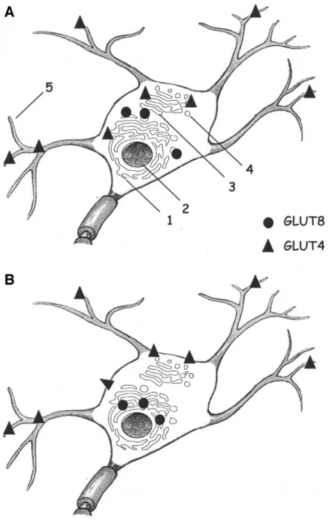
At the subcellular level, the transporters are mainly present in the neuronal cell bodies and the proximal apical dendrites. Contrary to GLUT8, GLUT4 is also abundantly present in the dendrites, in the dendrite spines, and at the synaptic level (23). In unstimulated conditions, GLUT4 immunoreactivity is localized on the membranes of trans-port vesicles, Golgi apparatus, and rough endoplasmic reticulum (ER) of all the brain areas analyzed (23) (Fig. 1A). Immunoblot analysis of hippocampal neurons re-vealed that GLUT8 is expressed in high-density micro-somes and low-density micromicro-somes, albeit at lower levels. Using electron microscopy, Piroli et al. (35) confirmed that GLUT8 was mainly present near and adjacent to the rough ER and absent in the Golgi apparatus (Fig. 1A). However, Ibberson et al. (31) demonstrated recently that GLUT8
immunoreactivity was differentially localized depending on the brain region analyzed. Indeed, GLUT8 was re-stricted to the nerve terminals and synaptic vesicles of the supraoptic nucleus, whereas it appeared associated with the cell surface in the cortex (and dentate gyrus of the hippocampus) (31).
Altogether, these studies demonstrated that 1) despite a specificity of expression in some brain regions, there is an important overlap between the cerebral areas where GLUT4 and GLUT8 are present, and 2) both transporters exhibit an intracellular localization suggesting a transloca-tion phenomenon to the plasma membrane. However, more investigations will be needed to determine whether GLUT4 and GLUT8 are colocalized in the same neurons and to determine more precisely the chemical nature of the neurons expressing the transporters.
Regulation of GLUT4 and GLUT8 expression.Insulin
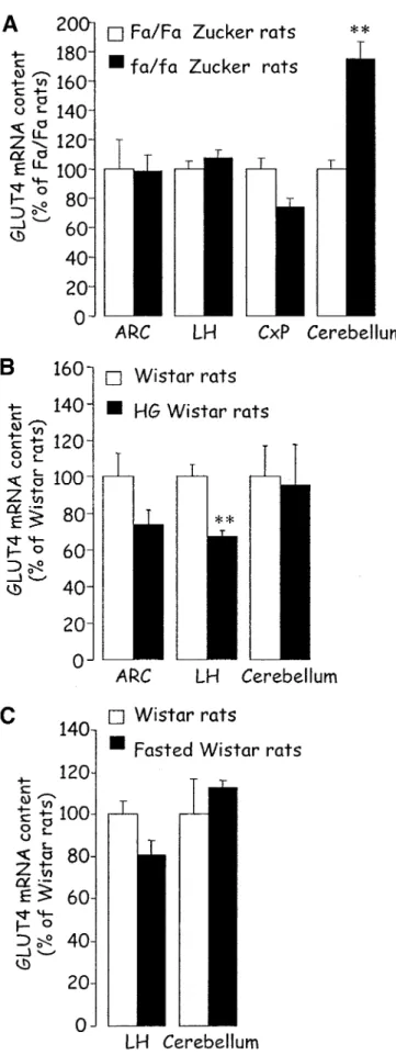
is one of the main regulators of GLUT4 expression at the mRNA and protein levels in the peripheral insulin-sensitive tissues, muscle, and adipose tissues (36). Indeed, a large body of literature has described changes of GLUT4 expres-sion in insulin-resistant states in humans and rodents. For instance, in rodents, the expression of GLUT4 is decreased in the adipose tissues of several hyperinsulinemic models such as diet-induced obese rats or obese Zucker fa/fa rats, whereas the protein levels are unchanged in muscle (37). Several studies have shown that the cerebral expression of GLUT4 was also modified in insulin-resistant models and demonstrated that the changes were specific to the area considered. For instance, Vannucci et al. (24) showed that GLUT4 protein levels were increased in the cerebel-lum of hyperinsulinemic-hyperglycemic db/db mice, whereas the expression was unchanged in the cortex or the olfactory bulbs. These data are in agreement with previous work of Campbell et al. (38), who demonstrated an increased GLUT4 cerebellar content in hyperinsuline-mic Milan rats. In support of these findings, our studies showed increased GLUT4 mRNA levels in the same area of hyperinsulinemic fa/fa Zucker rats without changes in the cortex (T.A., L.P., unpublished data; Fig. 2A). Finally, recent work by Komori et al. (39) showed increased levels of GLUT4 protein in the hypothalamus of ob/ob mice. However, at the mRNA level, GLUT4 expression was unchanged in the ARC and lateral hypothalamus of obese Zucker rats compared with lean Zucker rats (T.A., L.P., unpublished data; Fig. 2A). The reason for the discrepancy between the data in ob/ob mice and fa/fa rats is unknown but could be related to the species difference as well as the different metabolic phenotype. However, altogether, these studies demonstrate that chronic insulin-resistant states are associated with increased expression of GLUT4 in the cerebellum.
In our experiments, when insulin levels were acutely increased by a 48-h glucose infusion in rats, GLUT4 mRNA content was unchanged in the cerebellum, whereas GLUT4 expression was specifically decreased in the lateral hypothalamus and ARC (Fig. 2B) (40). When insulin levels were lowered acutely by streptozotocin treatment or ex-ercise in rats, the cerebellar GLUT4 protein levels were diminished, suggesting a clear correlation between circu-lating insulin levels and GLUT4 protein content in the cerebellum. However, at the mRNA level, hypothalamic (lateral hypothalamus) and cerebellar GLUT4 levels were unchanged in hypoinsulinemic-hypoglycemic fasted rats (unpublished data; Fig. 2C). All the data mentioned above are summarized in Table 1.
Altogether, these data point out that insulin may not be the only factor controlling GLUT4 expression in the brain, especially in the cerebellum, since depending on glycemia, the insulinemic state does not lead to identical results. Also, these data strongly suggest a region-specific control of the expression of GLUT4 in the CNS.
The regulation of the expression of GLUT8 in the brain has been poorly studied so far. The group of Reagan et al. (28) showed that GLUT8 mRNA levels were increased
in the hippocampus of hypoinsulinemic streptozotocin-treated rats. This increase was partially normalized by short-term restraint-induced stress in streptozotocin rats, but the GLUT8 protein content was unchanged in the same area (28,41). These data suggest that insulin and glucocor-ticoids may be involved in the regulation of GLUT8 expres-sion in the hippocampus.
Recent data have shown that GLUT4 and GLUT8 are expressed in the developing mammalian brain and that
FIG. 1. Basal and stimulated subcellular localizations of GLUT4 and GLUT8 in neurons. Although GLUT4 and GLUT8 are present in similar brain areas, no colocalization of the transporters in the same neuron has been reported yet. A: In the basal state, GLUT4 and GLUT8 are mainly present in the neuronal cell bodies. Whereas GLUT8 is mainly localized in the neuronal soma and proximal dendrites, GLUT4 is also abundantly present in vesicles in the dendrites, in the dendrite spines, and at the synaptic level. At the subcellular level, GLUT4 is associated with the membranes of the rough ER, Golgi apparatus, and transport vesicles in the vicinity of the plasma membrane. GLUT8, however, shows a more specific localization near or adjacent to the rough ER. B: In stimulated states, such as a glucose challenge, GLUT8 is translocated to the membranes of the rough ER. It has been proposed that this redistribution may ensure the transport of glucose from the ER lumen into the cytosol during protein glycosylation. The translocation of GLUT4 to the cell membrane has been recently demonstrated in vitro upon insulin stimulation, but the redistribution of the transporter in neurons in vivo has not yet been reported. The potential translocation of GLUT4 to the plasma membrane at the dendrite level is not represented here. 1, Rough ER; 2, nucleus; 3, Golgi apparatus; 4, transport vesicles; 5, dendrites.
their expression is modulated throughout the fetal to adult stages (42– 45). GLUT4 is present in the embryo brain as early as embryonic day 9 (E9) and E14 in rats, and the expression level peaks at E14 and is lower at E19 and remains stable thereafter up to adulthood (42,43). After birth, GLUT4 levels are increased during the suckling phase between postnatal day 14 (P14) and P21 in rats and mice and lower thereafter, similarly to GLUT8 (43,44). Gestational hypoxia or hyperglycemia during the last stages of gestation increased GLUT4 mRNA levels in the brain, suggesting that intervention modulating glucose or oxygen availability during pregnancy affects the expres-sion of the transporter in the fetal brain (42,45).
Whereas these data demonstrate that metabolic and hormonal parameters are involved in the control of GLUT4 expression, further investigations are needed to determine the specific factors involved in the control of GLUT4 and GLUT8 expression in the CNS.
Translocation of GLUT4 and GLUT8. To date, insulin
has been the best characterized stimulus involved in the translocation of GLUT4 in peripheral tissues (adipose tissues and muscles) and recently in the translocation of GLUT8 in the mouse blastocyst (29). However, other stimuli such as membrane depolarization or hyperosmotic shock have been shown to stimulate GLUT4 translocation in muscle or adipose cells (46). It is important to note that the translocation of GLUT4 in peripheral tissues requires the SNARE (soluble N-ethylmaleimide–sensitive factor attachment protein receptor) protein complex, in which proteins similar to those involved in exocytosis of synaptic vesicles in neurons are used (47,48).
GLUT4.The subcellular localization of GLUT4 in vivo in
intracellular vesicles close to the plasma membrane sug-gests that the transporter could be translocated to the cell surface (23) (Fig. 1A and B). In support of this, a recent report showed, using Western blot and immunocytochem-istry, that insulin induces the translocation of GLUT4 to the plasma membrane in human neuroblastoma cells (SH-SY5Y) (49). Also, the insulin-induced translocation of GLUT4 in SH-SY5Y cells enhances glucose uptake, and this augmentation is abolished in the presence of a phosphati-dylinositol 3-kinase inhibitor. These data suggest that GLUT4 redistribution in response to insulin is a phospha-tidylinositol 3-kinase– dependent mechanism similar to the mechanism described in muscle or adipose tissues.
Inter-FIG. 2. GLUT4 mRNA quantification by quantitative competitive RT-PCR in different rat brain regions. A: fa/fa obese Zucker rats and lean control fa/fa. B: 48-h glucose-infused Wistar rats and saline infused control rats. C: 4-day fasted Wistar rats and fed control rats. CxP, cortex parietal; LH, lateral hypothalamus. Data are meansⴞ SE. **P < 0.01 vs. control (unpaired t test).
TABLE 1
Regulation of GLUT4 mRNA and protein levels in different brain areas in animal models characterized by chronic or acute changes in circulating insulin levels
Hypo-thalamus Cortex Cere-bellum Hyperinsulinemic models Diabetic db/db mice ND % 1 Obese ob/ob mice 1 ND ND Obese fa/fa Zucker rat % % 1 HG Wistar rat 2 ND % Hypoinsulinemic models Streptozotocin-induced diabetic rat ND ND 2 Exercise in rat ND ND 2 Fasted rat % ND %
ND, not determined. HG, hyperglycemic rats induced by 48 h of glucose infusion.
estingly, leptin treatment also stimulates the redistribution of the transporter to the membrane (49). To our knowl-edge, this the first report describing the translocation of GLUT4 in neural cells in vitro. In collaboration with the group of Y. Lemarchand, a DsRed-tagged GLUT4 was used to investigate the localization and translocation of GLUT4 in primary cultures of fetal cortical neurons transfected by the construct. Our preliminary data showed that the localization of DsRed-GLUT4 was mainly somato-dendritic as the one described in vivo (23). The protein was present in the cytoplasm showing a perinuclear localization. How-ever, no change of this localization was observed upon insulin stimulation (4 mol/l during 20 min) (T.A., Y. Lemarchand, L.P., unpublished data). Cortical neurons in culture are known to express the insulin receptor, suggest-ing that, in contrast with the report of Benomar et al. (49), insulin does not stimulate the translocation of GLUT4 in vitro. However, our preliminary studies need further inves-tigations to be confirmed, and the effect of stimuli other than insulin needs to be investigated.
Although some GLUT4-enriched brain areas exhibit a high level of the insulin receptor, a direct demonstration of the insulin-induced translocation has not been reported in vivo yet. Several observations suggest that an insulin-induced translocation might occur also in the brain. A recent study showed that the insulin-regulated aminopep-tidase shows a brain distribution resembling that of GLUT4 and that the transporter is colocalized with insulin-regulated aminopeptidase in hippocampal neurons (50). In insulin-responsive tissues, insulin-regulated aminopepti-dase is a marker of GLUT4 vesicles and is involved in the trafficking of these specialized vesicles in adipocytes (51). Recently, the group of B. Levin demonstrated that 60 –75% of the glucose-sensitive neurons coexpressed GLUT4 and the insulin receptor mRNA in dissociated neurons of the ventromedial nucleus, strongly pointing out a role of insulin in GLUT4 translocation (11). Using electron mi-croscopy, Komori et al. (39) reported that in the ARC, the amount of GLUT4 associated with the plasma membrane was increased in hyperinsulinemic ob/ob mice (unstimu-lated conditions), suggesting a potential increase of GLUT4 translocation. However, the total amount of cellu-lar GLUT4 protein was also increased in the same nucleus, which could explain the increased GLUT4 content in the cell membrane.
GLUT8. In contrast to GLUT4, different groups have
investigated the translocation of GLUT8 in neural cells under the action of stimuli known to induce GLUT4 redistribution to the cell membrane in adipocyte or muscle cells (35,52,53). The only demonstration of the insulin-sensitive redistribution of GLUT8 from intracytoplasmic storage to the cell membrane has been in blastocysts. In mouse blastocysts, insulin exposure induced the translo-cation of GLUT8 to the plasma membrane, resulting in an increased glucose transport that ensured viability (29,54). However, in primary rat adipocytes, 3T3-L1 adipocytes, and CHO cells, insulin did not stimulate GLUT8 transloca-tion to the cell surface (55,56). The reason for this discrep-ancy is unknown, but the latter is in agreement with two studies using mouse neural cells in vitro. Recent work from the group of B. Thorens, using a myc-tagged GLUT8 construct transfected into PC12 cells and hippocampal neurons, demonstrated no change of GLUT8myc intracel-lular localization in response to insulin (53). Brain-derived neurotrophic factor, hyperosmotic shock, potassium- or glutamate-induced depolarization, glucoprivic stress
in-duced by 2-deoxyglucose, or AMP-activated protein kinase activation by 5-amino-imidazole-4-carboxamide-1--D -ribo-furanoside (AICAR) failed to stimulate the translocation of GLUT8myc (53). In agreement with these data, Shin et al. (52) showed an absence of translocation of a green fluorescent protein–tagged GLUT8 in N2A neuroblastoma cells in response to insulin, IGF-I, depolarization, or hyp-oxia treatment.
However, in vivo studies from Piroli et al. (35,41) showed that glucose administration, inducing increased serum insulin levels, stimulated the translocation of GLUT8 from light-density microsomes to high-density mi-crosomes in hippocampal neurons in vivo. Using electron microscopy, Piroli et al. (35) confirmed that glucose administration induces the redistribution of GLUT8 into the membranes of the rough ER (Fig. 1A and B). In contrast, streptozotocin-treated rats, characterized by hy-perglycemia and hypoinsulinemia, have a decreased amount of GLUT8 associated with the high-density micro-some fraction. These studies suggest that circulating insu-lin might stimulate the redistribution of GLUT8 from a cytoplasmic localization into the membranes of the rough ER in the neurons of the hippocampus. Because the translocation of GLUT8 has been only studied in the hippocampal formation in vivo, it is difficult to extend Piroli’s data to other brain areas. Importantly, GLUT8 was present in the nerve terminals and synaptic vesicles of the supraoptic nucleus and vasopressin neurons, suggesting that in these specific neurons, a redistribution of GLUT8 to the plasma membrane could occur upon a stimulus that remains to be identified (31).
Altogether, these findings demonstrate that any of the known signals inducing GLUT4 translocation in the pe-ripheral tissues are able to stimulate the redistribution of GLUT8 into the neuronal cell membrane, in vivo or in vitro. Consistently with the studies in neural cells, it was recently shown in 3T3-L1 adipocytes and CHO cells that GLUT4 and GLUT8 do not colocalize and that GLUT8 does not recycle to the plasma membrane, suggesting that GLUT8 is unlikely to respond to a stimulus that leads to GLUT4 translocation (56). This strongly points out that the role of GLUT8 may not be to increase neuronal uptake of extracellular glucose. These unexpected observations raise several questions concerning the physiological roles of GLUT4 and GLUT8 in the CNS that we will attempt to address in the following section.
FUNCTIONAL ROLE(S) OF GLUT4 AND GLUT8 IN THE BRAIN
GLUT4.The physiological role of GLUT4 is still unknown, but different hypotheses based on its neuroanatomical distribution can be proposed. First, GLUT4 is preferen-tially and highly expressed in brain areas associated with the control of motor activity such as the cortex and the cerebellum. In these areas, translocation of GLUT4 may provide additional glucose to motor neurons under condi-tions of high energy demand such as a high rate of neuronal firing. This hypothesis is consistent with data showing that physical exercise stimulates local cerebral glucose utilization in the motor system (57). Hence, the electrophysiological activity of the motor neurons may directly stimulate GLUT4 translocation to provide energy substrate required for neuronal firing. Also, in the basal forebrain, GLUT4 is mainly localized in cholinergic and GABAergic neurons (33). Because glucose serves as a
substrate for the synthesis of acetylcholine, it could be hypothesized that GLUT4 in these neurons may provide glucose under high firing activity to replenish acetylcholine contents. Also, GLUT4 is highly present in neuronal pro-cesses and at the synaptic level, pointing to a role in neurotransmission. Once again, the exact mechanisms of how GLUT4 is translocated in those neurons and may affect neuronal glucose homeostasis are not yet elucidated.
Second, the anatomical localizations of GLUT4 often relate with those of the insulin receptors, suggesting that the hormone could induce GLUT4 translocation and increase neuronal glucose transport. As we mentioned previously, whole brain glucose uptake is considered insulin independent. However, depending on the glycemia achieved during the hyperinsulinemic clamp, glucose up-take was either increased (13,14) or decreased (15–17) in different brain areas. The reason for such discrepancies is unknown, but when hyperinsulinemia was accompanied by hyperglycemia, glucose uptake was increased in several brain areas including the hypothalamus (14). Thus, we cannot rule out that insulin may modulate directly or indirectly (through modulation of neuronal firing) glucose uptake in discrete brain areas.
Finally, the localization of GLUT4 in the hypothalamus indicates that the transporter may be involved in glucose-sensing mechanisms. Those mechanisms are critical for the control of whole-body glucose homeostasis. Increases of glucose levels activate the hypothalamic glucose-ex-cited neurons, whereas they inhibit the glucose-inhibited neurons in the ARC or the ventromedial nucleus (58). Several studies, using electrophysiology, have demon-strated that central insulin modulates the activity of hypo-thalamic glucosensing neurons. Indeed, insulin inhibits the electrical activity of glucose-excited neurons in the ARC and the ventromedial hypothalamus in a glucose-depen-dent way (7,59). In the presence of 10 or 2.5 mmol/l glucose, insulin has no effect on glucose-excited neuron activity, whereas at 0.1 mmol/l glucose, insulin activates glucose-excited neurons of the ARC (7,59). The effect of insulin may be related to increased glucose transport into glucose-excited neurons. In support of this, the same group showed that 75% of the glucose-excited neurons in the ventromedial nucleus express mRNA coding for both the insulin receptor and GLUT4 (11). Interestingly, Fisher et al. (12) showed that the counterregulatory response to hypoglycemia was impaired in mice with a neuron-specific insulin receptor knockout, suggesting that central insulin action may be critical in the setting of low glucose concentrations. On the other hand, several rodent models characterized by altered glucose-sensing mechanisms have an alteration of GLUT4 expression in the hypothala-mus (39,40).
Taken together, these data suggest that central insulin action might be critical for glucose-sensing mechanisms. However, the precise role of GLUT4 in insulin effect and in glucose-sensing mechanisms is yet to be elucidated.
GLUT8. The results of several studies in neural and
non-neural cells have demonstrated specific features of GLUT8, such as a subcellular localization of GLUT8 in a cytoplasmic compartment that differs from the one de-scribed for GLUT4 (56), the absence of recycling (56), and translocation to the plasmic membrane upon different stimulations (52,53). Interestingly, studies from Piroli et al. (35,41) showed that the transporter is translocated from the cytoplasm into the rough ER upon glucose stimulation in vivo and that streptozotocin-induced diabetes disrupts
GLUT8 trafficking in the ER. This observation is of partic-ular interest in the context of recent studies demonstrating that obesity and diabetes causes ER stress (60 – 62). An essential function of the ER is the synthesis and process-ing of secretory and membrane proteins. Disruption of ER homeostasis, collectively termed ER stress, leads to the accumulation of unfolded protein and protein aggregates in the ER lumen, which is detrimental for cell survival (63). Based on their findings, Piroli et al. (35,41) proposed that in the hippocampus, GLUT8 transports the glucose mole-cules removed from glycoproteins during protein process-ing out of the rough ER lumen into the cytoplasm. Thus, it is tempting to speculate that somehow GLUT8 in the rough ER of hippocampal neurons may control ER glucose homeostasis, which is critical for glycosylation processes. Disruption of GLUT8 trafficking and the subsequent glu-cose equilibrium could lead to an ER dysfunction and stress affecting neuron viability (64). Studying the involve-ment of GLUT8 in ER glucose homeostasis is of particular importance, since neuronal damage observed during cere-bral ischemia/reperfusion or during neurodegenerative diseases is associated with ER stress (64,65).
GLUT8 is also expressed in the hypothalamus, and a potential role in hypothalamic glucose-sensing cannot be ruled out (31). However, similarly to GLUT4, this hypoth-esis has not yet been demonstrated.
As mentioned previously, GLUT4 and GLUT8 are ex-pressed in the developing mammalian brain. Dynamic changes of expression have been described throughout the fetal to adult stages (42– 45). For instance, GLUT4 levels peak at E14 in rat brain embryos, are further decreased at E19, and remain stable until adulthood (42). Brain GLUT8 expression in fetal stages has not been investigated. After birth, GLUT4 levels are increased during the suckling phase between P14 and P21 in rats and mice and further decreased until adulthood, similarly to the GLUT8 pattern of expression (43,44). The brain requires important sup-plies of fuel to support neuroglial growth and process formation during early postnatal development (66); thus, the increase in GLUT4 and GLUT8 expression during this period might be critical for glucose supply. In support of this, Cheng et al. (67) demonstrated that knocking out the neurotrophic factor IGF-I (whose expression peaks during postnatal brain development) in mice induces a dramatic decrease of glucose uptake in several brain regions of young mice (P10). Decreased glucose uptake was associ-ated with a specific decrease of GLUT4 expression, whereas GLUT1 and GLUT3 expression was unchanged in the brain of IGF-I knockout mice. Taken together, these observations strongly suggest that GLUT4 and probably GLUT8-mediated glucose transport may play critical roles during fetal and postnatal brain development.
While energy needs are highly increased during brain development, aging processes and neurodegenerative eases (Alzheimer’s, Parkinson’s, and Huntington’s dis-eases) are characterized by decreased glucose metabolism in the CNS (68 –70). Insulin and glucose are known to contribute to and stimulate cognitive functions such as learning and memory (71,72). Also, the insulin receptor, GLUT4, and GLUT8 are densely expressed in areas sup-porting cognition (such as the hippocampus). More inter-estingly, numerous lines of evidence suggest that an increased prevalence of insulin resistance similar to that seen in type 2 diabetes may contribute to the pathophys-iology of Alzheimer’s disease (69). Based on these obser-vations, it is tempting to speculate that insulin action on
cognitive functions is directly and/or indirectly dependent of GLUT4 and/or GLUT8 in those areas and that impair-ment of insulin effects during insulin-resistant states might be responsible for the decreased brain glucose metabolism observed in Alzheimer’s disease.
Finally, an alternative hypothesis that cannot be ruled out is that glucose is not the primary substrate for GLUT4 and GLUT8 in the brain and that it transports other substrates.
SUMMARY
It is now clear that the translocable glucose transporters GLUT4 and GLUT8 are present in neurons of several brain areas. Numerous studies have addressed the specific ana-tomical and subcellular localizations of both transporters, suggesting that GLUT4 and GLUT8 may be involved in different mechanisms within the neurons. Whereas much has been learned about their cellular features in the central nervous system, many questions remain unan-swered concerning their physiological functions. Based on their anatomical features, are the physiological roles of different brain areas specific? Do transporters participate in brain glucose uptake during basal states and/or during specific conditions such as brain development or high neuron firing? Are transporters involved in hypothalamic glucose sensing and control of energy balance? These and many other questions still need to be addressed. Gene knockout of GLUT4 and/or GLUT8 specifically in the brain and even more in specific nuclei or types of cells should provide new clues on the physiological role of both transporters in the CNS.
REFERENCES
1. Porte D Jr, Seeley RJ, Woods SC, Baskin DG, Figlewicz DP, Schwartz MW: Obesity, diabetes and the central nervous system. Diabetologia 41:863– 881, 1998
2. Obici S, Feng Z, Karkanias G, Baskin DG, Rossetti L: Decreasing hypotha-lamic insulin receptors causes hyperphagia and insulin resistance in rats.
Nat Neurosci5:566 –572, 2002
3. Obici S, Zhang BB, Karkanias G, Rossetti L: Hypothalamic insulin signaling is required for inhibition of glucose production. Nat Med 8:1376 –1382, 2002 4. Niswender KD, Morrison CD, Clegg DJ, Olson R, Baskin DG, Myers MG Jr, Seeley RJ, Schwartz MW: Insulin activation of phosphatidylinositol 3-ki-nase in the hypothalamic arcuate nucleus: a key mediator of insulin-induced anorexia. Diabetes 52:227–231, 2003
5. Niswender KD, Schwartz MW: Insulin and leptin revisited: adiposity signals with overlapping physiological and intracellular signaling capabil-ities. Front Neuroendocrinol 24:1–10, 2003
6. Woods SC, Seeley RJ: Adiposity signals and the control of energy ho-meostasis. Nutrition 16:894 –902, 2000
7. Spanswick D, Smith MA, Mirshamsi S, Routh VH, Ashford ML: Insulin activates ATP-sensitive K⫹ channels in hypothalamic neurons of lean, but not obese rats. Nat Neurosci 3:757–758, 2000
8. Peinado JM, Myers RD: Norepinephrine release from PVN and lateral hypothalamus during perfusion with 2-DG or insulin in the sated and fasted rat. Pharmacol Biochem Behav 27:715–721, 1987
9. Patterson TA, Brot MD, Zavosh A, Schenk JO, Szot P, Figlewicz DP: Food deprivation decreases mRNA and activity of the rat dopamine transporter.
Neuroendocrinology68:11–20, 1998
10. Alquier T, Leloup C, Atef N, Fioramonti X, Lorsignol A, Penicaud L: Cerebral insulin increases brain response to glucose. J Neuroendocrinol 15:75–79, 2003
11. Kang L, Routh VH, Kuzhikandathil EV, Gaspers LD, Levin BE: Physiolog-ical and molecular characteristics of rat hypothalamic ventromedial nu-cleus glucosensing neurons. Diabetes 53:549 –559, 2004
12. Fisher SJ, Bruning JC, Lannon S, Kahn CR: Insulin signaling in the central nervous system is critical for the normal sympathoadrenal response to hypoglycemia. Diabetes 54:1447–1451, 2005
13. Lucignani G, Namba H, Nehlig A, Porrino LJ, Kennedy C, Sokoloff L:
Effects of insulin on local cerebral glucose utilization in the rat. J Cereb
Blood Flow Metab7:309 –314, 1987
14. Orzi F, Lucignani G, Dow-Edwards D, Namba H, Nehlig A, Patlak CS, Pettigrew K, Schuier F, Sokoloff L: Local cerebral glucose utilization in controlled graded levels of hyperglycemia in the conscious rat. J Cereb
Blood Flow Metab8:346 –356, 1988
15. Grunstein HS, James DE, Storlien LH, Smythe GA, Kraegen EW: Hyperin-sulinemia suppresses glucose utilization in specific brain regions: in vivo studies using the euglycemic clamp in the rat. Endocrinology 116:604 – 610, 1985
16. Marfaing P, Penicaud L, Broer Y, Mraovitch S, Calando Y, Picon L: Effects of hyperinsulinemia on local cerebral insulin binding and glucose utiliza-tion in normoglycemic awake rats. Neurosci Lett 115:279 –285, 1990 17. Doyle P, Cusin I, Rohner-Jeanrenaud F, Jeanrenaud B: Four-day
hyperin-sulinemia in euglycemic conditions alters local cerebral glucose utilization in specific brain nuclei of freely moving rats. Brain Res 684:47–55, 1995 18. Minokoshi Y, Kahn CR, Kahn BB: Tissue-specific ablation of the GLUT4
glucose transporter or the insulin receptor challenges assumptions about insulin action and glucose homeostasis. J Biol Chem 278:33609 –33612, 2003
19. Kobayashi M, Nikami H, Morimatsu M, Saito M: Expression and localiza-tion of insulin-regulatable glucose transporter (GLUT4) in rat brain.
Neurosci Lett213:103–106, 1996
20. Leloup C, Arluison M, Kassis N, Lepetit N, Cartier N, Ferre´ P, Pe´nicaud L: Discrete brain areas express the insulin-responsive glucose transporter GLUT4. Mol Brain Res 38:45–53, 1996
21. Simpson IA, Li K, Koelher-Stec EM, Gibbs EM, Vannucci SJ: Regional expression of the GLUT4 glucose transporter in mouse brain. Soc Neurosci
Abstr22:1571, 1996
22. McCall AL, van Bueren AM, Huang L, Stenbit A, Celnik E, Charron MJ: Forebrain endothelium expresses GLUT4, the insulin-responsive glucose transporter. Brain Res 744:318 –326, 1997
23. El Messari S, Leloup C, Quignon M, Brisorgueil MJ, Penicaud L, Arluison M: Immunocytochemical localization of the insulin-responsive glucose transporter 4 (Glut4) in the rat central nervous system. J Comp Neurol 399:492–512, 1998
24. Vannucci SJ, Koehler-Stec ME, Li K, Reynolds HR, Clark R, Simpson IA: GLUT4 glucose transporter expression in rodent brain: effect of diabetes.
Brain Res797:1–11, 1998
25. Choeiri C, Staines W, Messier C: Immunohistochemical localization and quantification of glucose transporters in the mouse brain. Neuroscience 111:19 –34, 2002
26. El Messari S, Ait-Ikhlef A, Ambroise DH, Penicaud L, Arluison M: Expres-sion of insulin-responsive glucose transporter GLUT4 mRNA in the rat brain and spinal cord: an in situ hybridization study. J Chem Neuroanat 24:225–242, 2002
27. Ibberson M, Uldry M, Thorens B: GLUTX1, a novel mammalian glucose transporter expressed in the central nervous system and insulin-sensitive tissues. J Biol Chem 275:4607– 4612, 2000
28. Reagan LP, Gorovits N, Hoskin EK, Alves SE, Katz EB, Grillo CA, Piroli GG, McEwen BS, Charron MJ: Localization and regulation of GLUTx1 glucose transporter in the hippocampus of streptozotocin diabetic rats. Proc Natl
Acad Sci U S A98:2820 –2825, 2001
29. Carayannopoulos MO, Chi MM, Cui Y, Pingsterhaus JM, McKnight RA, Mueckler M, Devaskar SU, Moley KH: GLUT8 is a glucose transporter responsible for insulin-stimulated glucose uptake in the blastocyst. Proc
Natl Acad Sci U S A97:7313–7318, 2000
30. Corvera S, Chawla A, Chakrabarti R, Joly M, Buxton J, Czech MP: A double leucine within the GLUT4 glucose transporter COOH-terminal domain functions as an endocytosis signal. J Cell Biol 126:1625, 1994
31. Ibberson M, Riederer BM, Uldry M, Guhl B, Roth J , Thorens B: Immuno-localization of GLUTX1 in the testis and to specific brain areas and vasopressin-containing neurons. Endocrinology 143:276 –284, 2002 32. Ngarmukos C, Baur EL, Kumagai AK: Co-localization of GLUT1 and GLUT4
in the blood-brain barrier of the rat ventromedial hypothalamus. Brain Res 900:1– 8, 2001
33. Apelt J, Mehlhorn G, Schliebs R: Insulin-sensitive GLUT4 glucose trans-porters are colocalized with GLUT3-expressing cells and demonstrate a chemically distinct neuron-specific localization in rat brain. J Neurosci Res 57:693–705, 1999
34. Reagan LP, Rosell DR, Alves SE, Hoskin EK, McCall AL, Charron MJ, McEwen BS: GLUT8 glucose transporter is localized to excitatory and inhibitory neurons in the rat hippocampus. Brain Res 932:129 –134, 2002 35. Piroli GG, Grillo CA, Hoskin EK, Znamensky V, Katz EB, Milner TA,
McEwen BS, Charron MJ, Reagan LP: Peripheral glucose administration stimulates the translocation of GLUT8 glucose transporter to the
endo-plasmic reticulum in the rat hippocampus. J Comp Neurol 452:103–114, 2002
36. Charron MJ, Katz EB, Olson AL: GLUT4 gene regulation and manipulation.
J Biol Chem274:3253–3256, 1999
37. Kahn BB: Glucose transport: pivotal step in insulin action. Diabetes 45:1644 –1654, 1996
38. Campbell IW, Dominiczak AF, Livingstone C, Gould GW: Analysis of the glucose transporter compliment of metabolically important tissues from the Milan hypertensive rat. Biochem Biophys Res Commun 211:780 –791, 1995
39. Komori T, Morikawa Y, Tamura S, Doi A, Nanjo K , Senba E: Subcellular localization of glucose transporter 4 in the hypothalamic arcuate nucleus of ob/ob mice under basal conditions. Brain Res 1049:34 – 42, 2005 40. Alquier T, Leloup C, Arnaud E, Magnan C, Penicaud L: Altered Glut4 mRNA
levels in specific brain areas of hyperglycemic-hyperinsulinemic rats.
Neurosci Lett308:75–78, 2001
41. Piroli GG, Grillo CA, Charron MJ, McEwen BS, Reagan LP: Biphasic effects of stress upon GLUT8 glucose transporter expression and trafficking in the diabetic rat hippocampus. Brain Res 1006:28 –35, 2004
42. Royer C, Lachuer J, Crouzoulon G, Roux J, Peyronnet J, Mamet J, Pequignot J, Dalmaz Y: Effects of gestational hypoxia on mRNA levels of Glut3 and Glut4 transporters, hypoxia inducible factor-1 and thyroid hormone receptors in developing rat brain. Brain Res 856:119 –128, 2000 43. Vannucci SJ, Rutherford T, Wilkie MB, Simpson IA, Lauder JM: Prenatal expression of the GLUT4 glucose transporter in the mouse. Dev Neurosci 22:274 –282, 2000
44. Sankar R, Thamotharan S, Shin D, Moley KH, Devaskar SU: Insulin-responsive glucose transporters-GLUT8 and GLUT4 are expressed in the developing mammalian brain. Brain Res Mol Brain Res 107:157–165, 2002 45. Leloup C, Magnan C, Alquier T, Mistry S, Offer G, Arnaud E, Kassis N, Ktorza A, Penicaud L: Intrauterine hyperglycemia increases insulin binding sites but not glucose transporter expression in discrete brain areas in term rat fetuses. Pediatr Res 56:263–267, 2004
46. Bryant NJ, Govers R, James DE: Regulated transport of the glucose transporter GLUT4. Nat Rev Mol Cell Biol 3:267–277, 2002
47. Calakos N, Scheller RH: Synaptic vesicle biogenesis, docking, and fusion: a molecular description. Physiol Rev 76:1–29, 1996
48. Foster LJ, Klip A: Mechanism and regulation of GLUT-4 vesicle fusion in muscle and fat cells. Am J Physiol Cell Physiol 279:C877–C890, 2000 49. Benomar Y, Naour N, Aubourg A, Bailleux V, Gertler A, Djiane J,
Guerre-Millo M, Taouis M: Insulin and leptin induce Glut4 plasma mem-brane translocation and glucose uptake in a human neuronal cell line by a PI 3-kinase dependent mechanism. Endocrinology 147:2550 –2556, 2006 50. Fernando RN, Larm J, Albiston AL, Chai SY: Distribution and cellular
localization of insulin-regulated aminopeptidase in the rat central nervous system. J Comp Neurol 487:372–390, 2005
51. Waters SB, D’Auria M, Martin SS, Nguyen C, Kozma LM, Luskey KL: The amino terminus of insulin-responsive aminopeptidase causes Glut4 trans-location in 3T3–L1 adipocytes. J Biol Chem 272:23323–23327, 1997 52. Shin BC, McKnight RA, Devaskar SU: Glucose transporter GLUT8
trans-location in neurons is not insulin responsive. J Neurosci Res 75:835– 844, 2004
53. Widmer M, Uldry M, Thorens B: GLUT8 subcellular localization and absence of translocation to the plasma membrane in PC12 cells and hippocampal neurons. Endocrinology 146:4727– 4736, 2005
54. Pinto AB, Carayannopoulos MO, Hoehn A, Dowd L, Moley KH: Glucose transporter 8 expression and translocation are critical for murine blasto-cyst survival. Biol Reprod 66:1729 –1733, 2002
55. Lisinski I, Schurmann A, Joost HG, Cushman SW, Al-Hasani H: Targeting of GLUT6 (formerly GLUT9) and GLUT8 in rat adipose cells. Biochem J 358:517–522, 2001
56. Augustin R, Riley J, Moley KH: GLUT8 contains a [DE]XXXL[LI] sorting motif and localizes to a late endosomal/lysosomal compartment. Traffic 6:1196 –1212, 2005
57. Vissing J, Andersen M, Diemer NH: Exercise-induced changes in local cerebral glucose utilization in the rat. J Cereb Blood Flow Metab 16:729 – 736, 1996
58. Levin BE, Routh VH, Kang L, Sanders NM, Dunn-Meynell AA: Neuronal glucosensing: what do we know after 50 years? Diabetes 53:2521–2528, 2004
59. Wang R, Liu X, Hentges ST, Dunn-Meynell AA, Levin BE, Wang W, Routh VH: The regulation of glucose-excited neurons in the hypothalamic arcuate nucleus by glucose and feeding-relevant peptides. Diabetes 53:1959 –1965, 2004
60. Ozcan U, Cao Q, Yilmaz E, Lee AH, Iwakoshi NN, Ozdelen E, Tuncman G, Gorgun C, Glimcher LH, Hotamisligil GS: Endoplasmic reticulum stress links obesity, insulin action, and type 2 diabetes. Science 306:457– 461, 2004
61. Hotamisligil GS: Role of endoplasmic reticulum stress and c-jun nh2-terminal kinase pathways in inflammation and origin of obesity and diabetes. Diabetes 54:S73–S78, 2005
62. Nakatani Y, Kaneto H, Kawamori D, Yoshiuchi K, Hatazaki M, Matsuoka TA, Ozawa K, Ogawa S, Hori M, Yamasaki Y, Matsuhisa M: Involvement of endoplasmic reticulum stress in insulin resistance and diabetes. J Biol
Chem280:847– 851, 2005
63. Kaufman RJ: Stress signaling from the lumen of the endoplasmic reticu-lum: coordination of gene transcriptional and translational controls. Genes
Dev13:1211–1233, 1999
64. Paschen W: Endoplasmic reticulum: a primary target in various acute disorders and degenerative diseases of the brain. Cell Calcium 34:365–383, 2003
65. DeGracia DJ, Montie HL: Cerebral ischemia and the unfolded protein response. J Neurochem 91:1– 8, 2004
66. Vannucci RC, Vannucci SJ: Glucose metabolism in the developing brain.
Semin Perinatol24:107–115, 2000
67. Cheng CM, Reinhardt RR, Lee WH, Joncas G, Patel SC, Bondy CA: Insulin-like growth factor 1 regulates developing brain glucose metabo-lism. Proc Natl Acad Sci U S A 97:10236 –10241, 2000
68. Mattson MP, Pedersen WA, Duan W, Culmsee C, Camandola S: Cellular and molecular mechanisms underlying perturbed energy metabolism and neu-ronal degeneration in Alzheimer’s and Parkinson’s diseases. Ann N Y Acad
Sci893:154 –175, 1999
69. Watson GS, Craft S: The role of insulin resistance in the pathogenesis of Alzheimer’s disease: implications for treatment. CNS Drugs 17:27– 45, 2003 70. Browne SE, Beal MF: The energetics of Huntington’s disease. Neurochem
Res29:531–546, 2004
71. Park CR: Cognitive effects of insulin in the central nervous system.
Neurosci Biobehav Rev25:311–323, 2001
72. Messier C: Glucose improvement of memory: a review. Eur J Pharmacol 490:33–57, 2004