Single crystal to single crystal polymorphic phase transition of a silver nitrate 24-crown-8 complex and its pseudo-polymorphism {
Jerome Girard and Katharina Fromm*
A silver nitrate 24-crown-8 complex [Ag4(24-crown-8)(NO3)4] (1) was synthesized, showing a single crystal to single crystal polymorphic phase transition induced by temperature and leading to polymorph [Ag4(24-crown-8)(NO3)4]19. When this complex (1) is exposed to humidity or directly crystallized from a water containing solvent, a pseudo polymorph [Ag4(24-crown-8)(NO3)4(H2O)2] (2) with two additional water molecules is obtained. When this pseudo polymorph is heated above 100uC, it may be transformed back into the first structure (1).
Introduction
Crown ethers are a family of compounds widely used in chemistry for many different applications: they have high affinity for binding specific cations depending on their size, e.g. 18- crown-6 preferentially binds potassium ions,1while 12-crown-4 is well suited for lithium ions.2 Crown ethers are also used in supramolecular chemistry,e.g.in the construction of rotaxanes,3 or to generate synthetic ion channels for transport studies of specific cations.4Coordination of 24-crown-8 is one of the less studied of the crown ether family, as a search in the Cambridge Structural Database shows more than 2000 hits for the 18- crown-6 and more than 15 000 papers on SciFinder1 whereas the same research for the 24-crown-8 gives only one structure5 and about 1300 papers.
Single crystal to single crystal transformations have received a lot of attention in solid state chemistry.6–8Nevertheless only few examples are reported in the literature. Single crystal to single crystal transformations are usually induced by light,8tempera- ture7or guest introduction.9Most of these transformations are in fact polymorphisms. Polymorphism is a multidisciplinary term (biology, computing, chemistry…), referring in chemistry to the ability of a compound to exist in multiple forms in the solid state.
Different types of polymorphism10exist in crystallography: the first one is conformational polymorphism when the same compound can have different conformers. The second one is packing polymorphism, occurring when the molecule has the same conformation but a different packing. The last one is called pseudo-polymorphism and describes crystalline forms that differ
in nature or stoichiometry of included solvent molecules.
Pseudopolymorphs include examples with solvent on isolated lattice sites, in lattice channels as well as metal-ion coordinated solvates.11 This term is controversial: It is well recognized and used in the pharmaceutical area and recognized by the FDA.12In chemistry some researchers think that this term should be replaced by ‘‘solvate’’ or ‘‘hydrate’’,13 while others argue that this term is now too common and popular and should be used to designate solvates and hydrates.14Polymorphism in general can lead to very different physical properties of the material (e.g.
stability, solubility in solventetc.).15
In our group, we are interested in polymorphism16and pseudo polymorphism of antimicrobial silver-containing compounds17 in order to control their structure and silver-release properties for tuning antimicrobial properties and biocompatibility. We are also interested in crown ether compounds, particularly in the formation of one-dimensional channels obtained with the dibenzo-18-crown-6 in order to analyze ion transport.18In this work, we present a single crystal to single crystal transformation of a tetra silver nitrate 24-crown-8 complex, [Ag4(24-crown- 8)(NO3)4]: a reversible packing polymorphism induced by temperature, thus a phase transition, and also a reversible pseudo polymorphism induced by the presence of water to yield [Ag4(24-crown-8)(NO3)4(H2O)2].
Experimental
Materials and methods
All chemicals were standard reagent grade and were used without further purification. Air-sensitive and/or moisture- sensitive reactions were conducted under a dry argon atmo- sphere.1H and13C-NMR spectra were recorded with 300 MHz spectrometer with residual solvent signals used as reference. DSC analyses were done on a Mettler-Toledo DSC 30 with a TC 15 controller and TGA were performed on a TGA/SDTA 851 e. All single crystals were mounted on loops and all geometric and Department of Chemistry, University of Fribourg, Chemin du Muse´e 9,
CH-1700 Fribourg, Switzerland. E-mail: [email protected];
Fax: +41 263009738; Tel: +41 263008732
{Electronic supplementary information (ESI) available: Additional figures of packing, lists of Ag–O bond lengths, DSC analysis, figures with thermal ellipsoids (Figs. S9,10 and 11) and crystallographic data for 1,1’and2are given. CCDC reference numbers 875797–875799.
Published in "&U\VW(QJ&RPP"
which should be cited to refer to this work.
http://doc.rero.ch
intensity data were taken from one single crystal. Data collection, using Mo-Ka radiation (l = 0.71073 A˚ ) was performed at 273 K for 1 and 2, 200 K for 19 on a STOE IPDS-II diffractometer equipped with an Oxford Cryosystems open flow cryostat.19 Absorption correction was partially integrated in the data reduction procedure.20The structure was solved by SIR 200421or SHELX-97 and refined using full-matrix least-squares on F2 with the SHELX-97 package.21 All heavy atoms could be refined anisotropically. Hydrogen atoms were introduced as fixed contributors when a residual electronic density was observed near their expected positions. Powder X-ray diffractogram were collected on a Stoe StadiP using Cu-Ka1 radiation (l = 1.5406 A˚ ) or on a STOE IPDS-IIT diffractometer using Cu-Ka radiation (l = 1.54186 A˚ ) both equipped with an Oxford Cryosystems open flow cryostat.
Synthesis
24-crown-8 was synthesized following the procedure ofTalanov and Bartsch.22 Silver nitrate complex [Ag4(24-crown-8)(NO3)4] (1) was made by mixing 4 equivalents of silver nitrate and one equivalent of 24-crown-8 in methanol. Slow evaporation of methanol gives suitable crystals for X-ray measurement (yield
>90%).1H NMR (300 MHz, CD3OD)d3.70 (s, 32 H) IRn= 2923 (w), 2877 (w), 1452 (m), 1327 (s), 1269 (s), 1242 (m), 1280 (m), 1075 (s), 949 (s), 841 (m), 818 (m). Bulk product was analyzed by XRPD and does not show any impurity or byproducts.
Dihydrate silver complex [Ag4(24-crown-8)(NO3)4(H2O)2] (2) can be made by placing (1) in a humid environment, or by reacting four equivalents of silver nitrate and one equivalent of 24-crown-8 in a mixture of methanol and water (yield >90%).1H NMR (300 MHz, CD3OD)d3.71 (s, 32 H) 4.87 (s, 4 H) IRn= 3520 (w), 3350 (w), 2929 (w), 2880 (w), 1630 (w), 1455 (m), 1303 (s), 1279 (s), 1283 (m), 1075 (s), 949 (s), 844 (m), 813 (m). Bulk product was analyzed by XRPD and does not show any impurity or byproducts.
Results and discussion
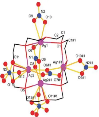
Analysis of product 1 by single crystal diffraction at 273 K reveals a 24-crown-8 ligand coordinating to four silver nitrate entities in the orthorhombic space group Ibca( Fig. 1).The asymmetric unit is composed of half of a crown ether molecule, two silver atoms, one full nitrate and two half nitrate anions. The other half is constructed by a 2-fold axis passing through O1 (0, J, 0.0092(2)) and O5 (0,J, 0.2443(2)) of the crown ether ligand.
Each silver ion is binding to three oxygen atoms of the crown ether with an average distance of 2.55(1) A˚ for Ag1 and 2.52(4) A˚
for Ag2 (all Ag–O distances until 3.05 A˚ are listed in Table S1 for the three structures described in this manuscript. Distances greater than 3.05 A˚ contribute to the bond valence sum with less than 0.04, and we choose thus this distance as a limit for the Ag–
O interactions23as derived from the software PLATON.24). Two nitrate anions (N1, O6, O7 and O8 and its symmetry equivalent) interact in a bidentate fashion with Ag1 (2.350(4) A˚ for Ag1–O6 and 2.433(5) A˚ for Ag1–O9) and Ag2 (2.364(4) A˚ for Ag2–O6 and 2.679(5) A˚ for Ag2-O8), O6 acting as bridge between the two silver ions. Ag1 is also connected to the nitrate anion around N2 with a distance of 2.432(4) A˚ for Ag1–O9 and 2.7268(6) A˚ for
Ag1–O10. Due to the rotational disorder around N3 of one of the nitrate anions (N3, O11, O12, O13) showing two different randomly distributed positions, Ag2 can be considered as six- or seven-coordinated, with contributions from three oxygen atoms of the crown ether ligand and three or four oxygen atoms from two different nitrate anions. Depending on the position of the disordered nitrate anion, the distance Ag2–O is shorter (2.19(3) A˚ ) upon monodentate binding than if the nitrate acts as bidentate ligand (2.60(3) A˚ and 2.80(2) A˚).
The average Ag–Ag distance between the metal ions of a
‘‘ring’’ is 3.91(1) A˚ . The inorganic part of the structure forms layers of silver nitrate along theb- and thec-axis, these layers are composed of silver nitrate chains along thec-axis (Fig. S1{). The crown ether ligand acts as connector between these chains and layers. Vice versa, one can consider the nitrate anions as Fig. 1 Excerpt of the coordination polymer of1. Crown ether atoms are shown as wire models and hydrogen atoms are omitted for clarity.
(#1:2x, 0.52y,z).
Fig. 2 Excerpt of the coordination polymer of 19. Crown ether molecules are shown in wire mode and hydrogen atoms are omitted for clarity. (#1: 12x, 0.5 +y,20.52z#2:21 +x,y,z#3:2x, 0.5 +y, 20.52z).
http://doc.rero.ch
connecting ligands between the crown ether-silver complexes, generating a 3D network (Fig. S2{). However, only two out of the four nitrate ligands act as bridging ligands between the crown ether complexes, while the other to only connect to silver ions of one complex. The packing of 1 shows small voids in the structure. Calculation of the contact surface of the voids gives 8 voids of 22 A˚3per unit cell for a total of 2.8% of the unit cell.
Upon cooling of a crystal of1to 200 K or less, it undergoes a phase transition into a monoclinic system, yielding19. The unit cell of 19 contains two crown ether molecules and eight silver nitrate units. The crown ether molecules maintain the same conformation as in1, and each ligand coordinates again to four silver atoms. Two silver atoms Ag1 and Ag5 are now hexacoordinated, while the six other silver ions are heptacoordi- nated. The two crown ether silver complexes of the unit cell are linked by a nitrate anion (N5, O29, O30 and O31) (Fig. 2).
For the first crown ether molecule (C1 to C16), two nitrate anions (around N1 and N4) are binding to silver ions (Ag1 to
Ag3) coordinated by this crown ether ligand, while the third nitrate anion around N2 forms two bidendate interactions with Ag4 (2.422(5) A˚ for Ag4–O20, 2.702(7) A˚ for Ag4–O21) and Ag2 (2.472(5) A˚ for Ag2–O20 and 2.527(5) A˚ for Ag2–O22), where O20 connects these two silver ions. The fourth nitrate ion around N3 interacts similarly with Ag1 (2.316(5) A˚ for Ag1–O23 and 2.800(7) A˚ for Ag1–O24) and Ag3 (2.293(5) A˚ for Ag3–O23 and 2.991(7) A˚ for Ag3–O25) with O23 as bridging atom between the two silver ions. Each of the four silver ions is also coordinated by three oxygen atoms of the crown ether moiety. The second crown ether ligand (C17 to C32), four silver atoms (Ag5 to Ag8) and four nitrate moieties. The coordination scheme of the silver ions is very similar to the one of the first entity: one nitrate anion (N7, O35, O36 and O37) is binding to Ag6 with distances of 2.342(7) A˚ for O35 and 2.862(7) A˚ for O37. A second nitrate (N6, O32, O33 and O34) forms two bidendate interactions with Ag6 (2.535(5) A˚ for Ag6–O32 and 2.517(6) A˚ for Ag6–O33) and Ag8 (2.641(6) A˚ for Ag8–O32 and 2.423(5) A˚ for Ag8–O34) with O32 bridging Ag6 and Ag8. The third nitrate (N8, O37, O38 and O39) interacts with Ag5 (2.909(7) A˚ for Ag5–O38 and 2.300(5) A˚
for Ag5–O39) and Ag7 (2.298(5) A˚ for Ag7–O39 and 2.985(8) A˚
for Ag7–O40) with O39 as bridging atom. A final nitrate anion (N5, O29 to O31) forms am2-g1:g2bridge between Ag2 and Ag5, connecting the two polyether-silver entities of the asymmetric unit.
All nitrate ions (around N1, N4 and N7), which only bind once or twice to silver ions of the asymmetric unit, also connect to silver atoms of neighbour crown ether molecules of the next asymmetric units (Fig. S3{), leading thus to a 3D-framework (Fig. S4{). The inorganic part of the structure is again composed of layers of silver nitrate along thea- and thec-axis, with chains of silver nitrate along thec-axis (Fig. S5{).
At first sight, one might link the difference between1and19 solely to the disorder of the nitrate ions described in 1. This would then represent a ‘‘partial melting’’ of the structure.
However, the main differences between1and19are not only the position of the nitrate anions, but also the way the nitrate anions link two crown ether moieties. If we assume the nitrate anion as a large sphere with the nitrogen atom as center (thus not taking into account the disorder of the O-atoms in1), the angles Ag–N–
Ag for 1 between silver ions and bridging nitrate anions are 153.8(2)uand 133.9(2)u, whereas for19these angles are 146.3(3)u, 144.5(3)u, 141.9(3)uand 129.9(3)u. This difference in the nitrate position is significant and can be easily seen by comparing the Fig. 3 Excerpt of the coordination polymer of2. Crown ether ligands
are shown in wire mode and some hydrogen atoms are omitted for clarity. (#1: 22x,y, 1.52z#2: 0.5 +x, 0.5 +y,z#3: 1.52x, 0.5 +y, 1.52z).
Table 1 Crystallographic parameters for1,19and2
1 19 2
Formula C8 H16 Ag2 N2 O10 C32 H64 Ag8 N8 O40 C16 H36 Ag4 N4 O22
Formula weight 515.97 2063.87 1067.97
Crystal system orthorhombic monoclinic monoclinic
Space group Ibca P21/c C2/c
Temp. (K) 273 200 273
a, A˚ 13.4204(6) 16.3286(8) 12.1262(13)
b, A˚ 16.5076(7) 13.4452(5) 17.6035(16)
c, A˚ 28.1263(13) 28.0645(14) 14.7902(12)
b, deg 90 90.800(4) 93.461(8)
V, A˚3 6231.1(5) 6160.7(5) 3151.4(5)
Z 16 4 4
R(int) 0.0456 0.0792 0.0848
R1(%) 3.82 4.29 3.55
http://doc.rero.ch
packing along thebaxis for1and thea-axis for19(Fig. S1 and S4{). It results in a dramatic loss of symmetry not only around the disordered nitrate ion around N3, but also around the second bridging nitrate ion around N2 of1 (compare Fig. S1 bottom with Fig. S5 bottom{).
To study the reversibility of this transformation, we performed a DSC analysis of compound1. An exothermic peak at 204 K is found for the transformation of1to19during the cooling and an endothermic peak at 214 K for the transformation of 19 to 1 during the heating cycle, this giving rise to a small hysteresis.
After fifty heating and cooling cycles, a conversion of1to19and back of 95% is still observed (Fig. S6{).
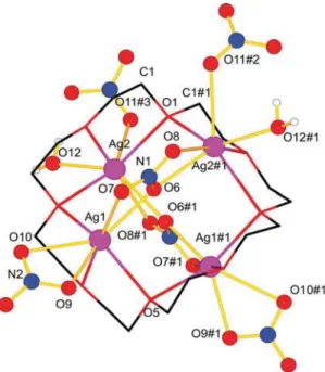
When1is exposed to humidity or when recrystallizing1from a mixture of water and methanol, we obtained a third structure [Ag4(24-crown-8)(NO3)4(H2O)2], 2, which crystallizes in the monoclinic space groupC2/c. One crown ether ligand and four silver nitrate entities are maintained per formula unit with, in addition, two water molecules (Fig. 3).
The asymmetric unit of2is composed of half of a crown ether ligand, two silver atoms Ag1 and Ag2, two nitrate ions and one water molecule. The other half of the complex is constructed by a 2-fold axis passing through O1 (1, 0.0389(3),L) and O5 (1, 0.4157(3),L). Ag1 and Ag2 are 7-coordinated each by three oxygen atoms of the crown ether with an average distance of 2.547(6) A˚ for Ag1 and 2.601(5) A˚ for Ag2. The nitrate (N1, O6, O7 and O8) forms bidendate interactions with Ag1 (2.453(4) A˚ for Ag1–O6 and 2.626(5) A˚ for Ag1–O7) and Ag2 across the
‘‘ring’’ (2.501(4) A˚ for Ag2–O6 and 2.609(4) A˚ for Ag2–O8) with O6 bridging the two silver ions. The second nitrate around N2 acts as bidentate ligand towards Ag1 (2.485(5) A˚ for Ag1–O9 and 2.699(6) A˚ for Ag1–O10) and as monodentate ligand to Ag2 of a next crown ether silver complex (2.862(5) A˚ for Ag2–O11).
This nitrate anion is thus the connector between the crown ether silver moieties. Finally, an oxygen atom of a water molecule, O12, is coordinated to Ag2 with a distance of 2.406(6) A˚ . The hydrogen atoms of the water molecules form H-bonds25with two bridging nitrate ions with a distance of 1.95(4) A˚ for H12B–O9 and 2.35(7) A˚ for H12A–O7. Four symmetry related nitrate anions (around N2) are thus binding to neighbour crown ether- silver complexes to form a two dimensional framework along the aandbaxes (Fig. S7).
In order to check if a dehydration of the complex will destroy the structure or not, we performed a DSC analysis (Fig. S8). The heating of the sample shows an endothermic peak at 370 K, which corresponds to the dehydration of 2.2then transforms into1, as confirmed by the subsequent cooling of the obtained dehydrated sample, showing an exothermic peak at 204 K similar to the one for the transformation of1to19. A following heating cycle shows again an endothermic peak at 214 K for the transformation of19to1.
The transformation from1to19leads to a small change in the unit cell, except forbwith a variation of 0.8u(Table 1). The cell axes a and b are exchanged and show the largest difference between theb-axis of 1 with 16.5076(7) A˚ and thea-axis of 19 with 16.3286(8) A˚ , which is significant.
In order to verify that all transformations are single crystal to single crystal transitions, a crystal of2was placed in an image plate x-ray diffractometer at 290 K. A few frames were recorded in order to confirm the unit cell, and then a powder
diffractogram was recorded to confirm the structure and its crystallinity. Then the crystal was heated to 390 K with a ramp of 4 K per hour, held at this temperature for 10 min and then cooled down to 290 K. Again, a few frames and a powder diffractogram were recorded. A similar procedure was used with a crystal of1, cooling it down to 150 K in order to observe its transformation to19(Fig. 4).
The transformation from2to1leads to microscopic damage inside the crystal. This is due to the evaporation of the water and is observed under the microscope in form of tiny bubbles in the Fig. 4 a) Powder diffraction of 2 before (top) and after (bottom) heating to 390 K. b) Powder diffraction of1at 300 K (top) and after cooling to 150 K (bottom). Recorded frames are shown in the corner.
http://doc.rero.ch
crystal. Nevertheless, the crystal still holds together in one piece, even if the recorded frames show a transition to a polycrystalline material. A slower heating ramp gives better results due to less strain applying on the crystal during the water evaporation process. Transformation from 1 to 19 does not show any microscopic damage of the crystal, and the recorded frames indicate diffraction patterns of a single crystal.
Conclusions
In summary, we have successfully synthesized a 24-crown-8 silver nitrate complex, which exists in two different polymorphs as a function of temperature. Transitions between these two phases occur reversibly in single crystal to single crystal transitions. A hydrated pseudo-polymorph is obtained upon exposure of1to humidity or by attempts of crystallizing 1 from a water- containing solvent. Two molecules of water are then additionally coordinated to the silver atoms. This pseudo polymorphism is also reversible, but single crystal damage occurs, while crystal- linity is maintained during the evaporation of water.
Acknowledgements
The authors thank the Swiss National Science Foundation, the University of Fribourg and the Fribourg Center for Nanomaterials FriMat for generous support.
References
1 L. X. Dang,J. Am. Chem. Soc., 1995,117, 6954–6960.
2 (a) Z. X. Shu,J. Electrochem. Soc., 1993,140, 922; (b) D. Ray, D.
Feller, M. B. More, E. D. Glendening and P. B. Armentrout,J. Phys.
Chem., 1996,100, 16116–16125; (c) I.-H. Chu, H. Zhang and D. V.
Dearden,J. Am. Chem. Soc., 1993,115, 5736–5744.
3 (a) P. R. Ashton, R. Ballardini, V. Balzani, I. Baxter, A. Credi, M. C. T.
Fyfe, M. T. Gandolfi, M. Gomez-Lopez, M.-V. Martinez-Diaz, A.
Piersanti, N. Spencer, J. F. Stoddart, M. Venturi, A. J. P. White and D. J. Williams,J. Am. Chem. Soc., 1998,120, 11932–11942; (b) J. D.
Badjic, C. M. Ronconi, J. F. Stoddart, V. Balzani, S. Silvi and A. Credi, J. Am. Chem. Soc., 2006,128, 1489–1499; (c) A. Harada,Acta Polym., 1998,49, 3–17; (d) M.-V. Martinez-Diaz, N. Spencer and J. F. Stoddart, Angew. Chem., Int. Ed. Engl., 1997,36, 1904–1907; (e) P. R. Ashton, P. T. Glink, J. F. Stoddart, P. A. Tasker, A. J. P. White and D. J.
Williams,Chem.–Eur. J., 1996,2, 729–736.
4 (a) D. A. Morton-Blake, B. Jenkins and I. Blake,Mol. Simul., 2011, 37, 1077–1084; (b) D. A. Morton-Blake and K. Korpela,Soft Matter, 2010,6, 558–567; (c) T. Nakamura, T. Akutagawa, K. Honda, A. E.
Underhill, A. T. Coomber and R. H. Friend,Nature, 1998, 394, 159–162; (d) V. Percec, G. Johansson, J. Heck, G. Ungar and S. V.
Batty,J. Chem. Soc., Perkin Trans. 1,1993, 1411–20.
5 N. Georges, S. J. Loeb, J. Tiburcio and J. A. Wisner,Org. Biomol.
Chem., 2004,2, 2751.
6 (a) M. Kawano and M. Fujita, Coord. Chem. Rev., 2007, 251, 2592–2605; (b) M. P. Suh, J. W. Ko and H. J. Choi,J. Am. Chem.
Soc., 2002,124, 10976–10977; (c) C.-L. Chen, A. M. Goforth, M. D.
Smith, C.-Y. Su and H.-C. Zur Loye,Angew. Chem., Int. Ed., 2005, 44, 6673–6677.
7 J.-P. Zhang, Y.-Y. Lin, W.-X. Zhang and X.-M. Chen,J. Am. Chem.
Soc., 2005,127, 14162–14163.
8 N. L. Toh, M. Nagarathinam and J. J. Vittal,Angew. Chem., Int. Ed., 2005,44, 2237–2241.
9 (a) T. K. Maji, G. Mostafa, R. Matsuda and S. Kitagawa,J. Am.
Chem. Soc., 2005,127, 17152–17153; (b) G. van Koten, M. Albrecht, M. Lutz and A. L. Spek,Nature, 2000,406, 970–974.
10 J. Bernstein, Polymorphism in molecular crystals, Clarendon Press, Oxford, 2002, vol.14.
11 (a) A. Y. Robin and K. M. Fromm,Coord. Chem. Rev., 2006,250, 2127–2157; (b) C. B. Aakero¨y, A. M. Beatty and D. S. Leinen,Cryst.
Growth Des., 2001,1, 47–52.
12 Guidance for Industry, ANDAs: Pharmaceutical Solid Polymorphism;
Food and Drug Adminstration, Silver Spring, 2007.
13 (a) K. R. Seddon,Cryst. Growth Des., 2004,4, 1087; (b) J. Bernstein, Cryst. Growth Des., 2005,5, 1661–1662.
14 (a) G. R. Desiraju,CrystEngComm, 2003,5, 466–467; (b) A. Nangia and G. R. Desiraju,Chem. Commun., 1999, 605–606.
15 (a) J. Bernstein,J. Phys. D: Appl. Phys., 1993,26, B66; (b) L. Huang, Adv. Drug Delivery Rev., 2004,56, 321–334.
16 (a) A. Y. Robin, K. M. Fromm, H. Goesmann and G. Bernardinelli, CrystEngComm, 2003,5, 405–410; (b) A. Crochet, E. Kottelat, A.
Fleury, M. Neuburger and K. M. Fromm,Z. Anorg. Allg. Chem., 2011,637, 672–675.
17 (a) J. L. Sague, M. Meuwly and K. M. Fromm,CrystEngComm, 2008,10, 1542–1549; (b) T. V. Slenters, J. L. Sague, P. S. Brunetto, S.
Zuber, A. Fleury, L. Mirolo, A. Y. Robin, M. Meuwly, O. Gordon, R. Landmann, A. U. Daniels and K. M. Fromm,Materials, 2010,3, 3407–3429.
18 (a) M. Dulak, R. Bergougnant, K. M. Fromm, H. R. Hagemann, A. Y. Robin and T. A. Wesolowski,Spectrochim. Acta, Part A, 2006, 64, 532–548; (b) K. M. Fromm and R. D. Bergougnant,Solid State Sci., 2007,9, 580–587; (c) M. G. Zolotukhin, M. d. C. G. Hernandez, A. M. Lopez, L. Fomina, G. Cedillo, A. Nogales, T. Ezquerra, D.
Rueda, H. M. Colquhoun, K. M. Fromm, A. Ruiz-Trevino and M.
Ree,Macromolecules, 2006,39, 4696–4703; (d) K. M. Fromm, R. D.
Bergougnant and A. Y. Robin, Z. Anorg. Allg. Chem., 2006,632, 828–836; (e) K. M. Fromm, E. D. Gueneau, H. Goesmann and C. G.
Bochet,Z. Anorg. Allg. Chem., 2003,629, 597–600; (f) K. M. Fromm, E. D. Gueneau, J.-P. Rivera, G. Bernardinelli and H. Goesmann,Z.
Anorg. Allg. Chem., 2002,628, 171–178.
19 J. Cosier and A. M. Glazer,J. Appl. Crystallogr., 1986,19, 105–107.
20 E. Blanc, D. Schwarzenbach and H. D. Flack,J. Appl. Crystallogr., 1991,24, 1035–1041.
21 (a) M. C. Burla, R. Caliandro, M. Camalli, B. Carrozzini, G. L.
Cascarano, L. de Caro, C. Giacovazzo, G. Polidori and R. Spagna,J.
Appl. Crystallogr., 2005,38, 381–388; (b) G. M. Sheldrick, SHELX- 97. Program for Crystal Structure Refinement, University of Go¨ttingen, Germany, 1997.
22 V. S. Talanov and R. A. Bartsch, Synth. Commun., 1999, 29, 3555–3560.
23 (a) N. E. Brese and M. O’Keeffe,Acta Crystallogr., Sect. B: Struct.
Sci., 1991, 47, 192–197; (b) I. D. Brown, The chemical bond in inorganic chemistry. The bond valence model, Oxford University Press, Oxford, 2009, vol.12.
24 A. L. Spek,Acta Crystallogr., Sect. D: Biol. Crystallogr., 2009,65, 148–155.
25 (a) T. Steiner,Angew. Chem., Int. Ed., 2002,41, 48–76; (b) M. C.
Etter,Acc. Chem. Res., 1990,23, 120–126.