Département de Physique
Université de Fribourg (Suisse)
L-subshell Coster-Kronig rates of Pd and Xe, electron-induced
L
3M double ionization of Pd, and KMM RAE of Ca
investigated by means of high-resolution x-ray spectroscopy
THÈSE
Présentée à la Faculté des Sciences de l’Université de Fribourg (Suisse)
pour l’obtention du grade de
Doctor rerum naturalium
Wei CAO (曹伟)
de la
Chine
Numéro de la thèse: 1696
Imprimerie Saint-Paul, Fribourg
Acceptée par la Faculté des Sciences de l’Université de Fribourg (Suisse) sur la proposition de:
Prof. Dr. Christian Bernhard, Université de Fribourg, Président de Jury, Prof. Dr. Jean-Claude Dousse, Université de Fribourg, Directeur de thèse, Prof. Dr. John L. (Iain) Campbell, University of Guelph (Canada), Rapporteur, Dr. Joanna Hoszowska, Université de Fribourg, Rapporteure.
Fribourg, 25 Novembre 2010
Le Directeur de thèse: Le Doyen:
Contents
Résumé 1
Abstract 4
Introduction 8
Part 1.
L-subshell Coster-Kronig yields of palladium determined via
synchrotron-radiation-based high-resolution x-ray spectroscopy 29
Part 2.
Synchrotron-radiation-based determination of Xe
L-subshell Coster-Kronig yields: A re-examination via
high-resolution x-ray spectroscopy 48
Part 3.
Double L
3M ionization of Pd induced by impact with
medium-energy electrons 61
Part 4.
High-resolution KMM radiative Auger emission spectra of calcium
induced by synchrotron radiation 86
Acknowledgements 104
Curriculum Vitae 107
Resumé
La présente thèse de doctorat comprend quatre projets de recherche dans le domaine de la physique des couches atomiques profondes. Les deux premiers projets sont consacrés à la détermination expérimentale des coefficients de Coster-Kronig des sous-couches L pour le palladium et le xénon. Dans le troisième projet, nous avons étudié la double ionisation L3M d’une cible de palladium bombardée par des électrons. Quant au quatrième projet,
il concerne les transitions radiatives Auger KM M du calcium. Les quatre projets ont été réalisées en utilisant la méthode de spectroscopie X de haute résolution.
Partie 1. Détermination des coefficients de Coster-Kronig des sous-couches L du palladium en utilisant la spectroscopie X de haute résolution et le rayonnement
synchrotron
Les coefficients de Coster-Kronig (CK) des sous-couches L du palladium ont été déter-minés en mesurant en haute résolution les raies X Lα1,2 (L3 − M4,5) et Lβ1 (L2 − M4).
Les spectres de rayons X L ont été collectés au moyen de spectromètres à cristaux cour-bés. Pour produire la fluorescence de la cible de palladium, un faisceau de rayonnement synchrotron d’énergie variable a été utilisé. Les coefficients CK ont été déterminés à partir des sauts d’intensité des raies X L apparaissant aux bords d’absorption. Pour déterminer la grandeur des sauts, la variation de l’intensité des raies L en fonction de l’énergie du fais-ceau de rayonnement synchrotron a été ajustée sur celle des sections efficaces d’absorption. Les intensités des raies X L ont été corrigées au préalable pour tenir compte des effets de corps solide. Ces derniers ont été déterminés en comparant les spectres d’absorption expéri-mentaux et théoriques. Grâce à la haute-résolution des spectromètres à cristaux utilisés, les coefficients CK partiels fL1L3M
13 ont pu être également déterminés à partir des intensités
relatives des transitions satellites résolues LαM . Pour les coefficients f23, f12 et f13, des
valeurs de 0.164 ± 0.033, 0.047 ± 0.001 et 0.730 ± 0.039 ont été trouvées. Pour les coefficients partiels fL1L3M
13 et f13L1L3N, des résultats de 0.406 ± 0.023 et 0.324 ± 0.032 ont été obtenus.
Partie 2. Réexamen des coefficients de Coster-Kronig des sous-couches L du xénon par spectroscopie X de haute résolution
Les valeurs des coefficients CK des sous-couches L du xénon ont été réexaminées par des mesures de haute résolution des transitions radiatives Lα1,2 (L3− M4,5) et Lβ1 (L2 − M4).
Les mesures ont été réalisées en utilisant un spectromètre à cristal incurvé de type Johansson et des faisceaux monochromatiques de rayonnement synchrotron d’énergie variable. Comme dans le projet précédent, les coefficients CK ont été déduits des sauts d’intensité des rayons X L apparaissant aux bords d’absorption. La dépendance en énergie de l’intensité des raies de fluorescence a été calquée sur celle des sections efficaces de photoabsorption L. Ces dernières ont été obtenues à partir du spectre d’absorption L du xenon, lequel a été préalablement mesuré. Des valeurs de 0.118 ± 0.029, 0.383 ± 0.037 et 0.096 ± 0.016 ont été trouvées pour les coefficients CK f23, f13 et f12. Le facteur de fluorescence de la sous-couche L1 a également
été déterminé à partir du rapport d’intensité des transitions Lβ4(L1−M2) et Lβ1 (L2−M4).
Ces transitions, très proches en énergie, ont pu être résolues grâce à la haute résolution du spectromètre à cristal. Une valeur de 0.059 ± 0.002 a été obtenue.
Ce travail a été publié dans Physical Review A 81, 012501 (2010).
Partie 3. Double ionisation L3M du Pd produite par bombardement d’électrons
d’énergie moyenne
Les sections efficaces de double ionisation L3M induite dans le Pd par bombardement
d’électrons ont été déterminées pour des énergies d’électrons comprises entre le seuil de dou-ble ionisation et 18 keV. Les sections efficaces ont été déterminées à partir des intensités rela-tives des raies satellites LαM par rapport aux raies diagrammes parentes Lα. L’échantillon a été bombardé par des électrons monoénergétiques produits par un canon à électrons de 20 kV. Les raies diagrammes et les satellites M ont été mesurés en haute résolution avec un spectromètre à cristal courbé von Hamos. Les sections efficaces partielles TS ont été déterminées en soustrayant les contributions de ’shake’ et des transitions CK L1 − L3M4,5
des sections efficaces totales. Malgré l’utilisation d’une cible épaisse, la variation des sections efficaces TS en fonction de l’énergie des électrons a pu être déterminée grâce à une nouvelle méthode de décomposition virtuelle de la cible en tranches d’épaisseur variable. Nous avons
pu démontrer que la dépendance en énergie des sections efficaces partielles TS était bien reproduite par le modèle semi-empirique de Pattard-Rost.
Ce travail a été soumis récemment à Physical Review A.
Partie 4. Spectres de haute résolution des transitions radiatives Auger KM M du calcium induites par rayonnement synchrotron
Les transitions radiatives Auger (RA) KM M du calcium solide induites par irradiation avec du rayonnement synchrotron ont été mesurées par la méthode de la spectroscopie X de haute résolution avec un spectromètre à cristal courbé von Hamos. Deux énergies différentes d’irradiation ont été utilisées, l’une juste au-dessus du seuil d’ionisation K et l’autre clairement au-delà du bord d’absorption K. L’analyse des données a montré que la structure spectrale et l’intensité relative des transitions RA KMM par rapport à la raie diagramme Kβ1,3 (K − M3,2) ne dépendaient pas de l’énergie du rayonnement synchrotron.
La structure RA observée ressemble à la densité des états externes s, p et d inoccupés. Toutefois, à cause d’effets de corps solide, les transitions RA associées aux processus de ’shakeup’ n’ont pas pu être mises en évidence. Une valeur de 0.053(3) a été trouvée pour l’intensité relative totale des transitions RA KM M par rapport à l’intensité de la raie diagramme Kβ1,3. Le résultat obtenu est comparé aux prévisions théoriques existantes ainsi
qu’à des valeurs expérimentales déterminées en utilisant divers types de projectiles pour l’excitation de la cible.
Abstract
The present Ph.D. thesis includes four research projects related to the field of inner-shell atomic physics. The first two are devoted to the experimental determination the L-subshell Coster-Kronig yields of xenon and palladium. In the third one, the electron induced two-step L3M double ionization cross sections for Pd were investigated. The fourth project concerns
the photon induced KM M radiative Auger x-ray transitions of calcium. For all projects the high-resolution emission x-ray spectroscopy technique was employed.
Part 1. L-subshell Coster-Kronig yields of palladium determined via synchrotron-radiation-based high-resolution x-ray spectroscopy
The experimental determination of the palladium L-subshell Coster-Kronig (CK) transi-tion yields via high-resolutransi-tion measurements of the Lα1,2(L3−M4,5) and Lβ1(L2−M4) x-ray
emission lines is presented. The L x-ray spectra were recorded by means of curved crystal spectrometers employing energy-tunable synchrotron radiation for fluorescence production. The CK yields were derived from the relative L x-ray intensity jumps at the L-edges by fitting the fluorescence intensities as a function of the photon energy to the photoionization cross sections. The L x-ray intensities were corrected for solid-state effects which were es-timated from the comparison of the measured and theoretical Pd L-edge x-ray absorption spectrum. Thanks to high-resolution, the partial CK yield fL1L3M
13 could be extracted from
the intensities of the resolved LαM satellite transitions. For f23, f12and f13CK rates, values
of 0.164 ± 0.033, 0.047 ± 0.001 and 0.730 ± 0.039 were found, respectively. For the partial CK yields fL1L3M
13 and f13L1L3N, results of 0.406 ± 0.023 and 0.324 ± 0.032, respectively, were
obtained.
This work was published in Physical Review A 80, 012512 (2009).
Part 2. Synchrotron-radiation-based determination of Xe L-subshell Coster-Kronig yields: A re-examination via high-resolution x-ray spectroscopy
The xenon L-subshell Coster-Kronig (CK) transition yields were revisited via high-resolution measurements of the Lα1,2 (L3− M4,5) and Lβ1 (L2− M4) x-ray emission lines.
and energy-tunable synchrotron radiation. The CK yields were derived from the relative L x-ray intensity jumps at the L edges by fitting the fluorescence intensities as a function of the photon energy to the L-subshell photoionization cross sections. The latter were obtained from the measured L-edge photoabsorption spectrum. Values of 0.118 ± 0.029, 0.383 ± 0.037 and 0.096 ± 0.016 were found for the f23, f13 and f12 CK yields, respectively. Thanks to
high resolution, the L1 fluorescence yield of 0.059 ± 0.002 was also determined from intensity
ratios of the well resolved Lβ4 (L1− M2) and Lβ1 (L2− M4) lines.
This work was published in Physical Review A 81, 012501 (2010).
Part 3. Double L3M ionization of Pd induced by impact
with medium-energy electrons
The electron-induced L3M two-step double ionization cross sections of metallic Pd were
determined experimentally for incident electron beam energies ranging from the double ionization threshold up to 18 keV. The double L3M ionization cross sections were derived
from the intensity ratios (ILαM : ILα) of the resolved M-satellites to the parent diagram
lines. The sample was bombarded with monoenergetic electrons from an energy tunable 20 kV electron gun. The diagram and M satellite x-ray lines were measured by means of high-resolution x-ray spectroscopy, using a reflection-type von Hamos bent crystal spectrometer. The two-step partial cross sections were determined by subtracting from the measured total double ionization cross sections the contributions due to the shake process and L1− L3M4,5
Coster-Kronig transitions. Despite the thick target employed in the present study, the dependence of the two-step cross sections on the incoming electron energy could be derived using a novel target slice decomposition method. It is shown that the obtained energy dependence can be well reproduced by the semiempirical parametrization model of Pattard-Rost.
Part 4. High-resolution KM M radiative Auger spectra of calcium induced by synchrotron radiation
The KM M radiative Auger (RA) x-ray spectra of solid Ca were induced by monochro-matic synchrotron radiation and measured with a high-resolution von Hamos spectrometer. Two excitation energies were employed, one in the near K threshold region and the second well above the K absorption edge. The RA spectral structure as well as the KMM RA intensity with respect to the diagram Kβ1,3 (K − M3,2) line are found to be independent of
the excitation energy. The overall RA structure resembles the density of unoccupied outer s-, p- and d- states. Due to solid state effects, however, spectral features resulting from the major discrete shake-up transitions could not be resolved. The obtained total KMM RA yield relative to the Kβ1,3 yield of 0.053(3) is compared to theoretical predictions and
available experimental data from various types of target excitation. This work will be submitted to Physical Review A.
When W. C. Röntgen discovered x-rays in 1895 [1], he must have expected his ex-citing observation to cause a stir among the scientific community. However, he probably did not realize how important this new type of radiation would be for basic and applied science. In the years following their discov-ery, x-rays have indeed played a major role in physics, chemistry, biology, medicine, cristal-lography and other natural sciences [2]. Fur-thermore, with the advent in the last two decades of third generation synchrotron radi-ation (SR) facilities, intense, monochromatic and energy-tunable x-ray beams have become available, which have brought a new boost to the domain. In atomic physics, x-rays are of prime importance to probe the inner- and outer-shell structure of atoms because their wavelengths are of the same order of magni-tude as the atomic dimensions.
I. X-RAY ABSORPTION AND
EMISSION
When interacting with matter, x-rays can either be absorbed or scattered by the atoms. The photoabsorption leads to an attenuation of the x-ray beam intensity because, as a re-sult of the photoelectric effect, the photons disappear, their whole energy being trans-ferred to bound atomic electrons, named pho-toelectrons, that are ejected into the
contin-uum. The kinetic energy of the photoelectron is equal to the difference between the energy of the incident photon and the binding en-ergy of the atomic subshell from which the photoelectron originates. Thus, in photoion-ization the photon vanishes, a photoelectron is emitted and a vacancy state is created in the corresponding atom. If the photon en-ergy is close to but smaller than the binding energy of the electron, the latter cannot es-cape from the atom, but it can be promoted into an outer incompletely filled level. This process is called photoexcitation.
Scattering processes may occur with or without energy losses of the scattered pho-tons. In the first case, called inelastic scat-tering, a part of the photon energy is used to promote a bound electron into an outer in-completely occupied bound state or to eject it into the continuum. After an inelastic scat-tering process, the atom is left in an excited or ionized state and the energy and direc-tion of the scattered photon have changed. In an elastic scattering only the photon di-rection is modified, the energy of the particle remaining unchanged. Thus, in both the elas-tic and inelaselas-tic scattering processes, there is no photon disappearance. Nevertheless, for collimated x-ray beams, scattering processes result also into a diminution of the incoming ray beam intensity because the deflected x-rays are removed from the collimated beam.
The elastic scattering cross sections decrease with growing energy. At low energies they are bigger than the inelastic scattering cross sections, but as the latter increase with the photon energy, both cross sections intersect at given energies that are specific to the ele-ments. For instance, for Al (Z=13) the elas-tic and inelaselas-tic cross sections curves cross at about 25 keV, whereas for Pd (Z=46) the crossing point lies at 85 keV. Above the K-edge, i.e., the binding energy of the K-shell electrons, the photoionization cross sections diminish with the photon energy. For ener-gies below about 100 keV, they are 1-2 orders of magnitude bigger than the scattering cross sections.
The method of measuring the intensity at-tenuation of a collimated x-ray beam by a given absorbing medium as a function of the energy of the incoming rays is called x-ray absorption spectroscopy (XAS). Depend-ing on the energy region of interest, close to the absorption edge or far above it, XAS is usually subdivided into XANES (x-ray ab-sorption near edge structure) and EXAFS (extended x-ray absorption fine structure). The absorption spectra can be also computed within the independent particle approxima-tion model (IPA) using relativistic Hartree-Slater (RHS) calculations. However, these calculations cannot reproduce the oscillations observed in the experimental XANES and
EXAFS spectra [3]. A good
understand-ing of these spectral fine structures which are beyond the IPA predictions is, however, mandatory for a correct interpretation of the experimental x-ray absorption spectra and a reliable and precise determination of the photoionization cross sections. Due to the fact that XANES permits to probe both the atomic core levels and the outer unoccupied electron states, unique signatures of a given material can be obtained from this tech-nique. XANES spectra also depend on the detailed atomic structure of the sample as well as on the electronic and vibrational prop-erties of the latter [4]. EXAFS is commonly understood in terms of high-order multiple-scattering processes. Based on the Fermi’s ’golden rule’ and the electric dipole transition approximation, an unified theory for XANES and EXAFS was elaborated which is based on the ’standard quasi-particle theory’ [5, 6].
Within the independent electron picture, a single-photon absorption accompanied by the simultaneous ejection of several bound electrons cannot be explained by photoion-ization. In the photoeffect, a single photon interacts indeed with a single bound electron. Other indirect effects such as the shakeoff (SO) and knock-out (KO) processes based on electron-electron interactions are thus needed to explain the multiple ionization resulting from single photon impact. In the shake
pro-cess [7], the second electron is ionized due to the sudden change of the effective nuclear charge resulting from the creation via the photoeffect of the first inner-shell vacancy. In the case of KO, the outgoing photoelectron knocks out the second electron, leaving the atom in a doubly-ionized state.
In contrast to photoionization, direct mul-tiple ionization can be produced by impact with charged particles because in this case, several bound electrons of the target atom can feel simultaneously the Coulomb po-tential of the charged projectile. The di-rect multiple ionization induced by impact with charged particles is particularly impor-tant in the case of heavy ions because the corresponding cross section grows with the squared charge of the incoming projectile. It is worth noting here that the shake process does not depend on the first ionization mech-anism. Thus, SO does also exist in particle-induced ionization and its contribution to the multiple ionization is the same as in pho-toionization. This is, however, not true for the KO process because the momenta of the first ionized electrons are in general smaller in collisions involving charged particles than in the case of photoionization. For instance the contribution of KO to the double K-shell ion-ization of Al was found to be about 3 times smaller in the case of electron impact than in photoionization, although similar electron
and photon beam energies were used [8]. XAS measurements can be performed in the transmission mode or in the fluorescence mode. In the first case, the intensities of the incoming and transmitted x-ray beams are measured simultaneously by two similar de-tectors, e.g. ionization chambers, as a func-tion of the photon energy. The absorpfunc-tion spectrum can be then obtained in a straight-forward way by dividing the measured trans-mitted intensity by the incoming one. In gen-eral, however, for practical reasons, the in-verse of the absorption spectrum is preferably determined, i.e., the intensity of the incoming beam is divided by the one of the transmit-ted beam. Around the K-edge or L-edges, an abrupt increase of the absorption is ob-served, which is due to the opening of the K-shell or L-subK-shells photoionization channels. Precise energies for the absorption edges can be obtained by calculating the first deriva-tive of the inverse absorption spectrum, the edge energy corresponding indeed to the en-ergy for which the first derivative is maxi-mum. In the inverse absorption spectrum a strong peak is usually observed just above the K-edge (white line). The latter is due to the presence of unoccupied discrete states close to the Fermi level.
In the fluorescence mode, the fluorescence x-ray emission spectrum of the absorbing sample is measured, using in most cases a
semiconductor detector. Information similar to that provided by the transmission mode can be obtained but there is no restriction on the thickness of the sample. In addition, some features related to inelastic scattering processes are observed in the fluorescence spectra but not in the corresponding absorp-tion spectra. As an alternative to the flu-orescence mode, the photoelectron spectrum of the sample can also be measured, provided that very thin samples are employed.
Due to the strong absorption of the incom-ing beam above the K-edge, the transmitted intensity is very weak. Intense x-ray sources and thin targets are thus needed to get reli-able results in XAS measurements performed in the transmission mode. In this respect, the performance of XAS has been drastically improved by using synchrotron radiation as the x-ray source. The improvements concern mainly the precision and reliability of the re-sults and the rapidity of the measurements. For these reasons, XAS has become nowadays a standard technique for the chemical char-acterization of materials.
The lifetimes of inner-shell vacancies are very small (e.g., 10−15-10−17 s, depending
on the element, for the K-shell). During these extremely short times, the core-holes are filled by outer shell electrons, bringing the ion to a lower more stable energy state. The energy ∆E released by the electron
tran-sition may lead to the emission of a pho-ton having the energy hν = ∆E. As in this process named radiative transition, a photon having an angular momentum of 1~ is cre-ated, the orbital quantum numbers of the two atomic levels involved in the transition should differ by |∆l| = 1. Radiative transitions satisfying this condition are called electric-dipole (E1) transitions. Electric-quadrupole (E2) and magnetic-dipole (M1) transitions which violate the |∆l| = 1 selection rule are strongly hindered and the intensities of the corresponding x-rays are therefore very weak. It is interesting to note that the radiative de-cay corresponds to the time-reversal of the photoexcitation process.
The transition energy ∆E may also be transferred to another bound electron which can then escape from the ion provided that the transition energy is bigger than the bind-ing energy of that electron. This alterna-tive atomic decay mode is called radiation-less or Auger transition. Auger transitions are autoionizing transitions since the num-ber of vacancies in the final atomic state is increased by one with respect to the initial state. Thus the creation of an atomic core-level vacancy results into cascades of radia-tive (fluorescence) and non-radiaradia-tive (Auger) transitions. As a consequence, one can in-vestigate the atomic relaxation by measur-ing either the emitted x-rays or the Auger
electrons. The method based on the observa-tion of the x-ray fluorescence is called x-ray fluorescence spectroscopy (XFS) and the one based on the measurement of Auger electrons Auger electron spectroscopy (AES). There is a further atomic decay channel, named radiative-Auger effect (RAE), which is much less probable than the two others but of in-terest in this thesis (see Part 4). In this exotic decay mode, the transition energy is shared between a photon and an Auger elec-tron which are emitted simultaneously [9, 10]. Diagram x-ray lines correspond to radia-tive transitions in which there is a single va-cancy in the initial and final atomic states. In inner-shell photoionization, however, dia-gram lines may be accompanied by satellite x-ray lines. The latter result from additional inner-shell vacancies that are present in the atom during the transition but are not di-rectly involved in it. For this reason these ad-ditional holes are called spectator vacancies. These holes, although inactive in the tran-sition, reduce the electronic screening of the nuclear charge. As the effect of the screening change decreases with the principal quantum number n, satellite x-ray lines are shifted to-wards higher energy with respect to their par-ent diagram lines. As a rule of thumb, one can say that the energy shift of a satellite decreases with the principal quantum num-ber of the shell in which the spectator
va-cancy is located and increases with the princi-pal quantum number of the shell from which the transition electron originates. For these reasons, energy shifts of M-satellites (specta-tor vacancy in the M-shell) are smaller than those of L-satellites and energy shifts of satel-lites of Kβ transitions are bigger than those of Kα transitions. However, these energy shifts remain small (a few eV only for light elements), so that high-resolution x-ray de-tectors are needed to resolve them from their parent diagram lines.
Depending on the specific aims of the measurements, XFS can be performed using energy-dispersive detectors or wavelength-dispersive spectrometers. Energy-wavelength-dispersive detectors are used preferably when high collection efficiency is needed and a moder-ate energy resolution is acceptable. With
wavelength-dispersive instruments whose
working principle is based on the Bragg law [11], more precise energies can be obtained and the resolving power is 10-100 times bet-ter but the price to pay is a poor efficiency as compared to energy-dispersive detectors. The poor efficiency of wavelength-dispersive spectrometers is due mainly to their small
solid angle. The latter can be improved
significantly by using cylindrically or spheri-cally curved crystals but, even in this case, the solid angle remains small (10−3-10−4
Despite this drawback, the use of crystal spectrometers is nevertheless mandatory whenever an energy resolution of a few eV is required.
II. THE PROJECTS
The present thesis belongs to the domain of atomic inner-shell physics. It is an ex-perimental work which contains four differ-ent projects. The first two projects con-cern the L-subshell Coster-Kronig yields of
Pd and Xe, the third one the L3M
dou-ble ionization of Pd induced by impact with medium-energy electrons, and the fourth one the KMM radiative Auger effect in Ca. The first two and the fourth projects were carried out using synchrotron radiation for the pro-duction of the sample fluorescence, whereas the measurements of the third project were performed at the University of Fribourg us-ing an electron gun. All four projects were carried out by means of high-resolution x-ray spectroscopy, employing either a Johansson (first two projects) or von Hamos (projects 3 and 4) curved crystal spectrometer.
A. L-subshell Coster-Kronig yields of Pd and Xe
LiLjX Coster-Kronig (CK) transitions
[12] are fast radiationless transitions, in
which a vacancy "bubbles up" from the sub-shell i to the higher subsub-shell j and an Auger electron is ejected from the outer shell X. The transition is energetically allowed only if the difference between the binding energies of the i and j subshells is bigger or equal to the binding energy of the electron in the subshell X. The vacancy transfer probabil-ity from the subshell i to the subshell j is called the CK yield fij. CK yields depend on
the initial- and final-state wave functions and are very sensitive to electron binding energies and solid-state effects [13, 14].
In general, L-subshell CK rates vary smoothly with the atomic number Z [15]. Ex-ceptions are observed when the binding en-ergy of the X electron becomes suddenly big-ger than the difference between the binding energies of the i- and j-subshells. Particu-larly interesting is the region around Z = 48 where a cutoff of the L1− L3 CK rate f13
ap-pears due to the closure of the L1 − L3M4,5
CK transition which becomes abruptly en-ergetically forbidden. The large discrepan-cies observed between theoretical CK yields [14, 16] and existing experimental values [17, 18] point out the need for new sets of re-liable and accurate experimental data. How-ever, the experimental determination of L-shell CK yields presents sizeable difficulties. For this reason, data are scarce and suffer of-ten from large uncertainties [19].
To date, to determine the f23 CK yields,
mostly the Kα-Lα x-ray coincidence tech-nique [19, 20] was used. The method is based on the fact that a L3 hole is formed after the
emission of a Kα1 x-ray (K − L3 transition).
This hole can then decay via a L3−M4,5
tran-sition with the emission of a Lα1,2x-ray.
Sim-ilarly, the decay of a L2 hole resulting from
the emission of a Kα2 x-ray (K − L2
tran-sition) may lead to the emission of a Lα1,2
x-ray provided that the L2 hole is transferred
beforehand to the L3-subshell by a L2L3X
CK transition. In this method, the CK rate f23 is thus determined by multiplying the
(Kα2 − Lα1,2) to (Kα1− Lα1,2) coincidence
rate ratio by the known Kα1to Kα2intensity
ratio. However, this ingenious method fails in the case of the L1-subshell CK yields
be-cause several transitions (e.g., the K −L1 and
L1−M4,5) that should be measured in
coinci-dence are forbidden by the above-mentioned |∆l| = 1 selection rule. For this reason, most available experimental data concerning the f12 and f13 CK yields were obtained from an
alternative photoionization method employ-ing γ−rays from intense radionuclide sources to produce the primary ionization [21].
By combining the advantages of energy-tunable monochromatic SR beams with the XAS and XFS techniques, a subshell-selective photoionization can be achieved which in turn can be used to determine all
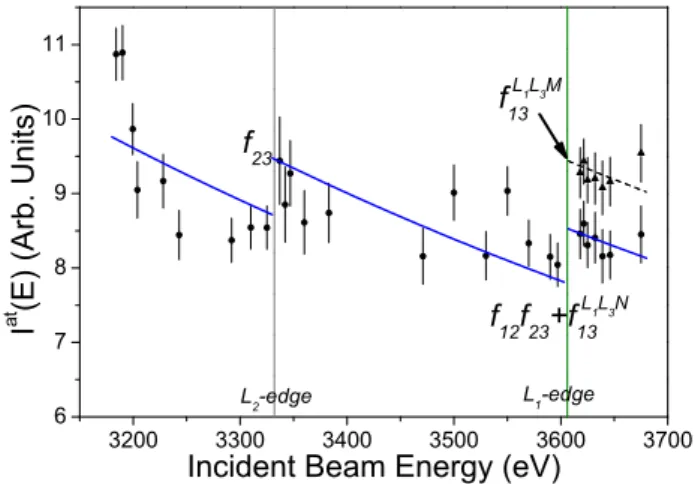
L-subshell CK yields [22]. By measuring a chosen fluorescence x-ray line at two beam energies tuned just below and just above a given absorption edge, a jump in the inten-sity of the fluorescence line is observed from which the CK yield can be deduced. For in-stance, measuring the Lα x-ray line at a SR beam energy tuned above the L3 edge but
below the L2 one, an intensity proportional
to the cross section of the L3-subshell
pho-toionization is found. Now, when the beam energy is tuned above the L2-edge but below
the L1 one, a step-like increase in the
inten-sity of the Lα x-ray line is observed which is due to the fact that part of the photoinduced L2 holes were transferred to the L3-subshell
by L2L3X CK transitions. The excess of
in-tensity found for the fluorescence line with re-spect to the previous measurement is simply proportional to the L2-subshell
photoioniza-tion cross secphotoioniza-tion multiplied by the CK yield f23. Similarly, by tuning the beam energy
above the L1-edge, a new intensity jump is
observed from which the f13CK yield can be
determined, provided the f12 rate is known.
The latter, however, can be determined by means of the same method, choosing a L2
x-ray line such as the Lβ1 (L2− M4 transition)
and tuning the SR beam energy below and above the L1 edge.
The above method requires a precise knowledge of the Li- and Lj- subshell
pho-toionization cross sections. Theoretical val-ues do exist but results from different IPA calculations are found to scatter up to 2% [23]. In addition, due to the hybridization of outer orbitals as a result of solid state ef-fects, for solid samples in the Z region of interest large discrepancies are observed be-tween the IPA predictions and the measured XANES spectra [24]. Furthermore, as for gaseous samples, for which the free atom pic-ture should apply, some oscillations are also observed in the XANES spectra, the discrep-ancies between experiment and theory cannot be explained entirely by solid state and crys-tal effects. In fact the observed deviations are not completely understood and different ex-planations are still under debate [25]. Hence precise measurements of the L-subshell pho-toionization cross sections were needed in our project.
In principle the fluorescence lines chosen as references for the determination of the CK yields can be measured by means of energy-dispersive detectors. However, as in L x-ray spectra of heavy elements many lines are overlapping, it is preferable to employ high-resolution crystal spectrometers to get reliable results. In addition, high-resolution spectroscopy offers the possibility to resolve the M−satellites from their parent diagram lines, which allows, as shown below, to dis-tinguish the L1L3−M4,5 CK transitions from
the L1L3− N ones.
In these two projects the Coster-Kronig transition yields were determined for metal-lic Pd (Z = 46) and gaseous Xe (Z = 54) by means of the above-mentioned synchrotron-radiation-based high-resolution photoioniza-tion technique. The measurements were per-formed at the XAFS beamline of the Italian Synchrotron Radiation Source Elettra, in Tri-este, Italy, using the Johansson bent crystal spectrometer of Ljubljana [26]. Complemen-tary measurements were performed later at the European Synchrotron Radiation Facil-ity (ESRF), in Grenoble, France, with the von Hamos curved crystal spectrometer of Fribourg [27].
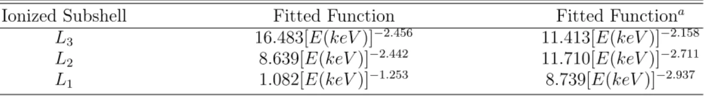
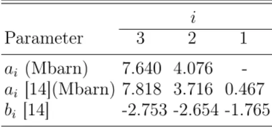
The x-ray absorption spectra of both sam-ples were measured over energy intervals cov-ering the L-subshell absorption edges and compared to the theoretical spectra obtained from IPA calculations. As discussed before, some significant deviations between the ex-perimental and theoretical XAS spectra were observed. In particular, the XANES oscil-lations observed in the experimental spec-tra were not reproduced by the calculations. For this reason, the measured L-subshell pho-toionization cross sections were employed for the determination of the CK yields. The vari-ation of the photoionizvari-ation cross section of a given subshell beyond the edge(s) corre-sponding to the next subshell(s) was
deter-mined by means of power-law extrapolations. For the determination of the parameters en-tering the power-law functions, the theoreti-cal IPA results were employed. The f23 and
f13 CK yields were derived from the Lα1,2
(L3 − M4,5) relative intensity jumps at the
L edges and the f12 CK yield from the Lβ1
(L2− M4) intensity jump at the L1 edge.
Thanks to high-resolution, the LαM satel-lite of Pd could be separated from its parent diagram line. This allowed us to determine the partial L1 − L3M4,5 CK yield from the
satellite-to-diagram line yield ratio. For Xe, the fluorescence yield ω1 of the L1-subshell
could also be determined from the intensity ratio of the resolved Lβ4 (L1− M2) and Lβ1
lines, which would not have been possible in low-resolution XRF measurements, the two lines being separated by 30 eV only.
B. Double L3M ionization of Pd induced
by impact with medium-energy electrons
For Pd, the intensity of the resolved LαM satellite is due mainly to the L1L3− M4,5 CK
transition. In fact, this assumption was made in the CK project to determine the partial CK yield fL1L3M4,5
13 . However, smaller
con-tributions to the satellite intensity may also arise from the SO and KO processes. With the aim of probing the contributions to the M-satellite yield of other double ionization
processes, a new project was undertaken in which the metallic Pd sample was bombarded with monoenergetic electrons of different en-ergies. The Lα1,2 x-ray spectra were
mea-sured at the University of Fribourg, using a 20 kV electron gun and the von Hamos crys-tal spectrometer [27]. The relative intensi-ties of the Lα1,2M satellite x-ray lines were
found to be different from those observed in the synchrotron radiation experiment. This is, however, not really surprising because, as mentioned before, the KO probabilities are not the same in electron-induced and pho-toinduced ionization. In addition, in the case of electrons, an additional process con-tributes to the L3M double ionization (DI),
namely the so-called two-step-two (TS2) pro-cess [28]. The TS2 mechanism corresponds to the knock-out of two bound electrons from the same atom by a single incoming electron. Note that in the standard KO process, named also two-step-one (TS1) process, the second electron is knocked out by the first ionized electron. In photoionization, the TS1 pro-cess does also exist, the first ionized electron being the photoelectron, but the TS2 process does not exist, the incoming photon being an-nihilated by the photoelectric effect.
The electron-induced TS process can be considered as a (e, 3e) collision, with a dou-bly ionized atom and three outgoing electrons in the exit channel. It corresponds to a three
body break-up process in the framework of the four-body Coulomb scattering, and per-mits to investigate the electron correlation ef-fects [29]. A powerful experimental method to study (e, 3e) processes consists of perform-ing inclusive coincidence measurements in which the momentum vectors of all outgoing particles are determined [30–33]. Such multi-coincidence measurements, however, result in very low coincidence rates, which makes the experiment difficult and time consuming. In addition for solid samples, extremely thin foils are needed.
An alternative experimental approach consists of observing by means of high-resolution x-ray spectroscopy the radiative
decay of the doubly ionized atoms. This
method was used for instance in the works reported in [34] and [8]. In these studies the double KL and double KK ionization in-duced in several light atoms by low-energy electrons were determined as a function of the incident electron energy. The energy dependent double ionization cross sections (DICSs) were derived from the measured intensity ratios of the resolved L satellite-or K hypersatellite-to-parent diagram x-ray lines, using available experimental values for the electron-induced single K-shell ioniza-tion cross secioniza-tions. A similar experimen-tal approach was employed in the present project concerning the electron-induced
dou-ble L3M ionization of Pd. The measured
relative intensities of the LαM satellite re-flecting the total DICSs, the partial TS cross sections were determined by subtracting from the measured satellite intensities the Coster-Kronig and shake contributions.
For Pd the average energy shift of the LαM satellite components with respect to the parent diagram line is ∼ 10 eV [35]. This energy separation is too small to be re-solved by an energy-dispersive detector, but using the von Hamos crystal spectrometer the two x-ray lines can easily be separated. The project was nevertheless challenging for the following three reasons: first, experimen-tal data for electron-induced L-subshell sin-gle ionization cross sections are scarce and existing data suffer from large uncertainties [36]. Analytical expressions [37] based on the distorted-wave Born approximation (DWBA) [38] provide values that are in satisfactory agreement with available experimental data but the predictions are poorly reliable in the near edge regions [39]. Secondly, the dou-ble LM ionization is more complicated than the double KL or KK ionization due to the L-subshell structures and the contribution to the measured DI of the LLM Coster-Kronig transitions. Last but not least, due to the low efficiency of crystal spectrometers, very thin targets cannot be used in high-resolution x-ray spectroscopy. Thin foils are also more
easily damaged when bombarded by intense medium-energy electron beams due to the re-sulting heat load. A relatively thick (about 100 µm) Pd foil was thus employed in our project. As a consequence, all employed elec-tron beams were stopped in the target and the measured diagram and satellite x-rays re-flected the single and double ionization in-duced by impact with electrons whose ener-gies varied in the sample from the nominal beam energy down to zero. This made the investigation of the TS process as a function of the electron energy more difficult.
The above mentioned difficulties, however, could be circumvented and the project could be achieved successfully. In particular, the energy dependence problem was solved by us-ing a novel method based on the decompo-sition of the thick target into slices of
vari-able thicknesses. The measurements were
performed for 15 different electron beam en-ergies ranging from 4 keV to 18 keV. From the observed intensity ratios of the resolved LαM satellites to the parent Lα1,2 diagram
lines, the partial ratios corresponding to the TS process were determined as a function of the nominal electron energy, the contri-butions to the double L3M ionization of the
L1 − L3M4,5 CK transitions [40] and
shake-off process having been subtracted before-hand. It was found that the variation of the TS cross sections as a function of the
in-coming electron energy was well reproduced by using the cross section energy dependen-cies proposed by Campos [37] and Pattard-Rost [41–43] for the parametrization of (e, 3e) collisions. On the other hand, the experi-mental TS cross sections could be also de-termined directly from the measured cross-section-integrals, using the novel slice decom-position method.
C. Ca KM M radiative Auger effect
The radiative Auger effect (RAE) is an ex-otic atomic decay channel in which both an x-ray photon and an Auger electron are emit-ted. In this process, the electron can also be promoted into an unfilled outer discrete level. The RAE, named sometimes ’semi-Auger’ process [44], represents thus an in-termediate or hybrid decay mode between a purely radiative and purely radiationless de-excitation. In the RAE, the transition en-ergy is shared between the emitted photon and the Auger electron. The energy range of the emitted photon extends thus from zero (if the whole transition energy is transferred to the Auger electron) to a maximum energy called onset energy which is equal to the tran-sition energy minus the binding energy of the subshell from which the Auger electron comes from. In the latter case, which is also the most probable one [45], the Auger electron
is emitted with a vanishing small kinetic en-ergy. As a consequence, the x-ray spectrum corresponding to the RAE is continuous with a sharp edge at the onset energy and a long decreasing tail on the low-energy side extend-ing theoretically down to zero. However, if the electron is not ejected from the atom but promoted to an outer discrete level, the en-ergy conservation implies that the photon is emitted with a well-defined energy equal to the transition energy minus the energy dif-ference between the initial and final levels of the excited electron. For this reason, RAE x-ray spectra may exhibit discrete structures above the onset energy.
Four electron levels are involved in a Ra-diative Auger (RA) transition, namely the level corresponding to the initial core hole, the level from which the transition electron originates and the initial and final levels of the Auger electron. The last level may cor-respond to a bound state or a state in the continuum. Thus, RA transitions are usu-ally denoted (nl, n0l0)n
AlA, n0Al0A if the Auger
electron is excited in an outer bound level or (nl, n0l0)n
AlA, ²AlA0 if the Auger electron is
ejected into the continuum. In the last case, a simpler three electron notation is often found in the literature, the two last quantum num-bers being omitted.
The RAE was predicted theoretically by F. Bloch and his coworker already in 1935
[9, 10] but the first RAE observation was done more than 30 years later by T. Åberg and J. Utriainen during a careful examination of the the low-energy tails of Kα x-ray lines (K − L transitions) of several light elements [46]. These RA transitions were called KLL RA transitions because they correspond to a K − L radiative transition with the simul-taneous emission or excitation of an Auger electron from the L shell. The RAE was ex-plained by Åberg as resulting from the shake-off (SO) or shakeup (SU) process following the change of the average atomic potential during the x-ray hole transfer [47]. This in-terpretation has led to the terms RA SO and RA SU transitions, depending on the final state of the Auger electron (a contin-uum state or a bound state). Åberg’s calcula-tions of the RA transition probabilities were thus derived within the Sudden Approxima-tion (SA) model using the same wavefunc-tion overlap integrals as those employed in the calculations of the SO and SU proba-bilities. Other models of calculations were proposed later, e.g, configuration interaction calculations (CI) [48] and radiative field cal-culations performed within the framework of the second order many body perturbation ex-pansion [49, 50].
The influence of the outer unoccupied sub-shells on the RA transitions, i.e., the sensi-tivity of the RAE to chemical and solid state
effects, was discussed already in the early sev-enties [44]. It was noticed later by J. Kawai that the KL2,3L2,3 RA spectrum resembles
the K XANES or K EXAFS ones [51]. For this reason, Kawai re-named the RA struc-ture occurring on the low-energy tail of the diagram lines ’extended x-ray emission fine structure’ (EXEFS) to emphasize the anal-ogy between the structures observed in the absorption spectrum (EXAFS) and the emis-sion one (EXEFS). Since then, the novel EX-EFS technique has proven to be a simple but powerful tool to analyze the chemical states of elements in compounds [52, 53]. In addi-tion, the EXEFS method does not need SR radiation and can thus be applied in in-house measurements using x-ray tubes.
Besides its use in the EXEFS method, the RA x-ray emission may also provide impor-tant information for the study of the many-particle interaction in atomic systems. The-oretically, the influence of the RAE on the atomic transition rates was taken into consid-eration in Scofield’s relativistic Hartree-Fock calculations [54]. Several experimental works [55, 56] also pointed out the importance of considering the RA transitions in the fits of x-ray spectra to get reliable transition rates in PIXE (particle-induced x-ray emission) or XRF (x-ray induced fluorescence) measure-ments. Furthermore, structures observed in EXEFS measurements [57] showed that some
RA transitions which are forbidden by the se-lection rules in the standard Auger process are allowed within the configurational inter-action model.
In principle the RAE can be investigated by x-ray emission spectroscopy (XES) or Auger emission spectroscopy (AES). In the case of RA SU transitions, however, AES can-not be employed since no electron is emitted. Thus, in order to get all RA transitions in a single measurement, XES is generally pre-ferred to AES. To get a reliable RA x-ray spectrum, a precise and detailed knowledge of the background is needed. The latter origi-nates mainly from the low-energy Lorentzian tails of the close lying diagram lines, from satellite x-ray lines and other inelastic scat-tering events. As mentioned before, satel-lite x-ray lines lie in most cases on the high-energy side of the parent diagram lines but their intensity should be fitted properly to get a correct value of the background. Wrong background values on the high energy sides of the diagram lines may indeed lead to back-grounds that are also incorrect in the RA re-gion. On the other hand, due to the fact that the spectator vacancies can be located in dif-ferent subshells and because of the numerous possibilities for the coupling of the vacancy angular momenta, satellite x-ray lines con-sist of many different transitions that scat-ter over a broad energy domain. Some of
these components may even be located on the low-energy side of the parent diagram transition. Such low-energy satellites were for instance observed in 3d transition ele-ments [58, 59], for which the outer-shell p and d electron orbitals are widely overlap-ping [60]. Inelastic x-ray scattering processes in which bound electrons are promoted into outer empty states just above the Fermi level may also result in the appearance of x-ray structures on the low-energy side of the di-agram lines. In general inelastic x-ray pro-cesses are weak except for incoming photons having an energy close to the absorption edge but just below it. In this case, the inten-sity of the x-ray spectrum corresponding to the inelastic x-ray scattering is resonantly en-hanced [61] and contributes thus significantly to the low-energy side background. For the K shell, this resonant inelastic x-ray scattering process (RIXS), named also x-ray resonant Raman scattering (XRRS), was observed re-cently and the corresponding cross section determined for Al, Si and their oxides [62, 63]. The background enhancement due to RIXS processes can, however, be avoided by tun-ing the energy of the incomtun-ing photons just above the K edge. In this case the intensity of the satellites due to SO and KO processes is also minimized since the photon energy is smaller than the threshold energy for DI or, at least, smaller than the energies at which
the variation of the SO and KO processes as a function of the photon energy evinces a rapid increase [64]. This background optimization is, however, hardly feasible in RAE measure-ments employing conventional x-ray sources like x-ray tubes [65, 66] or charged particle beams [56, 67].
The onset energies of RA spectra lying close below the Kα or Kβ x-ray diagram lines, measurements of the RAE by means of low-resolution x-ray spectroscopy using solid state detectors [68] or gas detectors [69], are difficult to perform and need very careful and sophisticated methods of analysis. Although such detectors present the advantage to per-mit the measurement of the whole diagram plus RA x-ray spectrum at once, they cannot resolve the RA transitions which are covered by the strong low-energy tails of the diagram lines. Therefore, to collect a clean RA x-ray spectrum a high instrumental resolution of the order of a few eV is needed. The instru-ment should also permit to cover a wide en-ergy range since the RA spectrum extends in principle down to vanishingly small ener-gies. As mentioned before, the background can be minimized by using energy tunable monochromatic x-ray beams tuned just above the threshold energy for single K-shell ioniza-tion. Such conditions are fulfilled at best by using SR for the production of the target flu-orescence and crystal spectrometers for the
measurement of the RA x-rays.
In the fourth project of the present Ph.D. thesis, the KM M RA x-ray spectrum of cal-cium (Z = 20) was measured. For the above mentioned reasons, SR and high-resolution x-ray spectroscopy were employed. The mea-surements were performed at the ID21 beam-line of the ESRF, using the von Hamos curved crystal spectrometer of Fribourg [27]. At first the absorption spectrum of Ca was measured around the K edge in the fluo-rescence mode. This allowed us to choose the best beam energy for the RAE mea-surements. The latter were performed using SR beam energies lying 50 eV and 1455 eV above the K edge. The aim of the measure-ment at the higher beam energy was to probe the dependence of the RAE effect and back-ground on the energy of the incoming pho-tons. The main KM M RA structures occur-ring between the Kα1,2 and Kβ1,3x-ray lines,
4 CCD positions were needed to cover the en-ergy domain of interest. The measured spec-tra were normalized by the beam intensity and corrected for the self-absorption in the target. Corrections for the solid angle of the spectrometer, CCD quantum efficiency and crystal reflectivity were also considered. The RA x-ray spectra were constructed by sub-tracting from the experimental spectra the Voigtians corresponding to the Kα and Kβ diagram lines and KαL and KβL satellites.
Numerical integrations of the residuals were applied to calculate the KM M RA yields. The low-energy tails of the RA transitions were fitted with exponential functions. The obtained KMM RA-to-Kβ1,3 intensity ratio
was found to be in good agreement with the theoretical value reported by Scofield [54].
III. THESIS ARRANGEMENT
The thesis contains four main parts which correspond each to a separate publication.
The first two parts concern the L-subshell Coster-Kronig yields of Pd and Xe which were determined by means of synchrotron-radiation-based high-resolution x-ray spec-troscopy. The experiments were carried out separately at ELETTRA using in both cases the Johansson bent crystal spectrometer of IJS Ljubljana. Besides the CK rates f12,
f13 and f23 that represented the main
ob-jective of the two experiments, the partial L1 − L3M4,5 CK yield of Pd and the L1
-subshell fluorescence yield of Xe could also be deduced from the measurements thanks to the high resolution of the employed crys-tal spectrometer. Both studies were already published as regular articles, the first one in Physical Review A 80, 012512 (2009) and the second one in Physical Review A 81, 012501 (2010).
in-house experiment devoted to the investi-gation of the L3M double ionization (DI)
in-duced in Pd by impact with medium-energy electrons. The particular aim of this study was to determine the contribution of the Two-Step (TS) process to the DI as a function of the energy of the incoming electrons. The measurements were performed with the von Hamos curved crystal spectrometer of Fri-bourg. The corresponding article was sub-mitted recently to Physical Review A.
The fourth and last part is related to the radiative Auger effect (RAE), a hybrid de-cay channel of atomic inner-shell vacancies in which both a photon and an Auger electron are emitted. This study represents a con-tinuation of former RAE investigations per-formed by the Atomic and X-Ray Physics
(AXP) group of Fribourg. In these former ex-periments, the KLM and KMM RAE prob-abilities were determined for 4d transition el-ements, using x-ray tubes and a Laue-type DuMond bent crystal spectrometer. In the present project, the KMM RA emission of Ca was measured, using the Bragg-type von Hamos crystal spectrometer and synchrotron radiation for the production of the sample fluorescence. The measurements were per-formed at the ESRF. The corresponding pa-per is near completion and will be submitted soon to Physical Review A.
The curriculum vitae and list of publica-tions of the author as well as the acknowl-edgements are enclosed at the end of the the-sis.
[1] W. C. Röntgen, Sitzungsberichte
der Physik.-med. Gesellschaft zu
Würzburg.1895. S, 132 (1896).
[2] C. N. Brown, Eng. Sci. Educ. J. 5, 105 (1996).
[3] D. C. Koningsberger and R. Prins, eds., X-ray Absorption : Principles, Applica-tions, Techniques of EXAFS, SEXAFS and XANES (Wiley, New York, 1988).
[4] A. Föhlischa, J. Hasselström, O. Karis, P. Väterlein, N. Mårtensson, A. Nilsson, C. Heske, M. Stichler, C. Keller, W. Wurth,
et al., Chem. Phys. Lett. 315, 194 (1999). [5] J. J. Rehr and R. C. Albers, Rev. Mod.
Phys. 70, 621 (2000).
[6] J. J. Rehr, Coord. Chem. Rev. 249, 131 (2005).
[7] T. Åberg, Phys. Rev. 156, 35 (1967). [8] K. Fennane, J.-Cl. Dousse, J. Hoszowska,
M. Berset, W. Cao, Y.-P. Maillard, J. Szla-chetko, M. SzlaSzla-chetko, and M. Kavčič, Phys. Rev. A 79, 032708 (2009).
[9] F. Bloch and P. A. Ross, Phys. Rev. 47, 884 (1935).
[10] F. Bloch, Phys. Rev. 48, 187 (1935). [11] W. L. Bragg, Nature 90, 410 (1912). [12] D. Coster and R. de L. Kronig, Physica 2,
13 (1932).
[13] W. Bambynek, B. Crasemann, R. W. Fink, H.-U. Freund, H. Mark, C. D. Swift, R. E. Price, and P. V. Rao, Rev. Mod. Phys 44, 716 (1972).
[14] M. H. Chen, B. Crasemann, and H. Mark, Phys. Rev. A 24, 177 (1981).
[15] M. O. Krause, J. Phys. Chem. Ref. Data 8, 307 (1979).
[16] S. Puri, D. Mehtad, B. Chand, N. Singh, and P. N. Trehan, X-ray Spectrom. 22, 358 (1993).
[17] B. L. Doyle and S. M. Shafroft, Phys. Rev. A 19, 1433 (1979).
[18] E. Rosato, Nucl. Instrum. Methods Phys. Res. B 15, 591 (1986).
[19] J. L. Campbell, At. Data Nucl. Data Tab. 85, 291 (2003).
[20] R. W. Dunford, E. P. Kanter, B. Kräs-sig, S. H. Southworth, L. Young, P. H. Moklerand, T. Stöhlker, S. Cheng, A. G. Kochur, and I. D. Petrov, Phys. Rev. A 74, 062502 (2006).
[21] J. L. Campbell, At. Data Nucl. Data Tab. 95, 115 (2009).
[22] W. Jitschin, G. Materlik, U. Werner, and P. Funke, J. Phys. B 18, 1139 (1985). [23] J. H. Hubbell, Phys. Med. Biol. 51, R245
(2006).
[24] T. K. Sham, Phys. Rev. B 31, 1888 (1985). [25] S. Botti, A. Schindlmayr, R. D. Sole, and L. Reining, Rep. Prog. Phys. 70, 375 (2007). [26] M. Kavčič, A. G. Karydas, and C. Zarkadas, Nucl. Instrum. Methods Phys. Res. B 222, 601 (2004).
[27] J. Hoszowska, J.-Cl. Dousse, J. Kern, and C. Rhême, Nucl. Instrum. Methods Phys. Res. A 376, 129 (1996).
[28] T. A. Carlson and M. O. Krause, Phys. Rev. 140, 1057 (1965).
[29] J. Berakdar, A. Lahmam-Bennani, and C. D. Cappello, Phys. Rep. 374, 91 (2003). [30] A. Lahmam-Bennani, C. Dupré, and A. Duguet, Phys. Rev. Lett. 63, 1582 (1989).
[31] M. A. Coplan, J. H. Moore, and J. P. Doer-ing, Rev. Mod. Phys. 66, 985 (1994). [32] Y. V. Popov, C. D. Cappello, B. Joulakian,
and N. M. Kuzmina, J. Phys. B 27, 1599 (1994).
[33] B. E. Marji, C. Schröter, A. Duguet,
A. Lahmam-Bennani, M. Lecas, and
L. Spielberger, J. Phys. B 30, 3677 (1997). [34] O. Mauron and J.-Cl. Dousse, Phys. Rev. A
66, 042713 (2002).
[35] M. Polasik, K. Kozioł, K. Słabkowska, M. Czarnota, and M. Pajek, J. Phys. Conf. Ser. 163, 012050 (2009).
[37] C. S. Campos, M. A. Z. Vasconcellos, J. C. Trincavelli, and S. Segui, J. Phys. B 40, 3835 (2007).
[38] S. Segui, M. Dingfelder, and F. Salvat, Phys. Rev. A 67, 062710 (2003).
[39] C. Tang, Z. Luo, Z. An, F. He, X. Peng, and X. Long, Phys. Rev. A 65, 052707 (2002). [40] W. Cao, J. Hoszowska, J.-Cl. Dousse,
Y. Kayser, M. Kavčič, M. Žitnik,
K. Bučar, A. Mihelič, J. Szlachetko, and K. Słabkowska, Phys. Rev. A 80, 012512 (2009).
[41] T. Pattard and J. M. Rost, Phys. Scr. T80, 295 (1999).
[42] T. Pattard, J. Phys. B 35, L207 (2002). [43] J. M. Rost and T. Pattard, Phys. Rev. A
55, R5 (1997).
[44] J. W. Cooper and R. E. LaVilla, Phys. Rev. Lett. 25, 1745 (1970).
[45] M. O. Krause, T. A. Carlson, , and R. D. Dismukes, Phys. Rev. 170, 37 (1968). [46] T. Åberg and J. Utriainen, Phys. Rev. Lett.
22, 1346 (1969).
[47] T. Åberg, Phys. Rev. A 4, 1735 (1971). [48] K. G. Dyall and F. P. Larkins, J. Phys. B
15, 4103 (1982).
[49] V. O. Kostroun and G. B. Baptista, Phys. Rev. A 14, 363 (1976).
[50] G. B. Baptista, J. Phys. B : At. Mol. Opt. Phys. 34, 389 (2001).
[51] J. Kawai, T. Nakajima, T. Inoue, H. Adachi,
M. Yamaguchi, K. Maeda, and S. Yabuki, Analyst 119, 601 (1994).
[52] H. Hayashi, N. Watanabe, and Y. Udagawa, J. Phys. : Condens. Matter 8, 37 (1996). [53] J. Kawai, Analy. Sci. 21, 733 (2005). [54] J. H. Scofield, Phys. Rev. A 9, 1041 (1974). [55] J. L. Campbell, A. Perujo, W. J. Teesdale, and B. M. Millman, Phys. Rev. A 33, 2410 (1986).
[56] M. Budnar, A. Mühleisen, M. Hribar, H. Janžekovič, M. Ravnikar, Ž. Šmit, and M. Žitnik, Nucl. Instrum. Methods Phys. Res. B 63, 377 (1992).
[57] I. Abrahams, D. S. Urch, B. Vrebos, and M. West, J. Phys. B : At. Mol. Opt. Phys. 32, L597 (1999).
[58] D. F. Anagnostopoulos, R. Sharon, D. Gotta, and M. Deutsch, Phys. Rev. A 60, 2018 (1999).
[59] R. Diamant, R. Sharon, W. A. Caliebe, C.-C. Kao, and M. Deutsch, J. Phys. B : At. Mol. Opt. Phys. 39, 651 (2006).
[60] R. D. Cowan, The Theory of Atomic Struc-ture and Spectra (University of California Press, Berkeley, 1981).
[61] F. Gel’mukhanov and H. Ågren, Phys. Rep. 312, 87 (1999).
[62] J. Szlachetko, J.-Cl. Dousse, J. Hoszowska, M. Pajek, R. Barrett, M. Berset, K. Fen-nane, A. Kubala-Kukus, and M. Szlachetko, Phys. Rev. Lett. 97, 073001 (2006).
[63] J. Szlachetko, J.-Cl. Dousse, J. Hoszowska, M. Berset, W. Cao, M. Szlachetko, and M. Kavčič, Rev. Sci. Instrum. 78, 093102 (2007).
[64] T. D. Thomas, Phys. Rev. Lett. 52, 417 (1984).
[65] A. Mühleisen, M. Budnar, and J.-Cl. Dousse, Phys. Rev. A 54, 3852 (1996). [66] C. Herren and J.-Cl. Dousse, Phys. Rev. A
56, 2750 (1997).
[67] S. P. Limandri, A. C. Carreras, R. D. Bonetto, and J. C. Trincavelli, Phys. Rev. A. 81, 012504 (2010).
[68] H. R. Verma, J. Phys. B: At. Mol. Opt. Phys. 33, 3407 (2000).
[69] D. Mitra, M. Sarkar, D. Bhattacharya, and L. Natarajan, X-Ray Spectrom. 37, 585 (2008).
W. CAO et al. Phy. Rev. A 80, 012512 (2009)
Part 1.
L-subshell Coster-Kronig yields of palladium
determined via synchrotron-radiation-based
high-resolution x-ray spectroscopy
W. Cao1, J. Hoszowska1, J.-Cl. Dousse1, Y. Kayser1, M. Kavčič2, M. Žitnik2, K. Bučar2 A.
Mihelič2, J. Szlachetko3, and K. Słabkowska4
1 Department of Physics, University of Fribourg, Ch. du Musée 3, Ch-1700 Fribourg,
Switzerland
2J. Stefan Institute, P.O. Box 3000, SI-1001 Ljubljana, Slovenia 3European Synchrotron Radiation Facility (ESRF), 38043 Grenoble, France 4Faculty of Chemistry, Nicholas Copernicus University, 87-100 Toruń, Poland
W. CAO et al. Phy. Rev. A 80, 012512 (2009)
I. INTRODUCTION
The Coster-Kronig (CK) transitions are special Auger transitions in which the initial and final atomic states are characterized by the presence of a vacancy in the same major shell. In the CK decay an initial vacancy in the i-subshell is transferred to a higher sub-shell j, and a bound electron is ejected simul-taneously. The vacancy transfer probability is described by the so-called CK yield fij.
The CK rates depend strongly on the over-lap between the initial and final state wave-functions, and are very sensitive to electron binding energies as well as solid-state effects [1, 2].
Experimental determination of L-shell CK yields is challenging. Data are scarce or nonexistent and often suffer from large un-certainties. Most measurements were per-formed by means of the widely used Kα-L x-ray coincidence method [1, 3] which has proved to be a powerful tool for determin-ing the f23 CK yield and the L2- and L3
-subshell fluorescence yields [3, 4]. For the L1
subshell this technique cannot be used since the KL1 radiative transition is dipole
forbid-den. In addition, the Kα and L x-ray coinci-dence detection is difficult to apply to low-Z and mid-Z elements. The alternative pho-toionization experimental method was lim-ited to the use of radio-nuclides [5]. About
two decades ago, with the advent of x-ray synchrotron radiation sources the photoion-ization method based on the selective pho-toionization of the L subshells by monochro-matic synchrotron radiation was introduced [6]. By this mean, the Coster-Kronig vacancy shifts to higher subshells can be switched on and off. By tuning the photon energy across the absorption L-edges and recording the x-ray fluorescence lines [7, 8] or detecting Auger electrons [9, 10] all CK yields can be ob-tained. So far, measurements of the x-ray fluorescence lines were performed by means of energy-dispersive semiconductor detectors. To the best of our knowledge, high-resolution x-ray emission spectroscopy has not been em-ployed.
The L-subshell CK transitions around Z=48 have drawn plenty of attention due to the predicted cutoff of the L1 − L3M4,5 CK
transitions in this region of the periodic table. For elements above the Z value correspond-ing to the cutoff, the CK transition probabil-ities vanish because the transitions are ener-getically forbidden. From theoretical calcu-lations [11] an abrupt cut-off between Z=49 and Z=50 is expected, however, experimen-tal data suggest that the cut-off is not sharp but extends over a certain range 48≤ Z ≤50 [12, 13]. Indeed, more experimental data are needed in this mid-Z regime.
high-W. CAO et al. Phy. Rev. A 80, 012512 (2009) resolution synchrotron-radiation-based
de-termination of the L-subshell f12, f13 and f23
Coster-Kronig yields of Pd (Z=46). The pal-ladium L-edge absorption spectrum [14, 15] and the L-shell emission lines [16] are well un-derstood, making it a good candidate for CK rate determination. The measurements of the Lα1,2 (L3 − M4,5) and Lβ1 (L2 − M4) lines
were carried out by means of high-resolution x-ray emission spectroscopy. From the mea-sured x-ray absorption spectrum the individ-ual L-subshell photoionization cross sections were determined and compared to theoretical values. The CK yields were derived from the variation of the Lα1,2or Lβ1fluorescence line
intensities at the absorption L-edges due to the onsets of CK vacancy transfers.
Moreover, from the intensities of the re-solved LαM satellite x-ray transitions the partial CK yield fL1L3M
13 was determined.
This was possible because for elements with Z ≤ 91 the L2− L3M CK transition is
en-ergetically forbidden. For illustration, the L1− L3M4,5 CK transition together with the
fluorescence decay following the CK process are shown in Fig. 1. This experimental result is new, since the low-resolution x-ray spec-troscopy method [8, 17] cannot give direct information on the LαM satellite lines result-ing from the L1− L3M4,5 CK process.
L1 L2 L3 Fermi Level M45 L3 L2 LαM L1 M4,5 Fermi Level
a)
b)
c)
M45 L1 L2 L3 Fermi Level ΕFIG. 1: The L1− L3M4,5 Coster-Kronig transi-tion and the LαM satellite x-ray emission. (a) Photoionization of the L1 electron, (b)
Coster-Kronig process, (c) LαM satellite transition.
II. EXPERIMENT
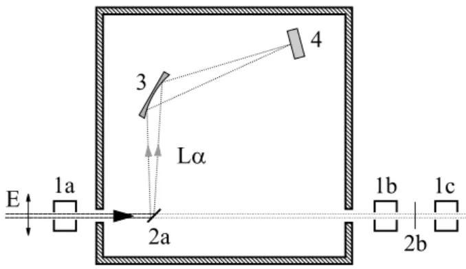
The experiments were carried out at the bending magnet XAFS beamline at the Elet-tra synchrotron, Trieste, Italy. The pri-mary x-ray beam was monochromatized by means of a double-crystal Si(111) monochro-mator. To suppress higher photon energies a Pt-coated mirror was used and the second monochromator crystal was detuned with re-spect to the first one. The energy bandwidth of the monochromatized radiation was 0.4 eV and the photon flux was ∼108ph/s. The
top-view of the experimental setup is shown in Fig. 2. For the x-ray absorption experiment


![FIG. 4: (color online) Experimental and the- the-oretical [18] photoabsorption for a 1.66 µm thick palladium sample](https://thumb-eu.123doks.com/thumbv2/123doknet/15027715.686375/40.892.480.778.134.375/color-online-experimental-oretical-photoabsorption-thick-palladium-sample.webp)