Suppression Subtractive Hybridization in an Equine
Model of Chronic Asthma
Jean-Pierre Lavoie1*, Josiane Lefebvre-Lavoie1, Mathilde Leclere1, Anouk Lavoie-Lamoureux1, Annie Chamberland2, Catherine Laprise2, Jacques Lussier3
1 Department of Clinical Sciences, Faculty of Veterinary Medicine, Universite´ de Montre´al, Saint-Hyacinthe, Quebec, Canada, 2 De´partement des Sciences Fondamentales, Universite´ du Que´bec a` Chicoutimi, Saguenay, Quebec, Canada,3 Department of Biomedicine, Faculty of Veterinary Medicine, Universite´ de Montre´al, Saint-Hyacinthe, Quebec, Canada
Abstract
Background:Gene expression analyses are used to investigate signaling pathways involved in diseases. In asthma, they have been primarily derived from the analysis of bronchial biopsies harvested from mild to moderate asthmatic subjects and controls. Due to ethical considerations, there is currently limited information on the transcriptome profile of the peripheral lung tissues in asthma.
Objective:To identify genes contributing to chronic inflammation and remodeling in the peripheral lung tissue of horses with heaves, a naturally occurring asthma-like condition.
Methods:Eleven adult horses (6 heaves-affected and 5 controls) were studied while horses with heaves were in clinical remission (Pasture), and during disease exacerbation induced by a 30-day natural antigen challenge during stabling (Challenge). Large peripheral lung biopsies were obtained by thoracoscopy at both time points. Using suppression subtractive hybridization (SSH), lung cDNAs of controls (Pasture and Challenge) and asymptomatic heaves-affected horses (Pasture) were subtracted from cDNAs of horses with heaves in clinical exacerbation (Challenge). The differential expression of selected genes of interest was confirmed using quantitative PCR assay.
Results:Horses with heaves, but not controls, developed airway obstruction when challenged. Nine hundred and fifty cDNA clones isolated from the subtracted library were screened by dot blot array and 224 of those showing the most marked expression differences were sequenced. The gene expression pattern was confirmed by quantitative PCR in 15 of 22 selected genes. Novel genes and genes with an already defined function in asthma were identified in the subtracted cDNA library. Genes of particular interest associated with asthmatic airway inflammation and remodeling included those related to PPP3CB/NFAT, RhoA, and LTB4/GPR44 signaling pathways.
Conclusions:Pathways representing new possible targets for anti-inflammatory and anti-remodeling therapies for asthma were identified. The findings of genes previously associated with asthma validate this equine model for gene expression studies.
Citation: Lavoie J-P, Lefebvre-Lavoie J, Leclere M, Lavoie-Lamoureux A, Chamberland A, et al. (2012) Profiling of Differentially Expressed Genes Using Suppression Subtractive Hybridization in an Equine Model of Chronic Asthma. PLoS ONE 7(1): e29440. doi:10.1371/journal.pone.0029440
Editor: Christian Scho¨nbach, Kyushu Institute of Technology, Japan
Received August 16, 2011; Accepted November 28, 2011; Published January 3, 2012
Copyright: ß 2012 Lavoie et al. This is an open-access article distributed under the terms of the Creative Commons Attribution License, which permits unrestricted use, distribution, and reproduction in any medium, provided the original author and source are credited.
Funding: Funding was provided by the Canadian Institutes of Health Research (MIF79636) and AllerGen NCE Inc., a member of the Networks of Centers of Excellence Canada program. The funders had no role in study design, data collection and analysis, decision to publish, or preparation of the manuscript. Competing Interests: The authors have declared that no competing interests exist.
* E-mail: [email protected]
Introduction
Inflammation and remodeling of the airway wall are charac-teristic features of asthma. The term ‘‘airway remodeling’’ in bronchial asthma is used to describe the structural changes that occur in conjunction with, or because of, chronic inflammation. A consequence of asthmatic airway remodeling is incompletely reversible, or even irreversible airway obstruction, bronchial hyperresponsiveness, and an accelerated decline in lung function [1]. Remodeling processes in asthma result from highly complex, and poorly defined interactions between inflammatory and
resident structural cells [2]. Therefore, the identification of the molecular pathways involved in the crosstalk between these cells is a prerequisite for the development of novel therapy to control airway remodeling.
Expression profile studies allow the discovery of transcripts correlated to disease phenotype and to generate hypotheses regarding genes and pathways underlying these phenotypic changes. Gene expression studies using human lung tissues have been primarily derived from the analysis of bronchial biopsies harvested from mild to moderate asthmatic subjects and controls [3]. These studies have identified candidate genes and pathways
the crosstalk between structural cells potentially less complex than in people.
Studies of comparative pulmonary morphology show that the horse’s lung closely resembles the human lung [7,8] and their lifespan (30–35 years) is closer to human than small rodents. Also, 10 to 20% of horses develop a condition called heaves that shares many features of ‘‘extrinsic’’ human asthma, including lower airway inflammation, reversible airflow obstruction, and bronchial hyperresponsiveness [9,10,11]. Heaves develop spontaneously in susceptible horses and, similarly to asthma, is associated with increased airway smooth muscle mass, goblet cell hyperplasia, and epithelial detachment and regeneration [12,13,14,15]. The horses size and temperament also allow for multiple sampling from the same animal to compare gene expression of the lung tissue under conditions of disease exacerbation and remission. Thus, equine heaves is an appealing model to study the complex inflammation-induced remodeling processes present in chronic asthma.
Suppression subtractive hybridization technique (SSH) is a highly sensitive PCR-based cDNA subtraction method [16] used to identify differentially expressed genes, including genes of relatively low abundances. It selectively amplifies differentially expressed cDNA fragments while suppressing nontarget cDNA amplification. SSH provides an approximately 1000-fold enrich-ment of low copy number genes related to defined phenotypes [17]. Compared to microarray analysis, SSH is more sensitive, sequence independent and yields relatively few false positive [18]. The goal of this study was to document the transcriptome associated with chronic asthmatic inflammation and tissue remodeling. We use SSH to subtract the lung transcriptome obtained from heaves-affected horses during clinical remission as well as from control horses with or without antigen exposure from lung cDNAs of horses with heaves after a 30-day antigen challenge.
Materials and Methods
Experimental animal model, tissue collection, and RNA isolation
Eleven horses (450–550 kg) including 9 mares and 2 geldings were studied. Six horses with heaves (mean 6 SD, 16.862.14 years of age) had a history (mean duration 6.462.9 years) of recurrent episodes of airway obstruction upon hay exposure, abnormal respiratory mechanic measurements, and increased neutrophils in bronchoalveolar (BAL) fluid. Control horses (n = 5, 13.862.39 years of age) had no history of respiratory diseases. Detailed description of the animals, their airway function and lung inflammation have been reported elsewhere [15]. All experimental procedures were performed in accordance with the Canadian Council for Animal Care guidelines and were approved by the Animal Care Committee of the Faculty of Veterinary Medicine of the Universite´ de Montre´al (06-Rech-1324).
All animals were kept together in a low antigenic environment (Pasture) for .3 months prior to the baseline measurements and
was used to calculate the RNA integrity number (RIN).
Suppression subtractive hybridization
SSH was used to compare gene expression in lung tissues of symptomatic heaves-affected horses during challenge (SH) versus heaves-affected horses at baseline and controls at both time points (all regrouped as ‘‘Ctls’’). Identical amounts of total RNA from each horse were pooled within SH (n = 6) and Ctls (n = 16) groups to decrease inter-animal variation (Figure 1.B). The SSH procedures were performed as previously reported [21] and are illustrated in figure 1.A and C. In brief, double-stranded cDNA were generated using the SMART PCR cDNA Synthesis Kit for both SH and Ctls samples according to the manufacturer’s instruction (user manual PT3041-1, Clontech Laboratories, Inc., Mountain View, CA, USA). One mg of total RNA from each pooled groups were reverse transcribed in a total volume of 10 ul with two primers (39 SMARTTMCDS Primer II A and SMART IITM A Oligonucleotide) and PowerScript reverse transcriptase (Clontech Laboratories, Inc., Mountain View, CA, USA), with the addition of 42 ng of T4 gene 32 protein (Roche Applied Science, Laval, QC, CA) to produce first cDNA strand. The resulting cDNA pools were diluted to 50ml in TE buffer (10 mM Tris pH 8, 1 mM EDTA). Double-stranded cDNA were obtained via PCR amplification of 19 cycles with 59PCR Primer II A using Advantage II DNA polymerase (Clontech Laboratories, Inc., Mountain View, CA, USA).
Subtracted forward (SH-Ctls) and reverse (Ctls-SH) reactions were generated by subtracting Ctls cDNAs from SH cDNAs and SH cDNAs from Ctls cDNAs, respectively, using the PCR-select cDNA Subtraction Kit (user manual PT1117-1, Clontech Laboratories, Inc., Mountain View, CA, USA). To perform the subtraction reactions, SH and Ctls cDNAs were digested with RsaI to obtain shorter, blunt-ended molecules suitable for adaptor ligation and optimal for subtractive hybridization.
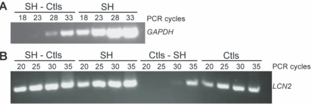
Subtraction efficiency was evaluated by comparing the abundance of known genes in subtracted and unsubtracted cDNAs population after different cycles of PCR using Advantage II DNA polymerase (Clontech Laboratories, Inc., Mountain View, CA, USA). Equine gene specific primers were designed for two genes; one that is constitutively expressed, glyceraldehyde 3-phosphate dehydrogenase (GAPDH); and lipocalin 2 (LCN2), a gene known for its association with lung diseases [22] (Table S1). The subtraction efficiency was indicated by the difference in the number of cycles needed to generate equal amplification of the corresponding PCR product before and after subtraction for these two genes.
Cloning of subtracted cDNAs
The purified (QIAquick PCR Purification kit, Qiagen, Toronto, ON, CA) subtracted (SH-Ctls) cDNAs were cloned into the pDrive-cloning vector (Qiagen, Toronto, ON, CA). Ligation products were then used to transform competent cells (DH5a, Invitrogen, Carlsbad, CA, USA), which were spread onto S-GalHLB agar
blend plate (Sigma-Aldrich Canada Ltd, Oakville, ON, CA) supplemented with kanamycin (40mg/ml). Individual colonies (950) were transferred into ten 96-well plates containing LB freezing media (8.8% glycerol, 55 mM K2HPO4, 1 mM MgSO4, 26 mM
KH2PO4, 15 mM NH4(SO4)) and incubated overnight to construct
the SH-Ctls subtracted library.
Differential hybridization screening
SH-Ctls subtracted macroarrays were established for differential screening, as previously described [21]. Briefly, PCR amplification was performed on the insert of each cDNA clone from the SH-Ctls subtracted library plates using AmpliTaq DNA Polymerase (Applied Biosystems, Foster City, CA, USA), with PCR-nested
Figure 1. Methodology for SSH. Schematic representation of the different steps described in Material and Methods (A), the sample pooling (n corresponds to the number of samples) (B), and the different hybridization steps performed with the SSH technique (C).
were obtained from unsubtracted (SH and Ctls) and subtracted (SH-Ctls and Ctls-SH) complex cDNA populations via secondary nested PCR amplification (PCR-select cDNA Subtraction Kit, Clontech Laboratories, Inc., Mountain View, CA, USA). Probes were then purified (QIAquick PCR Purification kit, Qiagen, Toronto, ON, CA), digested with AfaI, SmaI and EagI to remove adaptors and purified again (QIAquick PCR Purification kit, Qiagen, Toronto, ON, CA). One hundred nanograms of the cDNA probes were labeled by random priming with a32P[dCTP] (Megaprime DNA labeling System, GE Healthcare, Buckingham-shire, UK) as described previously [21]. Radio-labeled cDNA probes were purified (QIAquick Nucleotide Removal Kit, Qiagen, Toronto, ON, CA) and quantified with a liquid scintillation analyzer (Tri-Carb 2100TR, Packard BioScience Compagny, Meriden, CT, USA).
Each membrane was individually hybridized and washed as describe previously [21]. SH-Ctls macroarray membrane repli-cates were hybridized with identical amounts (cpm) of specific heat denatured cDNA probe (SH-Ctls, Ctls-SH, SH or Ctls). Washed membranes were exposed to a phosphor screen for 4 hours and the images were digitized (Storm 840, GE Healthcare, Buck-inghamshire, UK). cDNA clones with different hybridization pattern were characterized using DNA sequencing and gene expression analysis.
DNA sequencing and sequence analysis
The differentially expressed cDNA clones were amplified by PCR from the PCR products generated initially for the macroarrays using the Advantage II DNA polymerase (Clontech Laboratories, Inc., Mountain View, CA, USA) and the PCR-nested primers 1 and 2R. Amplicons were analyzed on 1.5% agarose gel with ethidium bromide to detect multiple PCR fragments. Single band amplicons were gel extracted (QIAquick Gel Extraction kit, Qiagen, Toronto, ON, CA) and sequenced via the dideoxy method (Big Dye Terminator 3.0, ABI Prism, Applied Biosystem, Foster City, CA, USA) by Ge´nome Que´bec using the PCR-nested primers 1 or 2R. Sequencing reactions were analyzed with an ABI Prism 310 sequencer (Applied Biosystem, Foster City, CA, USA). Nucleic acid sequences with at least 100 bp were aligned against GenBank database (NR and EST) using the Basic Local Alignment Search Tool (BLAST). The maximum expected (E) value accepted to be considered homologous was e230. Every match with higher E value score was aligned against the horse genome at the UCSC Genome Browser database using BLAT (BLAST-like alignment tool; http://genome.ucsc.edu/). Sequenc-es were classified into two groups: I) genSequenc-es with known sequence and function and II) genes with characterized sequence but unknown function. The genes from the 1st group were further classified through biological function categories using functional mapping tools (GeneOntology, http://www.geneontology.org/) and compared to available literature related to human asthma and other animal models. This classification allowed us to identify biological pathways, gene families or biological functions likely to
equine gene-specific primers (Table S1) were designed to span at least two intron-exon boundaries for the discrimination of contaminant genomic DNA. The absence of nonspecific products was confirmed by the analysis of the melting point curves and by electrophoresis in 1.5% agarose gels. All concentrations of target gene cDNA were calculated relatively to their respective standard curves. One microliter of cDNA template was added to the Quantitec SYBRHGreen PCR Kit master mix (Qiagen, Toronto, ON, CA). qPCR reactions were performed in a volume of 20ml using Rotor-Gene RG-3000 (Corbett Research, Sydney, AS) and qPCR conditions were similar for all primer sets (0.5mM, final concentration): denaturation 95uC for 10 min, cycling 95uC for 15 sec, 55uC for 25 sec and 72uC for 25 sec for a maximum of 40 cycles. Each reaction was run in duplicate with the appropriate negative control. Reference gene expression was evaluated using different analysis softwares: NormFinder [23], GeNormPLUS[24] and Rest 2009 [25].
Statistical analysis
Data are presented as mean 6 SD. Differences between groups were compared at each time point using Wilcoxon tests and differences within groups were evaluated using Mann-Whitney test. Unilateral tests were used to compare values with those of asthmatic horses after 30 days of antigen challenge because SSH technique predicted the direction of the effect. Bilateral tests were used for all other analyses. P,0.05 was considered significant.
Results
Experimental animal model
All horses had normal lung function prior to challenge, while only horses with heaves developed clinical signs of airway obstruction and persistent airway inflammation after antigen challenge (see [15] for detailed description of lung function and BAL fluid cytology in these animals).
Identification of differentially expressed genes
The quality of the RNA samples was confirmed by high RIN values (8.4760.58 SD) and electropherogram analysis. Subtrac-tion efficiency was evaluated using standard PCR for two genes; GAPDH and LCN2. GAPDH PCR products were detectable after only 18 cycles in the SH unsubtracted sample, whereas 10 more cycles were required to detect the PCR fragment in the SH-Ctls subtracted sample (Figure 2.A), a 40 fold reduction. LCN2 PCR products were detectable after 20 cycles in both subtracted SH-Ctls and unsubtracted SH samples, but the difference in intensity between the two signals indicates enrichment (Figure 2.B). Conversely, the LCN2 PCR products were detected after 20 cycles in the unsubtracted Ctls sample and after 10 more cycles (40 fold increase) in the reverse subtracted Ctls-SH sample.
Differential hybridization screening was performed using macroarrays in order to isolate genes implicated in heaves exacerbation from the 950 randomly selected clones. Differentially
expressed cDNA clones were identified based on the hybridization signal intensities observed between the four membranes. The positive clones had 1) a stronger hybridization signal with the SH-Ctls probe than with SH probe, 2) a weaker hybridization signal with the Ctls-SH probe than with the Ctls probe, and 3) a stronger hybridization signal with the SH-Ctls probe than with the Ctls-SH probe. Representative differential screening results are illustrated in Figure 3. Of the 950 cDNA clones screened, 294 were identified as strongly expressed and were analyzed on agarose gel. Sequencing performed on single band PCR amplicons generated a total of 224 clones with adequate sequencing results for BLAST analysis and Genbank deposition (accession numbers from GH613643 to GH613840).
The first group contained 167 sequences with known function, 20 of which were redundant. These sequences were further categorized based on their biological pathways (Table S2, 147 sequences). The second group contained 57 sequences previously characterized, but with unknown functions; 9 of these sequences were redundant (Table S2, 48 sequences). In group I, there were 14 genes related to regulatory proteins, 14 to immune signaling molecules, 13 to intracellular signaling component pathways, 10 to immune response, 5 to cell growth and proliferation, 5 to free radical metabolism (Table S2, 61 genes). There were 86 additional genes with known cell function (6 to transmembrane proteins, 10 to structural proteins, 4 to extracellular proteins, 1 to complement components, 4 to gene transcription, 2 to cell adhesion molecules,
Figure 2. Evaluation of subtraction efficiency. A: Reduction of GAPDH cDNA following subtraction in the SH-Ctls sample. PCR was performed on SH-Ctls subtracted and SH unsubtracted samples. GAPDH PCR products (760 pb) were detectable 10 cycles earlier in the unsubtracted sample (18 cycles) than in the subtracted sample (28 cycles). B: Enrichment of LCN2 cDNA following subtraction in the Ctls sample. PCR was performed on SH-Ctls and SH-Ctls-SH subtracted samples as well as SH and SH-Ctls unsubtracted samples. LCN2 PCR products (210 pb) were detected after 20 cycles for both SH unsubtracted and SH-Ctls subtracted samples, the difference in the intensity of the 2 bands indicate the enrichment compare to Ctls unsubtracted and Ctls-SH subtracted samples.
doi:10.1371/journal.pone.0029440.g002
Figure 3. Differential hybridization screening. Representative differential screening results of macroarrays of the SH-Ctls library. Four identical membranes were dot-blotted with PCR products obtained by SSH. The membranes were then hybridized with four different probes: SH-Ctls subtracted cDNAs (A), SH unsubtracted cDNAs (B), Ctls-SH subtracted cDNAs (C) and Ctls unsubtracted cDNAs. The arrow in the top left corner indicates the positive control (LCN2). The arrow head indicates an example of differentially expressed genes in SH compare with Ctls.
They included LCN2, collagen type I alpha 2 (COL1A2), collagen type III alpha 1 (COL3A1), protein phosphatase 3 catalytic subunit beta (PPP3CB), glypican 4 (GPC4), versican (VCAN), chemokine (C-C motif) ligand 5 ((C-C(C-CL5), decorin (D(C-CN), major histocompatibility complex class II invariant chain CD74 molecule (CD74), dedicator of cytokinesis 1 (DOCK1), fucosidase alpha-L-1 (FUCA1), mito-chondrial translational release factor 1-like (MTRF1L), NHL repeat containing 2 (NHLRC2), prostaglandin D2 receptor (PTGDR), leukotriene A-4 hydrolase (LTA4H), endothelin receptor type A (EDNRA), chemokin binding protein 2 (CCBP2), insulin-like growth factor I (IGF1), gamma actin (ACTG1), vimentin (VIM), TRPC4 associated protein (TRPC4AP) and Rho GTPase activat-ing protein 25 (ARHGAP25). LCN2, COL1A2, PPP3CB, VCAN, DCN, CD74, NHLRC2, IGF1, FUCA1 and LTA4H mRNA were significantly increased in heaves-affected horses after the challenge compared to baseline, in contrast to the mRNA expression in controls, which remained stable during the study. Similarly, PTGDR and MTRF1L also showed a significant increase in heaves-affected horses after challenge compared to baseline, but in addition, the baseline mRNA expression was significantly higher in the control group compared to the heaves-affected group. The expression of ARHGAP25 and ACTG1 mRNA were significantly decreased after challenge compared to baseline in control horses only. ARHGAP25, EDNRA and ACTG1 were also significantly different between groups at baseline, control horses having higher mRNA expression. There were no significant differences between groups and between time points in each group in COL3A1, GPC4, CCL5, DOCK1, CCBP2, TRPC4AP and VIM. Figure 4 represents the mRNA expression using qPCR for six of the genes found to be upregulated with SSH.
Quantification for genes of interest is expressed as absolute concentration because all reference genes tested showed significant (p,0.05) increase in the lung tissue of heaves-affected horses after the antigen challenge compared to baseline. The reference genes tested were GAPDH, ubiquitin C (UBC), b-glucuronidase (GUSB), ß2-microglobulin (B2M), peptidylprolyl isomerase A (PPIA), large ribosomal protein P0 (RPLP0), and ribosomal protein S9 (RPS9). Reference gene analysis using dedicated softwares further confirmed that the stability of these genes was highly dependent on horses’ clinical status, precluding their use for normalization. In view of these results, total RNA and reverse transcribe (RT) reactions were quantified using a spectrophotometer (NanoDrop ND-1000, NanoDrop products, Wilmington, DE, USA) and used to normalize cDNA quantity in the PCR reactions [26]. Lastly, to ensure that unidentified biases were not introduced during sample analysis, the upregulation of GAPDH, PPIA, LTA4H, PPP3CB and LCN2 in horses with heaves after antigen stimulation was confirmed (data not shown) using samples of lung tissues from the same animals but archived using a different technique of preservation (RNA later, Ambion, Austin, TX, USA), RNA extraction (TRIzolH Invitrogen, Carlsbad, CA, USA) and a different enzyme for RT reactions (AMV, Roche Diagnostics Corp, Laval, QC, CA).
Quantitative PCR confirmed the differential gene expression pattern in 15 of the 22 genes evaluated. The functions of many of these genes were related to inflammation, remodeling, and smooth muscle biology, possibly representing new therapeutic targets for asthma.
Genes associated with remodeling and smooth muscle contraction
Inflammation and repair of injured lung tissues in asthma results in an increased thickness of the airway wall leading to reduced baseline airway caliber and exaggerated airway narrowing, phenomena that are accentuated in allergen-induced broncho-spasm [28]. Of particular importance to asthma is the increased airway smooth muscle (ASM) mass observed in human subjects (reviewed by [29]) and in equine heaves [14,15]. Not only are ASM cells increased in number or size and contribute to the bronchospasm, but they show, at least in vitro, some phenotype plasticity in response to allergen challenge, including de-differen-tiation to a more synthetic type capable of producing an extracellular matrix (ECM), various cytokines, and growth factors (reviewed by [30]). However, the molecular pathways responsible for these changes are poorly defined. Herein, we identified at least 13 genes that have been linked to smooth muscle biology. Four genes (ARHGAP25, phosphatidylinositol transfer protein alpha (PITPNA), phosphatidylinositol-specific phospholipase C X do-main containing 3 (PLCXD3), and pyruvate dehydrogenase kinase isozyme 1 (PDK1)) identified in the lung tissues of heaves-affected horses in exacerbation are involved in the modulation of the RhoA pathway [31]. This pathway is necessary for the activation of factor serum response factor (SRF) by myocardin [32], one of its coactivators, which leads to the expression of contractile protein genes expression [33]. Interestingly, RhoA/Rho kinase is required for ASM contraction induced by endothelin-1 (EDN1) [34] and is upregulated by interleukin-4 (IL-4), a Th2 cytokine expressed in the airways of asthmatic patients [35]. Our results are thus in agreement and extend those of in vitro and animal studies, and further support the proposal of Rho kinase inhibitors as new targets for the treatment of airway bronchoconstriction and remodeling seen in asthma (reviewed by [36]). Conversely, it is the mitogen-activated protein kinase (MAPK) signaling pathway that promotes smooth muscle proliferation by modulating SRF-transcriptional activities via the activation Elk-1 [32,33]. MAPK1 identified in our SSH activates Elk-1 [37], suggesting that both ASM proliferation and differentiation may coexist in the asthmatic lungs.
PPP3CB (also known as calcineurin) and IGF1 identified by SSH share common signaling pathways also possibly contributing to smooth muscle phenotype switching and ECM remodeling in asthma [38,39,40,41,42]. The identification of PPP3CB, and one of its inhibitors, calcineurin homologous protein (CHP) [43], is of particular interest as PPP3CB/NFAT signaling is implicated in a wide range of biological responses relevant to asthma including lymphocyte activation, as well as neuronal and muscle development
[43,44,45]. While not yet investigated in lung tissues to our knowledge, PPP3CB activation results in muscle hypertrophy in response to increase workload in both the urinary bladder and in the heart [46,47,48]. Furthermore, alterations of the expression of the fast and slow myosin heavy chain isoforms in the obstructed bladder is PPP3CB-dependent [48]. Thus the PPP3CB pathway may participate in the increased ASM mass and the myosin heavy chain isoform switching observed in the asthmatic airways [49]. The expression of EDN1, a potent spasmogen for the bronchus, is increased in asthma [50], and single nucleotide polymorphisms (SNPs) have been associated with susceptibility to this disease [51]. One of its receptor, EDNRA, identified in our SSH, has previously been found to be upregulated in this animal model [52]. Interestingly, the PPP3CB/NFAT pathway discussed above has been shown to be required for at least some of the effects of EDN1 in cardiac myocytes [39,53], and thus, are further support for their possible modulation of ASM remodeling.
IGF1 plays a vital role in embryonic development and promotes the anabolism and the repair of various tissues in adults [54]. There are several evidences suggesting that IGF1 may also contribute to asthma. IGF1 is produced by human bronchial epithelial cells in response to IL-17F [55], a cytokine implicated in asthma. It was shown to induce the expression of alpha-smooth muscle actin and type-I collagen by human fetal lung fibroblasts [56], and to promote visceral myocyte differentiation into a contractile phenotype via the PPP3CB/NFAT pathway [57,58]. The increased expression of IGF1 in the peripheral lung tissue of horses with heaves during exacerbation is thus of interest to asthma, especially in the light that IGF1 neutralizing antibody inhibits airway obstruction and inflammation, while preventing airway wall thickening in a mouse model of asthma [59].
There is also alteration of various components of the ECM in the asthmatic airways. These changes vary depending on size of airways, and it has been shown that an increase in the degree of
Figure 4. Gene expression analysis. Analysis of mRNA expression using qPCR of six genes found up-regulated with SSH. LCN2 (A), COL1A2 (B), PP3CB (C), PTGDR (D), LTA4H (E) and IGF1 (F) were studied in six horses with heaves (black bars) and six control horses (white bars). When compared to baseline, the six genes were significantly increased in heaves-affected horses after the allergen challenge (p,0.05). PTDGR was also increased in control horses when compared to heaves-affected horses at baseline.
been proposed to be contributing to the increased ASM mass observed in asthma [61].
Genes associated with inflammation
Pulmonary inflammation is a characteristic finding in asthma and anti-inflammatory drugs are central for its control. Seven genes associated with leukotriene (LT)B4 metabolism or prosta-glandin (PG)D2 activity were identified as being overexpressed in the lungs of horses with heaves during exacerbation. Those included LTA4 hydrolase which metabolizes LTA4 in LTB4, PGF synthase that reduces PGD2 and PGH2 to PGF2, PTGDR (also named DP1), a PGD2 receptor involved in the regulation of Th2-type driven inflammation [62], and CNOT7 (CCR4-NOT transcription complex), a repressor of the retinoid X beta receptor (Rxrb) [63], which forms a heterodimeric complex with the nuclear receptors PPARs (peroxisome proliferator-activated receptor). LTB4 is an arachidonic acid metabolite synthesized by various cell types when activated by inflammatory stimuli. LTB4 was first described as a potent chemoattractant and activator of neutro-phils, the predominant airway cell population present in heaves, and in some asthmatic patients [64]. It is now recognized that LTB4 also exerts these effects on other cell types involved in airway inflammation [65] and it has also been suggested that it is implicated in T cell trafficking and asthmatic inflammation [66]. Further support for a role of LTB4 in asthma is its increase in exhaled breath condensate of affected patients [67], and the attenuation of allergic airway inflammation and hyperresponsive-ness by LTA4H inhibition [68]. However, the effects of LTA4H are complex, as it can also limit tissue damage-induced neutrophilia through its aminopeptidase activity which degrades proline-glycine-proline (PGP), a collagen breakdown product possessing potent neutrophil chemotactic activity [69]. PGD2 is also an arachidonic acid metabolite that is released in large quantities by mast cells during anaphylaxis. Other cell types present in lung tissues such has dendritic cells, macrophages, eosinophils, Th2
in the peripheral lung tissues of horses with heaves when antigen challenged. Genes previously associated with asthma as well as novel pathways were also identified. These genes encompass a range of biological processes with pathways related to ASM and ECM remodeling, and inflammation being notable. Our results suggest that targeting RhoA, PPP3CB, EDN1, and IGF1 signaling pathways may represent appropriate targets for anti-remodeling therapies, especially for the control ASM hypertrophy, while anti-inflammatory effects may possibly be achieved by drugs modulat-ing LTB4 and PGD2.
Supporting Information
Table S1 Sequences of primer pairs used for PCR analysis.
(DOCX)
Table S2 Identification and functional classification of differentially expressed transcripts in horses with heaves during airway obstruction when compared to healthy controls and asymptomatic asthmatic horses. (DOCX)
Acknowledgments
C.L. is the chairholder of the Canada Research Chair for genetic determinants in asthma (www.chairs.gc.ca). C.L. and J.-P.L. are members of the Respiratory Health Network (RHN) of the Fonds de la recherche en sante´ du Quebec (FRSQ).
Author Contributions
Conceived and designed the experiments: JPL JL. Performed the experiments: JLL ML ALL AC CL. Analyzed the data: JPL JLL AC CL. Contributed reagents/materials/analysis tools: JPL JL CL. Wrote the paper: JPL JLL.
References
1. Elias JA (2000) Airway remodeling in asthma. Unanswered questions. Am J Respir Crit Care Med 161: S168–171.
2. James A (2005) Airway remodeling in asthma. Curr Opin Pulm Med 11: 1–6. 3. Hansel NN, Diette GB (2007) Gene expression profiling in human asthma. Proc
Am Thorac Soc 4: 32–36.
4. Homer RJ, Elias JA (2005) Airway remodeling in asthma: therapeutic implications of mechanisms. Physiology (Bethesda) 20: 28–35.
5. Jeffery PK (1998) Investigation and assessment of airway and lung inflammation: we now have the tools, what are the questions? Eur Respir J 11: 524–528. 6. Karol MH (1994) Animal models of occupational asthma. Eur Respir J 7:
555–568.
7. McLaughlin RF (1983) Bronchial artery distribution in various mammals and in humans. Am Rev Respir Dis 128: S57–S58.
8. Magno M (1990) Comparative anatomy of the tracheobronchial circulation. Eur Respir J Suppl 12: 557s–562s; discussion 562s–563s.
9. van Erck E, Votion DM, Kirschvink N, Art T, Lekeux P (2003) Use of the impulse oscillometry system for testing pulmonary function during methacholine bronchoprovocation in horses. Am J Vet Res 64: 1414–1420.
10. Lowell F (1964) Observations on heaves: an asthma-like syndrome in the horse. J Allergy 35: 322–330.
11. Snapper JR (1986) Large animal models of asthma. Am Rev Respir Dis 133: 351–352.
12. Robinson NE (2001) International Workshop on Equine Chronic Airway Disease. Michigan State University 16–18 June 2000. Equine Vet J 33: 5–19. 13. Range F, Mundhenk L, Gruber AD (2007) A soluble secreted glycoprotein
(eCLCA1) is overexpressed due to goblet cell hyperplasia and metaplasia in horses with recurrent airway obstruction. Vet Pathol 44: 901–911.
14. Herszberg B, Ramos-Barbon D, Tamaoka M, Martin JG, Lavoie JP (2006) Heaves, an asthma-like equine disease, involves airway smooth muscle remodeling. J Allergy Clin Immunol 118: 382–388.
15. Leclere M, Lavoie-Lamoureux A, Gelinas-Lymburner E, David F, Martin JG, et al. (2010) Effect of Antigen Exposure on Airway Smooth Muscle Remodeling in an Equine Model of Chronic Asthma. Am J Respir Cell Mol Biol. 16. Cao W, Epstein C, Liu H, DeLoughery C, Ge N, et al. (2004) Comparing gene
discovery from Affymetrix GeneChip microarrays and Clontech PCR-select cDNA subtraction: a case study. BMC Genomics 5: 26.
17. Qin M, Zeng Z, Zheng J, Shah PK, Schwartz SM, et al. (2003) Suppression subtractive hybridization identifies distinctive expression markers for coronary and internal mammary arteries. Arterioscler Thromb Vasc Biol 23: 425–433.
18. McClintock TS (2002) High-throughput expression profiling techniques. Chem Senses 27: 289–291.
19. Relave F, David F, Leclere M, Alexander K, Bussieres G, et al. (2008) Evaluation of a thoracoscopic technique using ligating loops to obtain large lung biopsies in standing healthy and heaves-affected horses. Vet Surg 37: 232–240. 20. Bedard J, Brule S, Price CA, Silversides DW, Lussier JG (2003) Serine protease inhibitor-E2 (SERPINE2) is differentially expressed in granulosa cells of dominant follicle in cattle. Mol Reprod Dev 64: 152–165.
21. Fayad T, Levesque V, Sirois J, Silversides DW, Lussier JG (2004) Gene expression profiling of differentially expressed genes in granulosa cells of bovine dominant follicles using suppression subtractive hybridization. Biol Reprod 70: 523–533.
22. Ekberg-Jansson A, Andersson B, Bake B, Boijsen M, Enanden I, et al. (2001) Neutrophil-associated activation markers in healthy smokers relates to a fall in DL(CO) and to emphysematous changes on high resolution CT. Respir Med 95: 363–373.
23. Andersen CL, Jensen JL, Orntoft TF (2004) Normalization of real-time quantitative reverse transcription-PCR data: a model-based variance estimation approach to identify genes suited for normalization, applied to bladder and colon cancer data sets. Cancer Res 64: 5245–5250.
24. Vandesompele J, De Preter K, Pattyn F, Poppe B, Van Roy N, et al. (2002) Accurate normalization of real-time quantitative RT-PCR data by geometric averaging of multiple internal control genes. Genome Biol 3: RESEARCH0034. 25. Pfaffl MW, Horgan GW, Dempfle L (2002) Relative expression software tool (REST) for group-wise comparison and statistical analysis of relative expression results in real-time PCR. Nucleic Acids Res 30: e36.
26. Bustin SA (2002) Quantification of mRNA using real-time reverse transcription PCR (RT-PCR): trends and problems. J Mol Endocrinol 29: 23–39. 27. Anderson GP (2008) Endotyping asthma: new insights into key pathogenic
mechanisms in a complex, heterogeneous disease. Lancet 372: 1107–1119. 28. James AL, Pare PD, Hogg JC (1989) The mechanics of airway narrowing in
asthma. Am Rev Respir Dis 139: 242–246.
29. Bai TR (2010) Evidence for airway remodeling in chronic asthma. Curr Opin Allergy Clin Immunol 10: 82–86.
30. Hirota JA, Nguyen TT, Schaafsma D, Sharma P, Tran T (2009) Airway smooth muscle in asthma: phenotype plasticity and function. Pulm Pharmacol Ther 22: 370–378.
31. Mao J, Yuan H, Xie W, Wu D (1998) Guanine nucleotide exchange factor GEF115 specifically mediates activation of Rho and serum response factor by the G protein alpha subunit Galpha13. Proc Natl Acad Sci U S A 95: 12973–12976.
32. Lee SM, Vasishtha M, Prywes R (2010) Activation and repression of cellular immediate early genes by serum response factor cofactors. J Biol Chem 285: 22036–22049.
33. Wang Z, Wang DZ, Hockemeyer D, McAnally J, Nordheim A, et al. (2004) Myocardin and ternary complex factors compete for SRF to control smooth muscle gene expression. Nature 428: 185–189.
34. Yoshii A, Iizuka K, Dobashi K, Horie T, Harada T, et al. (1999) Relaxation of contracted rabbit tracheal and human bronchial smooth muscle by Y-27632 through inhibition of Ca2+ sensitization. Am J Respir Cell Mol Biol 20: 1190–1200.
35. Kay AB, Ying S, Varney V, Gaga M, Durham SR, et al. (1991) Messenger mRNA expression of the cytokine gene cluster, interleukin 3 (IL-3), IL-4, IL-5 and granulocyte/macrophage colony-stimulating factor, in allergen-induced late-phase cutaneous response in atopic subjects. J Exp Med 173: 775–778. 36. Schaafsma D, Gosens R, Zaagsma J, Halayko AJ, Meurs H (2008) Rho kinase
inhibitors: a novel therapeutical intervention in asthma? Eur J Pharmacol 585: 398–406.
37. Yang SH, Yates PR, Whitmarsh AJ, Davis RJ, Sharrocks AD (1998) The Elk-1 ETS-domain transcription factor contains a mitogen-activated protein kinase targeting motif. Mol Cell Biol 18: 710–720.
38. Ohkawa Y, Hayashi K, Sobue K (2003) Calcineurin-mediated pathway involved in the differentiated phenotype of smooth muscle cells. Biochem Biophys Res Commun 301: 78–83.
39. Kakita T, Hasegawa K, Iwai-Kanai E, Adachi S, Morimoto T, et al. (2001) Calcineurin pathway is required for endothelin-1-mediated protection against oxidant stress-induced apoptosis in cardiac myocytes. Circ Res 88: 1239–1246. 40. Xin X, Hou YT, Li L, Schmiedlin-Ren P, Christman GM, et al. (2004) IGF-I increases IGFBP-5 and collagen alpha1(I) mRNAs by the MAPK pathway in rat intestinal smooth muscle cells. Am J Physiol Gastrointest Liver Physiol 286: G777–783.
41. Veraldi KL, Gibson BT, Yasuoka H, Myerburg MM, Kelly EA, et al. (2009) Role of Insulin-like Growth Factor Binding Protein-3 in Allergic Airway Remodeling. Am J Respir Crit Care Med.
42. McWhinnie R, Pechkovsky DV, Zhou D, Lane D, Halayko AJ, et al. (2007) Endothelin-1 induces hypertrophy and inhibits apoptosis in human airway smooth muscle cells. Am J Physiol Lung Cell Mol Physiol 292: L278–286. 43. Crabtree GR (2001) Calcium, calcineurin, and the control of transcription. J Biol
Chem 276: 2313–2316.
44. Wu H, Peisley A, Graef IA, Crabtree GR (2007) NFAT signaling and the invention of vertebrates. Trends Cell Biol 17: 251–260.
45. Rao A, Luo C, Hogan PG (1997) Transcription factors of the NFAT family: regulation and function. Annu Rev Immunol 15: 707–747.
46. Nozaki K, Tomizawa K, Yokoyama T, Kumon H, Matsui H (2003) Calcineurin mediates bladder smooth muscle hypertrophy after bladder outlet obstruction. J Urol 170: 2077–2081.
47. Balakumar P, Jagadeesh G (2010) Multifarious molecular signaling cascades of cardiac hypertrophy: can the muddy waters be cleared? Pharmacol Res 62: 365–383.
48. Clement M, Delaney D, Austin J, Sliwoski J, Hii G, et al. (2006) Activation of the Calcineurin Pathway is Associated With Detrusor Decompensation: A Potential Therapeutic Target. The Journal of Urology 176: 1225–1229.
49. Leguillette R, Laviolette M, Bergeron C, Zitouni N, Kogut P, et al. (2009) Myosin, transgelin, and myosin light chain kinase: expression and function in asthma. Am J Respir Crit Care Med 179: 194–204.
50. Trakada G, Tsourapis S, Marangos M, Spiropoulos K (2000) Arterial and bronchoalveolar lavage fluid endothelin-1 concentration in asthma. Respir Med 94: 992–996.
51. Zhu G, Carlsen K, Carlsen KH, Lenney W, Silverman M, et al. (2008) Polymorphisms in the endothelin-1 (EDN1) are associated with asthma in two populations. Genes Immun 9: 23–29.
52. Costa LR, Eades SC, Venugopal CS, Moore RM (2009) Plasma and pulmonary fluid endothelin in horses with seasonal recurrent airway obstruction. J Vet Intern Med 23: 1239–1246.
53. Bao Y, Li R, Jiang J, Cai B, Gao J, et al. (2008) Activation of peroxisome proliferator-activated receptor gamma inhibits endothelin-1-induced cardiac hypertrophy via the calcineurin/NFAT signaling pathway. Mol Cell Biochem 317: 189–196.
54. Dai Z, Wu F, Yeung EW, Li Y (2010) IGF-IEc expression, regulation and biological function in different tissues. Growth Horm IGF Res 20: 275–281. 55. Kawaguchi M, Fujita J, Kokubu F, Ohara G, Huang SK, et al. (2010) Induction
of insulin-like growth factor-I by interleukin-17F in bronchial epithelial cells. Clin Exp Allergy 40: 1036–1043.
56. Chetty A, Cao GJ, Nielsen HC (2006) Insulin-like Growth Factor-I signaling mechanisms, type I collagen and alpha smooth muscle actin in human fetal lung fibroblasts. Pediatr Res 60: 389–394.
57. Hayashi K, Takahashi M, Kimura K, Nishida W, Saga H, et al. (1999) Changes in the balance of phosphoinositide 3-kinase/protein kinase B (Akt) and the mitogen-activated protein kinases (ERK/p38MAPK) determine a phenotype of visceral and vascular smooth muscle cells. J Cell Biol 145: 727–740. 58. Ohkawa Y, Hayashi Ki, Sobue K (2003) Calcineurin-mediated pathway
involved in the differentiated phenotype of smooth muscle cells. Biochemical and Biophysical Research Communications 301: 78–83.
59. Yamashita N, Tashimo H, Ishida H, Matsuo Y, Arai H, et al. (2005) Role of insulin-like growth factor-I in allergen-induced airway inflammation and remodeling. Cell Immunol 235: 85–91.
60. Benayoun L, Druilhe A, Dombret MC, Aubier M, Pretolani M (2003) Airway structural alterations selectively associated with severe asthma. Am J Respir Crit Care Med 167: 1360–1368.
61. Dutsch-Wicherek M (2010) RCAS1, MT, and vimentin as potential markers of tumor microenvironment remodeling. Am J Reprod Immunol 63: 181–188. 62. Pettipher R, Hansel TT, Armer R (2007) Antagonism of the prostaglandin D2
receptors DP1 and CRTH2 as an approach to treat allergic diseases. Nature Reviews Drug Discovery 6: 313–325.
63. Winkler GS, Mulder KW, Bardwell VJ, Kalkhoven E, Timmers HT (2006) Human Ccr4-Not complex is a ligand-dependent repressor of nuclear receptor-mediated transcription. EMBO J 25: 3089–3099.
64. Wenzel SE, Schwartz LB, Langmack EL, Halliday JL, Trudeau JB, et al. (1999) Evidence that severe asthma can be divided pathologically into two inflammatory subtypes with distinct physiologic and clinical characteristics. Am J Respir Crit Care Med 160: 1001–1008.
65. Watanabe S, Yamasaki A, Hashimoto K, Shigeoka Y, Chikumi H, et al. (2009) Expression of functional leukotriene B4 receptors on human airway smooth muscle cells. J Allergy Clin Immunol 124: 59–65, e51–53.
66. Luster AD, Tager AM (2004) T-cell trafficking in asthma: lipid mediators grease the way. Nature Reviews Immunology 4: 711–724.
67. Montuschi P, Barnes PJ (2002) Exhaled leukotrienes and prostaglandins in asthma. J Allergy Clin Immunol 109: 615–620.
68. Rao NL, Riley JP, Banie H, Xue X, Sun B, et al. (2010) Leukotriene A(4) hydrolase inhibition attenuates allergic airway inflammation and hyperrespon-siveness. Am J Respir Crit Care Med 181: 899–907.
69. Snelgrove RJ, Jackson PL, Hardison MT, Noerager BD, Kinloch A, et al. (2010) A critical role for LTA4H in limiting chronic pulmonary neutrophilic inflammation. Science 330: 90–94.
70. Ulven T, Kostenis E (2010) Novel CRTH2 antagonists: a review of patents from 2006 to 2009. Expert Opin Ther Pat 20: 1505–1530.