HAL Id: tel-01234310
https://tel.archives-ouvertes.fr/tel-01234310
Submitted on 26 Nov 2015HAL is a multi-disciplinary open access archive for the deposit and dissemination of sci-entific research documents, whether they are pub-lished or not. The documents may come from teaching and research institutions in France or abroad, or from public or private research centers.
L’archive ouverte pluridisciplinaire HAL, est destinée au dépôt et à la diffusion de documents scientifiques de niveau recherche, publiés ou non, émanant des établissements d’enseignement et de recherche français ou étrangers, des laboratoires publics ou privés.
Study of histone variants and chromatin dynamics in the
preimplantation mouse embryo
Ana Boskovic
To cite this version:
Ana Boskovic. Study of histone variants and chromatin dynamics in the preimplantation mouse embryo. Embryology and Organogenesis. Université de Strasbourg, 2014. English. �NNT : 2014STRAJ034�. �tel-01234310�
UNIVERSITÉ DE STRASBOURG
ÉCOLE DOCTORALE VIE ET SANTE
THÈSE
présentée par :Ana BOSKOVIC
soutenue le : 28 juillet 2014
pour obtenir le grade de :
Docteur de l’université de Strasbourg
Discipline/ Spécialité:
Aspects moléculaires et cellulaires de la biologieStudy of histone variants and chromatin
dynamics in the preimplantation mouse
embryo
THÈSE dirigée par:
Dr Maria-Elena TORRES-PADILLA, IGBMC, Illkirch
RAPPORTEURS:
Dr Didier Devys, IGBMC, Illkirch Dr Petra Hajkova, MRC, London
1
Acknowledgments
I would like to first thank the members of my thesis jury, Dr Petra Hajkova, Dr Saadi Khochbin and Dr Didier Devys, for accepting to read and evaluate my thesis.
I thank my supervisor, Dr Maria-Elena Torres-Padilla for all of her guidance, endless patience and help during my PhD. Thank you for giving me the opportunity to work in your lab, to learn and grow and explore, and for always being there to support me, professionally and personally. Thank you for all the fun times we had in the lab (Falcon-tube tequila shots), during lab retreats and conferences! You believed in me even when I did not believe in myself (which was quite oftenJ) and I am privileged to have had you as my supervisor.
All the members of the METP lab: thank you guys so much! Without you, the great experience I had during my PhD would not have been possible. Celine, thank you very much for all your help and kindness, for being the good spirit of the lab and for keeping the whole thing running smoothly. Adam, thank you for your friendship and all the help in the past years, and for interesting discussions on science/music/bananasJ Andre and Joanna, thanks for your enthusiasm and for the fun times we spent together, in and out of the lab! Takashi, thanks for your never-ending patience regarding my ‘I have a question’ moments, whether they were about potency, bags, gossip or McDonaldsJ Julien, it was cool working with you, even if it was only for a short time. All of you guys made everyday life and work fun and interesting at the same time. Thank you for all the scientific and not-so-scientific discussions (sailing vs. floating), great company and friendship!
Angele, Anas and Yusuke, I have learnt so much from all of you and I hope you know how much I appreciate knowing and working with you! Hopefully we will all see each other during the 10-year lab reunion!
A big merci goes to William, Alex, Hafid and all the ladies in the animal house, as well as Mark, Pascal and everyone in the Imaging center. Thank you for your perpetual positive attitude and help with mice and microscopes!
To our lab neighbours, particularly members of Labo Charlet and Seraphin, as well as Pascal Eberling and Jean-Marie Garnier, for being excellent company, always happy to lend (or borrow) equipment/advice/gossip. It’s been a pleasure knowing you all!
Thank you to the Vermot girls, especially Emily and Rita, for all the fun we had together over lunch and outside the lab! Lunch was definitely the highlight of the dayJ
Big thanks goes to my dearest Nucleosome 4D fellows – this network was just the best thing ever. Thank you Andreas and Corey for organizing such an amazing network. Thank you Iva, Ava, Gytis, Seb, Ivaylo (and all the rest!) for your friendship and great times we had together! Chromatin is great, I love chromatin!:)
Finally, to Helena and Nikolas, I cannot imagine what PhD would be like without you. Ever since interviews, you have always been there for me: through the good, the bad, the ugly and the Ylvis;) Nikolas Jose, thanks for your friendship, humor, Sunday night cinema and constant reminders that I should relax!
Helena de Fatima, thank you for moral and scientific support, millions of high fives, laughing to tears, barco Latino escapades, amazing trips - too many things to mention. Simply, thank you for being the best friend that a person can have, in every possible way!
Abstract
Mammalian development starts at fertilization, when two highly specialized cells, the sperm and the oocyte, fuse and create the totipotent zygote. Through subsequent cell divisions and differentiation during development, the zygote gives rise to every cell type in the organism. In mouse, 3.5 days after fertilization, the blastocyst forms. Despite the DNA content of all cells being identical, the blastocyst already comprises two distinct cell types: the pluripotent inner cell mass and the multipotent trophectoderm. This suggests that the mechanisms additionally to the DNA sequence itself play a role in regulating cell fate specifications, pointing out towards a key role for epigenetic regulation of the earliest stages of development. How the zygote acquires totipotency from two fully differentiated cells, and how cell fate decisions are made later in development is a pivotal biological question.
In eukaryotes, the DNA is associated with histones into a nucleoprotein complex called chromatin. Chromatin structure can range from very loose to highly compacted, and can be permissive to ‘reading’ of the DNA or antagonize it. Importantly, chromatin can be extensively modified, with functional implications in various biological processes, such as transcription and DNA-damage repair.
My doctoral studies were focused around two main subjects. Firstly, I was interested in understanding how chromatin composition and biochemical posttranslational modifications of histones influence early mouse development. In particular, I focused on a histone variant, H2A.Z, and posttranslational modifications associated with transcriptionally active chromatin in the early mouse embryo. This study resulted in a publication which is presented in Part 2 of my thesis. The importance of histone variants in the transitions in genome organization during spermatogenesis is outlined in
Publication 2. Some unpublished results and a general discussion on the importance of H2A.Z in mouse embryogenesis are also included.
Early embryogenesis is a period of intense chromatin remodeling, both biochemically and physically. I became interested in the dynamic properties of embryonic chromatin at different developmental stages to understand if there is a functional link between changes in chromatin plasticity and cell potency. A publication documenting histone mobility for the first time throughout early embryogenesis with complementary nuclear ultrastructure in the developing embryo is presented in Part 3 of my thesis.
My doctoral thesis contributed to the understanding of the dynamic events affecting embryonic chromatin during epigenetic remodeling after fertilization. Findings obtained from the embryo will surely prove useful in future investigations on the impact of chromatin structure on cellular differentiation and reprogramming.
Avant-propos
Le développement des mammifères commence à la fécondation, lorsque deux cellules hautement spécialisées, le sperme et l'ovocyte, fusionnent et créent le zygote totipotent. Grâce à la division cellulaire et la différenciation ultérieure au cours du développement, le zygote donne naissance à tous les types de cellules dans l'organisme. Chez la souris, 3,5 jours après la fécondation, il y a formation du blastocyste. En dépit que la teneur en ADN de toutes les cellules soit identique, le blastocyste comprend déjà deux types de cellules distinctes: la masse cellulaire interne pluripotente et le trophectoderme multipotent. Ceci suggère que des mécanismes indépendants de la séquence d'ADN jouent un rôle dans la régulation du cahier de charges du destin cellulaire, pointant vers un rôle clé pour la régulation épigénétique des premières étapes du développement. Comment le zygote acquiert la totipotence à partir de deux cellules complètement différenciées, et comment les décisions du destin cellulaire sont faites plus tard dans le développement sont des questions biologiques essentielles.
Chez les eucaryotes, l'ADN est associé avec les histones dans un complexe de nucléoprotéine appelé chromatine. La structure de la chromatine peut varier de très faible à très compact, et peut être permissive ou antagoniste à la «lecture» de l'ADN. Fait important, la chromatine peut être largement modifiée, avec des conséquences fonctionnelles sur divers processus biologiques, tels que la transcription et la réparation de l'ADN endommagé.
Mes études de doctorat ont porté sur deux sujets principaux. Tout d'abord, je me suis intéressée à comprendre comment la composition de la chromatine et modifications post-traductionnelles biochimiques des histones influencent le développement précoce de la souris. En particulier, je me suis concentrée sur une variante d'histone, H2A.Z, et les modifications post-traductionnelles associées à la chromatine transcriptionellement active dans l'embryon de souris. Cette étude a donné lieu à une publication qui est présenté dans la deuxième partie de ma thèse. L'importance des variantes d'histones dans les transitions de l'organization du
génome au cours de la spermatogenèse est décrite dans la publication 2. Certains résultats non publiés et une discussion globale sur l'importance de H2A.Z dans l'embryogenèse de la souris sont également inclus.
L’embryogenèse précoce est une période d'intense remodelage de la chromatine, à la fois physiquement et biochimiquement. Je me suis intéressée aux propriétés dynamiques de la chromatine embryonnaire à différents stades du développement pour comprendre s'il y a un lien fonctionnel entre les changements dans la plasticité de la chromatine et la potence de la cellule. Une publication qui documente la mobilité des histones pour la première fois tout au long de l'embryogenèse précoce en association avec l’ultrastructure nucléaire au cours du développement embryonnaire est présentée dans la troisième partie de ma thèse.
Ma thèse de doctorat a contribuée à la compréhension des événements dynamiques affectant la chromatine embryonnaire pendant le remodelage épigénétique après la fécondation. Les résultats obtenus à partir de l'embryon vont sûrement s'avérer utile dans les enquêtes futures pour étudier l'impact de la structure de la chromatine sur la différenciation cellulaire et la reprogrammation.
TABLE OF CONTENTS
Acknowledgments ……….. 1
Abstract ………. 3
Avant-propos ……… 5
Table of contents ……… 7
List of figures and tables ……….. 11
Abbreviations ………... 13
Part 1. Introduction
I. Structure and function of chromatin ……….. 181. Different levels of chromatin organization ……….. 18
a. 10-nm chromatin fiber and higher-order chromatin structures 20 b. Heterochromatin and euchromatin ………. 23
2. Studying chromatin dynamics in living cells ………... 25
3. Modulating chromatin organization and function ……….. 30
a. Chromatin remodeling complexes ………. 31
b. Post-translational modifications of histones ………. 33
c. Histone variants ………. 40
4. Heritability of chromatin marks ………. 42
II. Gametes, fertilization and mouse preimplantation development 1. Oogenesis – general overview ………. 44
a. Regulation of gene expression during oogenesis ……… 47
2. Spermatogenesis ……… 48
a. Folliculogenesis ………. 49
b. Spermiogenesis ………. 50
4. Mouse preimplantation development ……….. 52
a. Fertilization and formation of the zygote ……… 52
b. 2-cell and 4-cell stage – start of embryonic transcription …… 54
c. Later stages of preimplantation development: 8-cell stage, morula and blastocyst ……….. 56 5. In vitro systems for studying pluripotency ……….. 60
a. Embryonic stem cells ……… 60
b. Inducted pluripotent stem cells – iPS cells ……… 62
c. Pluripotency network ……… 63
III. Chromatin organization during reprogramming ……… 65
1. DNA-methylation during preimplantation development ……… 66
2. Histone posttranslational modifications at the onset of development ……… 68 3. The components of chromatin change as development proceeds . 70 a. Histone variants as regulators of epigenetic information during reprogramming ……….. 72 b. H3.3 and de novo establishment of heterochromatin ……….. 76
c. Variants of H2A: the case of macroH2A ……… 79
d. H2A.Z shows dynamic localization during early reprogramming in embryos ……….. 83 e. High endogenous levels of phosphorylated H2A.X are characteristic of early embryos ……… 86 f. Barr body-deficient H2A: H2A.B ………. 88
Part 2. Study of roles of histone variants during early mouse
development
……….
91A. Publication 1
……….
92I.
Summary of Publication 1……….. 921. H2A.Z levels are dynamic during mouse preimplantation
development ………
2. H2A.Z is acetylated in mouse embryonic nuclei ……… 93
3. Profiling of euchromatic marks during development ………. 93
4. Conservation of chromatin modifications at the time of EGA ….. 94
5. Conclusions ………. 94
II.
Publication 1 ………. 95III.
Unpublished results ……… 971. Effects of H2A.Z overexpression in the zygote ………. 97
2. Other H2A.Z posttranslational modifications? ……… 100
a. Characterization of H2A.Z phosphorylation ……….. 104
IV. Discussion ……… 106
1. H2A.Z on embryonic chromatin – timing is key ………. 107
2. Marks of active chromatin follow unusual patterns in the mouse embryo ……… 109
B. Publication 2
………
110Part 3. Chromatin dynamics during early mouse
embryogenesis ...
B. Publication 3
………
112I.
Summary of Publication 3 ……… 1121. Investigating histone mobility in the embryo by in vivo FRAP …. 113 2. Canonical core histones are highly dynamic in 2-cell stage nuclei ……… 113 3. H3.3-GFP mobility in different subnuclear compartments at the 2-cell stage ………. 114 4. Chromatin ultrastructure of early embryos, revealed by TEM …. 114 5. Pluripotent cells of the blastocyst exhibit higher chromatin mobility compared to TE cells ……….. 115 6. Lineage allocation to the ICM causes increased H3.1 mobility .. 115
7. Transient modulation of histone marks did not significantly affect chromatin mobility in the embryo ………..
116
8. 2-cell like ES cells recapitulate high histone mobility observed in early embryos ……….
9. Conclusions ………. 117
II.
Publication 3 ………. 118III.
Unpublished results ……… 1191. ... 119
2. Effect of H1.0 overexpression on histone dynamics ………. 122 3. The mobility of core histones in ES cells in different culture
conditions ……….
124
IV. Discussion ………. 126
1. Chromatin dynamics and embryonic genome activation in the mouse embryo ………
126
2. H3.3 – variant of choice during early development ……….. 127 3. Linking cell fate decisions and chromatin mobility ……… 129 4. Cell plasticity versus potency of embryonic stem cells …………. 131 5.
What causes high histone dynamics in totipotent cells? ……….. 132
6. Is high chromatin dynamics necessary and/or sufficient for
totipotency? ……….
134
Conclusions ………. 137
List of figures and tables
Figure # Title Page
Figure 1. The nucleosome – basic unit of chromatin. 19 Figure 2. Levels of chromatin compaction. 21 Figure 3. Polymer melt model of chromatin structure. 22 Figure 4. Heterochromatin and euchromatin. 23 Figure 5. Illustration of the experimental setup for 3 commonly
used F-techniques, FRAP, FLIM and FDAP.
27
Figure 6. Schematic representation of 4 families of chromatin remodelers.
32
Figure 7. Most characterized PTMs of core histones. 35 Figure 8. Deposition and spreading of H3K9me3 on
chromatin.
36
Figure 9. Schematic representation of different H2A and H3 variants.
41
Figure 10. Schematic representation of meiotic events during female and male gametogenesis.
46
Figure 11. Changes in chromatin composition and genome packaging during spremiogenesis.
51
Figure 12. The stages of mouse preimplantation development. 53 Figure 13. Characteristic molecular events during mouse
preimplantation development.
57
Figure 14. Levels of DNA-methylation after fertilization. 67 Figure 15. Global levels of histone modifications during murine
preimplantation development.
69
Figure 16. Profiles of endogenous histone variants during zygotic development.
72
Figure 17. Global changes in the levels of histone variants during murine preimplantation development.
78
Figure 18. MacroH2A, a divergent H2A variant. 80 Figure 19. Experimental setup for investigating the effects of
H2A.Z overexpression on embryonic development.
98
Figure 20. H2A.Z expression in the zygote delays developmental progression.
99
Figure 21. Effects of expression of H2A.Z point mutants on development.
100
site on H2A.Z.
Figure 23. Characterization of anti-H2A.ZS9p antibody. 104 Figure 24. Region-dependent H3.3-GFP mobility at the 2-cell
stage.
119
Figure 25. Mobility of HP1b during preimplantation mouse development.
121
Figure 26. Effects of H1.0 overexpression in the zygote on H3.1-GFP mobility.
123
Figure 27. Investigating H3.1-GFP and H3.2-GFP mobility in mouse ES cells grown in different culture conditions.
125
Table 1. Most commonly used F-techniques for measuring protein dynamics in living cells.
26
Abbreviations
2i 2 inhibitors
ac acetyl
ATP adenosine tri-phosphate
bp base pairs
BSA bovine serum albumin
CDK cyclin-dependent kinase
CENP-A entromere protein A
CHD1 chromodomain helicase DNA binding protein 1
DAPI 4',6-diamidino-2-phenylindole
DNA deoxyribonucleic acid
DNMT DNA methyltransferase EDTA ethylendiaminetetraacetic acid
EGA embryonic genome activation
EPI epiblast
FCS fluorescence correlation spectroscopy
FDAP fluorescence decay after photoactivation
FLIP fluorescence loss in photobleaching
FRAP flurescence recovery after photobleaching
FRET Försters resonance energy transfer
GFP green fluorescent protein
HAT histone acetyltransferase
HDAC histone deacetylase
HFD histone-fold domain
HJURP Holliday junction recognizing protein
HMT histone methyl-transferase
IAP Intracisternal A-particle
ICM inner cell mass
iFRAP inverse fluorescence recovery after photobleaching
INCENP inner centromere protein
Kb kilobase
kDa kilo Dalton
LAD lamina-associated domain
LIF leukemia inhibitory factor
LSD lysine(K)-specific demethylase
me methyl
MGA mid-preimplantation genome activation
min minutes
miRNA micro ribonucleic acid
mRNA messenger ribonucleic acid
MS mass spectrometry
MuERV-L murine endogenous retrovirus - leucine
NCP nucleosome core particle
nt nucleotide
p phospho
PCR polymerase chain reaction
PGC primordial germ cell
piRNA piwi-interacting ribonucleic acid
PrE primitive endoderm
PRC Polycomb-repressive complex
PTM posttranslational modification
RD replication-dependent
RI replication-independent
RNA ribonucleic acid
RNAi RNA interference
RSF remodeling and splicing factor
SCNT somatic cell nuclear transfer
SDS-PAGE sodium dodecyl sulfate polyacrylamide gel electrophoresis
siRNA small interference ribonucleic acid
SMC structural maintenance of chromosomes
TBS Tris Buffered Saline
TE trophectoderm
TF transcription factor
Ub ubiquityl
WB western blot
XCI X-chromosome inactivation
I.
Structure and function of chromatin
In a eukaryotic cell, genetic material is stored in a specialized organelle, the nucleus. Within the confined nuclear space, DNA is organized into a nucleoprotein complex called chromatin. In somatic cells, chromatin is composed of DNA and small, highly basic proteins, called histones. The wrapping of DNA around histones allows for the neutralization of negative charges of the DNA backbone, and the efficient condensation of genetic material, which (in human cells) if extended would be approximately 2 meters long. At the same time, organization of the DNA into chromatin interferes with the accessibility of regulatory DNA sequences, and chromatin is generally refractory to DNA-based processes that need reading of the genetic information.
1. Different levels of chromatin organization
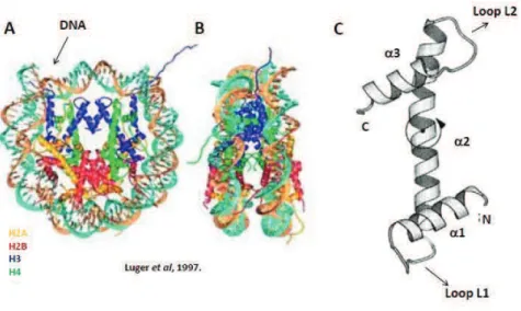
The basic and repeating unit of chromatin is the nucleosome. The nucleosome comprises about 146 base pairs of DNA wrapped around a histone octamer in 1.7 left helical turn. The Histone octamer contains 2 copies of each core histone H2A, H2B, H3 and H4 (Luger, Mader et al. 1997) (Figure 1A, B). Core histones can be divided into two categories: replication-dependent (RD, or ‘canonical’) and replication-independent (RI, or ‘variant’). Genes encoding for RD histones are organized into large co-regulated gene clusters (Marzluff, Gongidi et al. 2002), and are rapidly expressed during S-phase at high levels, coinciding with DNA-replication. RD histone mRNAs are the only known cellular non-polyadenylated mRNAs (Marzluff, Wagner et al. 2008). Canonical histones are namely H2A, H2B, H3 and H4, and they are incorporated into chromatin during DNA-replication through the action of specialized histone chaperones (Ellis 2013). Conversely, expression of RI histones, whose transcripts are polyadenylated and often contain introns, persists throughout the cell cycle. Histone variants include H2A.X, H2A.Z, H3.3 and others, and some have their own dedicated chaperones responsible
for their deposition and eviction onto and from chromatin (Weber and Henikoff 2014). Core histones consist of a highly conserved and structured central globular domain, called the histone fold domain (Figure 1C), and N- and C-terminal tails characterized by higher structural flexibility (Luger, Mader et al. 1997).
Figure 1. The nucleosome – basic unit of chromatin. (A and B) Crystal structure (at 2.8 Angstrom
resolution) of the nucleosome core particle (NCP), containing 2 copies of each core histone H2A, H2B, H3 and H4 (color coded) around which 146 base-pairs of DNA double helix are wrapped. (B) Lateral view of the NCP. (C) The conserved histone fold domain. Three a-helices (a1, a2 and a3) are connected by relatively unstructured linker loops (L1 and L2). Adapted from (Ramakrishnan 1997).
The majority of DNA-histone interactions are between structured histone regions, while the more disordered histone tails protrude from the nucleosomes and can interact with neighbouring nucleosomes and other factors. Histone tails are also subject to extensive posttranslational modifications, discussed below.
a. 10-nm fiber chromatin fiber and higher-order chromatin
structures
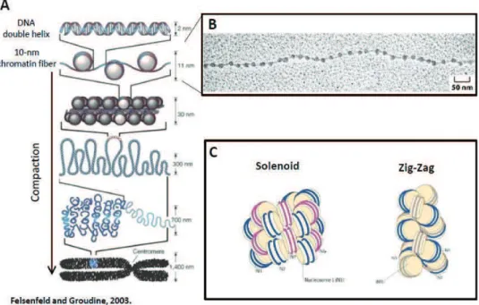
Nucleosomes are connected by short (10-80 nt) DNA segments, called linker DNA, and form nucleosomal arrays with a diameter of 10 nm. The existence of 10-nm fiber was observed by electron microscopy almost 40 years ago (Olins and Olins 1974) (Figure 2A, B). This so-called ‘beads on a string’ organization of nucleosomes is the first level of chromatin compaction (Kornberg 1974) and is generally permissive to transcription. Nucleosomal arrays can form short-range interactions with neighbouring nucleosomes to form chromatin fibers. Given the modularity of the nucleosomal composition and chemical modifications that the chromatin can be subject to, the possibilities to modify primary chromatin structure are virtually endless. Changes in nucleosome structure and stability can influence formation of higher-order structures.
Under physiological conditions, chromatin rarely exists in the simple and open mode of 10-nm fibres. Between the extended conformation of the 10-nm fiber and the highly compacted mitotic chromosomes there are several levels of chromatin organization that are less well understood. The secondary structure of chromatin includes internucleosomal contacts and comes about by folding of individual fibers into a defined fiber. Chromatin secondary structures are stabilized by linker histone H1 (or H5) and non-histone chromatin protein, such as HP1, Polycomb group proteins and others. In vitro experiments have shown that nucleosomal arrays can form helical structures of 30 nm in diameter, containing 6 to 11 nucleosomes per turn (Gerchman and Ramakrishnan 1987). This secondary structure, termed the 30-nm fiber, was proposed to be involved in chromatin compaction and transcriptional repression. Two models of 30-nm fibre structure have been proposed. The first one is the solenoid model, which proposed a single starting point of the 30-nm fiber, with a central axis of symmetry around which the fiber is formed (Finch and Klug 1976). Conversely, the zig-zag model predicts that the 30-nm fiber has 2 starting points and every other nucleosome interacts with each other
to stabilize the structure (Dorigo, Schalch et al. 2004)(reviewed in (Luger, Dechassa et al. 2012) (Figure 2C).
Figure 2. Levels of chromatin compaction. (A) Schematic representation of different levels of
chromatin compaction, ranging from the extended 10-nm fiber, to the fully condensed mitotic chromosomes. (B) Electron micrograph of the so-called ‘beads on a string’ 10-nm fiber. From Alberts B et
al, 2004. (C) Schematic of 2 proposed models of 30-nm fiber – the one-start solenoid model and the two-start zig-zag model. Adapted from (Luger, Dechassa et al. 2012).
Recent experiments designed to elucidate which of the models is predominant, including mesoscopic modeling, demonstrated that there is not one uniform type of helical fiber organization but rather conformational heterogeneity of nucleosome interactions (Grigoryev, Arya et al. 2009). It is now thought that the 30-nm fiber consists largely of zig-zag stacked nucleosomes interspersed with other structures (including solenoidal) with different levels of organization. Thus, it seems that the 30 nm fiber encompasses different chromatin structures which are not mutually exclusive. However, evidence for the 30-nm fibre existence in vivo remains elusive.
An alternative emerging concept is that chromatin in vivo is in a dynamically disordered state of a polymer melt, whereby linearly non-neighbouring nucleosomes can interact with each other (Sanyal, Bau et al. 2011).
Intramolecular interactions between secondary structures are thought to produce tertiary chromatin conformations, like such observed in mitotic chromosomes. The predominant view of how high condensation of chromatin in mitosis comes about was through sequential hierarchical coiling of the 30-nm fiber. This would presumably allow for the formation of non-random and constrained rod-like structures with reproducible dimensions. However, since the very existence of 30-nm fibers in vivo is questionable, a less-well defined organization of mitotic chromosomes is now proposed, in agreement with a study suggesting a disordered and random aspect of mitotic chromatin condensation (Nishino, Eltsov et al. 2012). In this model, the interactions of over-crowded and irregularly spaced nucleosomal arrays give rise to the physical arrangements of metaphase chromosomes similar to a ‘molten globule’ or ‘melted polymer’ state (Figure 3A). This model has one important shortcoming – it does not account for the rod-like structures observed in cytological preparations of mitotic cells. Most likely additional factors, such as condensin, influence the final physical properties of chromatin tertiary structures (Figure 3B).
Figure 3. Polymer melt model of chromatin structure. (A) Individual 10-nm fibers fold into different
types 30-nm structures. Nucleosomal concentration and crowding of 30-nm fibers causes interfiber interactions, which leads to the formation of disordered higher-order chromatin structures. Intramolecular contacts are further stabilized by the presence of divalent cations. (B) Mitotic chromosomes consist of disordered and diverse chromatin structures, which are stabilized by frequent inter and intranucleosomal protein-protein interactions and other factors, such as condensin.
b. Heterochromatin and euchromatin
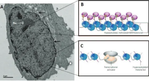
Chromatin structure must be viewed through the prism of specific biological functions. It can range from very distinctive micro-scale domains, such as centromeres, to the nanoenvironment of active promoters. From a functional point of view, chromatin can be roughly divided into 2 categories: accessible and inaccessible. The former, usually referred to as euchromatin, is gene rich, transcriptionally permissive, replicates early and is characterized by a loose chromatin structure. On the other hand, heterochromatin is gene poor, tightly packaged, replicates late and is generally refractory to transcription and DNA damage repair.
Figure 4. Heterochromatin and euchromatin. (A) Electron micrograph of a nucleus mouse embryonic
fibroblast (acquired by Andre Eid). Regions of high and low electron density can be observed, corresponding to heterochromatin and euchromatin, respectively. Nuclear membrane is showed by an arrow and the nucleolus by the letters Nu. (B and C) Schematic depiction of genome organization in heterochromatin and euchromatin. (B) Heterochromatin is characterized by high nucleosome density and chromatin condensation and lack of histone acetylation. (C) Euchromatic regions display less chromatin condensation and associated with histone hyperacetylation, and are accessible to transcription factors. B and C are adapted from (Grewal and Elgin 2002).
The dichotomy between euchromatin and heterochromatin on a nuclear level was observed almost a century ago, in 1928, in cytological experiments by Emil Heitz. Euchromatin and heterochromatin are generally associated with distinct sets of histone posttranslational modifications and chromatin factors (Figure 4). During S-phase, euchromatic regions are replicated first, while heterochromatic regions are replicated only at the end of the S-phase (Rhind and Gilbert 2013). However, it is important to note that chromatin organization in a cell is a lot more ‘fine tuned’ and specific. For instance, heterochromatin can further be subdivided into facultative and constitutive heterochromatin. Facultative heterochromatin is formed in gene rich regions to ensure proper regulation of developmental genes. Facultative heterochromatin can become ‘reactivated’ depending on developmental and signaling cues. On the other hand, constitutive heterochromatin is formed on centric, pericentric, telomeric regions that harbor repetitive DNA elements and imprinted genes, which are silenced in all cells of the organism in a heritable manner. Regions of constitutive heterochromatin remain condensed throughout the cell cycle and often associate with distinct subnuclear compartments (Zhao, Bodnar et al. 2009; Towbin, Gonzalez-Sandoval et al. 2013). Furthermore, constitutive and facultative heterochromatin are characterized by distinct sets of histone PTMs and associated proteins. Constitutive heterochromatin is marked by H3K9me3, H4K20me3 and HP1 binding (Grewal and Elgin 2002)). Regions of facultative heterochromatin are associated with Polycomb-group proteins, H3K27me3 and specific histone variants, like macroH2A. So, although both chromatin types repress gene activity, they are quite specialized, with different subnuclear localization, biochemical properties and functions. Interestingly, a comprehensive study of chromatin components in fly cells subdivided genome organization into 5 principal chromatin types with distinctive chromatin characteristics (Filion, van Bemmel et al. 2010). While the main division between heterochromatin and euchromatin remains, the researchers were able to further distinguish specific types of transcriptionally repressive versus permissive chromatin. They also reveal that a large portion of the genome, about 48%, is associated with transcriptionally inert chromatin (so-called ‘black’ chromatin). Interestingly, ‘black’ chromatin was not enriched in canonical marks of constitutive heterochromatin (such as H3K9me3 or H4K20me3).
To make things even more complex, a temporal dimension needs to be taken into account. Changes into chromatin are introduced constantly, and genes can dynamically fluctuate between expression and repression during development and differentiation, and depending on environmental and/or intracellular signals.
Clearly, the structure of chromatin is set in place to ensure proper genome packaging and the transmission of genetic material to future generations, but at the same time needs to be flexible and reversible to allow for DNA-based processes, such as transcription and replication, to take place.
2. Studying chromatin dynamics in living cells
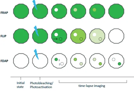
With the advance of optical and biophysical techniques, investigations into the dynamic properties of nuclear proteins were rendered possible. Microscopic techniques using fluorescently labeled molecules, termed F-techniques, have been particularly useful for such studies. A brief description of most commonly used F-techniques is outlined in table 1. Using FRAP (Fluorescence recovery after photobleaching) and FLIP (Fluorescence loss in photobleaching) (Figure 5), the kinetic properties of nuclear proteins involved in diverse processes, such as chromatin organization and rRNA processing, was determined in HeLa and BHK cells (Phair and Misteli 2000). The authors expressed GFP-tagged proteins (for instance HMG-17-GFP, fibrillarin-GFP) and photodestroyed flourescense in an area of the nucleus. Through measurments of fluorescence recovery in the bleached area over time, they could conclude on the dynamics of investigated proteins in living cells. In fact, rapid and complete recovery of fluorescence in bleached areas was observed for all proteins investigated, and ATP depletion did not affect protein recovery rates, suggesting energy-independent, diffusion-based processes and the absence of immobile protein fraction.
Table 1. Most commonly used F-techniques for measuring protein dynamics in living cells.
However, since the recovery was slower compared with GFP or reported dynamics of free solutes in the nucleus, it was hypothesized that protein-protein interactions within the nucleus cause this reduction in kinetics of the proteins analysed. These observations raised an interesting question: How do proteins reside within a subnuclear compartment and at the same time move throughout the nucleus in an unrestricted manner? The authors proposed that the compartments themselves are in constant turnover, and that proteins that occupy them roam (more or less) freely throughout the nucleoplasm in search for appropriate interactors.
Figure 5. Illustration of the experimental setup for 3 commonly used F-techniques, FRAP, FLIP and FDAP. All techniques are based on measuring changes in fluorescence levels in a defined region
over time. In FRAP and FLIP, flourescence is photodestroyed in a given area while in FDAP, laser power is applied to photoconvert a molecule from a non-fluorescent to fluorescent state. Regions of interest are marked by a circle and the laser photodestruction /activation by ( ).
The histone octamer is in the centre of the nucleosome, with 146 base pairs of DNA wrapped around it and it provides the structural basis for chromatin. Thus, the mobility of histone proteins in the octamer is generally very low, with recovery rates of hours (Kanda, Sullivan et al. 1998), (Kimura and Cook 2001). Even so, differential mobilities can be observed if one compares H2B-GFP and or H4-GFP. In HeLa cells, the H3-H4 tetramer is very stable on chromatin and there is very little exchange of H3-H3-H4 tetramers, as observed by FRAP experiments (Kimura and Cook 2001), while a small pool of H2B-GFP on the nucleosome surface is more mobile and exchanges continually. Nonetheless histone-GFP recovery curves are often used as controls in FRAP (and other F-techniques) experiments, since they represent virtually immobile proteins, as anticipated by their function in chromatin organization and nucleosomal DNA wrapping. Conversely, many chromatin-associated proteins, including HP1
proteins, linker histone H1 and a number of transcription factors, show relatively low residence time on chromatin and exchange rapidly.
Early FRAP experiments showed that HP1a is very mobile in cell a nucleus (Cheutin, McNairn et al. 2003), which was suggested to be a mechanism of heterochromatin maintenance. A more recent study by P.Hammerich and colleagues combined FRAP with FCS to provide a more detailed characterization of HP1a/b/g kinetics in living cells (Schmiedeberg, Weisshart et al. 2004). Initial FCS measurements revealed at least 2 populations of HP1a molecules in the nucleoplasm – a highly mobile fraction with uniform GFP signal, and a much less mobile one concentrated in bright GFP spots, which likely represent large and stable structures where a high fraction of fluorescent HP1s accumulate. Further investigation into these HP1 foci revealed 2 populations of HP1 molecules and a presence of a small but consistent immobile fraction (~5%), which was not observed in euchromatin. Interestingly, upon transcription inhibition or chromatin condensation, this very slow population of HP1 molecules can increase to ~20%. Interestingly, differences between different HP1 isoforms can be observed regarding their mobilities. During interphase, HP1g is the fastest isoform, while in mitosis only HP1a is associated with chromatin, enriched at pericentromeres where it exhibits very slow mobility and contributes to heterochromatin maintenance. The presence of three distinct binding sites for HP1a/b was later confirmed in a comprehensive study using fluorescence fluctuation microscopy. One binding site is present everywhere on chromatin with low residence time, one enriched in heterochromatin and one found only in heterochromatin. Interestingly, the enrichment of HP1a/b in heterochromatin was correlated with the presence of H3K9me2/3 and the corresponding Suv39 methyltransferases (Muller, Erdel et al. 2009).
Most of the studies on chromatin mobility have been performed in fully differentiated or transformed cell lines, such as HeLa cells or 293HEK, presumably due to their extensive characterization, availability and ease of manipulation. These studies have provided invaluable insight into the behavior of nuclear factors and chromatin organization. However, a shortcoming in using these systems is that they provide little
information about the putative changes in chromatin dynamics during transitions in cellular states, like those observed during differentiation or lineage allocation in vivo.
This limitation was partially overcome by using pluripotent mouse ES cells, which have the ability to self-renew but also the potential to differentiate into various other cell types. Interestingly, the chromatin structure of ES cells was shown to be different from that of differentiated cells, and lacked the typical prominent DAPI dense heterochromatin foci enriched in H3K9me3 and HP1a (Meshorer, Yellajoshula et al. 2006). When the behaviour of architectural chromatin proteins was investigated by FRAP in ES cells, it was observed that most core histones, linker histone H1 and HP1a in heterochromatin exhibit a highly dynamic and loose binding to chromatin. This behavior was attributed to pluripotency as it was not observed in differentiated cells which underwent lineage commitment. The authors confirmed the decreased binding of H3-GFP and H2B-GFP in biochemical essays and argue that loose association of core histones to chromatin contributes to the maintenance of pluripotency of ES cells. Recently, it was shown that H2A and H2A.Z mobility decreases upon ES cell differentiation (Subramanian, Mazumder et al. 2013), and that H2A.Z dynamics are at least partly controlled by its acidic patch. ES cells in which the linker H1 histone was tightly associated with chromatin failed to differentiate, suggesting the importance of dynamic exchange of linker histone in ES cell differentiation and lineage commitment. Interestingly, the histone variant H3.3 exhibited low mobility in both ES cells and neural progenitor cells (NPC) after differentiation. This finding was quite unexpected as H3.3 has long been associated with promoters of active genes and known to disrupt the stability of the nucleosome core particle (Jin and Felsenfeld 2007). A potential explanation for this observation is that H3.3 is indeed present on active genes, where it needs to be retained to mark their activity. This is indeed demonstrated by genome-wide mapping of H3.3 in ESCs, which revealed that H3.3 is enriched along the body of transcribed genes (Goldberg, Banaszynski et al. 2010). However, the genes in question are different in distinct cell types, such as ES cells and NPC cells.
Studying chromatin dynamics also entails investigating overall physical movements of chromatin fibres. Studies in budding yeast by the lab of Susan Gasser and others have
shown that chromatin fibres are constantly moving in the nucleus, and not only due to the temperature-dependent random Brownian motion (Dion and Gasser 2013). In fact, various factors, including ATP levels, protein-protein interactions and nucleoplasmic content affect chromatin fiber ‘walking’. Interestingly, chromatin remodeling complexes play an important role in chromatin fiber dynamics. It was recently shown that homology search upon double-stand break repair increases the movements of chromatin fibers and this increase depends on the Ino80 remodeling complex (Seeber, Dion et al. 2013). Therefore, when investigating overall chromatin dynamics, one should take into account local association of proteins to chromatin, but also the three-dimensional displacements of chromatin structures themselves. With the advance of optical techniques, such as 3D-FRAP, accurate measurements of overall chromatin dynamics will be possible. Indeed, 3D-FRAP provides a unique possibility to measure not only lateral diffusion, but also recovery of fluorescence within a 3D volume (Braeckmans, Peeters et al. 2003).
3. Modulating chromatin organization and function
Chromatin structure is by no means static and ‘locked’ – it is constantly challenged by cellular events, such as passage of the replication fork or the transcriptional machinery as well as the result of DNA damage and repair. Indeed, ever growing body of evidence suggests that structural uniformity is not a predominant feature of chromatin. To ensure that chromatin retains its correct state but also the ability to change upon stimuli, several mechanisms have evolved. Chromatin structure is modulated by three main players, which are discussed below. Of note, the role of non-coding RNAs in chromatin regulation through recruitment of histone methyltransferases (Volpe, Kidner et al. 2002), HP1a (Maison, Bailly et al. 2011) or chromatin condensation (Verdel, Jia et al. 2004), (Moazed 2009) is rapidly emerging. Prominent examples of long non-coding RNAs important for facultative heterochromatin are Xist and its antisense counterpart Tsix, which directly influence transcriptional silencing of one of
the X-chromosomes in female cells (Avner and Heard 2001; da Rocha, Boeva et al. 2014).
a. Chromatin remodeling complexes
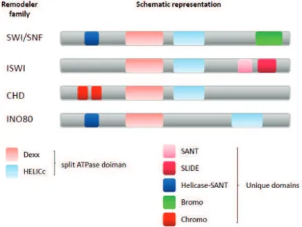
Chromatin remodelers use the energy of ATP hydrolysis to slide, destabilize or evict nucleosomes and thus change the local chromatin organization rendering it more or less accessible. It should be noted, however, that nucleosomes can also undergo spontaneous sliding, splitting and dissociation, without the need of remodelers (Miyagi, Ando et al. 2011). Although chromatin remodelers are divided into four families and differ in biological output and targeting mechanism, all of them use energy to introduce changes into chromatin (Figure 6). Furthermore, all remodelers contain a regulated DNA-dependent ATPase domain, recognize histone modifications (discussed in the next subsection) and prefer nucleosomes to DNA itself as a substrate. The 4 known families of chromatin remodelers are: SWI/SNF, ISWI, CHD and INO80 (Clapier and Cairns 2009). Some families, such as SWI/SNF, promote disruption and ejection of nucleosomes, while others, exemplified by the ISWI family, reassemble and organize chromatin (Clapier and Cairns 2009). Within individual families certain remodelers promote chromatin compaction while others loosen up the tight nucleosomal packaging. For instance, ISWI family members ACF and CHRAC promote chromatin assembly and repress transcription (Whitehouse and Tsukiyama 2006), while NURF assists RNAPII activation through randomizing nucleosomal spacing. These findings underscore the importance of domains other then the catalytic one as well as of proteins associated to the remodeler in mediating diverse functions on the chromatin. Often it is the non-catalytic domains, such as bromodomains or PHD domains which target or anchor the remodeling complexes to a particular locus. Many chromatin remodelers have particular functions. SWR1, a member of the INO80 remodelers, is unique in its ability to restructure the nucleosome by incorporating the H2A variant H2A.Z (discussed below) (Kobor, Venkatasubrahmanyam et al. 2004). Furthermore, during Drosophila
development, Hox gene expression is maintained by the action of NURF and its association with H3K4 trimethylation (Badenhorst, Voas et al. 2002). On the other hand, Mi2, a member of the CHD family of remodelers, is required for achieving the Polycomb-based repression of Hox genes (Kehle, Beuchle et al. 1998).
Figure 6. Schematic representation of 4 families of chromatin remodelers. Chromatin remodelers
contain the conserved split ATPase domain and various specific domains. Members of INO80 families are characterized by an extended linker region between the Dexx and HELICc domains. Different remodeler families are associated with unique domains contributing to their specificity and function.
It is important to note that complex chromatin processes such as chromosome segregation and DNA repair, often require the concerted action of several distinct remodeling complexes. The vastness of cellular processes that require chromatin remodeling makes it logical for so many different types of remodelers and associated proteins to evolve. It is then not surprising that disruption or aberrant function of chromatin remodeling complexes can lead to a number of diseases, including cancer.
b. Post-translational modifications of histones
Histones and chromatin-associated proteins are subject to posttranslational modifications (PTMs). These modifications range from covalent attachment of simple chemical groups, such as the methyl group, to addition of big globular proteins such as ubiquitin or SUMO. There are more than 10 types of different histone PTMs, which are outlined in Table 2. While many types, such as phosphorylation and methylation have been known for a long time, with the development of sensitive proteomics methods many more histone PTMs are coming to light, with their functions still unknown. It is becoming increasingly clear that the number of PTMs as well as their target sites on histones have been greatly underestimated (Tan, Luo et al. 2011).
Table 2. Known histone PTMs and their functions. There are 13 different types of
posttranslational modifications, and some of them can occur on the same residue or exist in several forms (e.g. mono-, di- and tri-methylation). Modified from (Kouzarides 2007).
Histone PTMs can act in 2 distinct ways. First, they can directly affect chromatin structure by altering the charge of a residue, and therefore the affinity of histones to DNA or impair internucleosomal contacts. The best studied case for this is acetylation of H4K16. It was shown that H4K16ac disrupts higher order chromatin structure by neutralizing the positive charge of the lysine and thereby changing the affinity of the H4 tail to the acidic patch of the neighboring nucleosome (Dorigo, Schalch et al. 2003; Shogren-Knaak, Ishii et al. 2006). Not surprisingly, H4K16ac was found incompatible with formation of highly condensed mitotic chromosomes and this modification is removed from chromatin during G2/M phase of the cell cycle. More generally, histone hyperacetylation is associated with regions of open chromatin which are transcriptionally active. Another example is H3K122 acetylation. Lysine 122 on histone H3 is located within the histone fold domain, on the lateral surface of the histone octamer. The authors have shown that the H3K122 acetylation is sufficient for transcriptional stimulation, and they attribute this to direct effects of H3K122ac on weakening histone-DNA binding (Tropberger, Pott et al. 2013).
Secondly, histone modifications can act indirectly, by creating a docking site(s) for effector proteins which contain specific domains or modules that recognize modification or their combinations. For instance, bromodomain-containing proteins recognize acetylated histones, while PHD fingers and chromodomains dock onto methylated sites (Yun, Wu et al. 2011). These proteins are often enzymes and can then recruit other proteins, change the structure of chromatin or help maintain it in its original state.
A prominent example of such action is H3K9me3 (Figure 7). H3K9 trimethylation is catalysed by SUV3-9 histone methyltransferases. Once this modification is set, it is recognized by HP1a through its chromodomain. HP1a also contains a chromo-shadow domain, through which it can associate with SUV3-9 to promote more H3K9me3, but also with the SUV4-20 HMT, which catalyses H4K20me2/me3. This leads to signal amplification and the expansion of heterochromatic domains (Grewal and Elgin 2002).
Figure 7. Deposition and spreading of H3K9me3 on chromatin. Suv3-9 represents the HMT
responsible for H3K9me3 deposition. H3K9me3 is recognized by the chromodomain of HP1 proteins. HP1s can interact with Suv3-9 through their chromoshadow domain and recruit it to pre-existing H3K9me3. This feedback loop allows for spreading of H3K9me3 and expansion of heterochromatic domains.
Many, but not all, of the PTMs occur on the unstructured tails of histones H3, H4 and H2A, which protrude from the nucleosomes (Figure 8). This is particularly interesting since the tails of neighbouring nucleosomes can interact with each other and help organize chromatin into higher-order structures. Thus, changing the properties of histone tails can have profound effects on overall chromatin structure. However, new histone modifications located close to the positions of the nucleosome that are in direct contact with the DNA are being uncovered and their impact on nucleosome stability is a subject of intense research. Because these residues have the ability to form physical contacts with the DNA backbone, they can potentially directly regulate nucleosomal stability.
Figure 8. Most characterized PTMs of core histones. Globular histone fold domain is represented by
the round shape, while and C-terminal aminoacids are annotated. Most histone PTMs occur on the N-terminal tail, and fewer on C-N-terminal part and in the globular domain (not shown).
Addition of small chemical groups
Acetylation
Histone acetylation entails the addition of an acetyl-group to specific lysines on histones. Histone tails, which are lysine-rich, are often acetylated at several neighboring residues and their hyperacerylation contributes to the overall modulation of DNA-histone interactions.
The enzymes catalyzing histone acetylation are called histone acetyltransferases (HATs) while the reversal of this PTM is achieved by histone deacetylatese (HDACs). Both types of enzymes are often associated with multiprotein complexes, such as the transcriptional coactivator SAGA, which contains the HAT GCN5 (Timmers and Tora 2005).
Almost exclusively, histone acetylation is linked to active transcription and chromatin ‘openness’. Due to the net increase in negative charge, histone acetylation weakens histone-DNA electrostatic interactions and promotes looser chromatin structure. Histone acetylation is enriched at active promoters and euchromatic regions in general. In fact, histone H4 lysine 16 acetylation is one of the few histone marks shown to directly antagonize higher order chromatin folding (Shogren-Knaak, Ishii et al. 2006). Interestingly, newly synthesized histones are hyperacetylated before their incorporation, but also during the transition from nucleosomal to protamine packaging of the sperm genome (Pivot-Pajot, Caron et al. 2003). Like methylation, histone acetylation can also act indirectly, by creating docking sites for effector proteins containing acetyl-recognizing folds called bromodomains (Jacobson, Ladurner et al. 2000).
Phosphorylation
Phosphorylation is the chemical addition of a negatively charged phosphate group to serine/threonine/tyrosine aminoacid residues. This modification is catalysed by protein kinases and is readily reversible through the action of phosphatases. Phosphorylation is one of the most studied protein PTMsin cells, and it can regulate protein function and localization very fast. On histones, 2 prominent examples of phosphorylation have been described. Firstly, serine 10 on histone H3 is known to be phosphoylated in mitosis by aurora B kinase (Johansen and Johansen 2006). This PTM promotes chromatin condensation but also antagonizes the neighboring H3K9 trimethylation, in a process termed phospho-methyl switch (Fischle, Tseng et al. 2005). A similar situation occurs between H3T3 phosphorylation and H3K4me3. It was shown that binding of TFIID to H3K4me3 is weakened in the presence of phosphorylated H3T3 during mitosis, concomitant with mitotic inhibition of transcription (Varier, Outchkourov et al. 2010).
Secondly, the histone variant H2A.X is known to be phosphorylated on its C-terminus within the aminoacid motif SQEY in response to DNA damage. Phosphorylated H2A.X, then called gH2A.Z is one of the first markers of DNA damage and serves as a signal and docking platform for repair enzymes, including Rad51 and Brca2 (Kang, Ferguson
et al. 2005). Furthermore, gH2A.X also recruits enzymes responsible for its own phosphorylation (ATM and ATR) which leads to the rapid amplification of the damage signal up to 1 megabase around a single double-stranded break and to an increased efficiency of DNA-damage repair.
The curious case of methylation
The covalent attachment of a methyl group to histone tails is a very interesting example of the fine-tuning of chromatin states as well as the importance of the context in which PTMs occur. Methylation can occur on lysine and arginine residues on histones, predominantly on H3 and H4. The complexity of this seemingly simple modification comes partly from the fact that lysines can be –mono, -di or –trimethylated, while arginines can be –mono and –dimethylated (symmetrically or asymmetrically). The enzymes responsible for setting methylation marks are termed histone methyltransferes (HMT). The catalytic domain of most HMTs, the SET domain, is highly conserved in eukaryotes. Many HMTs have been extensively characterized, including members of the Polycomb and Trithorax complexes, regulating methylation states of developmental genes, as well as suppression of variegation (SuVAR) HMTs important for silencing of constitutive heterochromatin (Elgin and Grewal 2003)
The mechanisms of methylation reversal were elusive for a long time, and it was thought that methylation was a very stable (if not irreversible) PTM. However, several histone demethylases have recently been identified (reviewed in (Shi and Tsukada 2013)), indicating that histone methylation can be enzymatically reversed. It is also possible that the combined action of enzymatic demethylation and histone exchange regulates methylation states at a given genomic region.
Interestingly, methylation can function both in transcriptional activation as well as heterochromatin formation, depending of the modified residue as well as effector proteins recognizing the modification. H3K4me3, H3K36me3 and H3K79me3 are modifications associated with open and transcriptionally active regions of the genome.
On the contrary, H3K9me3, H4K20me3 and H3K27me3 are most often found in heterochromatin. Thus, methylating histone tails provides a myriad of combinatorial possibilities to precisely regulate the chromatin status of a given region. While the presence of an active mark at a promoter usually excludes the presence of a repressive PTM, this is not always the case. Bivalent promoters, found at many developmentally regulated genes in ES cells, contain both H3K4me3 as well as H3K27me3 (Bernstein, Mikkelsen et al. 2006). The presence of histone PTMs with opposing roles on a single promoter is probably important for maintenance of pluripotency in ES cells, allowing for low-levels of transcription but also the competence of a promoter to become fully active or silent upon differentiation cues. During differentiation, transcription of a bivalent gene can thus be strongly activated by removal of H3K27me3, or completely silenced by demethylation of H3K4me3.
Addition of globular proteins
Ubiquitylation
Ubiquitin and SUMO are large (~7 kDa) globular proteins which can be enzymatically attached to histone tails.
Two ubiquitylation events with opposing roles have been mostly investigated. Ubiquitylation of histone H2B on lysine 120 in metazoans is present on almost 5% of H2B molecules in the nucleus, and is a mark of active transcription. This mark, catalyzed by the Bre1-Rad6 E2-E3 ubiquitin ligases(Jentsch, McGrath et al. 1987; Hwang, Venkatasubrahmanyam et al. 2003), is transient in nature, and its removal is achieved through the action of the histone-acetyltransferase deubiquitylation (DUB) module of SAGA. H2BK120ub functions to facilitate the smooth passage of the transcription machinery, in two separate ways. Firstly, H2BK120ub promotes the formation of H3K4me3 and H3K79me3, both important marks of active transcription, possibly by creating a docking platform or ‘bridge’ for COMPASS and Dot1 HMT.
Secondly, it facilitates the work of the FACT remodeler in reestablishing chromatin structure after the passage of the RNAPII (Braun and Madhani 2012). How these distinct types of H2BK120ub action are achieved is still not fully understood. The possibility that the ubiquityl moiety (76 aminoacids) physically interacts with different HMTs and FACT provides an attractive explanation, which is yet to be experimentally confirmed.
Conversely, H2A monoubiquitylation on lysine 119 (in mammals) plays a role in chromatin compaction. H2A and its variant H2A.Z are monoubiquitylated by several E3 ligases, including the members of the Polycomb repressive complex 1 (PRC1), Ring1B and Ring1A. This histone PTM is located on many sites of facultative heterochromatin, including silenced developmental genes and the inactive X chromosome in females. As is the case with H2B, H2AK119ub is a transient mark, and it can be removed by the PR-DUB complex, making H2AK119ub a tightly regulated mark under the control of Polycomb group proteins (Osley 2006; Scheuermann, de Ayala Alonso et al. 2010). Monoubiquitylation of H2AK119 generally acts as a repressive mark. It was shown that it can cause RNAPII pausing whilst not affecting its initial recruitment to promoters (Stock, Giadrossi et al. 2007). Furthermore, H2AK119ub provides a recognition site for its catalyser, PRC1 complex and it can also recruit the PRC2 complex, which methylates H3K27 and helps to tighten the overall chromatin structure (Margueron, Justin et al. 2009).
Thus it seems that H2AK119ub and H2BK120ub function in a mechanistically similar way, providing docking sites for effector-proteins and changing chromatin accessibility, but with opposite biological outcomes.
c. Histone variants
Histone variants can replace canonical histones in chromatin and can assume different roles in the cell. They differ from their canonical counterparts in various ways. Their primary sequence can be very similar- as in the case of the replacement variant H3.3 compared to the canonical H3.1 and H3.2- or extremely divergent –as in
macroH2A compared to H2A- from the canonical histones (figure 9). Their genes are located outside of the histone clusters and often contain introns. Furthermore, they are synthesized and incorporated into chromatin throughout the cell cycle.
Figure 9. Schematic representation of different H2A and H3 variants. Upper panel (A) shows the 5
most studied somatic H2A variants. Regions of divergence between H2A variants are marked in different color compared to H2A. Lower panel (B) represents the main types of histone H3 variants in mammals. The aminoacids that differ in each of the H3 variants are annotated and numbered.
Considering their conservation, histone variants can be roughly divided into two subgroups. The highly conserved ones like H2A.Z and CenH3 have evolved to perform essential functions in cells (DNA damage response, heterochromatin boundaries, formation of the centromere) and cannot be replaced by their canonical counterparts. Others, like H2A.Lap1, tH2B (now (TS)H2B.1) and H2A.Bbd (now H2A.B) are evolving rapidly and are seemingly undergoing Darwinian selection. These histone variants are evolving quickly to fulfill specific roles in certain cells or tissues, and are often specific to the germline. Because of the diversity and the increasing number of histone variants identified to date in different organisms, a new unifying nomenclature has recently been proposed (Talbert, Ahmad et al. 2012), to which I will adhere throughout the text.
Because this constitutes a major interest of my thesis, histone variants and their roles in early mouse development are discussed at length in a separate chapter below.
4. Heritability of chromatin marks
In the post-genomic era, it became clear that the vastness of cellular processes and phenotypes cannot be explained only by the information encoded in the DNA.
Concomintanly, a myriad of chromatin modifications, as well as their combination, were discovered and have become the focus of intense research in trying to undestand how different functional outputs come about from simple nucleotide sequences. Factors contributing to phenotypic changes, not caused by changes in DNA sequence, including histone modifications, regulatory RNAs, histone variants and nuclear localization, were referred to epigenetic ('above' genetic).
From its first definitions (proposed by Conrad Waddington), what is considered
epigenetic has changed substantially. Currently, epigenetics is defined as the study of heritable changes in gene activity that are not caused by changes in DNA sequence. While modulation of chromatin structure can indeed result in altered gene expression and phenotypic changes, histone modifications are not always epigenetic in nature. Many histone PTMs are transient in their temporal character (such as histone
acetylation necessary for their incorporation during S-phase, or H3K36me3 after the passage of the transcriptional machinery), and are thus not transmitted to the next generation. On the other hand, some genomic regions, such as pericentromeres, are stably silenced in a heritable manner. Indeed, even after replication and dilution of the original chromatin marks, positive feedback loops like that described above for
H3K9me3 ensure that the information of transcriptional silencing of pericentromeres is transmitted to daughter cells.
Subnuclear localization of certain parts of the genome was also considered as epigenetic. A specialized form of chromatin domains are found at the nuclear lamina (NL), and are called lamina-associated domains (LADs). The Nuclear lamina provides the interface between the inner nuclear membrane, the nuclear pore complex and
nearby chromatin, and is usually a transcriptionally refractory region. LADs contact with NL is linked to transcriptional repression mediated through the G9a methyltransferase and H3K9me2. Strikingly, however, after cell division and on a single cell level, the positions of the LADs within the nucleus are not inherited but instead randomly rearranged. (Kind, Pagie et al. 2013)
Heritability of chromatin states can also be assessed on different timescales. It is clear that somatic cells retain and propagate their epigenetic states to daughter cells.
However, in mammals, genome-wide erasure and re-setting of chromatin marks takes place in the germline and after fertilization. Thus, transgenerational epigenetic