HAL Id: tel-01556082
https://tel.archives-ouvertes.fr/tel-01556082
Submitted on 4 Jul 2017
HAL is a multi-disciplinary open access archive for the deposit and dissemination of sci-entific research documents, whether they are pub-lished or not. The documents may come from teaching and research institutions in France or abroad, or from public or private research centers.
L’archive ouverte pluridisciplinaire HAL, est destinée au dépôt et à la diffusion de documents scientifiques de niveau recherche, publiés ou non, émanant des établissements d’enseignement et de recherche français ou étrangers, des laboratoires publics ou privés.
Automation and integration of a bioreactor for
continuous cell culture
Fabien Abeille
To cite this version:
Fabien Abeille. Automation and integration of a bioreactor for continuous cell culture. Biotechnology. Université de Grenoble, 2014. English. �NNT : 2014GRENS036�. �tel-01556082�
THÈSE
Pour obtenir le grade de
DOCTEUR DE L’UNIVERSITÉ DE GRENOBLE
Spécialité : Biotechnologie, instrumentation, signal et imagerie pour la biologie, la médecine et l'environnement (BIS)Arrêté ministériel : 7 août 2006
Présentée par
Fabien ABEILLE
Thèse dirigée par Nathalie PICOLLET-D’HAHAN codirigée par Vincent AGACHE
préparée au sein du CEA-LETI, Laboratoire Bio-Chip et bio−Packaging (LBCP)
en collaboration avec BGE, équipe Biomics, IRTSV, CEA
dans l'École Doctorale Ingénierie pour la Santé, la Cognition et l'Environnement (EDISCE)
Automatisation et intégration d’un
réacteur de culture cellulaire pour
un fonctionnement en continu
Thèse soutenue publiquement le 25 Novembre 2014 devant le jury composé de :
M. Eric LECLERC
Chercheur à l’Université de Technologie Compiègne (UTC), Président
Mme Elisabeth M. J. VERPOORTE
Professeur à l’Université de Groningen (RUG), Rapporteur
Mme Rosaria FERRIGNO
Professeur à l’Université Claude Bernard Lyon (UCBL), Rapporteur
M. Donald K. MARTIN
Professeur à l’Université Joseph Fourier (UJF), Examinateur
M. Olivier THEODOLY
Chercheur à l’Université Aix-Marseille (AMU), Examinateur
Mme Nathalie PICOLLET-D’HAHAN
Ingénieure-Chercheur au CEA, Directrice de thèse
M. Agache Vincent
Ingénieur-Chercheur au CEA-LETI, Co-encadrant
Table of contents
Remerciements ... 5
1 Introduction ... 7
1.1 Motivations ... 7
1.2 Context ... 8
1.3 Aim of the work ... 8
1.4 Cell culture ... 9
1.4.1 Cell models & applications ... 9
1.4.2 Standard cell culture ... 10
1.4.3 Industrial cell culture ... 17
1.4.4 Microfluidic systems for cell culture ... 27
1.5 Conclusion ... 41
2 Thesis objectives ... 43
3 Microbioreactor design and fabrication ... 45
3.1 Microbioreactor overview ... 45 3.2 Cartridge design... 46 3.3 Perfusion integration ... 47 3.3.1 Perfusion strategy ... 47 3.3.2 Type of membrane ... 49 3.3.3 Bonding ... 50 3.3.4 Glue biocompatibility ... 55 3.3.5 Leakage tests ... 57
3.3.6 Optical considerations/cell visualization enhancement... 58
3.3.7 Conclusions ... 59
4 Cell proliferation ... 60
4.1 Control and monitoring of the cell environment ... 60
4.1.1 Porous-membrane-based perfusion ... 60
4.1.2 Thermal management ... 67
4.1.3 pH regulation ... 72
4.2 Microcarrier-based cell culture ... 77
4.2.1 Microcarrier preparation ... 77
4.2.3 Bioreactor (microcarrier) cell culture ... 81
4.3 Conclusion ... 83
5 Cell harvest ... 85
5.1 The different strategies for cell harvest ... 85
5.2 Thermally-mediated cell harvest ... 88
5.2.1 Poly(N-isopropylacrylamide) ... 88
5.2.2 Materials and methods ... 91
5.2.3 Physical characterization of PNIPAM-coated substrates ... 100
5.2.4 Biological characterization of PNIPAM-coated substrates ... 100
5.2.5 Discussion ... 104
5.2.6 Conclusion ... 108
5.3 Microcarrier dissolution ... 109
5.4 Bead-to-bead cell transfer for subculture ... 111
5.5 Conclusion ... 112
6 General conclusion... 113
7 Perspectives and future work ... 115
Appendix A ... 117 Appendix B ... 118 Appendix C ... 120 Appendix D ... 121 Appendix E ... 123 Appendix F ... 125 Appendix G ... 128 Appendix H ... 131 Appendix I ... 132 Bibliography ... 133
Remerciements
Je tiens à remercier le CEA pour avoir financé cette thèse. Je remercie aussi le Département des micro-Technologies pour la Biologie et la Santé et son Service Bio-Systems on Chip, et l’Institut de Recherche en Technologies et Sciences pour le Vivant de m’avoir accueilli dans leurs laboratoires, d’avoir investi leur confiance en moi et de m’avoir soutenu matériellement et financièrement pour la réalisation de cette thèse.
Cette thèse n’aurait pas pu être aussi riche en résultats, développement professionnel et personnel sans un grand nombre de personnes des laboratoires LBCP, LBAM, LCM, Biomics, LISA, LIPhy et bien d’autres encore. Les rencontres et les collaborations ont été essentielles pour mes recherches ainsi que pour ma construction en tant que jeune chercheur. Elles ont aussi été sources de motivations et de créativité. Certaines d’entre elles ont même donné naissance à des amitiés, qui je l’espère dureront par la suite.
Je tiens vivement à remercier les différents chefs des laboratoires LBCP, LBAM, LCMI et Biomics. Christine Louis et Béatrice Icard, chefs qui se sont succédées au LBCP, ont toujours prêté une oreille attentive à mes besoins. J’ai beaucoup apprécié l’intérêt de Béatrice pour ma thématique. Je suis reconnaissant envers Guillaume Delapierre, chef du LBAM, qui m’a généreusement permis de présenter mes travaux et d’échanger avec ses équipes. Je remercie également, Gilles Marchand, chef du LCMI, qui m’a ouvert les portes de son laboratoire et m’a donné de précieux contacts pour résoudre certains des problèmes techniques que j’ai pu rencontrer. Xavier Gidrol, chef du laboratoire Biomics, a toujours su être présent pour donner une opinion utile à l’avancée de cette thèse.
J’exprime une profonde gratitude envers tous les ingénieurs-chercheurs du CEA qui m’ont apporté de nombreux conseils scientifiques, leur aide et leur soutien pendant ces 3 ans de thèse. Je remercie tout particulièrement Fabrice Navarro, Florence Rivera, Guillaume Nonglaton, Pascal Maillet, Cédric Goyer, Jean Bertier, Jean-Maxime Roux, Nicolas Verplanck, Anthony Corfa, Cédric Allier, Ruth Griffin-Shea, François Baleras, Frédéric Bottausci, Cédric Poulain, Antoine Hoang, Tarek Fathallah, Eric Sulpice, Vincent Haguet, Amandine Pitaval, Danielle Gulino et Yoann Roupioz.
Je remercie mon collaborateur du LIPhy, Lionel Bureau, qui a généreusement accepté de travailler avec moi et qui a pris du temps pour me transmettre son savoir et ses techniques.
De nombreuses réalisations et la maîtrise de plusieurs techniques n’auraient été possibles sans les techniciens des différents laboratoires. Je les remercie pour le temps, la patience et la conciliation dont ils ont su faire preuve à mon égard. J’ai une pensée particulière pour Mathilde Menneteau, François Boizot, Manuel Alessio, Martine Cochet, Marie Escude, Frédérique Kermarrec, Nadine David, Patricia Obeid, Sophie Gerbaud, Frédérique Mittler, Guillaume Costa, Thomas Bordy et Jean-Guillaume Coutard.
Je remercie les post-doc et thésards que j’ai pu rencontrer lors de mon séjour au CEA. Ils ont été sources de réconfort, de joies et autres bons moments passés ensemble. Merci à Monika Dolega, Guillaume Laffite, Jonathan Bruniaux, Thomas Courant, David Lefebvre, Emilie Bisceglia, Prisca Dalle, Rapahël Renaudot, Pierre-Henri Cazorla, Jessica Morlieras, Srikanth Vinjimore Kesavan, Frédéric Fantoni, Marjorie Vrignaud, Johannes Theisen, Claire Authesserre, Lisa Racine, Alan Hibbitts, Aurélie Jacquart, Anthony Daures.
Je voudrais chaleureusement remercier mes encadrants de thèse avec qui j’ai beaucoup appris de leur personnalité et de leurs méthodes. Ils ont su tirer le meilleur de moi-même. Grâce à Patrick Pouteau j’ai pu développer mon autonomie, mon assurance et mes capacités de communication. Je suis profondément reconnaissant envers Vincent Agache d’avoir accepté l’encadrement de ma thèse suite au départ de Patrick Pouteau. Un grand merci à Nathalie Picollet-D’hahan qui m’a guidé du début jusqu’ à la fin. Nathalie et Vincent ont su être des encadrants particulièrement humains, attentifs, à l’écoute et source de motivation.
Je voudrais remercier les enseignant-chercheurs du Master Nanotech (PHELMA-INPG) pour la formation de qualité que j’ai pu recevoir et qui m’a conduit à faire cette thèse.
Je remercie aussi mes amis, ma famille et ma belle-famille pour leur soutien.
Pour finir, je remercie de tout mon cœur ma partenaire de vie, Zuza, pour sa patience et tous ces moments de bonheur qui permettent de surmonter les inquiétudes et le stress que peut donner une thèse. Je lui dédicace cette thèse.
1 Introduction
1.1 Motivations
Cell culture is a simple process used to produce cells and cell products. It requires many manual steps such as subculture (or reseeding), each two or three days associated with lots of manipulations, a periodic renewal of the culture medium and a control over the cell environment (temperature, humidity , CO2/pH, O2). These operations often lead to variability
and contamination.
In order to improve the consistency of the cell culture process and to reduce the risks of contamination, bio-industries have developed automated cell culture systems. Automation of bioreactors has improved the productivity and the quality of cell lines. However industrial bio-production remains a cumbersome, lengthy and costly process. Microbioreactors are expected to provide solutions to ease, shorten and reduce the costs of some of the steps involved in bioprocessing. For instance, enhancement of the clone or strain selection step and the media optimization step will be possible by the capacity of automated microbioreactors to perform higher throughput tests and to better address the cell microenvironment compared to the use of standard flasks or well plates.
Another example concerns the scaling-up phase during bioprocessing. The previously selected clones or strains can behave differently with the increasing culture volume of the bioreactors. Therefore, there is a need to perform more reliable upscaling cultures using bioreactors that better reproduce the full-scale cultivation.
Expectations are also rising on the side of research which strives to develop culture models closer to the in-vivo. Indeed, conventional methods (e.g. 2D cell culture, spheroids culture, use of animal models) have become questionable either on an ethical point of view or because the cell behavior can significantly differ from the one in the human body. As mentioned, microbioreactors are able to provide controlled microenvironments that can be used to create more relevant culture models. Their parallelization can allow obtaining large amount of quantitative data.
Microscale culture systems appear as promising tools to solve many shortcomings for both industrial bio-production and research. Based on these considerations, a microbioreactor has been developed during this thesis.
1.2 Context
This thesis project was done in collaboration between two labs both part of the French Alternative Energies and Atomic Energy Commission (CEA): the Biomics lab, led by Ph. D. Xavier GIDROL and the BioChip and BioPackaging lab led by Ph. D. Béatrice ICARD.
The Biomics lab relies on micro-technologies, microfluidics, micromanufacturing and MEMS (MicroElectroMechanical Systems) to identify genetic and microenvironmental determinants that control the proliferation/differentiation balance and carcinogenesis using cell prostate cancer models.
The BioChip and BioPackaging lab designs, manufactures and packages components for microfluidic devices with chemical/biological reagents and for implanted and embedded medical devices (electrodes).
1.3 Aim of the work
The aim of this collaborative project was to:
· initiate, in the host laboratory, the development a microfluidic system to perform continuous cell culture of anchorage-dependent mammalian cells
· provide novel tools to automate and simplify the steps involved in cell culture
· control the culture conditions by the integration of means to monitor and regulate the cell environment
1.4 Cell culture
This section intends to give a general description of the state of the art of cell culture at a laboratory and industrial level. Through this description, the limitations of the standard cultures are highlighted in order to introduce the advantages that microfluidic bioreactors could offer to address the remaining challenges.
1.4.1 Cell models & applications
Cell culture is defined as the process by which dispersed cells are grown under controlled conditions in order to produce cells or cell products[1]. It has been used since the ancient times by many civilizations for the production of alcoholic beverages, cheese, yogurt, etc., although they were not aware of it[2]. Such culture leading to fermentation involves yeasts and bacteria. The implication of these organism cultures was only discovered by Louis Pasteur in 1857[3]. Later, in the early 20th century, the tissue culture of plants and animal cells started to develop
with their respective pioneer Gottlieb Haberlandt[4] and Granville Harrison[1]. Tissue culture consists in the study of non-disaggregated cells from a tissue fragment. Coming from this type of culture, cell culture soon appeared when cells could be disaggregated cells from tissues with the use of a proteolityc enzyme, called trypsin[5]. This major discovery was one of the many that contributes in popularizing cell culture. The other main breakthroughs were the development of the first continuous cell line (HeLa)[6] which lead to the development of other cell lines and cell line banks, the introduction of antibiotics to preserve cell culture from microbial contamination and the development of chemically defined media to specifically support the growth of cell lines. Cell culture of various cell types can be achieved currently; however, the meaning of the words “cell culture” is often reduced to the culture of eukaryotes and more particularly to the culture of animal cells. The culture of bacteria, yeasts, fungi and protozoa is often referenced as “microbiological culture”, while the culture of cell plants is referenced as “tissue plant cell culture”. This classification of the different types of cell culture is not related to the conventional classifications of cells[7] but, seems instead, coming from the different fields of microbiology, botany and sciences related to medicine, existing in the late 19th before cell culture emerged. The applications of cell culture are numerous and diverse and are summarized in Figure 1.
Figure 1. Cell culture applications. Reprinted from[1].
1.4.2 Standard cell culture
1.4.2.1 The cell culture process
The following description of the cell culture process stands for any type of cells. The process of cell culture can be divided into 3 consecutive steps: seeding, proliferation and subculture. These phases are synchronized according to the growth kinetics of the cell type being cultured (Figure 2). To initiate a culture, a small population of cells is placed in a controlled environment: this is the seeding step. The cells comes usually from the cell bank (from the laboratory or an external one: ATCC for the U.S.A, ECACC for Europe…) or from a donor tissue. Then, follows the step of proliferation: the cell population remains stable the time to adapt to their new environment[8] (lag phase) and then starts to increase as the cells divide (exponential phase). If there is no human intervention, the cell growth rate eventually slows until becoming null. The plateau phase is either:
· The result of a lack of space available for the cells to keep on dividing. Indeed, in cell culture two categories of cells exist: adhering cells which need a substrate to adhere to in order to divide, and floating cells which can be cultured in suspension. After multiple divisions, anchorage-dependent cells will occupy the entire available space forming a monolayer, this state is called confluence. At this point, cells usually lose their ability to divide due to contact inhibition[9].
· the consequence of the depletion of a nutrient · due to the accumulation of cell wastes.
Generally, the plateau phase is preferably avoided since it can later alter the cell phenotype[10], lower their growth rate and initiate cell death. Moreover, cell populations in the exponential
phase are more consistent and uniform, which is important to obtain reproducible cell culture tests. The subculture step, also often referred as “passage”, is required before reaching this phase. During this step, cells are harvested and a fraction of them are seeded in a new controlled environment to propagate and divide again. The remaining cells are either used for experiments or simply discarded.
Figure 2. Cell growth kinetics driving the cell culture process. Cell culture is initiated by seeding a small number of cells in a new environment. The cells first adapt to their environment (lag phase), then they proliferate (exponential phase) and their number increases. Eventually, before reaching a critical number of cells, after which cells will not proliferate properly and die (plateau phase), subculture is performed. A part of the cells will be used for experiments while others will be used to perform another seeding and starts a new cell culture cycle.
All cells are characterized by a sigmoidal growth curve. However, each cell type exhibits specific growth kinetics. The exponential phase, the major phase of the growth kinetics, is related to the speed with which cells can divide, in other words their doubling time (time needed to increase the cell number twofold in the exponential phase). The doubling time corresponds to the cell cycle duration which is specific to a cell line or strain. For instance, bacteria have a doubling time, or cell cycle duration, of about 1 h[11] while mammalian cells have a doubling time of about 24 h[12]. The environmental conditions in which cells are cultured can vary this cell cycle duration and even block it[13]. Therefore, optimal growth requires optimal culture conditions.
1.4.2.2 Cell culture duration
There are many ways to classify cells in cell culture. Of these, only two will be described for the purpose of a general understanding. One, already mentioned, classifies cells according to their dependence to adhere or not to a substrate in order to proliferate: anchorage-dependent cells are called adhering cells and anchorage-independent cells are called floating cells.
The other classification separates cells into two groups:
· Finite cell line, which refers to a uniform population of cells that has been obtained after the subculture of primary cells (i.e. cells taken from a living tissue). These cells naturally die after a finite number of cell divisions due to senescence (i.e. naturally programmed cell death within cell genes)
· Continuous cell line, which corresponds to a cell line that has become immortal through
in vitro transformation. Such a transformation can be spontaneous or physically,
chemically or virally induced, and allows cells to escape senescence.
From these definitions, one can understand that the culture of continuous cell lines can be indefinite. However, the culture of continuous cell lines is commonly performed a finite number of passages. After many passages, continuous cell lines are likely to undergo selective pressures that may induce genotypic or phenotypic variations, impacting result reproducibility and consistency[14]. The limit of passages to consider before stopping the culture of a continuous cell line depends on the cell line itself and the phenotype that must be preserved[15]. As a rule of thumb, the American cell bank ATCC recommends not to use continuous cell lines after 5 consecutive passages. The limit of passages for the culture of an animal cell line before senescence also varies according to the cell type and specie[16,17]. In fact, any eukaryote cell will undergo senescence[18] as well as prokaryote cells[19]. However, prokaryote cell cultures are usually limited for the same reasons as for continuous cell lines[20].
1.4.2.3 Laboratory cell culture: a practical example of mammalian cell culture
After detailing the general process of cell culture, it is interesting to have a practical insight into the culture performed routinely in research laboratories. The given example will describe how the culture of mammalian cells is performed since it will be the main concern in this thesis. However, the procedure remains analogous for any type of other cell culture except for a few minor variations.
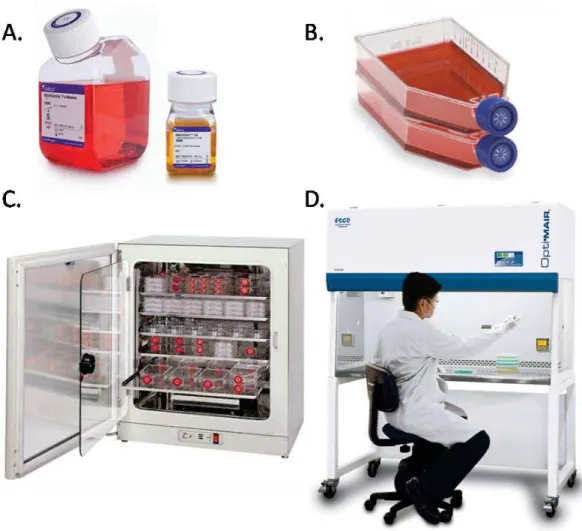
To initiate a culture, cells stored in a frozen vial are thawed. Then, they are seeded in a specific liquid culture medium (Figure 3.A) in a Petri dish or a culture flask (Figure 3.B). This gas permeable container is then stored in an incubator. The medium and the incubator (Figure 3.C) ensure that adequate conditions are provided to promote cell proliferation, which includes:
· Nutrients: The media contains, among other components, the nutriments required for the cell metabolism needs (see Appendix A for an example of the complete formulation of a culture medium). The more complex is the organism metabolism to be cultured, the more nutrients types are needed, which is the case for mammalian cells that have high nutritional needs. Their culture requires the presence of:
o Amino acids, the basic materials for cells to synthesize proteins
o Vitamins, which assists biochemical transformation through enzymatic activity o Glucose, which is a source of energy
o Inorganic ions, which play a role in cellular adhesion, cell signaling and the regulation of membrane potential and intracellular charges
o Metal traces are involved in the enzymatic activity of the cells
o Serum: serum is a complex mixture usually supplemented in the media. It is composed of hormones (regulating the uptake of glucose and amino acids), growth factors (having a beneficial impact on cell growth), proteins (acting as carriers for minerals, fatty acids and hormones) and other lipids, minerals and amino acids. The use of serum to supplement culture media is sometimes avoided as serum composition is variable and poorly defined from batch to batch, introducing potential cell culture results[21].
· Osmolality: Osmolality is mainly regulated by the salts, the glucose and the amino acids present in the media. Osmolalities between 260 mosmol/kg and 320 mosmol/kg are acceptable for mammalian cell culture. Higher or lower osmotic pressure will affect the cell membrane integrity and respectively induce the collapse or the explosion of the cells[22].
· Temperature: temperature is regulated by the incubator. The adequate cell culture temperature depends on the body temperature of the animal tissue (or the host for microbes). The target temperature is 37°C for most of the mammalian cells. Although mammalian cells can tolerate substantial drops in temperature, survive several days at 4°C, and be frozen to -196°C; they cannot endure more than about 2°C above normal for more than a few hours and will die quite rapidly at 40°C and over[23].
· pH: pH is regulated by the CO2 level in the incubator atmosphere and the hydrogen
carbonate ions contained in the media through acid-base equilibriums.
(Eq. 1)
(Eq. 2) The bicarbonate salt balances the dissolution of CO2 from the incubator atmosphere.
The CO2 partial pressure of the incubator is commonly regulated at 5% and the
concentration in bicarbonate is adapted to provide a buffering capacity to the media between pH 7.0 and 7.4. Some cell types may be best cultured slightly outside this range, and cells surely differ widely in their ability to tolerate significant deviations from
this level. Slow changes in pH are better tolerated than rapid changes that lead to apoptotic cell death. Most cells will tolerate a medium pH in the range of 6.5 to 7.8, but media much outside this range can degrade cell viability[22]. The buffer also compensates to some extent for any change in pH that would be induced by the wastes that cell produce such as lactic acid, CO2 or ammonia.
· Oxygen: more precisely dioxygen is needed for the respiratory system of the cells, even though some cells can be cultured in anaerobic conditions (i.e. deprived of oxygen). Mammalian cells have an oxygen dependent metabolism. The level of oxygen can be regulated in the incubator with the addition of an inert gas, such as nitrogen. Usually for mammalian cell culture, the level of CO2 in the incubator defines the level of oxygen. For
5% of CO2, the incubator atmosphere contains 18% of O2. However, the partial pressure
of O2 can also be actively regulated. The medium height is adapted to modulate the
diffusion of dioxygen according to the cell need[22]. Cells having high O2 requirement
are cultured in shallow media while cell with low O2 requirement are cultured in deep
media.
· Antibiotics are often added to prevent the cultures from being contaminated by other microorganisms. The most common is to use a combination of penicillin and streptomycin.
All requirements for cell culture are cell line and cell type specific. They have to be adapted accordingly.
Figure 3. The different components and equipment used for cell culture. A) Non supplemented culture medium (left) and serum (right) for cell culture. B) Vented flasks filled with culture medium. C) Incubator containing cell culture flasks. D) Operator working under a safety cabinet for cell culture.
As cells proliferate, they consume some of the medium constituents and release wastes, such as lactic acid, ammonia and CO2,proportionally to their density. Without human intervention, cells
will soon starve because of nutrient depletion and suffer from a drift of the pH due to the accumulation of cell wastes. Therefore, during proliferation, the medium is changed frequently (every 2 or 3 days for mammalian cells) depending on the cell metabolism and density.
Eventually, subculture is performed upon reaching a critical cell density. For adherent cells, cell harvest is achieved by enzymatic, chemical or mechanical action. The most common technique relies on the use of trypsin, a proteolytic enzyme that digests the cell membrane proteins involved in cellular adhesion. Floating cells are harvested simply by collection of the media in which they reside. A portion of the harvested cells are then placed in a new vessel containing medium and placed back into the incubator for proliferation. The remaining cells are used for experiments or are discarded.
The aforementioned steps of thawing, seeding, media change and subculture are all accomplished by an operator in a special outfit and under a laminar hood (Figure 3.D) to limit possible contaminations of the cells and the operator.
1.4.2.4 Manual laboratory cell culture limits
Cell culture is originally a manual laboratory practice extensively used in research that faces inherent challenges and limitations. As a manual technique, it is prone to human errors. Even if cell culture is performed by highly qualified operators with great care in order to preserve sterility, there is a non negligible risk for contamination due to repeated manipulations. Every cell culture laboratory has already experienced a microbial contamination.
Besides the frequent handling, media renewal and subculture expose cells to important environmental variations, such as thermal variations or dissolved species concentration variations. Indeed, cells need to be taken out of incubators for observation or to perform media exchange, exposing them to room temperature (20-25°C). Additional environmental variations may arise due to evaporation. Incubators are water vapor-saturated to limit media evaporation, but it still occurs. A reduction of the culture volume induces an increase in the concentration of salts and ions leading to osmolarity and pH drifts. All of these variations are not controlled in term of frequency and amplitude. It is established that they are likely to affect the cell phenotypes and to impact on the reproducibility of cell culture experiments[1,22].
Additionally, manual laboratory cell culture can hardly achieve mass production of cell or cell products. It also gives little and poor monitoring of the culture state (cell density, temperature, O2, pH, glucose, lactate, ammonia…).
As a consequence of the highlighted drawbacks, some laboratories and industries have turned to automated cell culture systems. These systems provide better sterile environment and reduce human intervention. They are usually coupled to a large number of probes that can regularly monitor the diverse culture parameters. Therefore, they can maintain the cell environment more accurately. Automated systems render cell culture less labor intensive and time consuming with the possibility to produce large amounts of cells and cell products[24]. Different classes of culture-automated systems exist. Some solely intend to provide cells to scientist for their research while others, called bioreactors or fermentors, aim at producing biomass or bioproducts. The next section will rather concentrate on the latter systems.
1.4.3 Industrial cell culture
1.4.3.1 Applications
Industrial cell culture concerns mammalian, microbial (meaning bacteria, yeast, fungi and some algae) and plant cells. The application of these cultures is diverse. They play a major role in:
· The food and the beverage industry with the production of food additives, food preservatives, alcohols…
· The chemical industry with the production of biopolymers, solvents, biofuels…
· The pharmaceutical industry with the production of recombinant products such as monoclonal antibodies, drugs, vitamins, hormones
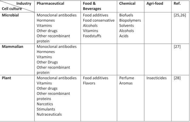
Table 1 gives a non-exhaustive list of product types synthesized by cells and used in the different industries.
Table 1. Specific use of cells in industries Industry
Cell culture
Pharmaceutical Food & Beverages
Chemical Agri-food Ref. Microbial Monoclonal antibodies
Hormones Vitamins Other drugs Other recombinant protein Food additives Food conservative Alcohols Vitamins Foodstuffs Biofuels Biopolymers Solvents Alcohols Acids [25,26]
Mammalian Monoclonal antibodies Hormones Vitamins Other Drugs Other recombinant protein [27]
Plant Monoclonal antibodies Vitamins Other drugs Other recombinant proteins Narcotics Stimulants Nutraceuticals Food additives Flavors Perfume Aromas Insecticides [28]
The main industry driving cell culture is the pharmaceutical industry[26] with global sales over US$120 billion per year, within which the production of monoclonal antibodies production represents a fifth of the sales with a growing trend[29].
Microbial cell culture dominates industrial cell culture because these cells are less sensitive to culture conditions, have simpler needs to grow, grow rapidly and produce in high yield (3-fold more than mammalian cells[30]).
However, plant and mammalian cells possess a higher level of expression which makes them capable of producing valuable pharmaceutical products that microbial cell culture cannot
achieve[26,31]. Although mammalian cells have been for a long time the most desired expression system to produce human compatible products[32], plant cell cultures are gaining interest and may provide in the future a more economical way to produce such human-like products[33,34].
Cells themselves can be produced as the end-product. For instance, microbial cells are used in wastewater treatment. Another example is stem cells, which are expected to impact medicine, healthcare and clinical applications for drug testing, tissue and organ repair and human disease curability[35].
1.4.3.2 Bioreactor culture modes
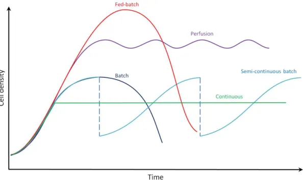
For the production of bioproducts, industrial cell culture relies on large-scale bioreactors. Higher volumes of culture mean higher number of cells and therefore higher quantity of product. Industrial bioreactors can be run in different ways. Four main operating modes exist and are from the least to the most complex to operate as follows: batch and semi-continuous batch, fed-batch, continuous, perfusion (Figure 4).
· Batch: Any culture mode is initiated with a batch culture. Cells and medium are dispensed inside the bioreactor and the culture volume remains constant over time. During the culture the cell population follows a sigmoid curve as described earlier in Figure 2. The medium nutrients, byproducts and products concentrations vary in time. The stability or the accumulation kinetics of the targeted product inside the bioreactor drive the time of harvest.
A variant of the batch is the semi-continuous batch which consists in repeated batch culture. Between two consecutive batches, a portion of the culture is kept to serve as an inoculum and fresh medium is added. This method presents the advantage to reduce the time and cost associated with cleaning, sterilization and re-seeding of the bioreactor. Runs over several months have been reported for the production of a monoclonal antibody using mammalian cells[36].
· Fed-batch: fed-batch culture is a batch culture during which the culture volume is continuously or periodically increased in time. The starting volume represents about 10% of the final volume reached at the end of the culture[37]. During this process, cells, nutrients, products and byproducts vary in time. This strategy aims at feeding the appropriate nutrients in order to maximize growth and product formation and lower the formation of toxic products such as ammonia or lactate. Fed-batch feeding strategies imply a good understanding of the cell metabolism and the rate of utilization by the cells of the nutrients. Complex organisms such as mammalian cells require more complex
mechanistic models of cell growth to provide the adequate feeding[38]. Higher cell densities (2.10^7 cells/mL) than in batch culture can be achieved as well as 10 to 50-fold higher titer (i.e. product concentration)[39].
· Continuous: In this mode there is constant flow in and out of the same amount of medium. Cell population, concentration of nutrients, product and byproduct remain constant during the culture. The feeding rate can be adjusted by controlling and measuring the biomass concentration; such bioreactors are called turbidostat. However, the most common type of continuous culture is the chemostat where the feeding of a limiting substrate regulates the biomass concentration. Other systems such as auxostats regulate the biomass concentration through a feedback loop based on the measurement of the pH, the dissolved oxygen tension or the product concentration (e.g. ethanol, sugar).
· Perfusion: As for continuous cultures, perfusion cultures are characterized by a constant supply and draw off of medium. Cells, nutrients, byproducts and products remain constant during the culture. The difference with the previous process is that cells are retained in the bioreactor. Perfusion process provides the highest volumetric productivities of all processes enhanced by about 10-fold compared to batch and fed-batch volumetric productions[40].
Figure 4. Evolution of cell density in time according to bioreactor culture modes. Adapted from[41]. Batch mode: fixed volume, no feeding. Fed-batch mode: gradual feeding until filling the entire bioreactor volume. Continuous mode: constant feeding, adjusted to keep a constant cell population. Semi-continuous mode: repetitive batch cultures operated from the same inoculum. Perfusion mode: continuous feeding, cells are retained in the bioreactor.
The choice of the culture mode depends on the experience of the companies, the type and the stability of the product to be produced and ultimately on process economics[25,37,39,42]. As mentioned, fed-batch, continuous and perfusion cultures are more complex and require a good understanding of the cell needs and metabolism as well as developed regulation systems based on complex mechanistic models. Microbial batch or fed-batch processes are preferred in the biopharmaceutical field since the producing organisms are highly mutated and tend to be replaced by fast growing and less effective producing organisms over long-term cultures. Besides, due to the low residence time in the bioreactor compared to batch and fed-batch, perfusion process offers advantages for the production of less stable products by improving their quality. To date, continuous processes have not been applied to the industrial culture of mammalian cells. Eventually, continuous and perfusion cultures requires smaller volume and less time to achieve comparable batch or fed-batch productivity but are more subjected to contamination and equipment failure.
1.4.3.3 Bioreactor designs
A wide diversity of bioreactors exists under different scales. For production, large-scale reactors are used and can reach up to 20,000 L[39]. Most of them can be operated in any of the modes described in the previous section and can cultivate any type of cells. The commercial systems
have been widely reviewed[39,43,44,45,46] and can be classified into five categories: stirred bioreactor, pneumatic bioreactor, fixed or fluidized bed bioreactor, dialysis bioreactor and shaken bioreactor. Among them, the stirred bioreactor, in the first place, and then the airlift bioreactor are the most common for production. This is principally due to the historical know-how acquired over the past decades and the ease with which these reactors can be scaled up[44]. A schematic representation of these bioreactors is given in Figure 5.
Figure 5. Schematic representation of bioreactors. A) Stirred bioreactor. B) Pneumatic bioreactor: airlift bioreactor[44]. C) Dialysis bioreactor: Hollow fiber bioreactor[44]. D) Fixed bed bioreactor. E) Fluidized bed bioreactor[44]. F) Shaken bioreactor: Orbital shaken bioreactor[47].
Despite the diversity of bioreactor designs, they all strive to provide the most homogeneous environment and fulfill the nutritional cell need in order to achieve robust and maximal cell growth and productivity in sterile conditions. Providing a homogeneous environment is a real challenge at large-scale. Indeed, large-volumes reactors require a mean by which mix the medium to maximize mass transport and avoid cell aggregation[35]. However, when medium is set in motion a shear stress is applied to the cells to which they may be sensitive, leading to their
death. Shear stress sensitivity depends on the type of microorganism: mammalian cells are extremely sensitive to shear stress, plants cells highly sensitive while microbial cells are poorly sensitive[48]. Strategies to provide good mixing with lower shear stress rely on impeller design and stirring rate optimization[49] (stirred bioreactor), shake rate optimization of the bioreactor[50] (shaken bioreactor), utilization of membranes (dialysis and fluidized bed bioreactor) or packing the cells in a porous matrix (fixed bed bioreactor)[44].
Bioreactor designs integrate engineered solutions to monitor and control the culture conditions as they impact on the process productivity and quality[44]. Figure 6 shows the influence of process operating strategies and parameters on the environmental conditions with their subsequent influence on process performance.
Figure 6. Diagram showing the influence of process operating strategy and parameters on process performance[51].
The maintenance of the cell environment (T°, pH, pO2, pCO2, osmolarity, nutrient
concentrations) is usually performed inside the culture vessel, except for dialysis bioreactors and some fixed bed bioreactor designs which externalize their regulation in another vessel. In the latter case, controlling the environment is more challenging[52]. Sensors are inserted inside the regulated vessel to monitor the different culture parameters and also obtain more information on the culture with respect to metabolite concentrations, cell density and cell viability… Common strategies are shared by bioreactors to regulate the cell environment. Temperature is regulated using a water jacket or a heating element around the bioreactor[53]. Regulation of the pH is usually performed by acid/base addition in combination with controlled CO2 sparging or
aeration of the medium. Cells can be very sensitive to pH variation; variations as small as 0.1 pH can impact on productivity and cell growth and metabolism[54,55] and a variation of 0.4 pH has shown a 10−fold decrease in antibody productivity (from 500 mg/mL to 50 mg/mL)[30]. The addition of acid or base requires the medium to be well mixed in order to avoid local concentration of acid/base. Oxygen levels are regulated by O2 sparging or aeration of the
medium[56]. Sparging can also lead to cell death. It has been demonstrated that when bubbles burst they damage the surrounding cells[57]. Bubble size and sparging rate have to be optimized to limit this effect. A surfactant, such as pluronic F-68 can be added to limit the impact of bubble burst[44].
Bioreactors are usually made in glass and/or steel. However, since the last 15 years, industries and research labs have increasingly adopted the use of disposable bioreactors[58]. They are usually used for the propagation of cells, development of larger non-disposable bioreactor, the preparation of inoculum or production at the smaller scale[39]. Their volumes vary from mL to 1000 L. The first disposable bioreactor still largely used up to now is the wave bioreactor. The rocking motion it exploits enhances mass transport at the air-liquid interface and allows effective mixing[59]. The other common bioreactor is a disposable version of the stirred bioreactor. Disposable bioreactors are particularly attractive due to their low cost, flexibility and simplicity of operation. These advantages provide important cost savings as well as reduced validation and process lifetime[43,58].
1.4.3.4 Towards the unique format of suspension cell culture using floating cells or microcarriers
It has already been introduced that cells, most particularly animal cells, can depend on the presence of a solid substrate to proliferate. These cells are usually cultured on the vessel walls. However, this strategy is impractical for scale-up and to achieve high density cell cultures, and therefore, exhibits insufficient productivity compared to suspension cell culture. As a consequence, two predominant strategies have been introduced to allow cells to be cultivated in suspension[44]. One of these strategies is to adapt cell lines to be cultured in suspension. Such adaptation requires time (several subcultures) and practical experience[39]. However, some cell lines simply cannot be adapted to suspension culture or will not be productive enough when they are attached to a solid substrate[60]. The other strategy is to rely on microcarriers, usually in the shape of microbeads (Figure 7).
Figure 7. Scanning electron microscopic images[61]: Left: macroporous microcarriers (Cytopore) Right: cells growing on a solid-filled microcarrier (Cytodex). Scale bar is 50 µm.
Microcarriers have a density similar to the medium and therefore can easily be set in suspension with standard bioreactor agitation. The fact that microcarriers settle relatively quickly (13cm/min) is both an advantage and a drawback[44]. On the one hand, settlement eases media exchange during cell culture and facilitates cell retention in bioreactors. On the other hand, agitation has to be increased compared to suspension culture to avoid gradients in cell density. Using microcarrier also increases the cost of cultures. Table 2 summarizes the different commercial microcarriers available on the market. Microcarriers can be divided into solid-filled (sometimes referred to as microporous) and macroporous. With microporous carriers cells are cultured on their outer periphery while with macroporous carriers cells are allowed to grow inside them. Macroporous carriers can reach higher cell densities and are a means to protect cells from shear stress during culture[60]. However, they are more subjected to limited mass transport in their inner part when cell density is high[43,62]. Microcarriers exist in different materials such as dextran, plastic, gelatin, glass, collagen, silicone, and cellulose, and with different surface modification (charge, fibronectin, collagen)[44,63]. The selection of the appropriate carrier depends on the cell line and the purpose of the culture[64]. The most common microcarriers are the Cytodex for the microporous case and Cytopore and Cultisphere for macroporous case.
Table 2. Non-exhaustive list of commercial microcarriers and their characteristics.
Type Name Material Surface modification Diameter (µm) Density (g/cm3) Ref. S o li d -f il le d
Cytodex 1 dextran charged
(N-diethylaminoethyl coated)
147–248 1.03 [44,60,63]
Cytodex 3 dextran collagen coated 141–211 1.04 [60,63]
Hillex II polystyrene charged 160-200 1.12 [44,63]
ProNectin polystyrene recombinant protein coated 125-212 1.03 Fact III polystyrene charged, collagen coated 125-212 1.03
Biosilon polystyrene 160–300 1.05 [60]
2D MicroHex polystyrene 125x25
(hexagonal)
1.05 [44]
Glass-coated polystyrene silica 125-212 1.03 [44]
Cellagen collagen 100-400 NA [63] M a cr o p o ro u s Cytopore cellulose 220-280 1.03 [44,63] Cytoline polyethylene, silica 120-220 1.32 [44,63] CultiSpher collagen 130-380 1.04 [44,63]
1.4.3.5 The need for enhanced scaled-down cultures
Despite the fact that higher productivity seems related to higher volume, much attention is being paid to small-scale culture systems particularly in pharmaceutical industry. To understand this point, one needs to understand the general process for bioproduction. The process is divided into 4 phases[51] (Figure 8):
· Cell line development: Cells are genetically transformed to create productive clones. Then they go through several consecutive selections, starting with a 100-1000 clones until only 1-2 remain. Clone selection is performed with screening studies based on the clone stability (for expression), cell growth specific productivity and volumetric productivity. After each selection, the volume of culture is increased from that of a 96-well plate (hundreds of µL) to that of a shake flask (a few mL) or a lab-scale bioreactor (hundreds of mL to a few L).
· Media and process optimization: different medium compositions are evaluated to support cell growth with low waste production, while at the same time achieving high titer ofhigh quality. The same evaluation is performed with medium feeding and process parameters which impact on the process outcomes (see Figure 6). Medium and process optimizations are known to be labor-intensive and time-consuming. High-throughput cell culture scaled-down systems and statistical design of experimental approaches are commonly combined to shorten such optimizations.
· Pilot-scale culture: Scalability tests and production at the manufacturing scale are carried out for preliminary tests (e.g. toxicological test, pre-clinical tests).
· Commercialization: This step involves process characterization, scale-up, technology transfer, and validation of the manufacturing process. A deep understanding of the process parameters on the product quality is undertaken (process characterization). Tools, equipment, materials and information are transferred to a manufacturing facility (technology transfer) to conduct scale-up production. Production is validated when it passes regulation approvals.
Figure 8. The 4 phases of a bioproduction process. A bioprocess is characterized by 3 successive development phases before reaching the ultimate manufacturing phase where a product is commercialized. These 3 development phases are: cell line development during which a clone is established and selected, the media/process optimization during which the best media and culture parameters (feeding, pH, T°, O2 …) are identified and the pilot
The whole process is very long and associated with high costs. For instance, in the case of pharmaceutical product, it takes almost 10 years before reaching the market and the investment of about €0.6 billion[65]. Therefore, industries are struggling to optimize the development phase which ends where commercialization begins. Many studies agree that such optimization lies in the improvement of scaled-down bioreactors, which play a key role in the development phase[51,65,66,67,68,69]. More precisely, cell line development and media/process optimization would benefit from new miniaturized high throughput bioreactors to early collect bioprocess data, identify more rapidly the best production clone, medium formulation and culture conditions while consuming less materials (biological and reagents) in order to get the best productivity and quality at large-scale[51]. In order to successfully address the aforementioned points, it is of the utmost importance that miniaturized culture systems demonstrate a high degree of controllability over process parameters to mimic the large-scale bioreactor environment[66]. The complex impact of the culture conditions on the productivity, product quality and cell metabolic profiles justify this criterion. The current test tubes, microtiter plates and shake flasks, commonly used in process development, miss this important feature[67]. Scaled-down models are also involved in characterization and validation studies during commercialization since the availability of large-scale reactors is limited and their operation for such studies expensive[51]. Microfluidic cell culture systems appear as promising scaled-down bioreactors and some have already reached the market[65,67,68]. Academic research demonstrates a similar interest in these devices as they can allow studies beyond standard culture ware[70] (see section 1.4.2.4 on Manual laboratory cell culture limits). Microfluidics applied to cell culture is hereafter introduced.
1.4.4 Microfluidic systems for cell culture
1.4.4.1 Microfluidics
Microfluidics is the science and technology of systems that process or manipulate fluids at the sub-millimeter length scale[71]. At this scale, some physical phenomena which were negligible at the macroscale enter in competition with those commonly know at the microscale. To evaluate this competition according to the scale, dimensionless parameters expressing the ratio of these phenomena have been introduced[72]. The most famous is the Reynolds number. It characterizes the relative importance of inertial forces over viscous forces. In microfluidics, fluid velocity field obeys the Navier-Stokes equation:
where ρ is the fluid density, u the fluid velocity field vector, p the pressure, t the time, µ the fluid dynamic viscosity and f body force densities.
The non-linear term is related to the inertial forces and the term to the viscous
force. Hence, the Reynolds number can be derived as:
(Eq. 4)
where U0 is the mean fluid velocity and dH the hydraulic diameter (or characteristic length). At high Re (≥2000) fluids are turbulent, they mix stochastically. Fluids at lower Re are conversely laminar; they usually exhibit a predictable parabolic profile. Microfluidic systems are almost always in the laminar regime since they are characterized by a low Re (Figure 9).
Two other important dimensionless numbers in microfluidics are the Péclet number and the capillary number. The Péclet number describes how molecular transport occurs.
Molecular transport can be described by the following equation:
(Eq. 5) where C is the concentration of the considered specie and D the diffusion coefficient of that specie.
The Péclet number is defined as the ratio of the convective term to the diffusive term:
(Eq. 6)
Figure 9. Laminar vs turbulent flow. At low Reynold’s number (<2000), fluids usually exhibit a predictable parabolic profile while at high Reynold’s number (>2000), they are turbulent and mix stochastically
For Pe<<1, diffusive transport dominates while for Pe>>1 convective transport prevails. The evaluation of the Péclet number helps in the design of multiple systems aiming at sensing, sorting or filtering[72].
The capillary number is of less importance in this thesis but still has a significant importance in droplet microfluidics (dealing with the generation, manipulation, and applications of droplets in microfluidic devices)[73]. It compares interfacial stresses with viscous stresses and is defined as follows:
(Eq. 7)
where γ is the interfacial tension between two immiscible phases (e.g. liquid-liquid, solid-liquid, air-liquid…). The design of droplet microfluidic devices heavily relies on this number.
Many other dimensionless numbers exist in microfluidics and have been reviewed by Squires and Quake[72].
As a summary, microfluidics systems are characterized by laminar flows, where fluid and particle behavior can be predictable[70].
1.4.4.2 Materials
Several classes of materials are used to fabricate microfluidic systems. They include, silicon, glass, plastics (thermosets, elastomers, thermoplastics), hydrogel and paper.
Originally, silicon and glass were used, as these materials were familiar to those in the microelectronic industry[71] from which microfabrication techniques have been borrowed. They are resistant to organic solvents, metal depositing is easy (to form electrodes for instance), and they have high thermo-conductivity and stable electroosmotic mobility[74]. However, these materials are expensive and require costly equipment, making them hardly accessible, to fabricate microfluidics devices in most labs. Besides, silicon is not compatible with conventional microscopic methods since it is opaque to visible light and ultraviolet. Between silicon and glass, glass is preferred to perform biological experiments, but it is not a long-term solution[70]. Microfluidics systems made of plastics have become more popular, especially for biological applications[75] because plastics are cheaper materials and their fabrication more accessible. They also have good optical properties and fairly good solvent resistance[74].
Three categories of plastics are used to fabricate microfluidic systems: elastomers, thermoplastics and thermosets.
Polydimethylsiloxane (PDMS) has been the elastomer that permitted the expansion of microfluidics[70]. PDMS is - questionably[70,76] - biocompatible, easy to set-up for fabricating
systems and its elastic properties allow the integration of valves and pumps directly on the microfluidic circuitry. Due to these features, PDMS is still a material of choice, especially in biology[74,75]. PDMS presents, however, certain amount of drawbacks. Improperly cured, it can leach toxic oligomers[76]. It is a porous material which is an advantage to supply gases during cell culture but a major disadvantage to control medium evaporation. PDMS also absorbs hydrophobic molecules and water. Both gas permeability and absorption capability can lead to a detrimental shift in osmolarity and other biomolecule concentration in the cell environment[77]. Berthier et al.[78] raise other concerns about PDMS as an appropriate material for cell-based studies, such as deformation and non-stable surface functionalization over time. Although many of the PDMS limitations have been overcome, these solutions require additional equipment, time and experience to be properly developed[76]. Finally, PDMS microfluidic systems are not suited to mass production and therefore commercialization.
Thermoplastics have gained a lot of interest in the microfluidic community since the deployment of more accessible fabrication techniques[70]. Moreover, these materials naturally overcome the shortcomings presented by PDMS. The most common thermoplastic materials are polymethylmethacrylate (PMMA), polycarbonate (PC) polystyrene (PS) and cyclic olefin (co-)polymer (COC, COP). A recent review discusses the use of thermoplastic materials over PDMS[78]. The authors highlight that PS should be preferred for cell-based experiments due to the wide knowledge from biologists in using such a material. From their analysis also arises that olefin polymers are showing great promises and could replace PS for biological applications. Thermosets are stable even at high temperatures, resistant to most solvents and optically transparent. Some example of thermoset microfluidic systems exist in the literature. However, their application in microfluidics is limited[74].
Hydrogels are only used for cell culture and most of the time for 3D cell culture[74]. In many cases, cells are encapsulated in hydrogels rather than adhered on top of it. The encapsulation of cells in hydrogels allow re-creating a 3D in vivo-like environment, with hydrogels mimicking the so called extra-cellular matrix (ECM); or cell scaffold. Through the porous matrix of hydrogels, cells can be supplied with nutrients, oxygen or other molecules provided, that the hydrogel is thin enough (<500 µm)[75]. Hydrogels are either injected in devices made of other materials or molded to form complete microfluidic systems.
Recently, there has been an increasing interest in developing paper-based microfluidic devices[70]. Very simple and inexpensive chips can be fabricated by using paper. Such devices are expected to provide solutions to fabricate portable, disposable and low-cost diagnostic systems to be deployed in emerging countries[74]. Paper microfluidics is also showing promises
for 3D cell culture[79]. Many challenges remain to improve and broaden the use of paper-based microfluidics such as sensitivity of detection, evaporation from open channels, demonstration of the fabrication of more common microfluidic functions (valves, pumps, droplets) and density integration (i.e. fabrication of channels smaller than 200 µm)[74].
1.4.4.3 Fabrication of microfluidic chips: structuring
Microfluidic chip fabrication can be divided into two steps. The first one is the structuring of the materials constituting the device. The second is the bonding as microfluidic chips are usually made of several layers of homogeneous or heterogeneous materials to incorporate diverse functions or simply to close the structures previously formed.
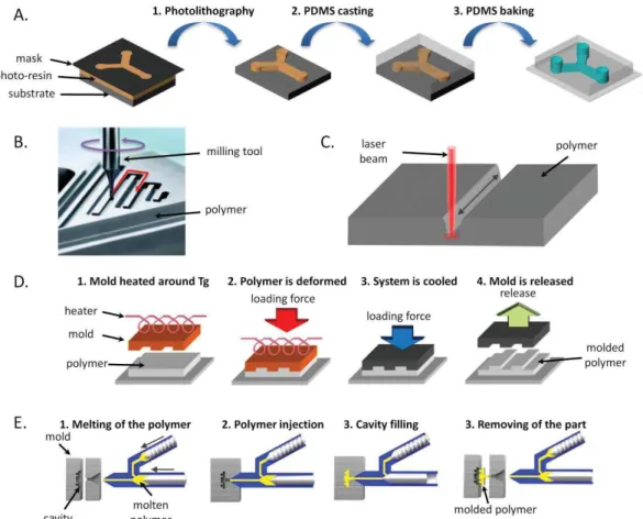
The fabrication sections will focus on the methods used to fabricate plastic microfluidic systems, as they have previously been identified as the main material used to fabricate microbioreactors. Structuring can be performed by using diverse techniques depending on the materials, the resolution one wants to achieve, the costs and the time needed to structure. It can be distinguished soft lithography, micromachining, laser ablation and micro molding methods such as hot embossing and micro injection molding can be distinguished (Figure 10). The first method, soft lithography, is adapted to the fabrication of PDMS devices while the other methods are usually employed for thermoplastics.
Figure 10. Schematic representation of the principle of the different methods used to structure plastic microfluidic chips. A) Soft lithography. B) Micromilling; C) Laser ablation. D) Hot embossing. E) Micro injection molding.
Soft lithography uses a structured mold onto which the elastomer is poured. The mold is fabricated by photolithography or micromachining and contains a negative replicate of the final structures to be formed. After pouring, thermal curing follows to crosslink the elastomer which is then peeled off from the mold. Highly resolved structures can be created due to the high resolution (sub micron) when photolithography is used to create the mold[80].
Micromachining is an abrasive method. A micromilling tool mounted on a computer assisted machine is guided by a computer software along the desired path to create the structures. Holes are made using microdrilling tools. This technique suffers from wall surface roughness and poor tolerances[81].
Laser ablation utilizes the energy of a pulsed laser beam to remove some materials of a solid surface by breaking the polymeric bond. The depth and width of the channels are defined by the laser intensity and the repetitive beam exposures along the same path[80].
The hot embossing technique gradually presses a structured mold into a polymeric substrate. At the same time, the mold is heated slightly above the glass transition temperature of the polymer (Tg) to soften the polymer so that it fills the mold. The mold microstructures are therefore
replicated with details into the polymer. The mold and the substrate are cooled before withdrawing the plastic[82].
Micro injection molding is an adaptation of the macroscopic injection molding. A mold cavity containing microstructures is closed and heated above the Tg of the polymer to be shaped. The polymer is heated in a separate unit and forced to flow in the mold cavity. The set-up is then cooled below the substrate Tg to demold the polymeric part[82].
Molding techniques are more suitable for mass production since they can rapidly produce polymeric components. The costs associated with mold fabrication make these methods hardly accessible to research labs or other low-volume production units. Micromachining, laser ablation or soft lithography are more adapted for smaller production or when the device design is not fixed[83].
1.4.4.4 Fabrication of microfluidic chips: bonding
Bonding methods can be classified in two classes: direct bonding and indirect bonding[84]. They differ by the presence (indirect) or not (direct) of an intermediate materials to seal the chip layers. The mechanism of bonding involves in general the interaction of charges (electrostatic, chemical (covalent bonding, acid/base interactions or Van der Waals forces) or the inter-diffusion of the substrates at the bonding interface[85,86].
The choice of the bonding methods depends on the material and the bond strength one wants to achieve. For instance, microfluidic systems that will be operated with fluids under high pressures will need to have a bond strength that can withstand these high pressures. The preservation of the surface integrity can additionally drive the choice of the bonding method in the case where the substrates have been functionalized (chemically or biochemically) and may be altered by the bonding process[83].
Indirect bonding is reduced to adhesive bonding. For adhesive bonding, a UV- or thermal-curable glue is deposited in between the material surfaces to be bonded. The glue can be deposited using different methods such as contact printing, screen printing or capillary-mediated gluing. During contact printing a thin glue layer is transferred onto the surface. Screen printing transfer glue dots on the surface by pushing the glue across a fine mesh. During capillary-mediated gluing, glue is introduced and fills a small volume right above the surface by capillarity. The main challenge with adhesive bonding is to avoid clogging of the structures by undesirable infiltration of glue. Sacrificial channels can be used to contain the excess of glue that might be deposited, preventing structure clogging. Additionally, the glue must wet the surfaces to achieve a proper bonding. However, the common glues used exhibit poor wetting on some plastics such as COC or PS.
Direct bonding includes thermal bonding, solvent-assisted bonding, local welding or the use of surface treatments[84]. Thermal bonding is the most common methods for bonding microfluidic thermoplastics components due to its simplicity and the high bond strength achievable[84]. It uses pressure and heat to mate substrates. The polymer is heated to around its Tg and the parts pressed together to create an intimate contact force by the inter-diffusion of the polymer chains to form a strong bond. The challenge with this technique is to control both the pressure and the temperature to avoid structure collapse while ensuring sufficient bond strength.
Solvent-assisted bonding is performed by exposing the thermoplastic surfaces to a solvent in its vapor or liquid phase. Vapor phase usually permits a better control of the solvent. The solvent induces the solvation of the surface polymer chains. They become mobile and can diffuse readily across the solvated layer forming a bond exceptionally strong, stronger than adhesive and thermal bonding. Care must be taken to avoid structure deformation by selecting the right solvent(s) and by controlling the exposition time to the solvent. Pressure and/or temperature can be used after solvent exposition to enhance the bond strength, as well as deep UV irradiation.
Localized welding uses ultrasonic waves, microwaves or infrared (IR) light to induce heat and soften the thermoplastic interface to bond. The utilization of microwaves requires the insertion of a thin metallic layer to absorb the wave’s energy while the utilization of IR light imposes one of the substrate to be opaque to IR light in order to induce the energy absorption at the interface. Localized welding has the advantage to create local or uniform bonding. However, this technique is not very practical, due to the complexity introduced by the need of a special design to direct the energy at the weld points, or the constraints associated with the requirement to deposit a metal layer, or the constraints to utilize polymers with different absorption characteristics.
Surface treatment is carried out by exposing the polymer surfaces to UV, UV/ozone or plasma. Such treatments increase the surface energy and by breaking chemical bonds can form reactive radicals. This enables the inter-diffusion of polymer chains and the possibility to form electrostatic interactions or even to create covalent bonds. High bond strength can be obtained. Surface treatments induce a modification of the surface which can be an advantage or a drawback. As a drawback, surface modification producing charges can undesirably interact with analytes. On the other hand, it can be used to obtain more hydrophilic surfaces or further enable other molecular grafting. Surface treatment can be combined with thermal bonding. The pressure and the temperature needed are lower, though.
![Figure 5. Schematic representation of bioreactors. A) Stirred bioreactor. B) Pneumatic bioreactor: airlift bioreactor[44]](https://thumb-eu.123doks.com/thumbv2/123doknet/14493251.717972/22.918.184.771.288.823/schematic-representation-bioreactors-stirred-bioreactor-pneumatic-bioreactor-bioreactor.webp)
![Figure 6. Diagram showing the influence of process operating strategy and parameters on process performance[51]](https://thumb-eu.123doks.com/thumbv2/123doknet/14493251.717972/23.918.148.739.392.777/figure-diagram-showing-influence-operating-strategy-parameters-performance.webp)


![Table 3. Basic requirements for cell culture, and improvements when microfluidic methods are used[76]](https://thumb-eu.123doks.com/thumbv2/123doknet/14493251.717972/37.918.135.755.165.400/table-basic-requirements-cell-culture-improvements-microfluidic-methods.webp)
