ORIGINAL ARTICLE
Assessing aesthetic outcomes after trigonocephaly correction
Philipp Metzler&Wolfgang Zemann&Christine Jacobsen&Heinz-Theo Lübbers&
Klaus Wilhelm Grätz&Joachim Anton Obwegeser
Received: 30 January 2013 / Accepted: 6 February 2013 / Published online: 17 February 2013 # Springer-Verlag Berlin Heidelberg 2013
Abstract
Purpose This study analysed the aesthetic outcome assess-ments after trigonocephaly correction using different assessor groups.
Methods Twenty-four patients (9 males, 15 females) with a surgical age between 8 and 10 months were included. Standardised photographs showing different facial views of the patients between ages 3 and 6 years were evaluated in terms of aesthetics by three study groups: surgeons, medical students, and lay persons. Each photograph was scored as follows: 1 (normal), 2 (acceptable, no need for revision), or 3 (unacceptable, needs revision).
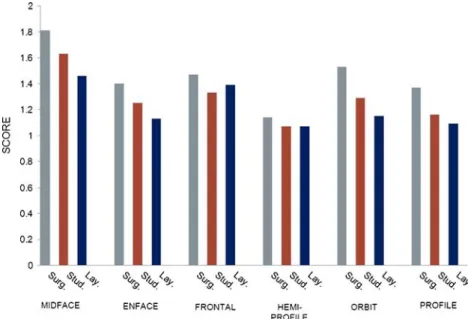
Results The mean surgical age was 9.1 ±0.4 months. Based on the en-face images, the mean scores assigned by the surgeon, student, and lay groups were 1.4 ±0.49, 1.25 ±0.44, and 1.13 ±0.34, respectively. Based on the patients’ profiles, the mean scores assigned by the surgeon, student, and lay groups were 1.37 ±0.49, 1.16 ±0.37, and 1.09 ±0.29, respectively. The scores of the hemi-profile evaluation were 1.14 ±0.35, 1.07 ±0.26, and 1.09 ±0.31, respectively. The scores of the frontal region were 1.47 ±0.54, 1.33 ±0.49, and 1.39 ±0.49, respectively. Within the orbital area, the surgeon, student, and lay groups assigned mean scores of 1.53 ±0.56, 1.29 ±0.46, and 1.15 ±0.36, respectively. The midface analysis showed mean scores of 1.8 ±0.66, 1.63 ±0.52, and 1.46 ±0.5, respec-tively. In all areas, there were significant differences (P<0.05) among the assessor groups.
Conclusion The expectations regarding aesthetic outcome differ considerably between experts and non-experts. The
need for correction did not concern the reshaped bone but rather the soft tissue epicanthal area.
Keywords Craniosynostosis . Trigonocephaly . Suture . Metopic . Cranial vault . Fronto-orbital advancement
Introduction
Premature fusion of the metopic suture impairs the growth of the skull and results in trigonocephaly. The deformity can be mild with only a slight prominence of the metopic ridge or severe with the formation of a keel-shaped fore-head [1, 2]. Metopic suture synostosis is rare, with an incidence of one to seven per 2,500 live births [2–5], and accounts for 7–23 % of all craniofacial disorders [3]. Approximately 10–20 % of cases of trigonocephaly are reported to be part of a syndrome combined with malfor-mation outside the skull [2].
Isolated trigonocephaly seems to have a genetic back-ground, with an autosomal dominant trait that has a very low penetrance [6,7]. The Ephrin-A4 (EFNA4) gene reportedly plays a role in non-syndromic craniosynostosis [7,8]. The occurrence of premature fusion of the metopic suture has increased dramatically worldwide in the last two decades, suggesting the role of additional environmental factors [9]. While mild manifestations of metopic suture craniosynos-tosis can be managed non-surgically, more severe deformities require surgical intervention. The deformity is progressive and worsens with age [10–12]. Single-suture craniosynostosis is not purely an aesthetic problem [13,14]; it also poses a risk for cognitive deficits [14,15]. The primary goal of surgery for trigonocephaly is anterior cranial vault expansion [7]. The technique used most commonly is fronto-orbital advancement with moulding of the supraorbital rims in combination with a cranioplasty that reshapes the frontal area of the skull. So far, P. Metzler (*)
:
W. Zemann:
C. Jacobsen:
H.-T. Lübbers:
K. W. Grätz
:
J. A. ObwegeserDepartment of Cranio-maxillofacial and Oral Surgery, University of Zurich, Frauenklinikstrasse 24,
8091 Zurich, Switzerland e-mail: [email protected]
trigonocephaly as rated by maxillofacial surgeons, medical students, and lay persons.
Materials and methods
This retrospective study included patients with premature fusion of the metopic suture treated at the Department of Cranio-maxillofacial and Oral Surgery of the University Hospital of Zurich between January 2002 and December 2010. The study design met the criteria of paragraphs 4a and b of the guidelines (ver. 13.03.2012, http:// www.kek.zh.ch/internet/gesundheitsdirektion/kek/de/
vorgehen_gesuchseinreichung.html, accessed at
2013-01-23) of the Ethics Committee of the Canton of Zurich and was therefore exempt from institutional review board ap-proval. The design also met the Declaration of Helsinki guidelines concerning ethical principles for medical re-search involving human subjects.
The inclusion criteria were (1) a diagnosis of isolated non-syndromic trigonocephaly, (2) surgery performed as simultaneous fronto-orbital advancement and cranioplasty of the forehead, (3) surgery performed between the ages of 8 and 10 months, and (4) no revisional surgery.
Surgical treatment
All surgical interventions were performed by one experi-enced cranio-maxillofacial surgeon and an experiexperi-enced neurosurgeon.
The surgical procedure was similar in all patients. After a bicoronal incision, a forehead flap was created at a subperiosteal level. After exposing the frontal and orbital re-gions, a fronto-orbital osteotomy was performed according to Marchac and Renier [16]. The fronto-orbital bandeau was reshaped and repositioned so as to overcorrect the hypoplastic temporal region and orbital rims. The frontal bone was split at the midline. The two segments were exchanged and rotated by 90°, also described by Marchac et al. [17]. For fixation, resorbable polydioxanone sutures (Ethicon, Johnson-Johnson Co., New Brunswick, NJ, USA) and resorbable plates and pins (Sonic Weld Rx® CMF; KLS Martin, Tuttlingen, Germany) were used.
Aesthetic evaluation
For the aesthetic evaluation, standardised photographs of patients with surgically treated trigonocephaly were used. The patients’ ages at the time of the photographs were
Two lay persons, two fifth year medical students, and two maxillofacial surgeons rated the appearance using a score of 1 to 3, where 1=normal, 2=acceptable (no need for correction), and 3=unacceptable (needs correction). None of the surgeons who evaluated the aesthetic outcome had performed the sur-gery or was part of the operating team. The experimental setup was according to that of Hilling et al. and Ozlen et al. [3,15]. Statistics
Mean values and standard deviations were calculated for the scores of the three groups. Multi-factor analysis of variance was used to assess the significance of differences among the groups. P-values of ≤0.05 were considered to indicate sta-tistical significance. For the stasta-tistical analysis, SPSS (ver. 18.0 for Mac; Chicago, IL, USA) was used.
Results
This study included 24 patients (nine males, 15 females). Surgery was performed between the ages of 8 and 10 months, at a mean age of 9.1±0.4 months. Surgery and the postoperative period were uneventful in all patients. En-face evaluation
Based on the en-face images, the mean scores of the sur-geon, student, and lay groups were 1.4±0.49, 1.25±0.44,
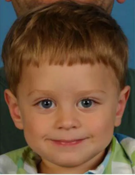
Fig. 1 En-face view of a patient after correction of trigonocephaly; age, 3.1 years; scoring: surgeons, 1.5; students, 1; laymen, 1
and 1.13±0.34, respectively. None of the groups suggested the need for corrective surgery for any patient.
Profile evaluation
On evaluating the patients’ profiles, the mean scores of the surgeon, student, and lay groups were 1.37±0.49, 1.16± 0.37, and 1.09 ±0.29, respectively. No group assigned a score of 3, which would have indicated a need for revisional surgery.
Hemi-profile evaluation
The results of the hemi-profile evaluation differed slightly from the profile results. The respective scores were 1.14±
0.35, 1.07±0.26, and 1.09±0.31. No group indicated the need for revisional surgery.
Frontal region evaluation
The surgeon group gave higher scores for the frontal region, with a mean score of 1.47±0.54. Corrective surgery was suggested for two patients. The mean scores assigned by the student and lay groups were 1.33 ±0.49 and 1.39 ±0.49, respectively, and neither group indicated a need for surgical revision.
Orbital region evaluation
Evaluating the orbital region, the surgeon group gave a mean score of 1.53±0.56 and suggested revisional surgery for three patients. Neither the student (mean score, 1.29± 0.46) nor lay group (mean score, 1.15±0.36) indicated a need for corrective surgery.
Central midface evaluation
After analysing the central midface photographs, the sur-geons reported a mean score of 1.8±0.66 and recommended revisional surgery for three patients. The students (mean score, 1.63±0.52) suggested corrective surgery for two pa-tients, whereas the lay group (mean score, 1.46±0.5) had no suggestions for corrective surgery.
Overall
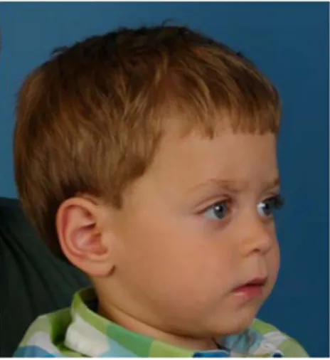
In all of the evaluations, significant differences (P<0.05) were found among the rating groups. The scores reported by all groups for the en-face, profile, hemi-profile, central Fig. 2 Profile of a patient after correction of trigonocephaly; age,
3.1 years; scoring: surgeons, 1; students, 1; laymen, 1
Fig. 3 Hemi-profile of a patient after correction of trigonocephaly; age, 3.1 years; scoring: surgeons, 1; students, 1; laymen, 1
Fig. 4 Frontal region of a patient after correction of trigonocephaly; age, 3.1 years; scoring: surgeons, 1; students, 1; laymen, 1
Fig. 5 Orbital region of a patient after correction of trigonocephaly; age, 3.1 years; scoring: surgeons, 2; students, 1.5; laymen, 1
midface, frontal, and orbital regions are shown in Fig.7. All patients with a score of 3, indicating the need for additional surgical correction, had noticeable epicanthal folds. These were corrected using the technique of Spaeth [18], as shown in Figs.8,9,10, and11.
Discussion
Several surgical techniques have been described for expanding the temporal region and correcting the aesthetic impairment in patients with trigonocephaly [10]. The tech-nique used most commonly is the‘tongue in groove’ tech-nique introduced by Marchac [16,19,20]. Modifications of this technique have been reported to correct hypotelorism, epicanthus, flat supraorbital rims, and bitemporal constric-tions [10, 21]. Nevertheless, aesthetic shortcomings can remain after correcting trigonocephaly. This indicates the need to critically review the outcomes. In patients with residual aesthetic impairment, one should ask whether the impairment justifies surgical correction or whether it is of minor impact. The primary goals of surgical intervention are
to design the present study to determine whether profession-al maxillofaciprofession-al surgeons with experience in craniofaciprofession-al surgery have the same expectations for the aesthetic out-come after correcting trigonocephaly as non-experts. We found considerable differences.
Notably, the surgeon group was more critical than the student and lay groups. This is not surprising as surgeons are more aware of what to focus on. However, the ratings differed between aesthetic evaluations of a patient’s entire face (en-face, profile, and hemi-profile views) and those of the smaller sections of the face. The ‘full-face’ evaluation did not indicate the need for revisional surgery in any case. Even when differences among the observer groups were evident, the appearance of all patients was deemed accept-able. By contrast, when analysing smaller regions, the sur-geon group suggested the need for corrective surgery in some cases, especially based on the central midface images. For this region, the surgeon group recommended corrective surgery in three cases and the student group in two cases. There were no suggestions for revision in the en-face eval-uations, so hypotelorism was obviously not objectionable. This may be explained by the symmetric nature of the pathology, making it less eye-catching than an asymmetric configuration.
A prominent epicanthal fold appeared to be the relevant aesthetic impairment in these images. This correction is quite simple using the technique of Spaeth [18], which gives excellent results and can be performed in a day-care unit. In our opinion, there is no age limitation for this technique, and it can also be performed during primary surgery.
Fig. 6 Central midface of a patient after correction of trigonocephaly; age, 3.1 years; scoring: surgeons, 2.5; students, 1.5; laymen, 1
Fig. 7 Distribution of the ratings for various regions according to the observer groups. Surg. surgeon, Stud. student, Lay. laymen
Based on the images focused on the frontal area (frontal region, profile, and hemi-profile), there was no suggestion for correction, although this area is most affected by the pathology. Nevertheless, all three observer groups (sur-geons, medical students, and lay persons) scored some pa-tients as‘acceptable’, but not as ‘normal’, which indicates that stigmata remained after the fronto-orbital advancement. Assuming sufficient correction during primary surgery, this implies some relapse.
Hilling et al. and Ozlen et al. used a comparable scale to rate the outcomes of patients treated for trigonocephaly [3,
15], addressing the following regions: forehead shape, hypotelorism, and temporal depression. The two study groups used different surgical techniques, but both showed a marked improvement of the characteristic stigmata in trigonocephaly patients. However, the techniques used in the current study cannot be reliably compared with those used by Hilling et al. and Ozlen et al. The inhomogeneous surgical age (6–15 and 4–40 months), follow-up period (12– 144 months and 5 months to 19 years), and various osteosynthesis techniques (suture material and rigid metal miniplates) of the latter study protocols would considerably bias the results. Furthermore, these previous studies evalu-ated the earlier-mentioned pathognomonic characteristics, whereas the current study rated the postsurgical aesthetics of various craniofacial regions.
Lwin et al. reported the relapse of fronto-orbital advance-ment in an anterior–posterior direction within 5 months postoperatively in 65 % of the analysed patients [23]. In the trigonocephaly group, they reported an operative ad-vancement loss of up to 57 %, which seems very high. Those authors recommend significant over-advancement of the fronto-orbital advancement. Although we cannot quan-tify relapse in our patients, as this was not the study aim, we
strongly support this recommendation and overcorrect the fronto-orbital advancement in all of our patients.
Our subjects were 3 to 6 years old, which seems young to make a reliable statement concerning the long-term aesthetic outcome. However, the configuration of the cranial vault remains stable about 2 years after craniofacial procedures in single-suture cases [24]. Furthermore, the number of pa-tients was rather small.
A three-point scale has limitations for rating the full amount of aesthetic impairment. However, we tried to keep the evaluation as simple as possible, principally so as not to overtax the lay group. In addition, this three-point scale was chosen because it had been suitable in previous studies [3,
15]. To the authors’ knowledge, this is the first study to
include lay persons in an observer group, which is of interest because of their non-medical approach. The lay group rep-resents, more or less, the perception of the social environ-ment and may tend to compensate for possible overly critical scoring by the surgeon and student groups. Inclusion of a lay group provides a valuable aspect of the current study. The commonly used Whitaker score [25] was deemed too specific for non-craniofacial surgeons. Engel et al. used the Whitaker classification to evaluate 54 trigonocephaly patients after treatment [2]. Although it is difficult to com-pare the Whitaker score with our scale, we believe that our study results were comparable to theirs and that category II of the Whitaker classification, i.e., ‘soft-tissue or lesser bone-contouring revisions advisable apt to be performed on an outpatient basis or requiring a maximum of 2-day hospitalisation’ [2,25], roughly corresponds with our score of 3.
However, only a small percentage of patients were deemed to benefit from additional corrective surgery as far as aesthetics were concerned. The need for correction did not concern the reshaped bone but rather was seen in the soft tissue epicanthal area.
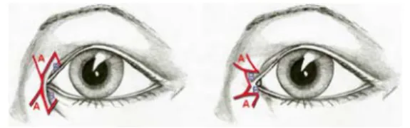
Fig. 8 Schematic of epicanthus correction according to Spaeth [18]
Fig. 9 Clinical picture before epicanthus correction
Fig. 10 Intra-operative view of epicanthus correction
1. Aesthetic scores differed significantly among surgeons, medical students, and lay persons.
2. The evaluation showed that there was no need for fur-ther bone correction.
3. The need for additional correction was identified in the soft tissue epicanthal folds.
Acknowledgments The authors thank Ruth Gottmann for the sche-matic drawings.
Conflict of interest The authors declare that they have no conflicts of interest.
References
1. Fearon JA, Singh DJ, Beals SP, Yu JA (2007) The diagnosis and treatment of single sutural synostosis: are CT-scans necessary? Plast Reconstr Surg 120:1327
2. Engel M, Thiele OC, Mühling J, Hoffmann J, Freier K, Castrillon-Oberndorfer G, Seeberger R (2012) Trigonocephaly: results after surgical correction of nonsyndromic isolated metopic suture syn-ostosis in 54 cases. J Craniomaxillofac Surg 40:347–353 3. Ozlen F, Kafadar AM, Abuzayed B, Ulu MO, Isler C, Dashti R,
Erdincler P (2011) Surgical treatment of trigonocephaly: technique and long-term results in 48 cases. J Neurosurg Pediatrics 7:300–310 4. Aryan HE, Jandial R, Ozgur BM, Hughes SA, Meltzer HS, Park MS, Levy ML (2005) Surgical correction of metopic synostosis: 107 cases. Childs Nerv Syst 21:392–398
5. Bottero L, Lajeunie E, Arnaud E, Marchac D, Renier D (1998) Functional outcome after surgery for trigonocephaly. Plast Reconstr Surg 10:952–958
6. Collmann H, Sorensen N, Krauss J (1996) Consensus: trigonocephaly. Child’s Nerv Syst 12:664–668
7. Steinbacher DM, Bartlett SP (2013) In: Neligan PC, Rodriguez ED (eds) Nonsyndromic craniosynostosis. Plastic surgery, 3rd edn. Elsevier Saunders, London, pp 726–748
8. Merrill AE, Bochukova EG, Brugger SM, Ishii M, Pilz DT, Wall SA, Lyons KM, Wilkie AO, Maxson RE Jr (2006) Cell mixing at a neural crest–mesoderm boundary and deficient ephrin-Eph signal-ing in the pathogenesis of craniosynostosis. Hum Mol Genet 15:1319–1328
treatment of single-suture metopic synostosis. Ann Plast Surg 59:6–13
11. Sidoti E, Marsh J, Marty-Grames L, Noetzl MJ (1996) Long-term studies of metopic synostosis: frequency of cognitive impairment and behavioral disturbances. Plast Reconstr Surg 97:276
12. Shimoji T, Shimabukuro S, Sugama S, Ochiai Y (2002) Mild trigonocephaly with clinical symptoms: analysis of surgical results in 65 patients. Childs Nerv Syst 18:215–224
13. Nischal K (2007) Is non-syndromic single suture craniosynostosis purely an aesthetic problem? Develop Med Child Neurol 49:565 14. Ricci D, Vasco G, Baranello G, Salerni A, Amante R, Tamburrini
G, Dickmann A, Di Rocco C, Velardi F, Mercuri E (2007) Visual function in infants with non-syndromic craniosynostosis. Develop Med Child Neurol 49:574–576
15. Hilling DE, Mathijssen IMJ, Vaandrager M (2006) Aesthetic re-sults of fronto-orbital correction in trigonocephaly. J Craniofac Surg 17:1167–1174
16. Marchac D, Renier D (1982) Craniofacial surgery for craniosyn-ostosis. Little, Brown, Boston
17. Marchac D (1978) Radical forehead remodeling for craniostenosis. Plast Reconstr Surg 61(6):823–835
18. Spaeth EB (1956) Further consideration of the surgical correction of blepharophimosis and epicanthus. Am J Ophthalmol 41(1):61– 71
19. Marchac D, Renier D (1979)“Le front flottant” traitement précoce des faciocraniosténoses. Ann Chir Plast 24:121
20. Marchac D, Renier D, Jones BM (1988) Experience with the floating forehead. Br J Plast Surg 41:1–15
21. Fearon JA (2008) Beyond the bandeau: 4 variations on fronto-orbital advancements. J Craniofac Surg 19:1180–1182
22. Pope AW, Ward J (1997) Self-perceived facial appearance and psychosocial adjustment in preadolescents with craniofacial anom-alies. Cleft Palate Craniofac J 34:396–401
23. Lwin CTTJW, Richardson D, Duncan C, May P (2011) Relapse in fronto-orbital-advancement: a pilot study. J Craniofac Surg 22:214–216
24. Metzler P, Zemann W, Jacobsen J, Grätz KW, Obwegeser JA (2013) Cranial vault growth patterns of plagiocephaly and trigonocephaly patients following fronto-orbital advancement: a long-term anthro-pometric outcome assessment. J Craniomaxillofac Surg. doi:10.1016/j.jcms.2012.11.035
25. Whitaker L A, Bartlett SP, Schut L, Bruce D (198 7) Craniosynostosis: an analysis of the timing, treatment and com-plications in 164 consecutive patients. Plast Reconstr Surg 80:195–2012