HAL Id: tel-02613807
https://tel.archives-ouvertes.fr/tel-02613807
Submitted on 20 May 2020HAL is a multi-disciplinary open access
archive for the deposit and dissemination of sci-entific research documents, whether they are pub-lished or not. The documents may come from teaching and research institutions in France or abroad, or from public or private research centers.
L’archive ouverte pluridisciplinaire HAL, est destinée au dépôt et à la diffusion de documents scientifiques de niveau recherche, publiés ou non, émanant des établissements d’enseignement et de recherche français ou étrangers, des laboratoires publics ou privés.
autoimmune Myasthenia Gravis
Melanie Cron
To cite this version:
Melanie Cron. Implication of microRNAs in the pathophysiology of autoimmune Myasthenia Gravis. Immunology. Sorbonne Université, 2018. English. �NNT : 2018SORUS395�. �tel-02613807�
Tout d’a o d, je tie s à e e ie les e es du ju d’a oi a epté de lire ce manuscrit et d’ t e présents pour ce jour particulier, le Dr. Florence Appa aill , ui a pu oi l’é olutio de o t a ail, et le P . Philippe Geo gel e leu ualité de appo teu s, le D . “ophie De e et e ta t u’e a i at i e de mon travail et le Pr. Olivier Benveniste, président du jury.
Je remercie très chaleureusement le Dr. Rozen Le Panse, qui a accepté de me prendre en stage pour o M il a uel ues a ées et ui à l’épo ue e sa ait pas u’elle e ga de ait jus u’e fi de thèse, soit presque 5 ans mis bout à bout. Ces années ont été extrêmement riches en apprentissage, j’ai eau oup g a di et p i ipale e t g â e à toi. To soutie , to aide ’o t été e t e e t p é ieu , et le fait ue tu ’aies eau oup ise e aleu pe da t es a ées a é o é e t o pté pou oi. J’esp e a oi épo du à tes atte tes et fait avancer le travail du laboratoire avec mon petit out de he i . Tu es uel u’u ui sait fai e g a di les ge s et les aide da s leu éfle io , je e se s u peu plus p te à ele e d’aut es halle ges g â e à toi. M e si es de ie s ois o t été une so te de ise à l’ép eu e ps hologi ue, je se ais p te à aff o te e ui a i e da s le futu . Tout e t a ail ’au ait ie été si tu ’a ais pas été là.
Je tiens à dire aussi un grand merci au Dr. Sonia Berrih-Ak i , ui ’a ue é olue u peu plus en retrait ais ui a toujou s eu les ots justes au o o e t pou e pe ett e d’a a e . Me i pou les diffé e tes oppo tu ités ue ous ’a ez appo tées ai si ue pou ot e soutie tout au lo g de es a ées. J’esp e a oi aidé à la ohésio de l’équipe que vous avez construite et menée.
U g a d e i, ais telle e t i suffisa t pa appo t à e ue tu ’as appo té, “ol e ! Tu ’as é o é e t soute ue, tout au lo g de es a ées u’o a pa tagées, ua d j’étais st essée, t iste, é e ée ’est a i é uel ues fois ua d e… ais tu as aussi pa tagé es joies, es i es et es o fide es. J’esp e t’a oi appo té auta t ue e tu ’as appo té, es a ées au aie t été eau oup oi s suppo ta les si tu ’a ais pas été là. Tu es uel u’u de gé ial, e laisse ja ais pe so e t’e faire douter.
Bérengère, Marie, nos discussions et débats dans la cuisine, nos rires, vos enfants (presque à tous), vos histoires… A oi t a aillé a e ous ’a o t é u’u e o e a ia e e à de elles e o t es et à u t a ail peut t e u peu oi s effi a e… :) Cloé, pour ces rires, ces discussions (avec Solène) sans intérêt et sans fin le vendredi après-midi (ou le jeudi, ou le e edi… et es séa es de spo t pour se défouler un bon coup.
Je souhaite e e ie les e es de l’é uipe ue je ’ai pas ités jus u’à p ése t, F édé i ue, sa s ui e la o e tou e ait pas o d, toujou s dig e de o fia e et fia le, tu es uel u’u su ui o peut o pte sa s hésite et j’esp e a oi u e emplaire dédicacé des Aventures de Paul dès sa sortie !). Nadine, avec qui je prends plaisir à discuter de choses plus ou moins sérieuses, José, ma caution hispanophone qui aime se moquer gentiment de notre côté très Français mais aussi Julien, Jean-Thomas et Jé ô e. Je ’ou lie pas Clai e et Ale a d a ui e ’o t uasi e t que vue assise à mon bureau pendant tout leur stage de M2.
Je tiens également à remercier Jordan Mecca et Jean-François Darrigrand, les deux autres membres fondateurs et actifs de la M oCoop’, ot e asso iatio ui j’esp e, su i a à os dépa ts. Je t ou e u’o a is ot e pie e à l’édifi e e éa t ette asso pou la ohésio du Ce t e et j’ai été heu euse de partager ça avec vous, ce fut une sacrée aventure.
Je remercie aussi les e es de l’é uipe au 5, pa tis ou e o e i i, Wladi i , Lea d o, Gaëlle, Céli e, Nad ge, Loïs, a e ui j’ai pa tagé de t s o s o e ts.
Un énorme merci à ma famille et en particulier à mes parents, leur soutien indéfectible, leur amour, leurs efforts et leur présence sont des piliers essentiels dans ma vie. Une petite pensée pour eux qui ne comprendront pas grand- hose à e a us it édigé e a glais, ais ui o t u e olo té d’e fe pou s’i pli ue .
Et puis, si dans la vie il y a des moments et des e o t es u’o ’ou lie pas, tu fais é ide e t partie de ceux-là, Da ie . Je ois u’il a peu de ots pou dé i e e u’o it et o e t le desti , le karma ou que-sais-je nous a réunis. Tu as su mettre un sourire sur mon visage quand lui clairement
é e e e ts, es sautes d’hu eu et o st ess au lo g de es a ées. Plus haut, j’ai dit ue j’a ais eau oup g a di pe da t tout e te ps, ’est é o é e t g â e à toi, tu ’as eau oup po tée. Je souhaite que tu sois à mes côtés pendant encore de très nombreuses années, où la thèse et mon passage au la o ’au o t été ue le dé ut de plei d’aut es hoses. Me i pou tout.
LIST OF TABLES AND FIGURES ... 8
ABBREVIATIONS ... 9
LIST OF PUBLICATIONS... 12
I. STATE OF THE ART ... 13
1. miRNAs: what are they, how do they work and for what purpose? ... 14
1.1. Discovery and first characterization of RNA interference ... 14
1.2. Characterization of miRNAs ... 15
1.2.1. Nomenclature ... 15
1.2.2. miRNA biogenesis pathways ... 16
1.2.3. Regulation of miRNA biogenesis ... 21
1.3. Modes of action and biological roles of miRNAs ... 24
1.3.1. Target recognition and mRNA-miRNA interactions ... 24
1.3.2. Mechanisms of post-transcriptional regulation of mRNA by miRNAs ... 26
1.3.3. Biological roles of miRNAs ... 30
1.3.4. Degradation of miRNAs ... 32
1.3.5. Clinical applications of miRNAs ... 32
2. Myasthenia Gravis ... 38
2.1. History of the disease: from the first cases to recent findings ... 38
2.2. Clinical features ... 40
2.2.1. Epidemiology ... 40
2.2.2. Clinical classification and associated symptoms ... 41
2.2.3. Treatments ... 45
2.3. Pathophysiology of MG ... 48
2.3.1. Consequences of the autoantibody attack at the neuromuscular junction (NMJ) 48 2.3.2. Thymic abnormalities in AChR+ MG patients ... 55
2.3.3. Immune dysregulation ... 62
2.3.4. Etiological mechanisms of MG ... 63
2.3.5. Alternative triggering events for MG ... 67
2.4. Animal models for MG ... 68
2.4.1. Induced animal models ... 68
2.4.2. The MG-NSG model ... 69
3.2. Dysregulated miRNAs associated with MG ... 71
3.2.1. miRNAs in the serum of MG patients ... 71
3.2.2. miRNAs in circulating cells of MG patients ... 72
3.2.3. In the thymus of MG patients ... 73
4. Conclusion and objectives ... 76
II. RESULTS ... 77
ARTICLE 1 ... 79
ARTICLE 2 ... 95
ARTICLE 3 ... 119
III. DISCUSSION AND SYNTHESIS ... 155
1. Wide analysis of dysregulated miRNAs in the thymus of MG patients ... 156
2. miRNAs and the IFN-I signature characteristic of MG: involvement of the miR-29 family and viral miRNAs ... 159
3. miR-150-5p dysregulation and function in MG ... 162
4. Conclusion ... 165
IV. BIBLIOGRAPHY ... 168
8
Figure 1: Canonical biogenesis pathway of miRNAs ... 19
Figure 2: Non-canonical biogenesis pathways: miRtrons and DICER/TRBP-independent pathway ... 21
Figure 3: miRNA-mRNA interactions ... 25
Figure 4: Possible mechanisms of miRNA-mediated repression ... 27
Figure 5: Functions of miRNAs: robustness during development and noise reduction ... 31
Figure 6: miRNAs as therapeutic targets ... 36
Figure 7: Major discoveries in MG... 40
Figure 8: Clinical classification according to the MGFA... 42
Figure 9: The neuromuscular junction ... 50
Figure 10: Mechanisms following antibody fixation on AChR... 53
Figure 11: The thymus and the T-cell selection ... 59
Figure 12: The thymus in MG ... 61
Figure 13: Summary diagram of investigated miRNAs during this PhD ... 166
Table 1: Antibody-specific characteristics of each type of MG. ... 44
9 3’UTR 3’ u t a slated egio
ACh Acetylcholine
AChE Acetylcholine esterase AChR Acetylcholine receptor
ADAR Adenosine deaminase acting on RNA AID Autoimmune disease
AKT Serine-Threonine Protein Kinase ASO Antisense oligonucleotide BCR B-cell receptor
bp Base pair
CCL C-C motif chemokine ligand CCR C-C motif chemokine receptor CCR4-NOT Carbon catabolite repressor 4-NOT CD Cluster of differentiation
CDS Coding DNA sequence CFA Co plete F eu d’s adju a t ciRs-7 Circular RNA sponge for miR-7
CTLA4 Cytotoxic T-lymphocyte associated protein 4 CXCL C-X-C motif chemokine ligand
CXCR C-X-C motif chemokine receptor DC Dendritic cell
DGCR8 DiGeorge critical region 8
DN Double negative
DP Double positive dsRNA Double-strand RNA
EAMG Experimental autoimmune myasthenia gravis EBV Epstein-Barr virus
eIF4 Eukaryotic initiation factor 4 EOMG Early-onset myasthenia gravis EpCAM Epithelial cell adhesion molecule
10 GWAS Genome wide association study
HDAC Histone deacetylase
HET Heterozygous
HEV High endothelial venule
HIV Human immunodeficiency virus HLA Human leucocyte antigen IFNAR IFN-I receptor
IFN-I/-α/-β/-γ Type-I interferon / interferon alpha / beta / gamma IVIg Intravenous immunoglobulin
KO Knock-out
LEV Lymphatic endothelial venule LNA Locked nucleic acid
LOMG Late-onset myasthenia gravis LPS Lipopolysaccharide
LRP4 Low density lipoprotein receptor-related protein 4 MAPK Mitogen-activated protein kinase
MG Myasthenia gravis
MGFA Myasthenia gravis foundation of america MHC Major histocompatibility complex MIR Main immunogenic region
miRNA MicroRNA
mRNA Messenger RNA
MuSK Muscle specific kinase NMJ Neuromuscular junction
NSG NOD scid gamma
nt Nucleotide
PABP Poly(A) binding protein
PAN2/3 Poly(A) specific ribonuclease subunit 2/3 pDC Plasmacytoid dendritic cell
11 PTPN22 Protein tyrosine phosphatase, non-receptor type 22
RISC RNA-induced silencing complex RLCV Rhesus lymphocryptovirus RNAi RNA interference
siRNA Silencing RNA
SLO Secondary lymphoid organ SNP Single nucleotide polymorphism ssRNA Single-strand RNA
TCR T-cell receptor TEC Thymic epithelial cell
TGF-β Transforming growth factor beta TK Thymidine kinase
TLO Tertiary lymphoid organ TLR Toll-like receptor TNF Tumor necrosis factor TNFAIP3 TNF alpha induced protein 3
TNFRSF11A TNF receptor superfamily member 11a
TNIP1 TNF alpha induced protein 3 interacting protein 1 TRBP Trans-Activation Responsive RNA-Binding Protein TSA Tissue specific antigen
TUT Terminal uridyltransferase
WT Wild-type
XPO5 Exportin 5
12
Cron, M.A., Maillard, S., Robinet, M., Fadel, E., Guihaire, J., Liston, A., Berrih-Aknin, S. and Le Panse, R.
"Involvement of miR-29s in the pathogenesis of autoimmune Myasthenia Gravis." in preparation
Cron, M.A., Maillard, S., Truffault, F., Gualeni, A. V., Gloghini, A., Fadel, E., Guihaire, J., Behin, A.,
Berrih-Aknin, S. and Le Panse, R.
"Causes and consequences of miR-150-5p dysregulation in Myasthenia Gravis." submitted
Cron, M. A., Maillard, S., Delisle, F., Samson, N., Truffault, F., Foti, M., Berrih-Aknin, S. and Le Panse, R.
(2018).
"Analysis of microRNA expression in the thymus of Myasthenia Gravis patients opens new research avenues." Autoimmun Rev 17(6): 588-600.
Cron, M. A., Maillard, S., Villegas, J., Truffault, F., Sudres, M., Dragin, N., Berrih-Aknin, S. and Le Panse,
R. (2018).
"Thymus involvement in early-onset myasthenia gravis." Ann N Y Acad Sci 1412(1): 137-145. Review
Robinet, M., Villeret, B., Maillard, S., Cron, M.A., Berrih-Aknin, S. and Le Panse, R. (2017).
"Use of Toll-Like Receptor Agonists to Induce Ectopic Lymphoid Structures in Myasthenia Gravis Mouse Models." Front Immunol 8(1029).
Robinet, M., Villeret, B., Maillard, S., Cron, M.A., Berrih-Aknin, S. and Le Panse, R. (2016).
"Review on Toll-Like Receptor Activation in Myasthenia Gravis: Application to the Development of New Experimental Models." Clin Rev Allergy Immunol. 52(1): 133-147. Review
13
14 This thesis aims at evaluating and characterizing the implication of various microRNAs (miRNAs) in the context of an autoimmune disorder, Myasthenia Gravis (MG). miRNAs are small molecules of non-coding RNA, characterized as epigenetic factors thanks to their ability to interact with messenger RNA (mRNA). miRNAs are being investigated in many diseases, including autoimmune diseases (AIDs). AIDs’ etiology is still largely unknown and unfortunately, MG is not an exception. In this context, I investigated the etiological and pathophysiological implications of miRNAs in MG with different approaches.
1.
miRNAs: what are they, how do they work and for what purpose?
1.1. Discovery and first characterization of RNA interference
In 1984 Izant and Weintraub conducted the first experiment, aimed at inhibiting the expression of a gene with the formation of an RNA duplex [1]. The authors used plasmid constructs coding for the thymidine kinase (TK) to be expressed in TK-deleted mouse cell lines (TK- L cells). In parallel, they used
a recombinant DNA, coding for the antisense RNA of the TK gene, so that the cells could express antisense RNA by themselves. With this design, the authors could evaluate in which proportion the RNA antisense was able to downregulate TK expression, without background noise. The authors saw a decrease in the TK activity in cells injected with antisense RNA construct. However, they could not determine the efficiency of the inhibition, the stability of the antisense RNA or the localization of the inhibition.
In the late 9 ’s, A.R. “uitje’s tea des i ed ha ges in the pigmentation pattern of petunia and tobacco flowers after the i t odu tio of a a tise se hal o e s thase ge e , a gene coding for a key enzyme of the flavonoid biosynthesis pathway that is involved in the pigmentation of the flowers [2]. The authors suggested that these changes were due to the presence of RNA duplexes in the flowers tissue. They also highlighted that differences observed in some patterns were the result of different integration positions of the antisense chalcone synthase gene, pointing out the importance of the transgene positioning. Late o , R. Jo ge se ’s team came to the same conclusions [3].
15 I 99 , A. Fi e’s tea a aged to i hi it ge e e p essio i the us le of the o Caenorhabditis elegans by injecting plasmids coding for antisense RNA of myofilament proteins in C. elegans oocytes. These injections resulted in disorganization of the myofilaments and thus slow movements of the animals. Good integration of the antisense RNA allowed the authors to see these defects in the next generations [4]. Still in C. elegans, the temporal down-regulation of the lin-14 gene by lin-4 was explained in wild-type (WT) worms [5, 6]. These studies showed for the first time that a RNA duplex could result in a lower protein expression in vivo.
The te RNA i te fe e e (RNAi) was used for the first time by A. Fire and C. Mello in 1998, in a study where they prove that the injection of double-strand RNA (dsRNA) is more efficient than the injection of a single-strand RNA (ssRNA) to induce the inhibition of endogenous myofilament proteins, compared to other previous studies, including their previous one [4, 7]. This study was the beginning of many investigations about RNAi. A. Fire and C. Mello received the Nobel Prize in Physiology or Medicine in 2006 for their discoveries.
1.2. Characterization of miRNAs
Among RNAi, two main types of different RNA can be distinguished: small interfering RNAs (siRNAs) and microRNAs (miRNAs). siRNAs are exogenous ssRNAs, coming from transposons movements, infections or transgene activity [8]. miRNAs are endogenous dsRNA processed into ssRNAs and involved in the regulation of physiological phenomena [9]. There are several miRNA-biogenesis pathways: the canonical pathway (the most common pathway) and alternative pathways for specific miRNAs and miRNA families [10]. It is noteworthy that long non-coding RNAs (around 200-nt long) are not considered as RNAi as they do not directly act on mRNA but act epigenetically [11].
1.2.1. Nomenclature
iRNAs a e a ed the p efi iR a d a si gle ide tif i g u e iR-150, miR- … alo g ith its coding gene, according to the gene nomenclature of the organism (MIR150, MIR7 for human per example). Ortholog miRNAs are named identically among species. If needed, three letters referring to
16 the species can be placed before the prefix: hsa fo Homo sapiens, mmu fo Mouse musculus, el for C. elegans…
If two miRNAs share a common sequence but are transcribed from different loci and have distinct precursors, a number is placed at the end (miR-7-1, miR-7- … . “i ila l , if two miRNA sequences differ only by one or two nucleotides (nt), they will be noted miR-29a, miR-29b, miR-29c...These rules can be added if the conditions require, such as the miR-29 family: miR-29a, miR-29b-1, miR-29b-2, miR-29c. Finally, orientations are very often noted at the end of the name: miR-150-5p, miR-150-3p. If the guide and the passenger strand can be differentiated among the two strands, the passenger strand will be noted with a star (*). It is noteworthy that the passenger strand can either be the -5p or the -3p strand [12, 13].
An exception is made for the lethal-7 (let-7) family. Let-7 was first discovered in C. elegans as a time controller for the development of the worm. Later on, orthologs were found in humans and other species but the denomination of the miRNA was not changed. In humans, the let-7 family is composed of 10 mature miRNAs [14].
1.2.2. miRNA biogenesis pathways Canonical miRNA-biogenesis pathway
As shown in Figure 1, miRNAs are transcribed from specific miRNA genes by the RNA polymerase II. The resulting transcript is a primary miRNA (pri-miRNA) characterized by a hairpin, a very specific form adopted by miRNAs during their biogenesis. The pri-miRNA can reach 1 kilobase (kb) length and is composed of a 33-35 base pair (bp) stem ending by a loop and two ssRNA ith 5’ a d 3’ e d fla ki g the stem [15]. Next, the pri-miRNA is cleaved by DROSHA, a RNase III, and its partner, DiGeorge Critical Region 8 (DGCR8), together called the microprocessor. The association of these nuclear proteins is called the microprocessor, with a single DROSHA molecule for two molecules of DGCR8. Here, the role of DROSHA is to set the cleaving point on the pri-miRNA, around 11bp from the basal junction and 22 bp from the apical loop. DGCR8 stabilizes DROSHA and enhances precision and efficiency of the cleavage [16].
17 After cleavage, the 60nt-length precursor miRNA (pre-miRNA) is exported from the nucleus to the cytoplasm by the protein exportin 5 (XPO5). XPO5, which is associated with a Ran-guanosine triphosphate (Ran-GTP) cofactor to enhance the export, binds to the pre-miRNA in the nucleus and releases it in the cytoplasm, along with the hydrolysis of the Ran-GTP, becoming Ran-GDP [17, 18]. Pre-miRNAs have the particularity to have a 2 nt 3’ o e ha g that allo s the e og itio of the RNase III DICER in the cytoplasm. DICER, thanks to its PAZ domain allowing it to identify ssRNA, will recognize the pre-miRNA and will cut out the loop, resulting in a miRNA duplex of 20-23 nt. DICER’s pa t e , the Trans-Activation Responsive RNA-Binding Protein (TRBP), has a similar role with DICER that DGCR8 has with DROSHA, which is helping DICER to stabilize the cleavage of the pri-miRNA by increasing its half-life [19].
Subsequently, the miRNA duplex is loaded into the RNA-Induced Silencing Complex (RISC). The RISC is a two-step transient association involving DICER, TRBP, AGO proteins and heat-shock proteins acting as chaperones, whose role is to select the leading strand to induce mRNA silencing. In mammals, we have four distinct AGO proteins (1-4) but only AGO2 displays a catalytic activity. First, the miRNA duplex is loaded from DICER onto AGO2, which is associated with two chaperones, HSC70 and HSP90, allowing it to adapt its conformation to the duplex. Next, the duplex is unhybridized by AGO2, leaving the non-chosen strand, called the passenger strand, out of the RISC to be discarded. Once the leading strand, in its final miRNA form, is loaded into AGO2, the chaperones leave the complex [20, 21]. The choice of the leadi g/passe ge st a d is ot a it a . The st a d ith the lo e 5’ the od a i sta ilit will preferentially be the leading (or guide) strand he eas the st a d displa i g a highe 5’ sta ilit will be the passenger strand [8, 22].
The ultimate goal of miRNAs is to prevent the expression of target proteins by acting on mRNA. According to the perfection of the sequence matching between a miRNA and its target mRNA, the mechanism of inhibition of protein expression will not be the same. In plants, miRNA-mRNA pairing is totally or nearly perfect. Consequently, the RISC proteins, AGO proteins in particular, function as endonucleases, slicing the mRNA complementary to the guide strand [23]. In mammals, the base
18 pairing is almost never perfect on the whole miRNA length. However, as detailed by Filipowicz et al., so e ules eed to be respected in order to have a correct translational repression: the pairing must be perfect between the nt to of the iRNA alled the seed egio a d the 3’ e d of the RNA 3’UnTranslated Regio o 3’UTR), pairing et ee the 3’ e d of the iRNA and the 5’ e d of the mRNA should be sufficient enough to stabilize the reaction and mismatches should be present to prevent any cleavage from AGO2 [24].
19 Figure 1: Canonical biogenesis pathway of miRNAs
miRNAs are transcribed from specific genes by the RNA Polymerase II, a 12-subunit enzyme involved in the transcription of mRNAs. When transcribed, the transcript, called pri-miRNA, adopts a particular conformation in a hairpin. DROSHA and DGCR8 will isolate the hairpin from the rest of the transcript and XPO5 will mediate the transport from the nucleus
to the cytoplasm. Then, DICER and TRBP will cut the apical loop of the pri-miRNA, now becoming miRNA. The pre-miRNA is a duplex of pre-miRNA, a d a ordi g to ’-end thermodynamic stability, only one strand (the leading strand,
without *) will be loaded in the RISC, composed, among other proteins, of AGO2. AGO2 is the only member of the family displaying catalytic activity, allowing it to dissociate the miRNA duplex. The RISC will lead the miRNA to its mRNA
20
DROSHA/DGCR8-independent biogenesis pathway
Also alled the iRt o path a , this alte ati e ioge esis pathway was the first to be described Ba tel a d Lei’s tea s i [26, 27]. As shown in Figure 2A, miRtrons are derived from spliced introns with a strong hairpin formation potential. Briefly, splicing is catalyzed by the spliceosome, composed of 5 ribonucleoproteins (U1, 2, 4, 5 and 6) associated with other proteins. Splicing consists in two major steps: 5’ lea age a d la iat fo atio of the i t o a d 3’ lea age a d e o asso iatio [28]. Then, the lariat takes a a o i al p e-miRNA form after the intervention of the lariat debranching enzyme DBR1 [29, 30]. Afterwards, the pre-miRNA can follow a classical biogenesis as detailed above.
DICER-independent biogenesis pathway
So far, this biogenesis pathway has been described only for miR-451, a highly conserved miRNA among vertebrates [31-33]. As shown in Figure 2B, miR-451 is processed like any other miRNA following the canonical pathway. After the first cleavage by DROSHA/DGCR8 and the export to the cytoplasm, the pre-miR-451 is too short to be processed by DICER and TRBP (around 20 nt long). Pre-miR-451 is loaded onto AGO2, which will exert its slicing activity to isolate the leading strand, helped by the exonuclease PARN [34].
21 Figure 2: Non-canonical biogenesis pathways: miRtrons and DICER/TRBP-independent pathway
(A) Some miRNAs are transcribed from introns. In this case, these miRNAs result from a spliced intron, isolated from flanked exons by the spliceosome. Once under the « classical » pre-miRNA form, they bypass the DROSHA/DGCR8 step
and are exported by XPO5 and are able to follow the canonical biogenesis pathway. (B) For now, only miR-451 was described as bypassing the DICER/TRBP step for its biogenesis. It is transcribed and processed by DROSHA and DGCR8 and then exported to the cytoplasm. miR-451 is not long enough to be processed by DICER and TRBP (17 nt stem and 4
nt loop) and therefore, is loaded in AGO2, which will isolate the mature form of miR-451 with the help of the exonuclease PARN. Adapted from [15, 35].
DROSHA/DGCR8 and DICER-independent biogenesis pathway
Called agotrons , these egulato s all RNAs a e de i ed f om short introns and are processed directly through AGO2, bypassing the DROSHA/DGCR8 and DICER/TRBP steps. Very recently described, these RNAs act similarly to miRNAs by binding to targets with seed complementarity [36]. However, fu tio s of su h RNAs eed to e fu the i estigated. Ha se ’s g oup suggests that agotrons could serve as a guide to avoid off-target effects or be useful as stabilizers of Ago proteins [10].
1.2.3. Regulation of miRNA biogenesis
miRNA biogenesis can be regulated at each step detailed above. As the canonical biogenesis pathway is the most common, I will focus on the regulation of the elements of this particular pathway.
22
1.2.3.1 Regulation of miRNA gene transcription
miRNAs are transcribed from specific miRNA genes. These genes can be transcribed independently (intergenic miRNAs) or inside the intron of a host gene (intronic miRNAs), supposedly under the control of the host gene promoter [37, 38]. However, 35% of intronic miRNAs might have their own regulatory elements functioning as promoters [39]. The identification of such promoters is difficult but can be facilitated by the detection of CpG islands, transcription start sites and conserved transcription factors binding sites. These elements imply that epigenetic modifications can happen in this particular region, suggesting the presence of promoters.
Methylation is a broadly spread mechanism to regulate gene expression but also miRNA expression. It adapts the chromatin conformation to induce or to repress the transcription of a region by adding or removing methyl groups thanks to DNA methyltransferases (such as DNMT1 or 3). So far, methylation of miRNA promoters was mainly described as one of the causes of several cancers [40], but this mechanism is most likely applied in physiological conditions too.
miRNA transcription can also be regulated via histone deacetylases (HDACs). HDACs act as modifiers of the chromatin conformation, along with histone acetylases, to facilitate or prevent the transcription. Some transcription factors also act on the transcription of particular miRNAs such as MYC, P53 or the MYOD1, among others [15, 41].
1.2.3.2 Regulation of the miRNA microprocessor
It is very important that DROSHA and DGCR8 remain stable in the context of the canonical biogenesis pathway. They are involved in a loop where they stabilize and interact with each other, even when they are not involved in the cleavage of the pri-miRNA [42]. Both DROSHA and DGCR8 also undergo post-transcriptional modifications. For example, DROSHA is phosphorylated by the Glycogen Synthase Kinase 3 β (GSK3-β) to ensure its nuclear localization [43, 44], just as DGCR8 is phosphorylated by the extracellular signal-regulated kinases (ERK) 1/2, in order to stabilize it [45]. DGCR8 is at the heart of a tight balance of acetylation where deacetylation by HDAC increases the affinity for pri-miRNAs and acetylation prevents degradation [46]. This particular and rapid form of regulation is useful if the
23 stoichiometry of the microprocessor is affected. Indeed, if DGCR8 is expressed three times more than DROSHA, this will result in a decreased processing activity of DROSHA [47].
The regulation of the export following the DROSHA/DGCR8 processing was less investigated and it will be interesting to have a look at it, as well as more generally at the regulation of Ran-GTP proteins.
1.2.3.3 Regulation of the DICER/TRBP complex
TRBP, as the major partner of DICER, is tightly regulated, so that DICER can assume its functions. It is phosphorylated by MAPK (ERK 1/2) to be stabilized. It is noteworthy that TRBP and DICER stability is increased when they are bound together [45]. DICER can be affected and regulated by exogenous signals, such as infections (mimicked by polyinosinic-polycytidylic acid (Poly(I:C)) and type-I interferon (IFN-I)), but also cellular stress (such as reactive oxygen species) [48]. Ha & Kim comprehensively reviewed a majority of DICER regulators especially proteins binding to the pre-miRNA at the DICER/TRBP step [15]. It is interesting that the adenosine deaminase acting on RNA family (ADAR, composed of ADAR1 and ADAR2) promotes gene editing and is complexed with DICER [49]. ADAR proteins catalyze a reaction that turns adenosine into inosine, read as guanosine. This results in a destabilization of the binding (a A:G association is less stable than a A:T association) leading to an increased susceptibility of a dsRNA to become single-stranded [50]. In the context of ADAR1/DICER association (ratio 1:1), ADAR1 helps to generate more miRNAs by increasing DICER efficiency, as TRBP [49].
1.2.3.4 Regulation of Argonaute proteins and accessibility to target mRNAs
AGO2 is reported to be stabilized by hydroxylation by the type I collagen prolyl-4-hydroxylase and its localization in the cytoplasm and in particular in P-bodies is ensured by its phosphorylation by AKT3 [51, 52]. Moreover, the epidermal growth factor receptor (EGFR) phosphorylates AGO2 in response to hypoxia. This phosphorylation results in the dissociation of AGO2 from DICER to diminish the production of miRNAs [53]. Another modification recently described is the regulation via Poly(ADP-ribose) polymerases. These enzymes will affect the accessibility of the AGO2/miRNA complex to the target mRNA by acting on electrostatic charges, especially upon viral and bacterial infections [54, 55].
24
1.3. Modes of action and biological roles of miRNAs
miRNAs are highly conserved among vertebrates, flies and worms. In mammals, they represent about 2% of the genome but can possibly regulate 60% of protein-coding genes [56]. The following part will describe how miRNAs interact with their mRNA targets and to what extent they act on protein output and other biological roles.
1.3.1. Target recognition and mRNA-miRNA interactions 1.3.1.1 Basic principles
iRNAs a e k o to i d to RNA i thei 3’UTR 5’ e d of the iRNA [57]. However, studies show that 5’UTR is just as effi ie t as 3’UTR to i du e iRNA-mediated gene silencing on mRNAs, hence, to be more accurate, authors introduced the term miRNA responsive element , as a binding site of miRNAs on mRNAs [58]. Some studies also showed transcripts with miRNA binding sites in their open reading frames but thought they were too rare to be noted [59]. As a result, the different databases listed below allow researchers to look for miRNA responsive elements i 3’UTR, i odi g DNA sequence (CDS) or in genomic sequences.
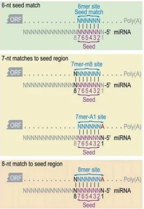
As mentioned in paragraph 1.2.2, miRNA-mRNA interaction relies on the perfect matching of the entire sequence between the nt 2 and 7 of the miRNA, called the seed region. Actually, four types of seed sites on mRNA are listed, as shown in Figure 3:
the 6mer, the seed region itself,
the 7mer-m8 site, consisting of the seed region and an additional match at nt 8, the 7mer-A1 site, consisting of the seed region and an adenosine in position 1,
the 8mer, consisting of a mix of the two previous sites, i.e. the seed match, an additional match in position 8 of the miRNA, and an adenosine in position 1.
The recognition of one of these sites may be sufficient to induce an effect on the mRNA target, but the additional pairing on the mRNA seems to enhance mRNA recognition by miRNAs. The extensive pairing of the nt 12 to 17 of the miRNA and in particular of nt 13 to 16 could balance imperfect matching in
25 the seed region [60]. It is worth noting that, the 6mer seed regions display a lower efficiency in targeting mRNA than 7mer-m8 [56]. The seed region was considered a major condition for the matching of miRNA on the mRNA because it is conserved among organisms [61], the majority of mRNA were under selective pressure and evolved to maintain the pairing with miRNA thanks to the seed region [62] and mutations or bulges of a single nt in the seed (typically G:U base pair) resulted in a less efficient targeting [57].
Figure 3: miRNA-mRNA interactions
In mammals, the perfect matching between miRNA and targeted mRNA almost never occurs. However, to induce miRNA- ediated sile i g, a perfe t at hi g ust e do e et ee the seed regio at the ’ e d of the iRNA a d
the mRNA. “tri tl speaki g, the seed regio represe ts the si u leotides at positio 2 to 7 of the iRNA gree fra e . “pe ifi it of the at hi g a e i reased if the seed is e larged o e or t o u leotides. Two conformations involving 7 nucleotides and one conformation involving 8 nucleotides are found. Adapted from [60].
1.3.1.2 Prediction of target sites and diversity of databases
After the characterization of miRNAs and their interaction with mRNA, an important point is to know which miRNA can target which mRNA, and inversely. It is interesting that a single miRNA can have several mRNA targets and mRNAs display several binding sites for different miRNAs [63]. To make those data more easily available, research teams created different databases. Here, I will describe
26 three databases/algorithms that I used during my PhD among the existing ones, comprehensively reviewed in [56, 64]:
TargetScan was the first to be created in the field DB. Ba tel a d CB. Bu ge’s g oup [61]. TargetScan is a tool allo i g use s to fi d iRNAs at hi g ith the 3’UTR of RNAs usi g the seed region; it extends the pairing to additional nt (as detailed above) and assigns a rank to 3’UTR a o di g to se e al the od a i pa a ete s. TargetScan also takes the conservation of the binding site on the mRNA among several species into account [62]. One of the most used algorithms is miRanda [65], available through the i o a.o g
website [66]. The algorithm is based on the at hi g et ee iRNAs a d the 3’UTR of the mRNA. miRanda has the advantage of displaying a score evaluating the impact of the miRNA on the mRNA (mirSVR score); furthermore, it does not limit mat hi g sea hes to 3’UTR ut also includes genomic sequences [64].
The DIANA-microT database, which is specialized for searching matching CDS a d 3’UTR, also indicates the recognition between a miRNA and targets containing a single miRNA responsive element [67]. DIANA-microT also takes the G:U mismatches and the proteins associated with the miRNAs (from the RISC) into account, whose conformation could interfere with the theoretical matching [64].
1.3.2. Mechanisms of post-transcriptional regulation of mRNA by miRNAs
iRNAs’ ai ode of a tio to p e e t the e p essio of spe ifi p otei s is post-transcriptional regulation. As detailed previously, when the matching between the miRNA and its target is perfect, mRNA will be cleaved immediately. This phenomenon, which is very common in plants, almost never happens in mammals. After their fixation on mRNA, miRNAs induce several mechanisms including mRNA destabilization and repression of translation. It is not quite clear how miRNAs directly induce these mechanisms but some evidence was found. The main mechanisms are illustrated in Figure 4.
27 Figure 4: Possible mechanisms of miRNA-mediated repression
Initiation of translation begins with the circularization of mRNA. This binding will induce several possible mechanisms to repress the translation. mRNAs can be deadenylated by the recruitment of the CCR4-NOT complex by GW182 or can induce the blo kade of the re og itio of the ’ ap eIF E left . After i itiatio of tra slatio , iRNAs ight e a le to induce ribosome dissociation, the degradation of the newly produced peptide (right) or PAPB displacement (middle).
28
1.3.2.1 Deadenylation and decay of miRNA targets
Destabilization and deadenylation of the miRNA targets are the most widespread mRNA decay mechanism in mammals. The mechanisms of mRNA decay after miRNA fixation are reviewed by Jonas and Izaurralde [69]. To summarize, after the fixation of the miRNA on its mRNA target, AGO2 recruits members of the P-body, or GW-body proteins. P-bodies are small granules localized in the cytoplasm and enriched in proteins involved in the mRNA metabolism, including deadenylation and decay [24, 70]. These adaptor proteins then recruit the Poly(A) specialized Nuclease 2 & 3 complex (PAN2-PAN3), proteins specialized in the deadenylation of mRNA (whether it is miRNA-mediated or not) by binding to the poly(A) tail. GW182, a protein of the P-bodies, also recruits the Poly(A) Binding Protein (PABP), which will be fixed on the poly(A) tail and will further induce the circularization of the mRNA and the mRNA decapping. PAN2, thanks to its catalytic activity, is able to rapidly induce the shortening of the poly(A) tail. Nonetheless, the PAN2-PAN3 complex is known to be part of the initial phase of deadenylation and sometimes, for reasons that are not clear, it will not complete the full degradation. The second phase of the deadenylation process is fulfilled by the CCR4-NOT complex, another complex recruited by GW182. Composed of nine subunits, including two catalytic subunits, these proteins are known to form the main mRNA-deadenylation complex. The CCR4-NOT complex shares several characteristics with the PAN2-PAN3 complex: their localization in the cytoplasm, their catalytic activity, and being components of P-bodies. The functions of the two complexes can be redundant but compensate for loss-of-function mutations [71]. These redundancies can be explained by the need to prevent any absence during processes, as the proteins involved in the mRNA metabolism and decay play an essential role.
Once deadenylated, mRNAs are uridylated the e z es TUT a d TUT at the 3’ e d. This uridylation is considered as a mark for downstream proteins to pursue the decay [72]. Next, mRNAs undergo de appi g at the 5’ to e pose the to exonucleases. This step is performed by the enzyme Decapping Protein 2 (DCP2) and one of its many cofactors, Decapping Protein 1 (DCP1) [73]. In the end, the e z e 5’-to-3’ e o i o u lease XRN ill fi alize the RNA de a totall deg adi g it [69].
29
1.3.2.2 Inhibition of the translation of the miRNA target
In addition to the mRNA deadenylation and decay, miRNAs mediate silencing through translation initiation inhibition according to four distinct mechanisms: PABP movement mediated by GW182, recruitment of translational repressors, non-recognition of the cap by eIF4E and ribosome drop-off [74].
Izau alde’s tea sho ed that PABP is disso iated f o t a slatio i itiatio fa to s the CCR4-NOT complex recruited by GW182, resulting in the non-circularization of the mRNA. As this conformation being compulsory for the degradation of mRNA, the mRNA is not degraded but its translation is impaired [75].
Independently of mRNA deadenylation, deadenylases are also able to induce translation ep essio . Wi ke s’ g oup de o st ated that CAF , a p otei of the CCR -NOT complex, could induce the repression of translation by itself, whether the mRNA is deadenylated or not [76]. CAF1 and CCR4 are also able to suppress poly(A) residues upstream of the poly(A) tail, possibly previously deadenylated. This observation suggests that these enzymes play another role in miRNA-mediated translational repression [77].
When translation is initiated, eIF4E is associated with eIF4G to induce mRNA circularization by binding to PABP [68]. T. P eiss’ g oup de o st ated that iRNA-mediated repression of translation could happen during initiation by targeting eIF4E, whether by inducing the
non-i dnon-i g to the 5’ ap of the RNA o lo knon-i g non-its fu tnon-io he ou d to the ap [78]. Several miRNAs were shown to act at the post-initiation stage of the translation to induce the
ribosome drop-off. At the translation initiation, the 40S and 60S ribosomal subunits assemble on the mRNA to form polysomes (or polyribosomes), leading to translation and protein p odu tio . PA. “ha p’s g oup de o strated that miRNAs could prematurely induce the dissociation between ribosomes and targeted mRNA [79], which was contradicted by studies led JD. Ri hte a d TW. Nilse . The o se ed that i oso es e e still atta hed to RNA and hypothesized that miRNAs prevent the association of polypeptides from ribosomes
30 [80, 81]. Nonetheless, Iwakawa and Tomari confirmed the theory of Petersen and colleagues in vitro in plants [82]. I the ea ti e, Ta o i’s tea i te esti gl de o st ated that GW could block the 80S ribosomal complex association in Drosophila (D.) Melanogaster by the upstream dissociation of eIF4A, a RNA helicase [83, 84].
However, the functional mechanisms underlying the translational repression are very unclear. The evidence detailed above provides clues to decipher the different translational repression mechanisms, but to my knowledge, no clear pathways have been described.
1.3.3. Biological roles of miRNAs
1.3.3.1 miRNAs as regulators of developmental processes
miRNAs often act to reinforce cellular destinies by acting on developmental and/or homeostasis pathways. It is the case in C. elegans during neurogenesis, where miR-124 is expressed during the transition of neuro-ectodermal progenitors to mature neuronal tissue. In the future neuronal tissue, miR-124 is highly expressed, resulting in a low expression of its target mRNAs. However, in epidermal cells, derived from the same progenitors and that do not express miR-124, miR-124 targets are increasingly expressed compared to the neuronal tissue. With such a process, epithelial ge es targeted by miR-124 are repressed in the neuronal tissue, ensuring that the differentiation occurs correctly [85].
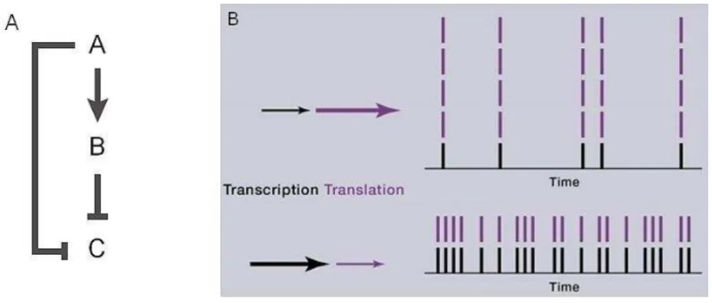
miRNAs act following the coherent feedforward loop (Figure 5A). This motif is a typical model where the expression of the target mRNA is inversely correlated with the expression of a miRNA and the most widespread model recapitulating miRNA-mediated regulation. For example, element A (a given miRNA-inducer gene) activates element B (a miRNA) and both inhibit element C (target gene). Element B, induced by element A, accumulates and is able to compensate any disturbance in element A production by acting on element C with the same effect [9].
31
1.3.3.2 miRNAs acting as noise reducers in gene expression
As raised by Schmiedel et al, how do miRNAs, which weakly repress a vast number of targets and whose knockdown often does not result in a pronounced phenotype [86, 87], remain conserved among species? The hypothesis is that miRNA regulation of gene expression is very tight and precise and that miRNAs are able to reduce background noises [88]. Elowitz et al. described two types of noises: intrinsic and extrinsic. Intrinsic noise corresponds to biochemical processes during gene expression whereas extrinsic noise reflects events happening in other cellular compartments [89]. Overall, noise arises from random events such as cell-to-cell expression variation and fluctuations over time of protein degradation, translation etc…[90]. To what extent can miRNAs act on noise reduction? For intrinsic noise, an important parameter to take into account is the transcription and translation rates that will influence protein levels. If the transcription rate is low (a few mRNA transcribed), the translation rate will be more important to maintain a certain pool of proteins. Knowing that translational repression represents 16% of miRNA-mediated repression [91], miRNAs will preferentially tend to reduce intrinsic noises by acting on the mRNA destabilization and decay.
Figure 5: Functions of miRNAs: robustness during development and noise reduction
(A The ohere t feed for ard loop represe ts the t pi al ase here iRNA a d its target’s e pressio s are i ersel correlated. This way, action on element C is reinforced and changes in the expression of element A or B can be
compensated. B) miRNAs act as noise reducers and act preferentially during transcription to induce mRNA destabilization and decay, rather than inducing translation repression. Adapted from [9, 92].
32
1.3.4. Degradation of miRNAs
Mainly because of their small size and by the protection RISC proteins brings, miRNAs are not easily accessible from nucleases and have the reputation of being stable. In spite of the difficulty in evaluating the half-life of miRNAs, some studies investigated them and their effective stability. MP. Gantier and colleagues followed the lifespan of several miRNAs following Dicer1 ablation in mice. Briefly, the authors estimated the average half-life of miRNAs of 115h (about five days) (varying from 100h for let-7b and miR-155 to 225h for miR-125b), 10-fold higher than the average mRNA half-life [93].
However, actual degradation of miRNAs first lies in its destabilization. miRNAs can be rapidly destabilized during changes of cell environment and behavior such as cell cycle [94], luminosity [95] or hormonal influence [96]. As detailed in paragraph 1.3.2.1, 5’ o 3’ u p ote ted e ds esults i a apid de a of RNAs. The sa e p o ess is o se ed i iRNAs. I the ase he e 5’ o 3’ e ds of the miRNAs were not protected by the RISC, miRNAs are susceptible to be tagged by uridylation and to be degraded. This phenomenon can happen to the passenger strand, which is not chosen by the RISC to induce miRNA-mediated silencing. Adenylation was also found to be a stabilizer of miRNAs so adenylases could act to destabilize them before decay [97].
Nucleases attacking miRNAs were reported to be substantially the same as the ones targeting mRNAs, such as XRN2. The small RNA degrading nuclease 1 (SDN1) was also found to degrade miRNAs effi ie tl f o 3’ to 5’ i Arabidopsis thaliana and C. elegans and remains to be studied in mammals [98, 99].
1.3.5. Clinical applications of miRNAs 1.3.5.1 miRNAs as biomarkers
Besides being present in cells and organs, miRNAs are detectable in body fluids such as urine, serum or saliva. Their presence in accessible body fluids and their easy quantification makes them good candidates as biomarkers. A biomarker is any substance, structure or process that can be measured in the body or its products and influences or predicts the i ide e of out o e o disease , as defi ed by the World Health Organization [100]. In body fluids, miRNAs are mostly carried in exosomes [101].
33 Exosomes are extracellular vesicles ranging from 40 to 100 nm diameter budding from the plasma membrane of endosomes [102]. Moreover, miRNAs can also be carried by ribonucleoprotein complexes such as AGO2, making them highly resistant to ribonuclease attacks [103]. Like other endocytosis vesicles, exosomes are coated with adhesion molecules and major histocompatibility complex (MHC) molecules that are involved in antigen presentation [104].
However, a biomarker, or in this particular case, miRNAs, must be expressed differently enough between controls and patients to establish a threshold where controls cannot be considered as patients, and inversely. Knowing that miRNA fold change is generally no more than 3, except for some individual miRNAs in particular situations, miRNAs can hardly be considered as good biomarkers or diagnostic tools on their own. Still, they can be invaluable in the case of no or bad diagnostic in rare diseases such as cancers or autoimmune diseases [105, 106]. For example, miRNAs can be used to classify patients in HIV infection [107], and to monitor the evolution of a disease or the response to treatments [108].
1.3.5.2 miRNAs as therapeutic targets
Three major techniques were developed to act against miRNAs, before and after biogenesis: miRNAs sponges, antisense oligonucleotides (ASOs, or antimiRs) and pharmacological molecules [35].
miRNA sponges, first described and investigated by MS. Ebert, JR. Neilson and PA. Sharp, are designed to act as competitive inhibitors of miRNAs [109]. They are naturally present in the organism or result from artificial constructs [110, 111]. Briefly, sponges correspond to RNA sequences that display several miRNA-binding sites that will be transcribed. These sequences will attract miRNAs so that they will not interact with target mRNAs (Figure 6A). miRNA sponges have to be highly expressed under a strong promoter to achieve their function, such as U6 or CMV promoters. Naturally present miRNA sponges can be circular [111] or long non-coding transcripts and may be called competitive endogenous RNAs (ceRNAs) [112, 113]. Artificial constructs can be delivered in vivo by viral vectors and can be designed to be under the influence of a tissue-specific promoter (reviewed in [110]).
34 ASOs, also named antimiRs, may be the most intuitive approach to target miRNAs. Their mechanism consists in designing an RNA sequence complementary to a specific miRNA to neutralize it (Figure 6B). ASOs will interact with the miRNA into the RISC so that miRNAs cannot reach the target mRNAs [35]. However, unmodified ASOs are useful in in vitro conditions but remain difficult to exploit in in vivo experiments due to nucleases present in serum. 2’-O-methyl modifications were demonstrated to be the most potent way to stabilize of ASOs [114, 115] but such modified ASOs, also called antagomiRs [116] were shown to be very sensitive to nucleases [117]. Moreover, KA. Lennox and MA. Behlke concluded that locked nucleic acids (LNA)- ’OMe-phosphorothioate combination was the better alternative to a maintained efficiency in vivo. LNA are RNA structures whe e the ’-oxygen is bound to the ’-carbon of the ribose, making it more stable and increasing its temperature of melting (Tm) [118]. Phosphorothioate modifications consist of exchanging an oxygen by a sulfur at the phosphodiester bond between two nucleotides (Figure 6C) [117]. The first clinical trial involving ASOs was developed in 2010 to test the effect of the Miravirsen, an LNA- phosphorothioate modified ASO sequestrating miR-122 in the context of hepatitis C virus infection. The authors confirmed the efficiency of the treatment, as after 14 weeks, four out of nine patients treated with the highest dose and one out of nine patients treated with a medium dose did not display any residual hepatitis C virus RNA levels and viral resistance was not seen in all patients [119]. Since then, several phase I or II clinical trials were open for other miRNAs in various disorders. It should be noted that the difference between ASOs and antimiRs is not clearly defined. R. Rupaimoole and FJ. Slack defined antimiRs as structurally similar to ASOs without being more precise [116].
Some pharmacological compounds selectively inhibit the expression of certain miRNAs, such as azobenzene for miR-21 [120] and benzimidazole for miR-96 [121]. Some compounds also act on the biogenesis pathway. For example, enoxacin enhances the production of miRNAs by acting on TRBP affinity [122]. Some other compounds can be found by active screening of mature and precursors miRNA libraries (Figure 6D) [121].
35 Along with the development of all these techniques to act on miRNAs in order to treat pathophysiological situations, it might be wise to have wider look at manipulating miRNA expression, knowing that they are tightly regulated.
36 Figure 6: miRNAs as therapeutic targets
miRNA sponges (A) are expressed artificially from a reporter coding for the miRNA responsive element of a target miRNA. This way, when expressed, miRNAs will preferentially target these sponges and not their initial targets. (B) AntimiRs are unmodified ssRNAs meant to bind to the mature miRNA strand, thus leading to a functional blockade. (C) AntimiRs were chemically modified, by binding oxygen and carbon atoms of the ribose together (locked nucleic acids or LNA) or by replacing an oxygen atom by a sulfur (phosphorothioate). Finally, some pharmacological compounds are
able to act on the miRNA biogenesis (D). Adapted from [35].
1.3.5.3 miRNAs as therapeutic tools
The other way to use miRNAs in a pathophysiological context is as tools to treat or to ameliorate a disorder. In pathologies, we often observe an altered expression of one or several miRNAs compared to controls. In the case of an increase, ASOs (detailed in the previous paragraph) are more suitable to adjust the miRNA level to the control one. However, in case of a decrease, we will tend to use miRNA
37 mimics to increase their expression level. miRNA mimics are synthetic dsRNAs designed to bypass the biogenesis pathway until the loading in the RISC and to mimic a miRNA duplex [123]. They have the advantage of being processed in part by AGO2 and to later act as endogenous miRNAs. Contrary to ASOs, if they are too much modified, they will not be taken up by the RISC and not be efficient. However, to prevent degradation in vivo by nucleases, miRNA mimics are surrounded by modified liposomes: stable nucleic acid-lipid particles (SNALPs). However, cargo delivery in vivo has yet to be improved [124]. In serum and other biofluids, miRNAs are very stable. Knowing that, teams succeeded in modulating miRNAs in exosomes ex vivo or to address a specific type of exosomes-carrying miRNAs to particular cells for them to deliver their cargo very specifically [125, 126]. Combining these two approaches could be very promising, for example by bringing miRNAs to an organ lacking these miRNAs. An approach using miRNAs to limit the expression of genes, in a specific cell context, was investigated by Boisgerault et al. [127]. The authors used in their advantage the restricted expression of miR-142-3p in immune system cells to improve the transduction efficiency of their transgene and prevent an immune response towards the transgene. Briefly, they designed a vector with miR-142-3p target sequences coded with their transgene of interest. Therefore, when injected systemically, miR-142-3p will be fixed on the transcribed designed target sequences and prevent the expression of the transgene in immune cells, thus preventing an immune response.
38
2.
Myasthenia Gravis
The class of myasthenic syndromes gathers autoimmune Myasthenia Gravis (MG), the Lambert-Eaton myasthenic syndrome and congenital myasthenic syndromes. They are all characterized by defects at the neuromuscular junction (NMJ) but differ in terms of mechanisms.
Autoimmune Myasthenia Gravis (MG) is a rare autoimmune disorder, characterized by the presence of antibodies directed against several components of the post-synaptic membrane of the NMJ, leading to an impaired transmission signal to the skeletal muscle. This results in a generalized fatigability during effort, sometimes affecting respiratory and swallowing muscles. The Lambert-Eaton myasthenic syndrome is also an autoimmune disorder characterized by defects at the presynaptic membrane of the NMJ, caused by autoantibodies directed to voltage-gated calcium channels [128]. Congenital myasthenic syndromes are inherited disorders and must be distinguished from autoimmune MG and Lambert-Eaton Myasthenic Syndrome. Congenital myasthenic syndromes display similar symptoms as autoimmune MG, due to defects in neuronal transmission as the consequence of mutations of proteins of the NMJ.
During my PhD, I only focused on autoimmune MG; there will be no further mention of the Lambert-Eaton Myasthenic Syndrome or congenital myasthenic syndromes in this manuscript.
2.1. History of the disease: from the first cases to recent findings
There are some uncertainties concerning the first described MG case. Reportedly, Indian Chief Opechankanough, ho died i , see ed to ha e de eloped eak esses aki g hi u a le to alk a d aki g his e elids so eak the had to e held atte da ts . However, the sources came from correspondence between settlers in America and England and can be discussed [129]. In 1672, Thomas Willis published several reported cases in De Anima Brutorum, including the case of a woman that speaks freely and readily enough for a while, but after a long period of speech, she is not able to speak a word and is as mute as a fish. Her voice does not return for one or two hours [130, 131]. In the 19th century, Wilks, Erb and Goldflam described some clinical features of MG: fatigue, fluctuating
39 symptoms with relapses and remission cycles [132-134]. F. Jolly then showed that stimulation of the nerve innervating the patie t’s muscle caused reduced muscle contraction. After being named for the Erb-Goldflam disease a few times, Jolly introduced the name asthe ia g a is pseudoparalytica after her discovery [135]. The name can literally e de o posed as myo , greek for muscle, asthenia fo fatigue, gravis Latin for severe a d pseudoparalytica fo pseudo-paralysis. After several discoveries, such as the improvement of MG after anticholinesterase treatment [136] and the importance of acetylcholine (ACh) in the NJM, this evidence led to suggesting MG as an neuromuscular disorder. Thanks to the discovery of α-bungarotoxin, a snake toxin that binds specifically and irreversibly to AChR [137], Fambrough et al. showed that defects in ACh receptor (AChR) at the NMJ in MG patients may be the cause of the disease [138]. After the immunization of rabbits with purified AChR by J. Patrick and J. Lindstrom, the rabbits developed MG symptoms by producing specific antibodies to purified AChR (later called Experimental Autoimmune Myasthenia Gravis, EAMG) [139], suggesting that MG could be due to autoantibodies directed at AChR. Later on, additional evidences about the presence and the role of AChR in MG emerged: detection of anti-AChR antibodies in the serum of patients [140], transfer of human serum to mice led to the development of MG in the animals [141], improvement of symptoms after plasma exchange in patients [142] and co-localization of the lytic complement component C9 and AChR on the motor endplate [143].
For 30 years, AChR was the only antigen characterized in MG. It was only at the beginning of the 21st
century that antibodies against the muscle associated receptor tyrosine kinase (MuSK) were described in 70% of seronegative MG patients [144]. Ten years later, another antigenic target was described, the low-density lipoprotein receptor-related protein 4 (LRP4), with antibodies leading to MG symptoms [145]. Several other antigens were found in MG patients such as titin [146], ryanodine [147], agrin [148, 149] and cortactin [150], but they were found in AChR+ and MuSK+ patients as well and were not
exclusive to MG. The role at the NMJ of the different antigenic targets involved in MG will be further explained in paragraph 2.3.1. Major discoveries regarding MG are summed up in figure 7.
40 Figure 7: Major discoveries in MG
Adapted from [131].
2.2. Clinical features 2.2.1. Epidemiology
I , J. M Co ille’s g oup o piled 55 epidemiological studies of MG that were published between 1951 and 2004. The studies included all MG patients whatever the serotype. The incidence rate, defined by the emergence of new cases during a certain period, ranges from 1.7 to 21.3 cases per million person-years but with a high heterogeneity between studies (35 studies). However, major differences can be observed when the serotype is taken into account. In AChR+ and MuSK+ MG specific
studies (eight and two studies respectively), the incidence rate ranges from 4.3 to 18.0 per million person-years in AChR+ MG and 0.21 per million person-years in MuSK+ MG.
The prevalence rate, defined by the number of cases identified at a precise point in time, ranges from 15 to 179 per million (44 studies), all MG forms included. For AChR+ MG, the prevalence rate ranges
from 70.6 to 163.5 per million (three studies) and is 1.9 per million in the Netherlands for MuSK+ MG
cases, the only country where the rate is known [151].
Regarding LRP4-positive MG patients (LRP4+ MG), 18.7% of double-seronegative patients displayed
41 10 countries. Interestingly, the authors noticed that some AChR+ MG and MuSK+ MG patients displayed
LRP4 autoantibodies, making them double-positive [152]. It is interesting that a given patient cannot be seropositive for AChR and MuSK.
All these epidemiological studies noted little global geographical variation except in Asia, where almost 50% of AChR+ MG cases, mostly ocular, were detected before the age of 15. Moreover, the prevalence
rate may seem to have doubled between the 1960s and the 1970s and between the 1980s and the 1990s but diagnosis methods may have influenced this rate as they probably improved during these periods [153].
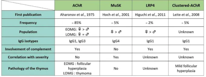
To sum up, AChR+ MG patients represent 80 to 85% of all MG patients, MuSK+ MG patients 5 to 4%
and LRP4+ MG patients 2%. Remaining patients are considered seronegative [154, 155]. 2.2.2. Clinical classification and associated symptoms
MG patients can be classified according to different criteria: the symptoms and the severity, the serotype, the age of onset and the implication of thymic pathologies.
2.2.2.1 Symptoms Ocular MG
This form may affect 15 to 20% of MG patients. Ocular weaknesses symptoms can be ptosis (fall of the upper eyelid) or diplopia (double vision of a single object). Only patients without any other symptoms during the two years following their diagnosis are classified as pure ocular MG patients. Fifty percent of these patients do not display classical detectable antibodies by radioimmunoassay (RIA) but low-affinity antibodies detectable when AChR is clustered (see below) [154, 156].
Generalized MG
The Myasthenia Gravis Foundation of America (MGFA) established strict criteria to define severity grades for patients, without regarding the serotype (Figure 8). Some clinical features are common between the different MG subclasses: ptosis or diplopia (without pupillary abnormalities), weaknesses of superior limbs and neck/cervical muscles and signs of bulbar defects. Fatigue worsens after exercise
42 and improves after rest. However, remitting and relapsing phases can happen during the day or over long periods of time. It should be noted that MG patients do not display central nervous system dysfunctions.
Figure 8: Clinical classification according to the MGFA
Symptoms are classified from class I to V, V being the more severe form of the disease. Classes II, III and IV display two subclasses (A and B) according to muscles affected. Adapted from [157].
Neonatal and juvenile MG
Pregnant MG patients can transiently induce MG to their newborns. This form of MG principally concerns AChR+ MG patients and results in hypotonia, respiratory troubles and impaired sucking as
well as swallowing. Symptoms last from one to five months after birth, when the antibodies of the mother disappear [154].
MG in children can happen within the first years of life. Patients display AChR antibodies and mainly purely ocular symptoms. Contrary to generalized MG, there is no specific gender distribution and usually, children patients show more spontaneous remissions than adults [158]. The juvenile form of MG is predominantly found in Asians [154].
2.2.2.2 Antibodies
43
AChR antibodies
85% of MG patients display AChR antibodies [154]. The majority of AChR antibodies binds to α subunits of the receptor, called the main immunogenic region (MIR). They are from IgG1 and IgG3 subclasses, thus able to activate the complement cascade.
Patients can be classified in three populations, according to the age of the onset: early-onset AChR+
MG patients (EOMG) with symptoms before the age of 50 and late-onset AChR+ MG patients (LOMG) after 50 years of age. Very late onset MG patients develop symptoms after the age of 60 and the onset may be linked to the aging population. In EOMG patients, the thymus is very often affected and develops histological and functional abnormalities (more precisely described in paragraph 2.3.3). Interestingly, more than three EOMG patients out of four are female.
The LOMG form is generally associated with the development of a thymoma, a tumor of thymic epithelial cells (TECs). The thymus does not present abnormalities like in EOMG patients, and thymectomy does not improve the symptoms of MG. However, thymectomy is mandatory to remove the tumor. In this case, MG is a paraneoplastic disease.
Low-affinity antibodies / clustered AChR
Patients seronegative for AChR, MuSK and LRP4 antibodies represent between 5 and 10% of MG patients. Some of these patients may display low-affinity antibodies for these proteins, detectable only when AChR is clustered [156]. These antibodies are of the same IgG subclass as AChR antibodies, i.e. IgG1 or IgG3 suggesting a common etiology and pathogenesis [159, 160]. This hypothesis is confirmed by patients being similar to lassi al AChR+ MG patients as regards their clinical symptoms, response
to treatment and thymic abnormalities [156].
MuSK antibodies
About 5% of MG patients display autoantibodies to MuSK. These patients are adults; only rare cases were described in children or elderly patients. Like with EOMG patients, most of those affected are female. MuSK+ MG patients do not display thymic abnormalities, thus thymectomy is not
44 weaknesses i.e. masticatory weaknesses, dysphonia (hoarseness), dysarthria (troubles in enunciating) and more respiratory weaknesses than EOMG patients. Overall the condition of MuSK+ MG patients
tends to be more severe, and patients have more myasthenic crises [161].
Interestingly, antibodies against MuSK are of the IgG4 subclass. They interfere with the clustering of the receptor and induce a presynaptic effect [162]. Contrary to IgG1 and IgG3, they do not bind the complement [160].
LRP4 antibodies
LRP4 autoantibodies are found in highly variable proportions from 7 to 32% of double-seronegative MG patients. Patients display ocular or mild generalized MG and bulbar and respiratory muscles can be involved. However, due to the recent findings about LRP4 autoantibodies, the pathogenesis of LRP4+ MG still needs to be investigated. The involvement of the thymus in the pathogenesis of LRP4+
MG remains unclear but some patients display ectopic GCs like AChR+ MG patients [152, 155]. Other antibodies
Some other antibodies are also found in MG patients. For example, LOMG patients can display autoantibodies directed at muscle or NMJ proteins, such as ryanodine, titin, cortactin, collagen Q (CollQ) or Kv1.4 (a subunit of the voltage-gated potassium channel) [150, 163, 164].
Table 1: Antibody-specific characteristics of each type of MG