HAL Id: cea-02937752
https://hal-cea.archives-ouvertes.fr/cea-02937752
Submitted on 14 Sep 2020HAL is a multi-disciplinary open access archive for the deposit and dissemination of sci-entific research documents, whether they are pub-lished or not. The documents may come from teaching and research institutions in France or abroad, or from public or private research centers.
L’archive ouverte pluridisciplinaire HAL, est destinée au dépôt et à la diffusion de documents scientifiques de niveau recherche, publiés ou non, émanant des établissements d’enseignement et de recherche français ou étrangers, des laboratoires publics ou privés.
anesthesia in non-human primate on the brain kinetics
and binding of [
18F]DPA-714, a PET imaging marker of
glial activation
Wadad Saba, Sébastien Goutal, Bertrand Kuhnast, Frédéric Dollé, Sylvain
Auvity, Yoan Fontyn, Jérôme Cayla, Marie-Anne Peyronneau, Héric Valette,
Nicolas Tournier
To cite this version:
Wadad Saba, Sébastien Goutal, Bertrand Kuhnast, Frédéric Dollé, Sylvain Auvity, et al.. Differential influence of propofol and isoflurane anesthesia in non-human primate on the brain kinetics and binding of [18F]DPA-714, a PET imaging marker of glial activation. European Journal of Neuroscience, Wiley,
Research reports
Differential influence of propofol and isoflurane anesthesia in non-human primate on
the brain kinetics and binding of [18F]DPA-714, a PET imaging marker of glial
activation
Wadad SABA*, Sébastien GOUTAL, Bertrand KUHNAST, Frédéric DOLLÉ, Sylvain AUVITY, Yoan FONTYN , Jérôme CAYLA, Marie-Anne PEYRONNEAU, Héric VALETTE and Nicolas TOURNIER
Address: CEA, DSV, I2BM, Service Hospitalier Frédéric Joliot, Orsay, F-91406, France Institution: Inserm / CEA / Université Paris Sud, UMR 1023 - ERL 9218 CNRS, IMIV, Orsay, F-91406, France
* Corresponding author:
wadad.saba@cea.fr
CEA, DSV, I2BM, Service Hospitalier Frédéric Joliot, Orsay, F-91401, France Phone number: +33 1 69 86 77 52 Fax number: +33 1 69 86 77 68
Running Title: Influence of anesthesia on TSPO brain PET imaging
Number of words
- Whole manuscript: 5858 - Abstract: 250
- Introduction: 506
Keywords: neuroinflammation, anesthetic, baboon, peripheral benzodiazepine receptor,
positron emission tomography, in vitro binding
ABSTRACT
Translocator protein 18kDa (TSPO) expression at the mitochondrial membrane of glial cells is related to glial activation. TSPO radioligands such as [18F]DPA-714 are useful for the non-invasive study of neuroimmune processes using positron emission tomography (PET). Anesthetic agents were shown to impact mitochondrial function and may influence [18 F]DPA-714 binding parameters and PET kinetics.
[18F]DPA-714 PET imaging was performed in Papio Anubis baboons anesthetized using either intravenous propofol (n=3) or inhaled isoflurane (n=3). Brain kinetics and metabolite-corrected input function were measured to estimate [18F]DPA-714 brain distribution (VT). Displacement experiments were performed using PK11195 (1.5 mg/kg). In
vitro [18F]DPA-714 binding experiments were performed using baboon brain tissue in the absence and the presence of tested anesthetics.
Brain radioactivity peaked higher in isoflurane anesthetized animals compared to propofol (SUVmax=2.7±0.5 versus 1.3±0.2, respectively) but was not different after 30 min.
Brain VT was not different under propofol and isoflurane. Displacement resulted in a 35.8±8.4
% decrease of brain radioactivity under propofol but not under isoflurane (0.1±7.0 %). In vitro, the presence of propofol increased TSPO density and dramatically reduced its affinity for [18F]DPA-714 compared to control. This in vitro effect was not significant with isoflurane.
Exposure to propofol and isoflurane differentially influences TSPO interaction with its specific radioligand [18F]DPA-714 with subsequent impact on its tissue kinetics and specific binding estimated in vivo using PET. Therefore, the choice of anesthetics and their potential
influence on PET data should be considered for the design of imaging studies using TSPO radioligands, especially in a translational research context.
INTRODUCTION
Translocator Protein 18 kDa (TSPO) is expressed at particularly high levels in steroid hormone producing tissues and parotid glands (Gatliff & Campanella, 2012). Conversely, expression of TSPO in the healthy brain is low and mainly dependent on glial cells, the largest population of macrophages of the central nervous system (CNS). Consequently, transition of astrocytes and microglia from the normal resting state to the activated one following CNS injuries and neuroinflammation results in an increased level of TSPO and binding sites for TSPO ligands (Lavisse et al., 2012). This neuroimmune process is used as a surrogate marker of neuronal dysfunction with minimally-invasive positron emission tomography (PET) imaging and TSPO radioligands (Scarf & Kassiou, 2011). Therefore, several TSPO-selective radioligands have been developed.
Only few compounds have reached the clinical application, among which [18 F]DPA-714 (Arlicot et al., 2012). Early clinical evaluation of TSPO radioligands has raised new questions about the TSPO ligands binding interactions: it was reported that genetic polymorphism (Guo et al., 2013), multiple binding sites (Guo et al., 2012), variable conformational states and complex interaction of TSPO with its molecular environment complicates PET data interpretation (Scarf & Kassiou, 2011). In summary, a growing literature reports that variation in TSPO ligand binding detected in vivo using PET may not be simply interpreted as a change in TSPO density.
General anesthesia is almost inevitable in animal studies and is sometimes necessary for patients who are unable to tolerate a prolonged imaging session and may affect PET data
of anesthetics on mitochondrial metabolism and function has been widely studied (Kajimoto et al., 2014) and may explain the deleterious effects of anesthetics in patients suffering from mitochondrial diseases (Finsterer & Segall, 2010). TSPO is a partner of the mitochondrial permeability transition pore (mPTP) and a regulator of the mPTP complex. Recent reports suggest that isoflurane may induce the opening of mPTP via increasing levels of reactive oxygen species (Zhang et al., 2012). Only few studies have addressed the influence of anesthesia on TSPO radioligands brain kinetics. It was reported that propofol anesthesia decreased the brain distribution of the TSPO ligand [11C]PBR28 measured in the Human brain using PET (Hines et al., 2013). Similarly, isoflurane was shown to reduce the binding of [11C]DPA-713, a [18F]DPA-714 derivative, in the monkey brain (Tsukada et al., 2014). These data suggest that these anesthetics may differentially affect TSPO binding parameters which may possibly influence TSPO radioligands brain kinetics.
To address this question, we studied in vivo the differential influence of propofol and isoflurane on [18F]DPA-714 kinetics and binding parameters. [18F]DPA-714 PET data obtained in the brain and the parotid gland (a TSPO-rich region) were compared in non-human primates anesthetized using either intravenous propofol or inhaled isoflurane. The influence of anesthetics on the [18F]DPA-714 peripheral pharmacokinetics and metabolism was also assessed. In vitro binding experiments were also performed on baboon brain tissue and brain mitochondrial preparation to elucidate PET data and to compare with [18F]DPA-714 binding parameters in the absence of anesthetics.
MATERIAL AND METHODS Animals
Papio Anubis male baboons (8 to 14 kg, 3-5 year old) were obtained from the primatology station (Rousset-sur-Arc, France). PET studies were carried out using three different animals for a total of 12 experiments. First, [18F]DPA-714 PET imaging was
performed in each animal, once under each of the two anaesthetic conditions (isoflurane or propofol) with an interval of at least 1 month between experiments. The order of anaesthetic conditions was varied with two animals receiving isoflurane followed by propofol (one and three months after, respectively) and one animal receiving propofol followed by isoflurane two months after. Then, [18F]DPA-714 displacement experiments were performed using the same animals under each of the two anaesthetic conditions. Two animals received propofol first (two and three months after, respectively) whereas the third animal received propofol followed by isoflurane, four months after. In vitro experiments were performed using baboon brain tissue samples obtained from another and previously euthanized animal. Animal use procedures were in strict accordance with the recommendations of the European Community (86/609/CEE) and the French National Committee (Décret 87/848) for the care and use of laboratory animals. The experimental protocol was evaluated by a local ethic committee (CETA Reference n° 10-004).
Drugs, chemicals and radiochemicals
Isoflurane and propofol were administered using the commercial drugs Forene® (Abbott, Rungis, France) and Propofol Fresenius® 200 mg/20 mL (Fresenius Kabi, Sèvre, France), respectively. Propofol without soja oil was purchased from Sigma Aldrich (France). Solutions of unlabeled racemic PK11195 (gift from Sanofi, Chily-Mazarin, France) were prepared by dissolving 2–3 mg of the compound in Dimethylsulfoxid (0.2 mL), followed by dilution with PEG 400 (1 mL) and NaCl 0.9% (2 mL). Similarly, solutions of unlabelled DPA-714 for i.v. injection were prepared by dissolving 2–3 mg of the compound in Dimethylsulfoxid (0.2 mL), followed by dilution with Poly(ethylene glycol) (PEG, Sigma-Aldrich), 400 (0.4 mL) and finally NaCl 0.9% (1.2 mL).
DPA-714 was labeled with fluorine-18 (half-life, 109.8 min) at its 2-fluoroethylmoiety using a tosyloxy-for-fluorine nucleophilic aliphatic substitution, according to slight
modifications of procedures already reported, and using a commercially available GE TRACERLab FX-FN synthesizer (GE Medical Systems, Buc, France) (Kuhnast et al., 2012). Typically, 10 GBq [18F]DPA-714 were routinely obtained within 60 min, including High-performance liquid chromatography (HPLC) purification and formulation. Ready-to-inject, >99% radiochemically pure [18F]DPA-714 (formulated in physiological saline containing less than 10% of ethanol) was obtained with 15-20% non-decay-corrected yields and specific radioactivities at the end of the radiosynthesis ranging from 37 to 148 GBq/μmol.
PET imaging
The influence of anaesthetics on [18F]DPA-714 brain distribution was assessed in baboons using a HR+ Exact positron tomograph (Siemens Healthcare, Knoxville, TN, USA). One hour before the PET acquisition the animal received ketamine (Ketamine Virbac®, Carros, France; 10 mg/kg i.m.) to induce anaesthesia and prepare the PET experiment. After being intubated, a catheter was inserted in a sural vein for tracer and unlabelled PK11195 or DPA-714 administration. Another sural vein catheter was dedicated to propofol infusion when necessary. A femoral artery catheter was inserted for blood sampling and input function assessment. After being positioned under the camera, animals were maintained anaesthetized with either i) inhaled isoflurane/nitrous-oxide mixture (1-2 % / 66% in oxygen), controlled by an Ohmeda ventilator (Ohmeda OAV 7710, Madison, WI, USA) (n=3) or ii) i.v. bolus of propofol (2mL) followed by a 16 to 22 mL/h i.v infusion under oxygen ventilation (n=3). The tidal volume was adjusted to achieve stable end-tidal carbon dioxide tension between 38 and 42 mmHg. Heart rate and rectal temperature were monitored throughout the experiment. The head of the baboon was positioned in the tomograph using a custom-designed stereotaxic headholder in order to image the brain and the parotid glands in the same field of view. Head transmission scans were acquired for 10 min using three retractable germanium-68 rod sources. Animals were i.v. injected with [18F]DPA-714 (231± 64 MBq) and imaged for 90
min. Displacement using unlabelled PK11195 (1.5 mg/kg) (James et al., 2008) or DPA-714 (1.5 mg/kg) was performed 40 min after tracer injection in each condition (n= 3). PET imaging was then continued for an additional 80 min.
Metabolite corrected input function
During the PET scan, arterial blood samples were withdrawn at selected times from the femoral artery. Metabolic profile of plasma extracts were obtained by radioactive-HPLC analysis using a reported method (Peyronneau et al., 2013). The influence of anesthetics on [18F]DPA-714 fraction bound to plasma protein was measured using an ultrafiltration method (MPS Micropartition Microcon® - YM-10 membrane from Millipore, Guyancourt, France) (Tournier et al., 2012).
PET data processing
Regional time activity curves (TACs) were generated by calculating the mean radioactivity in selected volumes of interests (VOIs): whole brain hemispheres and parotid glands corrected for fluorine-18 decay and injected radioactivity. PET data are expressed as standard uptake value (SUV) in each VOI. [18F]DPA-714 distribution volumes (VT ; mL.cm-3)
in both VOI were estimated using the Logan graphical method, considering the arterial plasma corrected for metabolites (PMOD® software, Zurich, Switzerland).
To quantify the extent of displacement, measured SUV at 120 min were normalized from SUV at 40 min (before displacement). The impact of displacement is expressed as the difference (in percentage) between non-displacement and displacement experiments at 120 min.
[18F]DPA-714 binding studies in vitro
In vitro binding experiments were performed on a baboon total brain cortex homogenate and on a mitochondrial enriched preparation of this homogenate. [18F]DPA-714
affinity constant (KD) and TSPO binding density (Bmax) were measured in the presence of
each tested anaesthetic agent and compared to the control condition (without anesthetic). Cortex tissue samples were obtained from a baboon euthanized via i.v. injection of an overdose of pentobarbital (100 mg/kg, i.v; Sanofi, France). Samples were stored at -80°C until use. Total brain cortex tissue was directly homogenized in an incubation buffer (50 mM HEPES buffer; pH 7.4) using a teflon glass homogenizer to provide the total brain cortex homogenate. The mitochondrial enriched fraction was prepared as previously described with minor modifications (Romeo et al., 1993). The brain cortex tissue sample was homogenized in 10 vol. of ice-cold 0.32 M sucrose, 10 mM HEPES buffer (pH 7.4), and centrifuged at 1,000 g for 10 min at 4°C. The supernatant was centrifuged at 9,000 g for 10 min at 4°C. Then, the pellet was washed by incubation buffer and finally centrifuged at 12,000 g for 10 min at 4°C. [18F]DPA-714 binding parameters were measured in vitro in the presence of i) 100 µM propofol, ii) 10 mM isoflurane and iii) in the absence of anesthetic agent in incubation buffer. Tested anesthetic concentrations were set on the previously reported concentrations found in rats after standard anesthesia with propofol (Shyr et al., 1995) and previous in vitro studies using isoflurane (Sugimura et al., 2001). Binding experiments were performed in sealed glass vials. Samples (~100 µg protein/vial; n=3 for each condition) were incubated with increasing concentrations of [18F]DPA-714 ranging from 0.3 to 30 nM at 37°C for 30 min. Nonspecific binding was determined by adding 10 µM of the reference TSPO ligand PK11195 to the incubation buffer. Incubation was terminated by rapid filtration through a GF/B glass fibre filters presoaked with 0.1% polyethyleneimine (Sigma-Aldrich). Then the filters were washed three times with 3 mL of the ice-cold incubation buffer, using a Brandel® 48-channel cell harvester (Inotech-IH-110, PerkinElmer, France). Radioactivity was measured using an automatic γ-counter (model 5000; Packard instrument CO., Downers
Grove, IL). Protein concentration was determined using a BCA-based Protein Assay Kit (Thermo Scientific, France).
Statistical analysis
The results are presented as means ± standard deviation. Statistical analysis of the data was performed using R version 3.1.2. Homogeneity of residuals was assessed using the Levene test to assume homoscedasticity (α=0.05). Then, data were analyzed using one-way analysis of variance (ANOVA) followed by Tukey's HSD (honest significant difference) post-hoc test. The null hypothesis is that there was no change in tested parameters (SUV, VT, percentage
displacement, KD, Bmax, Binding Potential) between groups (anesthetics). Level of
significance for all hypotheses was set at α=0.05.
RESULTS
PET study
[18F]DPA-714 distribution in the brain was rather homogenous regardless of tested anesthetic protocols (Fig. 1). The TACs obtained in the whole brain of propofol and isoflurane anesthetized baboons showed that radioactivity peaked rapidly and was followed by a rapid wash-out until 20 min post injection. Then, the radioactivity slowly declined until 120 min post injection. The main difference between anesthetics was observed in the early distribution phase, SUVmax being higher (F3,8= 21.88, p=0.026) in isoflurane (2.7±0.5) than in
propofol anesthetized animals (1.3±0.2). SUV measured at 30 min (0.7) were not significantly different between anesthetic (F3,8= 23.32, p=0.99), followed by parallel plateaus
until the end of the scan (Fig. 2).
PET kinetics in the parotid glands, a TSPO-rich region, varied between tested anesthetics: under isoflurane, radioactivity increased gradually, to reach SUVmax at 40 min (2.8±0.6)
although non-significant (F3,8= 21.88, p=0.20) and was achieved more rapidly, at 16 min
post-injection, followed by a slow decline of radioactivity. SUV values obtained 60 min after tracer injection were not significantly different (F3,8= 23.32, p=0.21) (Fig. 2).
[18
F]DPA-714 plasma kinetics is described by a distribution phase followed by an elimination phase, starting 8 min post injection (Fig. 2). Mean SUV measured from 40 to 90 min (elimination phase) were 0.08±0.05 and 0.13±0.05 for propofol and isoflurane, respectively. Radio-HPLC analysis showed no striking influence of tested anesthetics on [18F]DPA-714 metabolism (Fig. 2). The fraction of [18F]DPA-714 that is bound to plasma protein (fp) was 94.8±1.6 and 94.7±2.1 % under propofol and isoflurane respectively.
Estimated VT were not significantly different (F3,8= 31.13, p=1.0) using propofol and
isoflurane anesthesia in the brain (2.86±1.35 mL.cm-3 and 2.15±0.34 mL.cm-3, respectively)
(Fig. 2). As expected, VT in the parotid glands was higher compared to the VT in the brain
under either propofol (p= 3.3×10⁻⁴) or isoflurane (p= 0.015). However, VT in the parotid
glands was not different (p=0.37) between propofol and isoflurane (21.60±11.44 mL.cm-3 and 18.21±8.12 mL.cm-3, respectively) (Fig. 3).
Displacement of brain radioactivity with unlabeled PK11195 was obvious under propofol anesthesia (Fig. 4) and resulted in a 35.8±8.4% decrease in total brain radioactivity. Surprisingly, no displacement (0.1±7.0 %) could be observed under isoflurane anesthesia using either PK11195 (Fig. 3 and 4) or unlabeled DPA-714 (1.5 mg/kg; data not shown). Displacement obtained under propofol was observed in each animal (Fig. 4), regardless of tested condition sequence (propofol first or isoflurane first). After PK11195 injection, a transient increase in the brain radioactivity was observed, due to the peripheral release of [18F]DPA-714 from peripheral tissue into plasma (Fig. 4). Differential influence of displacement between anesthetics was confirmed by statistical analysis (F3,8=53.89, p=
radioactivity in the parotid glands which extent was not different in the propofol (62.2 ± 7.5 %) and isoflurane groups (60.0 ± 3.2 %) (p=0.97) (Fig. 3).
In vitro binding study
Differences in the displacement sensitivity observed in vivo prompted us to pinpoint the effect of anesthetic exposure on [18F]DPA-714 affinity for TSPO. In the absence of anesthetics, the binding of [18F]DPA-714 is saturable and interacted with a single binding site in the baboon brain (Fig. 5). Estimated KD values were not different in total brain homogenate
and in mitochondrial enriched preparation (F5,12=35.02, p>0.05), regardless of the presence of
anesthetic (Table 1). Incubation of baboon brain homogenate with propofol resulted in a significant increase in KD (p=3.0×10⁻⁶), indicating a reduction of the affinity of [18F]DPA-714
for TSPO binding site. Increase in KD found with isoflurane was not significant (p=0.58).
However, anesthetic effect on KD was found different between propofol and isoflurane
(p=2.69×10⁻⁵). Measure of Bmax was higher in the mitochondrial preparation compared to the
brain homogenate under propofol (F5,12=18.93, p=1.9×10⁻⁴) and isoflurane (p=0.047) but not
in the absence of anesthetics (p=0.37). Propofol exposure resulted in a significant increase in Bmax in the brain homogenate (p=3.7×10⁻⁵), a result that was not observed with isoflurane
(p=1.0). The effect of propofol on Bmax was also detected in the mitochondrial preparation
(p=3.7×10⁻⁴) whereas isoflurane effect was not significant (p=0.85) (Table 1). Mean binding potential (BP=Bmax/KD) measured in the brain homogenate in the absence of anesthetics was
BPcontrol=535.51±50.52. Binding potential was significantly lower in the presence of propofol
(BPpropofol=99.59±5.54, F5,12=33.2, p=9.4×10⁻⁵) and isoflurane (BPisoflurane= 256.36±35.07,
p=0.028). Difference in BP measured under propofol and isoflurane was not significant (p=0.357).
In this study, we used complementary in vitro and in vivo methods to show the differential influence of two major anesthetics on [18F]DPA-714 brain kinetics and binding parameters. Despite dramatic influence on measured TACs in the distribution phase, brain distribution at equilibrium (VT) was not different under propofol and isoflurane. However,
displacement of brain radioactivity was efficient under propofol but had no influence under isoflurane. In vitro, anesthetic protocols differentially impacted TSPO density and reduced its affinity for [18F]DPA-714 compared to control, even at the mitochondrial level. Throughout this work, several hypotheses were tested to provide an in-depth explanation of these results.
Methodological considerations
Nonhuman primates are widely used for neuroscience studies because they provide relevant data regarding brain function and allow fast translation of preclinical concepts into Humans (Roelfsema & Treue, 2014). Baboons are widely used for PET imaging, given the size of the brain compared to PET imaging spatial resolution (Tournier et al., 2015). Moreover, baboons allow suitable blood sampling for accurate measure of metabolite-corrected input function to test the hypothesis of a differential influence of anesthetic protocols on [18F]DPA-714 plasma kinetics. It was shown that [18F]DPA-714 baseline binding in the brain of healthy rat is very low (Chauveau et al., 2009). Therefore, we assumed that rat experiments may not allow suitable quantification and comparison between tested anesthetic procedures using [18F]DPA-714 PET imaging.
In nonhuman primate experiments, the choice of maintenance anesthesia is driven by the impact of anesthetic on measured outcome parameter (Bauer et al., 2007; Alstrup & Smith, 2013). In the present study, we chose to compare two widely used anesthetic protocols, including necessary ketamine induction (for nonhuman primate mobilization), followed by either isoflurane or propofol maintenance anesthesia (Alstrup & Smith, 2013). All monkeys received a same dose i.m ketamine, at least 1 hour before PET acquisition, in order to induce
anesthesia. It was reported slight influence of i.m ketamine on cerebral blood flow in cats compared to awake condition (Hassoun et al., 2003). Pharmacokinetic studies have reported a 3h elimination half-life of ketamine in Human (Clements et al., 1982). Therefore, the impact of residual ketamine on [18F]DPA-714 brain kinetics cannot be excluded and may require further investigations. Conversely, inhaled isoflurane or infused propofol were continuously administered during PET experiment. Therefore, we assume that observed effects of anesthetics on [18F]DPA-714 brain kinetics may preferentially reflect the impact of maintenance anesthesia.
Animals were allowed to rest for a minimum of 1 month between experiments, allowing complete clearance of anesthetics from the body. In the framework of this study, two monkeys received isoflurane first whereas the third animal received propofol first to take potential sequential effect of anesthetic protocols into account. Main outcome parameter (VT)
measured in the third animal (2.08) was of similar extent than those obtained in the others (1.84 and 2.52 mL.cm-3). Similarly, we show that propofol anesthesia, but not isoflurane influenced [18F]DPA-714 displacement sensitivity in the brain, regardless of the sequence of tested condition.
Peripheral pharmacokinetic parameters
First, the influence of propofol and isoflurane anesthesia on [18F]DPA-714 peripheral kinetics was tested in baboon. [18
F]DPA-714 input function cannot reasonably explain the marked change in [18
F]DPA-714 PET kinetics. Variability in the function of ABC-transporter is not likely to alter [18F]DPA-714 brain distribution (Tournier et al., 2011). Potential influence of anesthetic exposure on the blood-brain barrier (BBB) permeability may impact its brain uptake. However, variability in [18F]DPA-714 kinetics in the parotid glands, that are not protected by the BBB, suggest that observed effects of anesthetics cannot be exclusively attributed to any variation in [18F]DPA-714 brain availability. Altogether, these results
suggest that unspecific and non-TSPO related pharmacokinetic parameters cannot explain the effects of anesthetics on [18F]DPA-714 binding observed in vivo.
Differential influence of anesthesia on [18F]DPA-714 specific binding
The differences in the TACs observed in the brain mainly occurred in the distribution phase, which may be attributed to the differential effect of anesthetics on the cerebral blood flow (Masamoto & Kanno, 2012). Conversely, estimation of VT, that mostly reflects the
elimination phase, showed no significant difference between anesthetics. This result confirms that dynamic PET acquisition and suitable pharmacokinetic modelling are needed to estimate and compare TSPO availability in vivo (Yoder et al., 2015).
One major finding of this work is the difference of [18F]DPA-714 specific binding revealed in vivo using displacement procedure in non-human primate. We show that neither PK11195 nor unlabeled DPA-714 were able to displace [18F]DPA-714 brain radioactivity under isoflurane anesthesia. The same procedure revealed significant specific binding of [18F]DPA-714 in the brain under propofol. Conversely, displacement was substantial with both anesthetics in the parotid glands. This result suggests that propofol and isoflurane differentially impact [18F]DPA-714 binding parameters in the brain.
Our in vitro binding experiments performed using brain homogenate revealed that the [18F]DPA-714 BP is reduced in the presence of both propofol and isoflurane. This result may explain the reduced in vivo binding of TSPO ligands that was previously described under both i.v propofol and inhaled isoflurane anesthesia compared to conscious state (Hines et al., 2013; Tsukada et al., 2014). Lack of significant difference between BP measured in vitro under propofol and isoflurane is in agreement with the lack of difference in VT estimated in vivo.
The differential influence of propofol and isoflurane on the displacement of [18F]DPA-714 brain radioactivity cannot be solely explained by the differential influence of anesthetics on [18F]DPA-714 that we observed in vitro and may involve other in vivo parameters. However,
we confirm that exposure to anesthesia modulates TSPO binding parameters in vivo, with differential consequences of [18F]DPA-714 displaceable binding to the brain.
Biochemical hypothesis
First, we hypothesized that PET imaging, using [18F]DPA-714, was able to reveal the neuroimmunomodulatory properties of anesthetics and reflected their subsequent effects on glial function (Schneemilch et al., 2004; Schifilliti et al., 2010; Shen et al., 2013). However, anesthetic effects on [18F]DPA-714 binding were also observed in vitro using mitochondrial enriched preparation. This suggests that the TSPO, which is expressed at the mitochondrial membrane, may be differentially affected by the presence of propofol and isoflurane with subsequent impact on its interaction with [18F]DPA-714. Nevertheless, further investigations are needed to understand this interaction at a molecular level and test the hypotheses of a direct allosteric modulation of TSPO binding. The influence of anesthetics on mitochondrial membrane properties and the potential involvement of partners of the mPTP complex in the regulation of TSPO binding remain to be clarified using complementary approaches.
Consequences for TSPO PET imaging
Our results highlight the need to consider the choice of anesthetics to study neuroinflammation processes in PET using TSPO ligands such as [18F]DPA-714. The low basal binding of [18F]DPA-714 in the healthy brain under isoflurane may be advantageous to unveil a local increase in TSPO density, with exquisite contrast. However, the use of propofol, with clearly evidenced specific binding in the brain might be advantageous to study baseline TSPO regulation or test the reversibility of TSPO ligand binding to the brain using displacement experiments. Finally, this study shows that undercurrent parameters such as drug exposure may impact [18F]DPA-714 affinity for TSPO, which could be misinterpreted as a significant reduction in TSPO density using PET.
The authors wish to thank Salvatore Cisternino for helpful discussions and Hervé Lemaître for the statistical analysis. Nicholas Bernards kindly reviewed the English text.
ABBREVIATIONS
ANOVA: analysis of variance; BBB: blood brain barrier; Bmax: density of binding sites; BP :binding potential; CNS: Central nervous system; DMSO: Dimethyl Sulfoxide; KD: constant
affinity; mPTP: mitochondrial permeability transition pore; PEG 400: polyethylene glycol 400; PET: positron emission tomography; SUV: Standard uptake value; SUVmax: Maximum
Standardized Uptake Value; i.v : intravenous; TAC : time activity curve; VOI: volume of interest; TSPO: Translocator protein 18KDa; VT: distribution volume.
REFERENCES
Alstrup, A.K.O. & Smith, D.F. (2013) Anaesthesia for positron emission tomography scanning of animal brains. Lab. Anim., 47, 12–18.
Arlicot, N., Vercouillie, J., Ribeiro, M.-J., Tauber, C., Venel, Y., Baulieu, J.-L., Maia, S., Corcia, P., Stabin, M.G., Reynolds, A., Kassiou, M., & Guilloteau, D. (2012) Initial evaluation in healthy humans of [18F]DPA-714, a potential PET biomarker for neuroinflammation. Nucl. Med. Biol., 39, 570–578.
Bauer, A., Baschnegger, H., Renz, V., Brandl, U., Brenner, P., Thein, E., Reichart, B., Schmoeckel, M., & Christ, F. (2007) Comparison of propofol and isoflurane anesthesia in orthotopic pig-to-baboon cardiac xenotransplantation. Xenotransplantation, 14, 249–254.
Chauveau, F., Van Camp, N., Dollé, F., Kuhnast, B., Hinnen, F., Damont, A., Boutin, H., James, M., Kassiou, M., & Tavitian, B. (2009) Comparative evaluation of the translocator protein radioligands 11C-DPA-713, 18F-DPA-714, and 11C-PK11195 in a rat model of acute neuroinflammation. J. Nucl. Med., 50, 468–476.
Clements, J.A., Nimmo, W.S., & Grant, I.S. (1982) Bioavailability, pharmacokinetics, and analgesic activity of ketamine in humans. J. Pharm. Sci., 71, 539–542.
Elfving, B., Bjørnholm, B., & Knudsen, G.M. (2003) Interference of anaesthetics with radioligand binding in neuroreceptor studies. Eur. J. Nucl. Med. Mol. Imaging, 30, 912–915.
Finsterer, J. & Segall, L. (2010) Drugs interfering with mitochondrial disorders. Drug Chem. Toxicol., 33, 138–151.
Gatliff, J. & Campanella, M. (2012) The 18 kDa translocator protein (TSPO): a new perspective in mitochondrial biology. Curr. Mol. Med., 12, 356–368.
Guo, Q., Colasanti, A., Owen, D.R., Onega, M., Kamalakaran, A., Bennacef, I., Matthews, P.M., Rabiner, E.A., Turkheimer, F.E., & Gunn, R.N. (2013) Quantification of the specific translocator protein signal of 18F-PBR111 in healthy humans: a genetic polymorphism effect on in vivo binding. J. Nucl. Med., 54, 1915–1923.
Guo, Q., Owen, D.R., Rabiner, E.A., Turkheimer, F.E., & Gunn, R.N. (2012) Identifying improved TSPO PET imaging probes through biomathematics: the impact of multiple TSPO binding sites in vivo. NeuroImage, 60, 902–910.
Hassoun, W., Le Cavorsin, M., Ginovart, N., Zimmer, L., Gualda, V., Bonnefoi, F., & Leviel, V. (2003) PET study of the [11C]raclopride binding in the striatum of the awake cat: effects of anaesthetics and role of cerebral blood flow. Eur. J. Nucl. Med. Mol. Imaging, 30, 141–148.
Hines, C.S., Fujita, M., Zoghbi, S.S., Kim, J.S., Quezado, Z., Herscovitch, P., Miao, N., Ferraris Araneta, M.D., Morse, C., Pike, V.W., Labovsky, J., & Innis, R.B. (2013) Propofol decreases in vivo binding of 11C-PBR28 to translocator protein (18 kDa) in the human brain. J. Nucl. Med., 54, 64–69.
James, M.L., Fulton, R.R., Vercoullie, J., Henderson, D.J., Garreau, L., Chalon, S., Dolle, F., Costa, B., Selleri, S., Guilloteau, D., & Kassiou, M. (2008) DPA-714, a new translocator protein-specific ligand: synthesis, radiofluorination, and pharmacologic characterization. J. Nucl. Med., 49, 814–822.
Kajimoto, M., Atkinson, D.B., Ledee, D.R., Kayser, E.-B., Morgan, P.G., Sedensky, M.M., Isern, N.G., Des Rosiers, C., & Portman, M.A. (2014) Propofol compared with isoflurane inhibits mitochondrial metabolism in immature swine cerebral cortex. J. Cereb. Blood Flow Metab., 34, 514–521.
Kuhnast, B., Damont, A., Hinnen, F., Catarina, T., Demphel, S., Le Helleix, S., Coulon, C., Goutal, S., Gervais, P., & Dollé, F. (2012) [18F]DPA-714, [18F]PBR111 and
[18F]FEDAA1106-selective radioligands for imaging TSPO 18 kDa with PET: automated radiosynthesis on a TRACERLAb FX-FN synthesizer and quality controls. Appl. Radiat. Isot., 70, 489–497.
Lavisse, S., Guillermier, M., Hérard, A.-S., Petit, F., Delahaye, M., Van Camp, N., Ben Haim, L., Lebon, V., Remy, P., Dollé, F., Delzescaux, T., Bonvento, G., Hantraye, P., & Escartin, C. (2012) Reactive astrocytes overexpress TSPO and are detected by TSPO positron emission tomography imaging. J. Neurosci., 32, 10809–10818.
Masamoto, K. & Kanno, I. (2012) Anesthesia and the quantitative evaluation of neurovascular coupling. J. Cereb. Blood Flow Metab., 32, 1233–1247.
Peyronneau, M.-A., Saba, W., Goutal, S., Damont, A., Dollé, F., Kassiou, M., Bottlaender, M., & Valette, H. (2013) Metabolism and quantification of [(18)F]DPA-714, a new TSPO positron emission tomography radioligand. Drug Metab. Dispos., 41, 122–131. Roelfsema, P.R. & Treue, S. (2014) Basic neuroscience research with nonhuman primates: a
small but indispensable component of biomedical research. Neuron, 82, 1200–1204. Romeo, E., Cavallaro, S., Korneyev, A., Kozikowski, A.P., Ma, D., Polo, A., Costa, E., &
Guidotti, A. (1993) Stimulation of brain steroidogenesis by 2-aryl-indole-3-acetamide derivatives acting at the mitochondrial diazepam-binding inhibitor receptor complex. J. Pharmacol. Exp. Ther., 267, 462–471.
Scarf, A.M. & Kassiou, M. (2011) The translocator protein. J. Nucl. Med., 52, 677–680. Schifilliti, D., Grasso, G., Conti, A., & Fodale, V. (2010) Anaesthetic-related neuroprotection:
intravenous or inhalational agents? CNS Drugs, 24, 893–907.
Schneemilch, C.E., Schilling, T., & Bank, U. (2004) Effects of general anaesthesia on inflammation. Best Pract. Res. Clin. Anaesthesiol., 18, 493–507.
Shen, X., Dong, Y., Xu, Z., Wang, H., Miao, C., Soriano, S.G., Sun, D., Baxter, M.G., Zhang, Y., & Xie, Z. (2013) Selective anesthesia-induced neuroinflammation in developing mouse brain and cognitive impairment. Anesthesiology, 118, 502–515.
Shyr, M.H., Tsai, T.H., Tan, P.P., Chen, C.F., & Chan, S.H. (1995) Concentration and regional distribution of propofol in brain and spinal cord during propofol anesthesia in the rat. Neurosci. Lett., 184, 212–215.
Sugimura, M., Kitayama, S., Morita, K., Irifune, M., Takarada, T., Kawahara, M., & Dohi, T. (2001) Effects of volatile and intravenous anesthetics on the uptake of GABA, glutamate and dopamine by their transporters heterologously expressed in COS cells and in rat brain synaptosomes. Toxicol. Lett., 123, 69–76.
Tournier, N., Cisternino, S., Peyronneau, M.-A., Goutal, S., Dolle, F., Scherrmann, J.-M., Bottlaender, M., Saba, W., & Valette, H. (2012) Discrepancies in the P-glycoprotein-mediated transport of (18)F-MPPF: a pharmacokinetic study in mice and non-human primates. Pharm. Res., 29, 2468–2476.
Tournier, N., Saba, W., Goutal, S., Gervais, P., Valette, H., Scherrmann, J.-M., Bottlaender, M., & Cisternino, S. (2015) Influence of P-Glycoprotein Inhibition or Deficiency at the Blood-Brain Barrier on (18)F-2-Fluoro-2-Deoxy-D-glucose ( (18)F-FDG) Brain Kinetics. AAPS J.,7,652-659
Tournier, N., Valette, H., Peyronneau, M.-A., Saba, W., Goutal, S., Kuhnast, B., Dollé, F., Scherrmann, J.-M., Cisternino, S., & Bottlaender, M. (2011) Transport of selected PET radiotracers by human P-glycoprotein (ABCB1) and breast cancer resistance protein (ABCG2): an in vitro screening. J. Nucl. Med., 52, 415–423.
Tsukada, H., Nishiyama, S., Ohba, H., Kanazawa, M., Kakiuchi, T., & Harada, N. (2014) Comparing amyloid-β deposition, neuroinflammation, glucose metabolism, and mitochondrial complex I activity in brain: a PET study in aged monkeys. Eur. J. Nucl. Med. Mol. Imaging,11,2127-2136
Yoder, K.K., Territo, P.R., Hutchins, G.D., Hannestad, J., Morris, E.D., Gallezot, J.-D., Normandin, M.D., & Cosgrove, K.P. (2015) Comparison of standardized uptake values with volume of distribution for quantitation of [(11)C]PBR28 brain uptake. Nucl. Med. Biol., 42, 305–308.
Zhang, Y., Xu, Z., Wang, H., Dong, Y., Shi, H.N., Culley, D.J., Crosby, G., Marcantonio, E.R., Tanzi, R.E., & Xie, Z. (2012) Anesthetics isoflurane and desflurane differently affect mitochondrial function, learning, and memory. Ann. Neurol., 71, 687–698.
TABLE
Table 1: Influence of anesthetics on in vitro binding parameters of [18F]DPA-714 in
baboon brain tissue
Bmax KD (fmol/mg proteins) (nM) Brain Homogenate Control 409.40±22.09 0.65±0.12 Isoflurane 389.63±6.79 1.55±0.45 Propofol 537.27±69.96*** 3.99±0.89***/### Brain Mitochondrial Preparation Control 624.83±32.55 0.77±0.11 Isoflurane 744.43±41.52 1.54±0.21 Propofol 1258.57±300.35***/## 5.42±0.93***/##
Binding parameters measured in vitro in the absence (control) and the presence of tested anesthetics (propofol 100 µM and isoflurane 10mM). In vitro binding experiments were performed using either baboon brain tissue or brain mitochondrial preparation (n=3 for each condition). Each value represents mean ± S.D. (*** p<0.001 compared to control; ## p<0.01, compared to isoflurane using one way ANOVA and Tukey's HSD post-hoc test).
FIGURE LEGEND
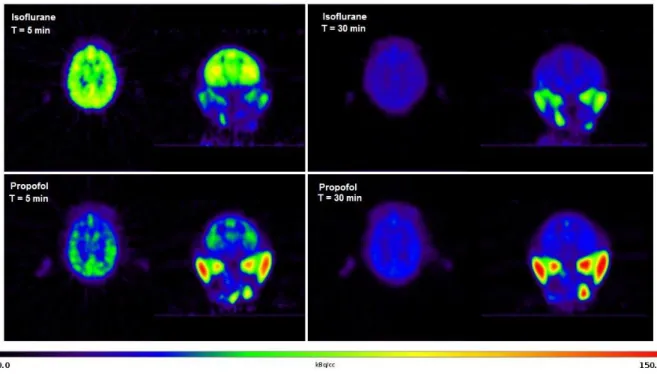
Fig. 1 Representative PET images obtained using the same baboon after injection of
[18F]DPA-714 anesthetized with either propofol or isoflurane. Animal was injected 277
MBq and 285 MBq under propofol and isoflurane anesthesia, respectively. Images obtained at 5 min and 30 min post-injection are shown in both conditions.
Fig. 2 Influence of anesthetics on [18F]DPA-714 PET kinetics and metabolism in baboon.
PET data were obtained after i.v. injection of 231±64 MBq [18F]DPA-714 in baboon under either propofol (×) or isoflurane () anesthesia (n=3 in each condition). PET data are represented as mean time-activity curves (SUV) obtained in the brain (A) and parotid glands (B). Parent [18F]DPA-714 measured in arterial plasma (C) was calculated given [18F]DPA-714 metabolism in arterial plasma showing the percentage of parent [18F]DPA-714 versus time (D). SUV obtained in the brain at 5 and 30 min post injection, and those obtained at 60 min in the parotid glands were statistically compared using one way ANOVA and Tukey's HSD post-hoc test (* p<0.05; n.s p>0.05).
Fig. 3 Influence of anesthetics on [18F]DPA-714 kinetic parameters. Data are shown as
mean VT (mL.cm-3) and displacement efficacy (%) measured under either propofol or
isoflurane anesthesia (n=3 in each condition). Data were statistically compared using one way ANOVA and Tukey's HSD post-hoc test (* p<0.05; ** p<0.01, *** p<0.001; n.s p>0.05).
Fig. 4 Influence of anesthetics on [18F]DPA-714 specific binding in vivo. Time-activity
curves (SUV) obtained after displacement experiment in baboons anesthetized using either propofol (n=3) or isofurane anesthesia (n=3) in the brain and parotid glands. Intravenous injection of [18F]DPA-714 was followed by displacement procedure using unlabeled racemic PK11195 (1.5 mg/kg) performed 40 min after tracer injection.
Fig. 5 Influence of anesthetics on [18F]DPA-714 binding parameters measured in vitro.
Represented binding experiments were performed on mitochondrial enriched preparation obtained from baboon brain cortex using [18F]DPA-714 and unlabeled DPA-714 (0.3 to 30 nM). A is a representative saturation experiment performed in control condition (absence of anesthetics). Representative Scatchard’s plot obtained in control condition (absence of anethetics, Δ), in the presence of propofol 100 µM (×) and isoflurane 10 mM () are shown in B. Mean data with corresponding statistical analysis are reported in table 1.
![Fig. 2 Influence of anesthetics on [ 18 F]DPA-714 PET kinetics and metabolism in baboon](https://thumb-eu.123doks.com/thumbv2/123doknet/13404880.406581/23.892.122.774.493.933/fig-influence-anesthetics-dpa-pet-kinetics-metabolism-baboon.webp)
![Fig. 3 Influence of anesthetics on [ 18 F]DPA-714 kinetic parameters. Data are shown as mean V T (mL.cm -3 ) and displacement efficacy (%) measured under either propofol or isoflurane anesthesia (n=3 in each condition)](https://thumb-eu.123doks.com/thumbv2/123doknet/13404880.406581/24.892.463.744.299.558/influence-anesthetics-parameters-displacement-efficacy-isoflurane-anesthesia-condition.webp)
![Fig. 4 Influence of anesthetics on [ 18 F]DPA-714 specific binding in vivo. Time-activity curves (SUV) obtained after displacement experiment in baboons anesthetized using either propofol (n=3) or isofurane anesthesia (n=3) in](https://thumb-eu.123doks.com/thumbv2/123doknet/13404880.406581/25.892.131.746.336.894/influence-anesthetics-specific-displacement-experiment-anesthetized-isofurane-anesthesia.webp)
![Fig. 5 Influence of anesthetics on [ 18 F]DPA-714 binding parameters measured in vitro](https://thumb-eu.123doks.com/thumbv2/123doknet/13404880.406581/26.892.127.734.448.661/fig-influence-anesthetics-dpa-binding-parameters-measured-vitro.webp)
