EXPERIMENTAL RESEARCH
Elevated level of endothelin-1 in cerebrospinal fluid
and lack of nitric oxide in basilar arterial plasma associated
with cerebral vasospasm after subarachnoid haemorrhage
in rabbits
Volker Neuschmelting&Serge Marbacher&
Ali-Reza Fathi&Stephan M. Jakob&Javier Fandino
Received: 16 February 2008 / Accepted: 14 October 2008 / Published online: 5 May 2009 # Springer-Verlag 2009
Abstract
Background The role of endothelin-1 (ET-1) and nitric oxide (NO) as two important mediators in the development of cerebral vasospasm (CVS) after subarachnoid haemor-rhage (SAH) is controversial. The objective of this study was to determine whether local levels of ET-1 and NO in cerebral arterial plasma and/or in cerebrospinal fluid (CSF) are associated with the occurrence of CVS after SAH. Methods CVS was induced using the one-haemorrhage rabbit model and confirmed by digital subtraction angiography of the rabbits’ basilar artery on day 5. Prior to sacrifice, local CSF and basilar arterial plasma samples were obtained by a transclival approach to the basilar artery. Systemic arterial plasma samples were obtained. ET-1 levels were determined by immunometric technique (pg/ml ± SEM) and total nitrate/nitrite level spectrophotometrically (µmol/l ± SEM).
Findings Angiographic CVS was documented after SAH induction (n=12, P<0.05). The ET-1 level in CSF was significantly elevated by 27.3% to 0.84±0.08 pg/ml in SAH animals (n=7) in comparison to controls (0.66±0.04 pg/ml,
n=7, P<0.05). There was no significant difference in ET-1 levels in systemic and basilar arterial plasma samples of SAH animals compared to controls. A significant lack of local NO metabolites was documented in basilar arterial plasma after SAH (36.8±3.1 µmol/l, n=6) compared to controls (61.8±6.2 µmol/l, n=6, P<0.01).
Conclusion This study demonstrates that an elevated ET-1 level in CSF and local lack of NO in the basilar arterial plasma samples are associated with CVS after experimental SAH.
Keywords Subarachnoid haemorrhage . Cerebral vasospasm . Endothelin-1 . Nitric oxide
Abbreviations
ET-1 endothelin-1 NO nitric oxide CVS cerebral vasospasm SAH subarachnoid haemorrhage CSF cerebrospinal fluid SEM standard error of the mean DSA digital subtraction angiogram CI of diff confidence intervals of difference iNOS induced nitric oxide synthase nNOS neuronal nitric oxide synthase eNOS endothelial nitric oxide synthase
Introduction
Cerebral vasospasm (CVS) develops in 30–70% of patients suffering from subarachnoid haemorrhage (SAH) [29] and still remains an unsolved complication leading to cerebral
DOI 10.1007/s00701-009-0350-1
V. Neuschmelting
:
S. Marbacher:
A.-R. Fathi:
J. Fandino Department of Neurosurgery, University Hospital Berne, Berne, SwitzerlandV. Neuschmelting
:
S. Marbacher:
A.-R. Fathi:
S. M. Jakob:
J. FandinoDepartment of Intensive Care Medicine, University Hospital Berne,
Berne, Switzerland J. Fandino (*)
Department of Neurosurgery, Kantonsspital Aarau, Tellstrasse,
5001 Aarau, Switzerland e-mail: [email protected]
ischaemia, permanent neurological dysfunction and death [21]. Despite substantial progress in the understanding of the development of CVS after SAH, its pathogenesis remains unclear with regard to its complexity. In addition to haemolytic metabolites recently discovered to be pathogenetic factors, such as oxyhaemoglobin and bilirubin oxidation products [4], the probably most important factors contributing to the narrowing of cerebral arteries after SAH are dysfunctions observed in the endothelin-1 (ET-1) and nitric oxide (NO) system as a result of haemolytic processes in the subarachnoid space [21].
ET-1 is the most potent vasoconstrictive endogenous peptide produced by various cells, including endothelial cells, smooth muscle cells, astrocytes and neurons [13]. In both experimental and clinical studies, elevated levels of ET-1 in either cerebrospinal fluid (CSF) and/or peripheral plasma have been observed to be associated with the occurrence of CVS [9,12,35]; however, none of the studies has focused on the local cerebral plasma levels as the bridge between CSF and the periphery. The therapeutic approach in affecting the system by selective endothelin A receptor antagonism seems to be promising according to preliminary clinical results [3,31]. However, ET-1’s source and cause of release remain unclear. Some researchers propose that ET-1 is induced by cerebral ischaemia and released from astrocytes, leading to elevated levels in CSF [12, 22]. Others propose that elevated ET-1 levels observed in peripheral plasma samples are directly associated with CVS as a result of endothelial dysfunction [9,10].
Nitric oxide synthase mediates oxygen-dependent conversion from l-arginine to radical NO, leading to vasodilation through the cyclic guanylate monophosphate pathway [2]. Decreased bioavailability of endogenous NO has been widely accepted as an important part of the pathogenesis of delayed CVS after SAH, more likely caused by decreased production than by increased scav-enging by haemolytic metabolites as was initially assumed [21]. Controversies about the origin of decreased avail-ability remain because of conflicting data published about NO metabolite levels in the CSF and peripheral plasma [8,
21,25].
On the basis of a novel sampling approach, our study seeks to include local cerebral plasma levels as a further variable to focus on for bridging a gap between conflicting data published about CSF and peripheral plasma levels of ET-1 and NO. With the use of an experimental SAH model, it compares local basilar arterial plasma levels of ET-1 and NO metabolites with those in CSF as well as compares them to systemic arterial plasma levels. This may possibly contribute to the clarification of conflicting data published on the source of release of ET-1 and reduced bioavailability of NO.
Methods and materials
In vivo
The protocol of this study was reviewed and approved by the Swiss Institutional Animal Care and Use Committee and meets the Swiss guidelines for laboratory animal use (Canton Bern, Switzerland, approval no. 15/06).
Experimental groups
A total of 24 adult New Zealand white rabbits of either sex weighing 2.6 to 3.6 kg were randomly assigned to two groups (12 animals each). One group underwent experi-mental SAH on day 0, and one served sham-operated as control.
Angiography
After induction of general anaesthesia by intramuscular administration of ketamine (30 mg/kg) and xylazine (6 mg/kg), digital subtraction angiograms (DSA) were performed on day 0 prior to SAH and on day 5 post SAH. The rabbits’ left (day 0) or right (day 5) subclavian artery was microsurgically cannulated using a 0.5-mm plastic tube. The catheter was fixed distal to the vertebral branch. Subsequently, DSA was performed by intraarte-rial bolus injection of non-ionic iopamidol (0.6 ml/kg) as contrast agent. Meanwhile, the arterial blood gas status was analysed to monitor ventilation parameters from a collected arterial blood sample. Images of the vertebro-basilar system were obtained using a rapid sequential angiographic technique with the small focal spot at 66 kV 125 mA (DFP 2000A, Toshiba Corporation®, Japan). The average diameter of the basilar artery was digitally calculated in μm using the automatic measure-ment tool of ImagePro Discovery® analysis software (MediaCybernetics, Bethesda, MD).
Cerebral vasospasm model
Following baseline DSA on day 0, the rabbits’ atlan-tooccipital membrane was exposed by a microsurgical posterior approach in prone position, and a 25-gauge needle was inserted into the cerebromedullar cistern while maintaining general anaesthesia. After relief of 1.0 ml CSF, an equal amount of unheparinised autolo-gous arterial blood in the SAH group was injected into the cistern under microscopic view to imitate SAH in order to induce CVS. An equal amount of isotonic saline solution instead was injected in controls. Postsurgical pain relief was managed by subcutaneous administration of buprenorphine (0.1–0.2 mg/kg).
Collection of samples
Following post-treatment DSA on day 5, the rabbits underwent coniotomy to keep up general anaesthesia by inhalation of an isoflurane/oxygen/air mixture (2.5/5/ 92.5 vol%). Systemic arterial plasma samples were collected via the central arterial catheter. Basilar arterial plasma and CSF samples were collected by a transclival approach. In brief, the rabbits’ head was reclined and fixed at a 20° angle, the trachea and oesophagus were cautiously laterally retracted and the musculature under-neath bluntly microsurgically dissected. The paraverte-bral venous plexus exposed in the depth was coagulated using a pair of bipolar forceps. The exposed dorsocaudal edge of the clivus was followed frontocranially by cautious dissection until the clivus’ surface on the base of the skull could fully be displayed. A compression of the carotid arteries and the descending vagus nerves was avoided throughout the procedure.
An ellipsoid osteotomy (2-mm diameter) with the use of a high-speed microdrill (Fine Science Tools, North Vancouver, BC, Canada) exposed the dura and the basilar artery underneath. The CSF sample was collected by transdural insertion of a 26-gauge bent needle into the subarachnoid space. The part of the dura exposed was fully excised with microscissors in order to allow a direct approach to the distal segment of the basilar artery. The basilar arterial blood sample was collected by intraluminal insertion of a 29-gauge bent needle into the basilar artery. The collected blood samples were centrifuged for 15 min at 1,500 rpm, and the plasma remnants and CSF samples were stored at -80° C. The animals were killed by intraarterial bolus injection of sodium thiopenthal (40 mg/kg). All surgical procedures were performed under sterile conditions.
In vitro
Detection of ET-1
In seven randomly chosen animals of each group, ET-1 systemic and basilar plasma levels as well as CSF levels were quantitatively determined by the extraction-free human ET-1 chemiluminescent sandwich immuno-assay technique (QuantiGlo QET00B, R&D Systems, Minneapolis, MN) as previously validated [18]. The amino acid sequence of human 1 matches rabbit ET-1 exactly. The quantitative range of the assay is 0.32 to 1,000 pg/ml. According to the manufacturer, cross-reactions of antibodies used are observed for human 1 (100%), human big 2 (0.01%), human big ET-38 (0.019%), human ET-3 (7.8%) and human ET-2 (27.4%).
Measurement of NO metabolites
In six random animals of each group, nitrite/nitrate levels were quantitatively determined as a relative measure for basilar, systemic plasma and CSF levels of NO produc-tion, as is common practice. Following reduction of nitrate to nitrite with a modified Griess method, the total nitrite level was spectrophotometrically detected at 540 nm (QuantiChrom Nitric Oxide Assay Kit, Bioassay Systems, Hayward, CA) as previously validated [24].
Drugs
The following drugs were used: buprenorphinum (Temgesic® 0.3 mg/ml, Essex Chemie, Lucerne, Switzerland), iopamidol (Iopamiro® 300 mg/ml, Bracco, Milan, Italy), isoflurane (Forene® 99%, Abbott Laboratories, Abbott Park, IL), ketamine HCl (Ketalar®50 mg/ml, Pfizer AG, Zurich, Switzerland), sodium thiopenthalum (Pentothal® 25 mg/ml, Abbott Laboratories, Abbott Park, IL) and xylazine HCl (Xylapan® 20 mg/ml, Vetoquinol AG, Bern, Switzerland).
Statistical methods
Values are expressed as mean ± SEM (n=number of animals). Statistical difference between two means and multiple means was determined by Student's unpaired t-test and one-way ANOVA followed by Bonferroni’s post-testing, respectively. The statistical significance was considered if the P-value was less than 0.05 (p<0.05) in context of the 95% confidence intervals of the difference examined (95% CI of diff).
Results
Arteriographic vasospasm
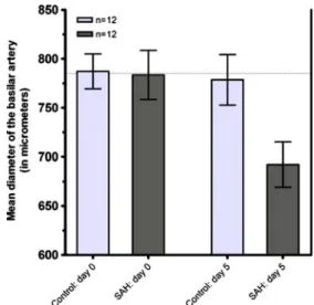
On day 5 after SAH, the diameter of the basilar artery was significantly narrowed by 11.7% to 692.2 ± 23.0 µm (n= 12) compared to the animals’ baseline angiogram on day 0 (783.5 ± 25.1 µm, n=12, P<0.01, 95% CI of diff: 15.4 to 167.2 µm); on the same day, the basilar artery diameter remained unchanged (778.5 ± 25.8 µm, n=12) in the control group compared to the baseline on day 0 (787.2 ± 17.8 µm, n=12, P>0.05, 95% CI of diff: -84.6 to 67.2 µm) (Fig. 1).
The animals’ initial blood pressure at baseline angiogra-phy (SAH group: 71.1 ± 2.8 mmHg in mean arterial pressure, n=12; controls: 68.8 ± 2.0 mmHg, n=12) was not different from the values monitored on day 5 in both the SAH group (70.6 ± 2.2 mmHg, n=12, P>0.05) and the controls (70.8 ± 2.1 mmHg, n=12, P>0.05).
Correspond-ingly, the initial blood gas parameters such as pO2 (SAH
group: 57.9 ± 3.6 mmHg; controls: 56.8 ± 2.1 mmHg), pCO2 (SAH group: 50.6 ± 1.1 mmHg; controls: 50.1 ±
1.3 mmHg) and pH (SAH: 7.39 ± 0.01; controls: 7.38 ± 0.01) did not change compared to the values monitored during follow-up angiography on day 5 in both the SAH (P>0.05) and control group (P>0.05) and did not statisti-cally differ between the two groups (P>0.05).
Local ET-1 levels
The ET-1 level in CSF samples was significantly elevated by 27.3% to 0.84 ± 0.08 pg/ml in SAH animals (n=7) in comparison to controls (0.66 ± 0.04 pg/ml, n=7, P<0.05,
95% CI of diff: 0.01 to .35 pg/ml). There was a slight increasing tendency observed in plasma samples as well, but no significant difference of ET-1 levels was remarked in systemic or basilar arterial plasma samples of SAH animals compared to controls. Local levels of ET-1 in the basilar arterial plasma samples did not statistically differ from those in the systemic plasma in SAH (n = 7, P≥ 0.05, 95% CI of diff: -0.12 to 0.53 pg/ml) or control groups (n=7, P≥0.05, 95% CI of diff: -0.05 to 0.44 pg/ml), nor did they statistically differ from those in CSF of the SAH (P≥0.05, 95% CI of diff: -0.51 to 0.10 pg/ml) or control groups (P≥0.05, 95% CI of diff: -0.51 to 0.09 pg/ml) (Fig. 2).
Local levels of NO metabolites
The level of total nitrate/nitrite in the basilar arterial plasma samples was significantly reduced, by 40.4% to 36.8 ± 3.1 µmol/l in SAH animals (n = 6) compared to controls (61.8 ± 6.2 µmol/L, n = 6, P < 0.01, 95% CI of diff: 9.5 to 40.3 µmol/l). A slightly decreasing tendency of the local NO metabolite level was also observed in CSF (n = 6) and systemic arterial plasma (n = 6) samples in comparison to the controls (n = 6), but remained insignificant (P > 0.05). The local level of total nitrate/ nitrite in basilar arterial plasma samples did not differ statistically from the level in systemic samples for both the SAH group (n = 6, P > 0.05) and the control group (n = 6, P > 0.05). It was significantly higher than the level detected in CSF samples for both the SAH group (n = 6, P < 0.001, 95% CI of diff: 15.9 to 37.1 µmol/l) and the control group (n = 6, P < 0.001, 95% CI of diff: 28.5 to 62.7 µmol/l) (Fig. 3).
Fig. 2 Box plot with SAH vs. control: ET-1 levels in CSF, basilar and systemic arterial plasma on day 5 after SAH (in pg/ml statistically differ SEM). (*P<0.05; ns=non-significant)
Fig. 3 Box plot with SAH vs. control: total nitrite levels in CSF, basilar and systemic arterial plasma on day 5 after SAH (in µmol/l +/-SEM). (*P<0.01)
Fig. 1 Box plot with the absolute change of the basilar artery in diameter: Baseline angiography in SAH and control group on day 0 vs. on day 5 (in µm ± SEM). (*P<0.05; ns=non-significant)
Discussion
Origin of increased bioavailability of ET-1
ET-1 is produced by various cells, including neurons, glial cells, smooth muscle cells and endothelial cells, within minutes after being stimulated by hypoxia, ischaemia and shear stress [13]. Its release from vascular endothelial and smooth muscle cells is more likely to follow a paracrine than an endocrine pattern, as ET-1 abluminally binds to ETA receptors in the smooth muscle cell layer to mediate long-lasting vasoconstriction [13, 32]. Despite broad acceptance of ET-1 as a contributing mediator of CVS, conflicting data have been reported about the correlation of ET-1’s plasma level and CVS. On one hand, ET-1’s peripheral plasma level has been found to correlate with the severity of vascular diseases [13], as shown in clinical studies of Juvela et al. [9, 10] and Suzuki et al. [30] in regard to the occurrence of delayed cerebral ischaemia and vasospasm after SAH. On the contrary, other authors were not able to correlate ET-1 plasma levels to CVS [11, 12,
27].
In our experimental study neither the local ET-1 level in the basilar arterial nor the one in systemic plasma samples after SAH differed significantly from those in controls. These findings support the clinical observations reported elsewhere [11,12,27]. In addition, our data did not show significant differences between the local ET-1 level obtained from basilar arterial plasma samples compared to the systemic samples within either the SAH or the control group. Nevertheless, our results demonstrated a trend towards lower levels of this peptide in the basilar arterial samples than in the peripheral plasma in both groups. This observation might suggest that the elevated peripheral plasma levels of ET-1 observed in clinical studies are rather unlikely to be caused by increased release of ET-1 into the cerebral circulation [9, 10, 30]. One reason might be that the systemic plasma ET-1 concentration could be signifi-cantly influenced by physiological and pathological conditions in structures other than the brain and cerebral vessels, as shown, for example, in systemic hypertension and heart failure [13,35].
Instead, our data demonstrated a significantly elevated ET-1 level in CSF on day 5 after experimental SAH. This observation supports the hypothesis of mediated CVS partly due to elevated ET-1 levels in CSF. In experimental studies on felines and canines, intracisternal bolus injec-tions of ET-1 have previously demonstrated ET-1’s potency in mediating vasoconstriction from the abluminal site [16]. Pluta et al.’s experimental study in primates found the ET-1 level in CSF to be dependent on astrocyte release [22]. Our finding of an elevated level of ET-1 in CSF confirms the clinical results reported earlier [11,12,14,27,28]. Among
these reports the correlation of the CSF to the plasma levels of ET-1 in SAH-induced CVS is still controversial. With respect to the apparently very limited specificity and sensitivity of ET-1 in systemic plasma to correlate with CVS, some authors showed ET-1’s peripheral plasma concentration during CVS to act differently from the one in CSF [12,27], while Kastner et al. demonstrated them to be relatively similar in concentration averaged over time [11]. Keeping in mind that ET-1 levels of local cerebral plasma and CSF had not been previously examined, we documented the basilar arterial plasma levels of ET-1 to be statistically indifferent from the ones in CSF. These findings might possibly be explained epiphenomenally due to the ET-1 conversion rate in the endothelial basilar arterial layer or due to ET-1 release from the CSF into the cerebral circulation. This hypothesis might also be supported by Menon et al. who found high arterio-jugularvenous ET-1 gradients in patients suffering from SAH-induced CVS [15].
Though our study results tend to agree with these hypotheses, one needs to keep in mind that the SAH model used in this study is certainly limited in mimicking the complex clinical conditions of an aneurysmal SAH in humans. Due to single time point assessment during the peak time of CVS, the study is also limited in providing data about possible metabolite level changes during the development of CVS.
Origin of reduced NO bioavailability
Endogenous NO is a radical product of the oxidation of l-arginine to citrulline by nitric oxide synthase, which causes vasodilation through activation of the soluble guanylate cyclase pathway. Three isoenzyme forms are known to play a role in vasculature-mediating NO release: induced nitric oxide synthase (iNOS), periadven-titial neuronal nitric oxide synthase (nNOS) and endo-thelial nitric oxide synthase (eNOS). While iNOS possibly plays a pathophysiological role in the acute and early states after SAH, there is debate over whether the activity of nNOS and eNOS has a role in the delayed development of CVS after SAH [5, 21]. After a short half-life, radical NO is metabolised by being oxidised to stable nitrite and nitrate. This serves as an acceptable relative measure of NO level in both experimental and clinical research. Decreased bioavailability of NO has been shown to be associated with the development of delayed CVS after SAH, more likely caused by deprived production rather than just increased scavenging by haemolytic metabolites such as oxyhaemoglobin as ini-tially assumed [5,21].
This study demonstrated the novel finding that locally reduced NO availability in basilar arterial plasma is
associated with CVS after SAH. It hypothesises that there is decreased production of NO in the endothelial layer of local cerebral vasculature. This local lack of NO in the basilar plasma might hypothetically be explained by dysfunctional endothelium [7] or by inhibited enzyme activity of eNOS [8]. Whether these results are caused by downregulation of eNOS expression itsself, however, remains uncertain because of the conflicting data reported: Hino et al. [6] found decreased eNOS mRNA in cerebral arteries of primates and Park et al. [20] in the microvasculature of rats after induced SAH. On the contrary, Vatter et al. could recently not reproduce those findings by histochemical staining of eNOS in the rats’ basilar artery after SAH [33]. In support of our hypothesis, Jung et al. found that the level of asymmetric dimethylarginine, an endogenous competi-tive inhibitor of nitric oxide synthase and a metabolisation product of bilirubin, was elevated in CSF during delayed CVS in an experimental primate model [8]. It is suggested to contribute to the endogenous inhibition of eNOS [8] and, thus, possibly to the lack of NO in the basilar arterial plasma found in our study.
Uncertainty remains about conflicting data reported for NO metabolites after SAH concerning the CSF level. While some authors reported in clinical studies with small sample sizes that NO metabolite levels in CSF were increased after SAH [17, 25, 29, 34]; others observed CVS to be associated with reduced CSF levels of nitrite [26], suggest-ing possible affection of nNOS [8,23]. However, our data do not confirm any significant change in nitrite level in CSF in either direction. Possible explanations for the conflicting data published are a lack of large clinical sample sizes, different time courses, and different spots of vessel catheterisation and sample collection.
Our data also do not demonstrate a significant difference between nitrite levels in systemic plasma after SAH compared to controls, in agreement with clinical data provided by Suzuki et al. [29]. It suggests that systemic plasma may be too unspecific to detect a locally limited pathophysiological process in the cerebrovascular NO system.
Interaction of ET-1 and NO
Experimental data have been published suggesting that there may be a direct or indirect interaction between the ET-1 and NO systems on the basis of a yet unclear mechanism. Ohkita et al. provided evidence for inhibi-tion of ET-1 producinhibi-tion by NO, assuming a modulatory role of NO [19]. Impairment of this possible modulatory role of NO on ET-1 during CVS after SAH has been found by Alabadi et al. [1] and supports our observation that an elevated ET-1 level in CSF accompanies a lack of local NO.
Acknowledgments and funding This study was supported by the Cerebrovascular Research Fund from the Departments of Neurosur-gery and Intensive Care Medicine (Account Nr. 34–160), University of Bern, Switzerland, and from an unrestricted grant from Actelion Ltd, Allschwil, Switzerland.
We gratefully thank H.R. Widmer, PhD, and J. Schmid, RN, from the Department of Neurosurgery, University Hospital Bern, for their professional laboratory support.
Financial disclosure/conflict of interest None of the authors has any duality of interest to declare.
References
1. Alabadi JA, Torregrosa G, Miranda FJ, Salom JB, Centeno JM, Alborch E (1997) Impairment of the modulatory role of nitric oxide on the endothelin-1-elicited contraction of cerebral arteries: a pathogenetic factor in cerebral vasospasm after subarachnoid hemorrhage. Neurosurgery 41:245–252 discussion 252-243 2. Andresen J, Shafi NI, Bryan RM Jr (2006) Endothelial influences
on cerebrovascular tone. J Appl Physiol 100:318–327
3. Barth M, Capelle HH, Munch E, Thome C, Fiedler F, Schmiedek P, Vajkoczy P (2007) Effects of the selective endothelin A (ET(A)) receptor antagonist Clazosentan on cerebral perfusion and cerebral oxygenation following severe subarachnoid hemorrhage—preliminary results from a randomized clinical series. Acta Neurochir (Wien) 149:911–918 discussion 918 4. Clark JF, Sharp FR (2006) Bilirubin oxidation products (BOXes) and their role in cerebral vasospasm after subarachnoid hemor-rhage. J Cereb Blood Flow Metab 26:1223–1233
5. Hanggi D, Steiger HJ (2006) Nitric oxide in subarachnoid haemorrhage and its therapeutics implications. Acta Neurochir (Wien) 148:605–613 discussion 613
6. Hino A, Tokuyama Y, Weir B, Takeda J, Yano H, Bell GI, Macdonald RL (1996) Changes in endothelial nitric oxide synthase mRNA during vasospasm after subarachnoid hemor-rhage in monkeys. Neurosurgery 39:562–567 discussion 567–568 7. Iuliano BA, Pluta RM, Jung C, Oldfield EH (2004) Endothelial dysfunction in a primate model of cerebral vasospasm. J Neuro-surg 100:287–294
8. Jung CS, Iuliano BA, Harvey-White J, Espey MG, Oldfield EH, Pluta RM (2004) Association between cerebrospinal fluid levels of asymmetric dimethyl-L-arginine, an endogenous inhibitor of endothelial nitric oxide synthase, and cerebral vasospasm in a primate model of subarachnoid hemorrhage. J Neurosurg 101:836–842
9. Juvela S (2000) Plasma endothelin concentrations after aneurys-mal subarachnoid hemorrhage. J Neurosurg 92:390–400 10. Juvela S (2002) Plasma endothelin and big endothelin
concen-trations and serum endothelin-converting enzyme activity following aneurysmal subarachnoid hemorrhage. J Neurosurg 97:1287–1293
11. Kastner S, Oertel MF, Scharbrodt W, Krause M, Boker DK, Deinsberger W (2005) Endothelin-1 in plasma, cisternal CSF and microdialysate following aneurysmal SAH. Acta Neurochir (Wien) 147:1271–1279 discussion 1279
12. Kessler IM, Pacheco YG, Lozzi SP, de Araujo AS Jr, Onishi FJ, de Mello PA (2005) Endothelin-1 levels in plasma and cerebro-spinal fluid of patients with cerebral vasospasm after aneurysmal subarachnoid hemorrhage. Surg Neurol 64(Suppl 1):S1:2–5 13. Levin ER (1995) Endothelins. N Engl J Med 333:356–363 14. Mascia L, Fedorko L, Stewart DJ, Mohamed F, terBrugge K,
endothelin-1 concentrations and cerebral vasospasm in patients with aneurysmal subarachnoid hemorrhage. Stroke 32:1185–1190 15. Menon DK, Day D, Kuc RE, Downie AJ, Chatfield DA, Davenport AP (2002) Arteriojugular endothelin-1 gradients in aneurysmal subarachnoid haemorrhage. Clin Sci (Lond) 103 (Suppl 48):399S–403S
16. Mima T, Yanagisawa M, Shigeno T, Saito A, Goto K, Takakura K, Masaki T (1989) Endothelin acts in feline and canine cerebral arteries from the adventitial side. Stroke 20:1553–1556
17. Ng WH, Moochhala S, Yeo TT, Ong PL, Ng PY (2001) Nitric oxide and subarachnoid hemorrhage: elevated level in cerebrospinal fluid and their implications. Neurosurgery 49:622–626 discussion 626–627
18. Oechslin E, Kiowski W, Schindler R, Bernheim A, Julius B, Brunner-La Rocca HP (2005) Systemic endothelial dysfunction in adults with cyanotic congenital heart disease. Circulation 112:1106–1112
19. Ohkita M, Takaoka M, Shiota Y, Nojiri R, Matsumura Y (2002) Nitric oxide inhibits endothelin-1 production through the suppres-sion of nuclear factor kappa B. Clin Sci (Lond) 103(Suppl 48):68S–71S
20. Park KW, Metais C, Dai HB, Comunale ME, Sellke FW (2001) Microvascular endothelial dysfunction and its mechanism in a rat model of subarachnoid hemorrhage. Anesth Analg 92:990–996 21. Pluta RM (2005) Delayed cerebral vasospasm and nitric oxide:
review, new hypothesis, and proposed treatment. Pharmacol Ther 105:23–56
22. Pluta RM, Boock RJ, Afshar JK, Clouse K, Bacic M, Ehrenreich H, Oldfield EH (1997) Source and cause of endothelin-1 release into cerebrospinal fluid after subarachnoid hemorrhage. J Neuro-surg 87:287–293
23. Pluta RM, Thompson BG, Dawson TM, Snyder SH, Boock RJ, Oldfield EH (1996) Loss of nitric oxide synthase immunoreactivity in cerebral vasospasm. J Neurosurg 84:648–654
24. Ridnour LA, Sim JE, Hayward MA, Wink DA, Martin SM, Buettner GR, Spitz DR (2000) A spectrophotometric method for the direct detection and quantitation of nitric oxide, nitrite, and nitrate in cell culture media. Anal Biochem 281:223–229 25. Sadamitsu D, Kuroda Y, Nagamitsu T, Tsuruta R, Inoue T, Ueda
T, Nakashima K, Ito H, Maekawa T (2001) Cerebrospinal fluid and plasma concentrations of nitric oxide metabolites in postop-erative patients with subarachnoid hemorrhage. Crit Care Med 29:77–79
26. Sakowitz OW, Wolfrum S, Sarrafzadeh AS, Stover JF, Dreier JP, Dendorfer A, Benndorf G, Lanksch WR, Unterberg AW (2001) Relation of cerebral energy metabolism and extracellular nitrite and nitrate concentrations in patients after aneurysmal subarach-noid hemorrhage. J Cereb Blood Flow Metab 21:1067–1076 27. Seifert V, Loffler BM, Zimmermann M, Roux S, Stolke D (1995)
Endothelin concentrations in patients with aneurysmal subarach-noid hemorrhage. Correlation with cerebral vasospasm, delayed ischemic neurological deficits, and volume of hematoma. J Neurosurg 82:55–62
28. Suzuki H, Sato S, Suzuki Y, Takekoshi K, Ishihara N, Shimoda S (1990) Increased endothelin concentration in CSF from patients with subarachnoid hemorrhage. Acta Neurol Scand 81:553–554 29. Suzuki M, Asahara H, Endo S, Inada K, Doi M, Kuroda K,
Ogawa A (1999) Increased levels of nitrite/nitrate in the cerebrospinal fluid of patients with subarachnoid hemorrhage. Neurosurg Rev 22:96–98
30. Suzuki R, Masaoka H, Hirata Y, Marumo F, Isotani E, Hirakawa K (1992) The role of endothelin-1 in the origin of cerebral vasospasm in patients with aneurysmal subarachnoid hemorrhage. J Neurosurg 77:96–100
31. Vajkoczy P, Meyer B, Weidauer S, Raabe A, Thome C, Ringel F, Breu V, Schmiedek P (2005) Clazosentan (AXV-034343), a
selective endothelin A receptor antagonist, in the prevention of cerebral vasospasm following severe aneurysmal subarachnoid hemorrhage: results of a randomized, double-blind, placebo-controlled, multicenter phase IIa study. J Neurosurg 103:9–17 32. Vatter H, Konczalla J, Weidauer S, Preibisch C, Zimmermann M,
Raabe A, Seifert V (2007) Effect of delayed cerebral vasospasm on cerebrovascular endothelin A receptor expression and function. J Neurosurg 107:121–127
33. Vatter H, Weidauer S, Dias S, Preibisch C, Ngone S, Raabe A, Zimmermann M, Seifert V (2007) Persistence of the nitric oxide-dependent vasodilator pathway of cerebral vessels after experi-mental subarachnoid hemorrhage. Neurosurgery 60:179–187 discussion 187-178
34. Woszczyk A, Deinsberger W, Boker DK (2003) Nitric oxide metabolites in cisternal CSF correlate with cerebral vasospasm in patients with a subarachnoid haemorrhage. Acta Neurochir (Wien) 145:257–263 discussion 263-254
35. Zimmermann M, Seifert V (1998) Endothelin and subarachnoid hemorrhage: an overview. Neurosurgery 43:863–875 discussion 875-866
Comments
The authors used a well established animal model (rabbit single hemorrhage) of cerebral vasospasm (CVS) with the aim to investigate the source of ET-1 and NO in the development of CVS. They determined levels of ET-1 and nitrite in the basilar artery (BA), the systemic plasma and the CSF. They observed an increased level of ET-1 in the CSF after experimental SAH but no changes in the BA and the systemic circulation. For nitrate they found decreased levels in the BA after SAH but no changes in the systemic circulation and the CSF.
The investigation is well performed, the data is presented in an adequate and clear form. It supports the pathophysiological concept, that for the development of CVS relevant changes of the ET metabolismn are localiced inside the cereborspinal space but not in the systemic circulation. Furthermore, the present data support a decreased function of the NO metabilism mainly inside the cerebro-vasculature after SAH, which represents another factor for the development of CVS.
Harmut Vatter
Dept. of Neurosurgery, Goethe-University Frankfurt am Main, Germany
It has been more than 50 years since delayed cerebral vasospasm in patients after aneursymal subarachnoid hemorrhage (aSAH) has been linked to a poor outcome.[1] Most unfortunate is that this rather uncommon but most devastating form of stroke affects those patients who have already been treated by surgical or endovascular securing of the aneurysm. In extensive worldwide research, thousands of neuro-surgeons and scientists have examined innumerable chemicals and drugs using simple, sophisticated and ingenious in vivo and in vitro models testing numerous hypotheses hoping to cure or prevent delayed cerebral vasospasm. Still, it is an effort in vain with regard to finding the magic bullet but because of this effort, we have significantly advanced our knowledge of cerebral vessels physiology and developed a better understanding of the pathophysiological changes in the brain and the cerebral vessels evoked by aSAH. In spite of these advances, there appears to be an increasing uneasiness that the failure to find treatment may be due to a “barking up the wrong tree”.[2]
The discoveries that nitric oxide (NO), a toxic gas from fumes of gasoline engines is Furchgott’s endothelium-derived relaxing factor
(EDRF),[3] that endothelin-1 (ET-1), a venom from the sarafotoxin family, is produced in our bodies by endothelial cells as the most potent vasoconstrictor[4] and that both regulate cerebral blood have given new hope to develop a cure for vasospasm.
In the 1990s, numerous studies suggested that NO or ET-1 was involved in the development of delayed cerebral vasospasm,[5, 6] while other reports denied such a relationship.[7, 8] The controversy is still ongoing although preclinical and clinical trials using intracarotid infusion of NO gas solution,[9] intracarotid or intrathecal administra-tion of NO donors[10-12] and ET-1 receptor inhibitors have claimed clinical success.[13, 14]
However, better understanding of aSAH pathophysiology, discovery of new aSAHrelated events like cortical spreading ischemia[15] and, probably most importantly, the failure of Clazosentan, a selective ET-1 A receptor inhibitor, in Phase II clinical study[2] to improve the outcome despite prevention of vasospasm all lead to a paradigm shift with the focus on the possibility that a stroke after aSAH may be evoked by another nonvasospasm-related mechanism(s). Growing body of evidence suggests that these pathomechanisms are a result of the presence of an arterial blood clot in the subarachnoid space, increased ET-1 in CSF, and decreased availability of NO in cerebral arteries.[2, 15]
In their clever, well-planned and executed, but most confirmatory in vivo experimental study published in this issue of Acta Neuro-chirugica , V. Neuschmelting and colleagues simultaneously examined the role of both NO and ET-1 in the development of vasospasm in a rabbit model of aSAH. To assure the relevance of their findings, the authors used an innovative surgical approach. They collected blood samples directly from the basilar artery to compare to the peripheral arterial blood and CSF samples. The authors convincingly showed that the presence of ET-1 in the CSF and endothelial NOS dysfunction were associated with vasospasm development. These findings, while not altogether new,[6, 10, 11, 13, 14] are a novel approach to sample collection and shed new light on the pathophysiology of vasospasm after SAH by separating the extra- and intravascular events. Doing this further opens the door to a Kuhnian revolution in our understanding the role of vasospasm in stroke after aSAH and hopefully leads to successful treatment of this dreadful complication of a ruptured intracranial aneurysm.
Ryszard M. Pluta
National Institute of Neurological Disorders and Stroke National Institutes of Health