DOI 10.1007/s00417-004-1121-6 L. Kodjikian T. Richter M. Halberstadt F. Beby F. Flueckiger M. Boehnke J. G. Garweg Received: 1 August 2004 Revised: 2 September 2004 Accepted: 13 December 2004 Published online: 15 April 2005 # Springer-Verlag 2005
Toxic effects of indocyanine green, infracyanine
green, and trypan blue on the human retinal
pigmented epithelium
Abstract Background: Indocyanine green, infracyanine green, and trypan blue are frequently used as aids to visualize structures removed during vitreoretinal surgery. But they may have toxic effects on the retina. We therefore compared the acute and chronic toxicities of these stains on cultured human retinal pigmented epithelial (RPE) cells using clinically relevant concentrations and an identi-cal experimental setup for each agent. Methods: Monolayers of RPE cells were incubated with various concen-trations of indocyanine green, infra-cyanine green (each at 0.005%, 0.05%, and 0.5%) or trypan blue (0.05%, 0.06%, 0.1%, 0.15%, and 0.5%) for 5 min (acute exposure) or
6 days (chronic exposure). Using the propidium iodide assay, acute cyto-toxicity was monitored at 15-min intervals for up to 3 h. Chronic cyto-toxicity was assessed by monitoring cell calcein esterase activity, cell pro-liferation, and cell morphology (via-bility) after 6 days of exposure. Results: Indocyanine and infracya-nine green induced acute and chronic toxicities at a concentration above 0.05%. Trypan blue evoked no acute
toxicity, but it was chronically cyto-toxic at all tested concentrations. Conclusions: Despite thorough rins-ing after application, significant amounts of the not sufficiently water soluble indocyanine and infracyanine green are retained after surgery by the eye. Trypan blue, being more water-soluble than ICG, is probably retained to the least degree. This circumstance is fortunate given that trypan blue exhibits a chronic cytotoxicity com-parable to ICG at all clinically relevant concentrations. During vitrectomy, surgeons should aim to expose retinal tissue to only low concentrations of these stains and for as short a period as possible.
Keywords Acute and chronic toxicity . Cell viability assay . Human retinal pigmented epithelium . Indocyanine green . Infracyanine green . Toxicity . Trypan blue . Viability
Introduction
Great advances have been made in macular surgery during the past few years. Achievements include the repairing of macular holes by removing the internal limiting membrane (ILM) [44] and the treatment of diabetic macular edema
[10]. Perhaps one of the most important innovations has been the introduction of intraoperative staining for the ILM and epiretinal membranes [11, 12,24,36]. By improving the visualization and differentiation of these structures, their staining facilitates surgery, thereby reducing not only trauma, but also the operating time and hence retinal ex-This study received no funding, and the
authors had no financial or proprietary interest in it.
Laurent Kodjikian had full access to all available data and takes full responsibility for its integrity and for the accuracy of the analysis. The authors permit Graefe’s Archive for Clinical and Experimental Ophthalmology to review the data on request. L. Kodjikian . T. Richter .
M. Halberstadt . F. Flueckiger . J. G. Garweg (*)
Department of Ophthalmology, Inselspital, University of Bern, 3010 Bern, Switzerland e-mail: justus.garweg@insel.ch Tel.: +41-31-6329565 Fax: +41-31-6328539 L. Kodjikian . F. Beby Department of Ophthalmology, Croix-Rousse Hospital,
103 Grande rue de la Croix-Rousse, Lyon, 69004, France
M. Boehnke
Rothenbaumchaussee Clinic for Ophthalmology and Ophthalmosurgery, Hamburg, Germany
posure to light. Many conflicting observations have been reported concerning the possible cytotoxic effects of these dyes on the retina when locally applied [14]. The burden of evidence from experimental studies weighs in favor of acute and/or chronic toxicity at clinically relevant concentrations, although the functional or clinical relevance of the effects have not been evaluated [4,6,17,37,38]. Clinicians are nonetheless aware of a possible cytotoxicity, which is re-flected in the broad range of concentrations and contact times that have been deemed necessary or optimal for staining and in the attempts that have been made to remove the dyes [30]. Recently, several independent groups have reported that indocyanine green may persist within the retina for several months after its intraoperative application and removal. Hence, this dye might induce or enhance chronic cytotoxicity [35,39,43].
Three dyes have been clinically evaluated for intraop-erative staining of the ILM: indocyanine green, infracya-nine green, and trypan blue [11,12,24,36]. Indocyanine green contains iodine, which enhances its solubility in water, whereas infracyanine green, which contains no iodine, can be rendered soluble in water by dissolving it in 5% glucose. Trypan blue is a well-known water-soluble vital stain, which has been used in anterior segment surgery for several decades with no noted cytotoxic effects [31]. These three stains are complementary: trypan blue exhibiting a prefer-ential affinity for epiretinal membranes and the two green dyes for the ILM [36]. A direct comparison of the cytotox-icity of these three dyes in the preclinical or clinical setting has not been undertaken. In the present in vitro study, we therefore compared the acute and chronic toxic effects of these three stains on cultured human retinal pigmented ep-ithelial cells using clinically relevant concentrations and an identical experimental setup for each agent.
Materials and methods
Cell culturing
Human retinal pigmented epithelial (RPE) cells were har-vested from several donors and propagated under standard culturing conditions. Cells derived from passages 3–5 were used for all proliferation and metabolism assays in order to avoid the functional modification that occurs at later stages of culturing. The cells were cultured in Dulbecco’s modi-fied Eagle medium (MEM) containing 10% fetal bovine serum (Gibco; Invitrogen Life Technologies, Basel, Swit-zerland), L-glutamine (2 mM), penicillin (10,000 U/ml), streptomycin (10,000μg/ml), and amphotericin B (2 μg/ml) in a humidified atmosphere (5% carbon dioxide) at 36°C. The medium was exchanged twice a week.
Dye preparation
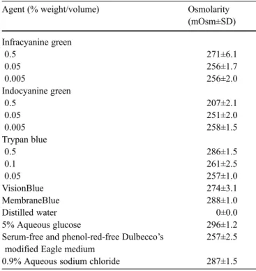
Indocyanine green (Pulsion, Muenchen, Germany) and in-fracyanine green (SERB, Paris, France) were prepared by dissolving 25 mg of the sterile powder in 1 ml of sterile distilled water and in 2.5 ml of 5% aqueous glucose, respec-tively. The dye solutions were mixed until fully dissolved, and then further diluted with serum-free and phenol-red-free MEM (Hospital Pharmacy, Inselspital Bern, Switzerland) at pH 7.4 to yield final concentrations of 0.05 mg/ml (0.005%), 0.5 mg/ml (0.05%), and 5 mg/ml (0.5%). Trypan blue (Fluka, Buchs, Switzerland) was first dissolved in 0.9% aqueous sodium chloride and then in serum-free and phenol-red-free MEM to yield final concentrations of 0.5 mg/ml (0.05%), 1 mg/ml (0.1%), and 5 mg/ml (0.5%). Two commercially available trypan blue preparations (DORC, Zuidland, The Netherlands), which are sold exclusively for intraocular use, were also tested. These preparations con-tain trypan blue at concentrations of 0.6 mg/ml ([0.06%] VisionBlue) and 1.5 mg/ml ([0.15%] MembraneBlue). The osmolarity of each preparation was measured using an au-tomatic semimicro osmometer (Knauer, Berlin, Germany) and is shown in Table1.
Experimental set-up
Cells were seeded onto 24-well Costar culture plates at a numerical density of 3,000 cells/well for acute toxicity tests and at 1,500 cells/well for chronic toxicity tests. They were
Table 1 Osmolarity of preparations used in the study Agent (% weight/volume) Osmolarity
(mOsm±SD) Infracyanine green 0.5 271±6.1 0.05 256±1.7 0.005 256±2.0 Indocyanine green 0.5 207±2.1 0.05 251±2.0 0.005 258±1.5 Trypan blue 0.5 286±1.5 0.1 261±2.5 0.05 257±1.0 VisionBlue 274±3.1 MembraneBlue 288±1.0 Distilled water 0±0.0 5% Aqueous glucose 296±1.2
Serum-free and phenol-red-free Dulbecco’s modified Eagle medium
257±2.5 0.9% Aqueous sodium chloride 287±1.5
grown overnight to subconfluency before introducing the dye to be tested. Cell monolayers were exposed to each dye for either 5 min (acute exposure) or 6 days (chronic ex-posure). One milliliter of diluted dye solution was added per well. The cultures were maintained in the dark during their exposure to the tested dye. Each experimental con-dition was run four times. Cell viability was determined using standardized tests that have been developed in our laboratory (see below). The mitotic rate, confluence and regularity of the cell cultures were also assessed. Individual cells were microscopically evaluated with respect to their
morphology, shape, adherence, nuclear density, and nucle-us/cytoplasm relationship. Controls were included in par-allel in all experiments on each plate to serve as the 100% baseline.
Acute cytotoxicity tests
Acute cytotoxicity was assessed according to a customized propidium iodide assay [41]. Propidium iodide (Sigma, Buchs, Switzerland) enters nonviable cells and binds
irre-Fig. 1 a Acute cytotoxicity. Temporal course of nuclear fluorescence in cultured human RPE cells following their exposure to different concentrations of indocyanine green (ICG) and trypan blue (TB) for 5 min. No acute cytotoxicity was observed using any of the tested concentrations of trypan blue. Indocyanine green and infracyanine green (data for the latter not presented) were cytotoxic (p<0.05) at the highest concentration tested (5 mg/ml) after 2 h of monitoring.
b Inverted-phase micrographs of human RPE cells at the end of the 3-h monitoring period after exposure to culture medium (left crograph), 0.5 mg/ml (center micrograph), and 5 mg/ml (right mi-crograph) indocyanine green. A dose-dependent increase in nuclear fluorescence resembling acute cytotoxicity is evident but signifi-cant only for a concentration of 5 mg/ml (magnification ×250).
versibly to their nucleus, whereupon it acts as a fluorescent dye. The amount of bound propidium iodide can then be quantified fluorometrically in a Cytofluor (Millipore, Bed-ford, MA, USA), using an excitation/emission wavelength coupling of 530 nm/645 nm.
For the testing of acute toxicity, cell monolayers were incubated with 1 ml of serum-free and phenol-red-free MEM containing propidium iodide (10μg/ml) and the dye to be tested at the desired concentration. The medium was changed after 5 min and thereafter lacked the dye. Fluo-rescence measurements were made at 15-min intervals dur-ing the 1st hour and every 30 min durdur-ing the followdur-ing 2 h. Only those measurements that were made after the removal of the dye were utilized for the evaluation, in order to rule out the possible influence of autofluorescence. A cytotoxic effect was deemed to have been incurred if fluorescence increased more than 15% above the baseline level in the control group. Fluorescence micrographs of the nuclei were recorded at the end of the 3-h monitoring period in an in-verted-phase microscope (Leica, Bern, Switzerland) using a high-resolution camera (CF 15 MCC; Kappa, Gleichen, Germany). The time-dependence of acute cytotoxicity was assessed every 15 min (after removal of the dye) during the 3-h monitoring period using the nonparametric Mann– Whitney test (SPSS for Windows, version 11.5; SPSS, Chi-cago, IL, USA). The level of significance for an acute cytotoxic effect was set at p<0.05.
Chronic cytotoxicity tests
Chronic cytotoxicity was assessed after a 6-day period of exposure to the tested dye by monitoring cell calcein es-terase activity and by determining changes in the mor-phology and numerical density of the cells. After 5 days of cell culturing in the presence of the tested dye, without a change of medium, the medium (still containing the same concentration of the tested dye) was replaced. Chronic cy-totoxic effects were assessed 1 day later.
Calcein-AM (Molecular Probes Europe, Leiden, The Netherlands) is a virtually nonfluorescent, cell-permeable, fluorogenic esterase substrate, which is converted intracel-lularly to free calcein by a cytoplasmic esterase. Calcein is a membrane-impermeable green fluorescent dye, which is retained only by living cells. It can be quantified by mea-suring the fluorescence emitted at 485 nm in a Cytofluor. Cell monolayers were washed in phosphate-buffered saline and then incubated with 200μl of serum-free and phenol-red-free MEM containing 1.5μM calcein-AM for 30 min. They were then rinsed and covered with 500μl of MEM (without phenol red or serum) for the measurement of fluo-rescence resulting from calcein uptake and activation.
After performance of the calcein-AM assay, the mono-layers were trypsinized and the cells harvested. The nu-merical density of cells was determined using a cell counter (Sysmex Europe, Norderstedt, Germany), which was cali-brated for this application.
Cell morphology was assessed in a phase-contrast mi-croscope (Leitz Fluovert FU; Leica, Bern, Switzerland) at a final magnification of ×200. Signs of pleomorphism, prom-inent nuclei, a shrunken cytosol, and disruption of the intercellular junctional complexes were sought.
Fluorescence measurements and cell counts for each experimental condition were statistically compared with the controls using the paired-sample t-test (SPSS for Windows, version 11.5; SPSS, Chicago, IL, USA). Since the results of the chronic cytotoxicity tests are likely to have a greater clinical relevance than those of the acute ones, the level of significance for an effect was set at p<0.01.
Results
Acute cytotoxicity
After an exposure time of 5 min, no toxic effects were ob-served within human RPE cells for either indocyanine green (up to 0.5 mg/ml), infracyanine green (up to 0.5 mg/ml), or
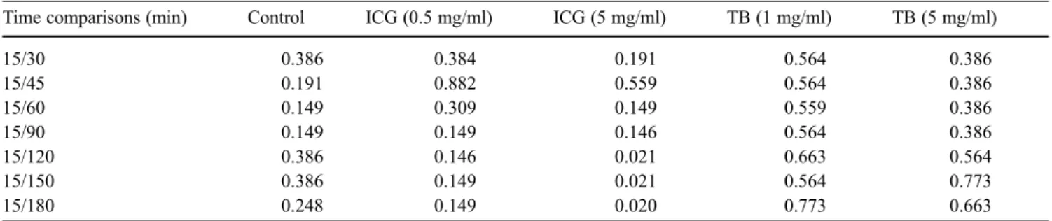
Table 2 Acute cytotoxicity: Mann–Whitney test comparison (p values) of absolute fluorescence measurements. Cultured human RPE cells were exposed to each of the tested dyes for a 5-min period. After rinsing, their viability was monitored during the ensuing 3 h. Acute cytotoxicity was assessed fluorometrically according to the propi-dium iodide assay. Only p values (nonparametric Mann–Whitney test) relating to the two higher concentrations of the dyes tested are
presented in this Table. Since data pertaining to indocyanine and infracyanine green were similar, the p values for the former dye alone (ICG) are displayed. No acute cytotoxicity was observed using any of the tested concentrations of trypan blue (TB) during the 3-h mon-itoring period. Indocyanine and infracyanine green were cytotoxic (p<0.05) at the highest concentration tested (5 mg/ml) after 2 h of monitoring
Time comparisons (min) Control ICG (0.5 mg/ml) ICG (5 mg/ml) TB (1 mg/ml) TB (5 mg/ml)
15/30 0.386 0.384 0.191 0.564 0.386 15/45 0.191 0.882 0.559 0.564 0.386 15/60 0.149 0.309 0.149 0.559 0.386 15/90 0.149 0.149 0.146 0.564 0.386 15/120 0.386 0.146 0.021 0.663 0.564 15/150 0.386 0.149 0.021 0.564 0.773 15/180 0.248 0.149 0.020 0.773 0.663
trypan blue (up to 5 mg/ml) up to 3 h later. At 5 mg/ml, both indocyanine green and infracyanine green induced an acute cytotoxic effect after 2 h according to the propidium iodide uptake assay (p=0.02; Fig. 1a, b; Table 2), although no morphological changes were apparent (data not shown).
Chronic cytotoxicity
Indocyanine green toxicity was morphologically evident after 6 days of continuous exposure to the dye at concen-trations above 0.5 mg/ml. The RPE cells had phagocytosed the dye and exhibited mild cytopathic changes. Similarly at
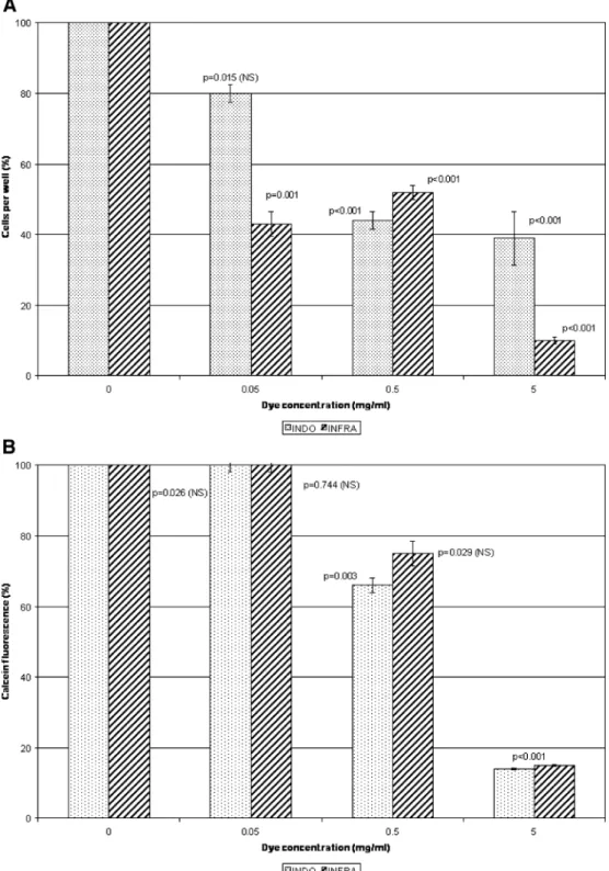
Fig. 2 a Chronic cytotoxicity. Numerical density of cultured human RPE cells after their ex-posure to different concentra-tions of indocyanine green (INDO) or infracyanine green (INFRA) for 6 days. The con-centration 0 corresponds to the included control. b Chronic cytotoxicity. Metabolic activity (calcein uptake and metabolism-induced fluorescence) of cul-tured RPE cells after their exposure to different concentra-tions of indocyanine green (INDO) or infracyanine green (INFRA) for 6 days. The con-centration 0 corresponds to the included control.
concentrations above 0.5 mg/ml, cell proliferation was in-hibited and calcein metabolism suppressed (Fig.2a, b).
Chronic cytotoxic effects of infracyanine green were likewise apparent at concentrations above 0.5 mg/ml, with cell proliferation and metabolism decreasing rapidly. At 0.05 mg/ml, cell proliferation, but not calcein metabolism, was inhibited (Figs. 2a, b, and 3). The dye was phago-cytosed by the RPE cells, which exhibited mild cytopathic changes already at the lowest concentration (0.05 mg/ml). Chronic cytotoxic effects of trypan blue were observed at all of the tested concentrations as well as with the two commercial preparations (MembraneBlue and VisionBlue) (Fig.4a, b). Cytoplasmic esterase activity was barely per-ceptible, indicating that most of the cells were dead. This finding was consistent with the observed morphological changes.
Discussion
A safe and effective means of rendering the ILM and epiretinal membranes more visible would greatly facilitate their surgical removal. The use of the dyes indocyanine green, infracyanine green, and trypan blue has been advo-cated for this purpose [11, 12, 24, 36]. However, when retinal breaks are present, as is the case in macular hole surgery, each of these dyes can potentially diffuse into the subretinal space [20] and have cytotoxic effects on the RPE. To simulate this clinical situation in vitro, we grew cultured human RPE monolayers to subconfluency and then ex-posed them to each of the dyes at clinically relevant con-centrations for either 5 min or 6 days. This experimental setup permitted us to directly compare the acute and chronic toxicities of these three drugs.
During posterior segment surgery, the dye is applied only for a few seconds or minutes before being removed by lavage of the vitreous cavity [11, 12, 28, 36]. Hence, in contrast to earlier in vitro studies, in which the dye was applied for 20 min [34], we wished to establish a situation that more closely resembled the clinical one. To evaluate acute cytotoxicity, we exposed the RPE cells to each of the dyes for 5 min, and then, after changing the medium, mon-itored them for the ensuing 3 h. The change of medium was necessary in order to avoid the spurious effects of dye autofluorescence [37].
It is well known in the case of indocyanine green that, despite thorough rinsing after application, significant amounts of the dye are retained by the eye for several weeks or even months after surgery [1,2,20,35,39,43]. The tissue con-centration in the early postoperative period is unknown. We assumed that 1% of the clinically used concentration could meet the clinical conditions for the first week. Until now, this has not been assessed in chronic toxicity assays. Hence, we also evaluated chronic cytotoxicity by exposing the RPE cells to each drug for 6 days in a low subclinical con-centration (0.05 mg/ml [0.005%]). In animals, a comparably low dose was shown to evoke significant functional damage of the retina after 10 days and 2 months of exposure [6].
The discussion about phototoxicity of ICG is contro-versial, but there exists no evidence for such in trypan blue [9,23]. A few articles have described ICG phototoxicity in different experimental setups [13, 25]. Meanwhile, more recent articles have demonstrated that addition of light for up to 72 h to the cultures did not significantly alter cell viability with either dye, except in combination with very low osmolarity at levels never found clinically (185 mOsm) [9,23]. Nevertheless, the effects of interaction of residual dye and transmitted natural light focused on the fovea for months is as yet unknown. In our experiments, human RPE cells were thus kept in the dark to allow comparison of drug toxicities independent of phototoxicity. Furthermore, all solutions used presented comparable, nontoxic osmolarities [21,23] (Table1).
Our study is the first to describe a chronic cytotoxic effect of trypan blue on cultured human RPE cells. On the basis of the acute results alone, this dye would appear to be fairly safe for intraoperative use in retinal surgery. This latter finding accords with the results of an earlier study which likewise indicated that short-term exposure to trypan blue elicited no toxic effects on cultured RPE cells [37]. Although trypan blue is water-soluble and should, in theory, be readily removed by rinsing, its spontaneous clearance from the vitreous cavity of rabbits is surprisingly slow, i.e., longer than 4 weeks [40]. This revelation prompted an in-vestigation of the dye’s chronic toxicity. Using a rabbit model, Veckeneer et al. [40] demonstrated that at a con-centration as low as 0.2%, trypan blue exerted toxic effects at the neuroretinal level after a contact time of 1 month. More than 30 years ago, Vlckova et al. [42] postulated that the two main color components of trypan blue, monoazo Fig. 3 Phase-contrast micrograph of human RPE cells showing a shift
in the nucleus/plasma ratio with prominent nucleoli (arrow), and spindle-shaped cells with intracytoplamic dye inclusion (double arrow) after a 6-day exposure to 5 mg/ml infracyanine green (magnifi-cation ×200).
and bisazo, might be responsible for its toxicity, although they were unable to rule out the possibility that impurities present in the commercial products might be responsible for the observed effects.
Three clinical studies have assessed the effects of trypan blue in vitreoretinal surgery [8,19,29]. However, the in-vestigators observed no negative effects or adverse reac-tions on retinal function after a mean follow-up time of 4
months and a maximum of 6 months. On the other hand, Kwok et al. [26], using indocyanine green, have reported the presence of glial and/or neural components on the retinal side of the ILM. These findings are in accord with those of a recent electron microscopic study of an ILM which had been exposed to indocyanine green. In this study, Gandorfer et al. [11] described significant morphological damage within the retina, which had no apparent functional Fig. 4 a Chronic cytotoxicity.
Numerical density of cultured human RPE cells after their ex-posure to different concentra-tions of trypan blue for 6 days. Differences between each of the dye concentrations tested and the control were significant with p<0.001. The concentration 0 corresponds to the included control. b Chronic cytotoxicity. Metabolic activity (calcein up-take and metabolism-induced fluorescence) of cultured human RPE cells after their exposure to different concentrations of try-pan blue for 6 days. Differences between each of the dye con-centrations tested and the control were significant with p<0.001. The concentration 0 corresponds to the included control.
consequences. This absence of functional consequences in association with morphological damage to the retina has been reported also by other investigators [27,28]. However, in other studies performed by Gandorfer’s group [15–17], a functional worsening was observed after the use of indo-cyanine green in macular hole surgery, although this effect was partially attributable to retinal damage caused by a dye-induced alteration in the cleavage plane to the inner retinal layers [11]. On the other hand, Eckardt et al. [5] observed clear signs of necrosis in the processes of Müller cells which were lodged in an ILM that had been removed with-out staining during macular hole surgery. And Da Mata et al. [3] observed no cellular elements upon the retinal surface of an ILM stained with indocyanine green.
In contrast to those regarding indocyanine green, very few studies have addressed the cytotoxic effects of infra-cyanine green. But our results indicate that these two dyes behave similarly. One recent ex vivo study has likewise reported the existence of no histopathological differences between these two stains [18].
Our findings indicate that indocyanine and infracyanine green should be used at a concentration of, or below, 0.5 mg/ml in dye-assisted surgery, since at this level they had no acute cytotoxic effects (up to 3 h after their removal) on cultured human RPE cells. However, chronic exposure to these dyes (6 days) reduced RPE cell viability, as was re-vealed using several parameters. A similar conclusion has been reached respecting the acute and chronic cytotoxic effects of indocyanine green on retinal ganglion cells in a recent in vitro study [22]. Also in accordance with our results, morphological and functional damage of the retina by intravitreous indocyanine green was demonstrated in rat eyes. After exposure times of 10 and 60 days, the retinal structure was affected with a concentration of 2.5 mg/ml or
more, whereas intravitreal indocyanine even at a low dose of 0.025 mg/ml induced electroretinographically evident damage [6].
It is important to minimize the time of retinal exposure to these dyes and thereby reduce tissue loading [33], since indocyanine and infracyanine green can persist within the eye for several months after their intraoperative application and removal [35, 39, 43]. Furthermore, the present study has demonstrated that these dyes can be phagocytosed by human RPE cells, and therein induce chronic cytotoxicity. In a recent case report, indocyanine green was shown to persist for more than 6 months in the subretinal space, and its presence was associated with atrophy of the surrounding RPE [20]. Unusual atrophic changes in the RPE have also been observed at the site of macular holes that have been treated by indocyanine-green-assisted peeling of the ILM [7]. Nevertheless, similar observations have also been re-ported before the advent of ICG-assisted macular surgery with ICG [32]. This effect on retinal cells could be aggra-vated by a potential indocyanine-green-enhanced photo-toxicity during, and possibly also after, surgery [25].
In conclusion, tissue exposure to each of the three dyes (indocyanine green, infracyanine green, and trypan blue) used in posterior segment surgery should be for as short a time as is commensurate with the needs of the clinical sit-uation. Even after an exposure period of only a few minutes, residual amounts of stain can be retained by retinal tissue after lavage of the vitreous cavity and be taken up by RPE cells to induce chronic cytotoxicity. Trypan blue, being the most water-soluble of the three dyes, is probably retained to the least degree, which is fortunate, since chronic exposure of human RPE cells to this stain induces cytotoxic effects at all clinically relevant concentrations.
References
1. Ashikari M, Ozeki H, Tomida K, Sakurai E, Tamai K, Ogura Y (2003) Retention of dye after indocyanine green-assisted internal limiting mem-brane peeling. Am J Ophthalmol 136:172–174
2. Ciardella AP, Schiff W, Barile G, Vidne O, Sparrow J, Langton K, Chang S (2003) Persistent indocyanine green fluorescence after vitrectomy for macu-lar hole. Am J Ophthalmol 136:174–177 3. Da Mata AP, Burk SE, Riemann CD,
Rosa RH Jr, Snyder ME, Petersen MR, Foster RE (2001) Indocyanine green-assisted peeling of the retinal limiting membrane during vitrectomy surgery for macular hole repair. Ophthalmology 108:1187–1192
4. Dietz FB, Jaffe RA (2003) Indocyanine green. Evidence of neurotoxicity in spinal root axons. Anesthesiology 98:516–520
5. Eckardt C, Eckardt U, Groos S, Luciano L, Reale E (1997) Removal of the internal limiting membrane in macular holes: clinical and morphological find-ings. Ophthalmologe 94:545–551 6. Enaida H, Sakamoto T, Hisatomi T,
Goto Y, Ishibashi T (2002) Morpho-logical and functional damage of the retina caused by intravitreous indocya-nine green in rat eyes. Graefe Arch Clin Exp Ophthalmol 240:209–213 7. Engelbrecht NE, Freeman J, Sternberg
P, Aaberg TM, Aaberg TM Jr, Martin DF, Sippy BD (2002) Retinal pigment epithelium after macular hole surgery with indocyanine green-assisted internal limiting membrane peeling. Am J Ophthalmol 133:89–94
8. Feron EJ, Veckeneer M, Parys-Van Ginderdeuren R, Van Lommel A, Melles GRJ, Stalmans P (2002) Trypan blue staining of epiretinal membranes in proliferative vitreoretinopathy. Arch Ophthalmol 120:141–144
9. Gale JS, Proulx AA, Gonder JR, Mao AJ, Hutnik CM (2004)
Comparison of the in vitro toxicity of indocyanine green to that of trypan blue retinal pigment epithelium cell cultures. Am J Ophthalmol 138:64–69
10. Gandorfer A, Messmer EM, Ulbig MW, Kampik A (2000) Resolution of diabetic macular edema after surgical removal of the posterior hyaloid and the inner limiting membrane. Retina 20:126–133
11. Gandorfer A, Haritoglou C, Gass CA, Ulbig MW, Kampik A (2001) Indo-cyanine green-assisted peeling of the internal limiting membrane may cause retinal damage. Am J Ophthalmol 132:431–433
12. Gandorfer A, Messmer EM, Ulbig MW, Kampik A (2001) Indocyanine green selectively stains the internal limiting membrane. Am J Ophthalmol 131:387–388
13. Gandorfer A, Haritoglou C, Gandorfer A, Kampik A (2003) Retinal damage from indocyanine green in experimental macular surgery. Invest Ophthalmol Vis Sci 44:316–323
14. Grisanti S, Szurman P, Gelisken F, Aisenbrey S, Oficjalska-Mlynczak J, Bartz-Schmidt KU (2004) Histological findings in experimental macular sur-gery with indocyanine green. Invest Ophthalmol Vis Sci 45:282–286 15. Haritoglou C, Gass CA, Schaumberger
M, Ehrt O, Gandorfer A, Kampik A (2001) Macular changes after peeling of the internal limiting membrane in mac-ular hole surgery. Am J Ophthalmol 132:363–368
16. Haritoglou C, Gandorfer A, Gass CA, Schaumberger M, Ulbig MW, Kampik A (2002) Indocyanine green-assisted peeling of the internal limiting mem-brane in macular hole surgery affects visual outcome: a clinicopathologic correlation. Am J Ophthalmol 134:836–841
17. Haritoglou C, Gandorfer A, Gass CA, Schaumberger M, Ulbig MW, Kampik A (2003) The effect of indocyanine-green on functional outcome of macular pucker surgery. Am J Ophthalmol 135:328–337
18. Haritoglou C, Gandorfer A, Gass CA, Kampik A (2004) Histology of the vitreoretinal interface after staining of the internal limiting membrane using glucose 5% diluted indocyanine and infracyanine green. Am J Ophthalmol 137:345–348
19. Haritoglou C, Eibl K, Schaumberger M, Mueller AJ, Priglinger S, Alge C, Kampik A (2004) Functional outcome after trypan blue-assisted vitrectomy for macular pucker: a prospective, randomized, comparative trial. Am J Ophthalmol 138:1–5
20. Hirata A, Inomata Y, Kawaji T, Tanihara H (2003) Persistent subretinal indocya-nine green induces retinal pigment epi-thelium atrophy. Am J Ophthalmol 136:353–355
21. Ho JD, Tsai RJF, Chen SN, Chen HC (2003) Toxic effect of indocyanine green on retinal pigment epithelium related to osmotic effects of the solvent. Am J Ophthalmol 135:258
22. Iriyama A, Uchida S, Yanagi Y, Tamaki Y, Inoue Y, Matsuura K, Kadonosono K, Araie M (2004) Effects of indocyanine green on retinal ganglion cells. Invest Ophthalmol Vis Sci 45:943–947 23. Jackson TL, Hillenkamp J, Knight BC,
Zhang JJ, Thomas D, Stanford MR, Marshall J (2004) Safety testing of indocyanine green and trypan blue using retinal pigment epithelium and glial cell cultures. Invest Ophthalmol Vis Sci 45:2778–2785
24. Kadonosono K, Itoh N, Uchio E, Nakamura S, Ohno S (2000) Staining of the internal limiting membrane in mac-ular hole surgery. Arch Ophthalmol 118:1116–1118
25. Kadonosono K, Takeuchi S, Yabuki K, Yamakawa T, Mekada A, Uchio E (2003) Absorption of short wavelengths of endoillumination in indocyanine green solution: implications for internal limiting membrane removal. Graefe Arch Clin Exp Ophthalmol 241:284–286
26. Kwok AKH, Li VWY, Pang CP, Lai TYY, Yam GHF, Chan NR, Lam DSC (2001) Indocyanine green staining and removal of internal limiting mem-brane in macular hole surgery: histology and outcome. Am J Ophthalmol 132:178–183
27. Kwok AK, Lai TY, Yuen KS, Tam BS, Wong VW (2003) Macular hole surgery with or without indocyanine green stained internal limiting membrane peeling. Clin Experiment Ophthalmol 31:470–475
28. Kwok AK, Lai TY, Li WW, Yew DT, Wong VW (2004) Trypan blue- and indocyanine green-assisted epiretinal membrane surgery: clinical and histo-pathological studies. Eye 18:882–888 29. Li K, Wong D, Stanga P, Groenewald C,
McGalliard J (2003) Trypan blue stain-ing of internal limitstain-ing membrane and epiretinal membrane during vitrectomy: visual results and histopathological findings. Br J Ophthalmol 87:216–219 30. Meyer CH, Rodrigues EB, Kroll P
(2004) Reduced concentration and in-cubation of intravitreal indocyanine green can improve the functional out-come in macular hole surgery. Am J Ophthalmol 137:386
31. Norn MS (1980) Per operative trypan blue vital staining of corneal endotheli-um: eight years’ follow up. Acta Ophthalmol Scand 58:550–558 32. Sakamoto T, Itaya K, Noda Y, Ishibashi
T (2002) Retinal pigment epithelial changes after indocyanine green-as-sisted vitrectomy. Retina 22:794–796 33. Schmidt JC, Rodrigues EB, Meyer CH,
Hoerle S, Kroll P (2004) A modified technique to stain the internal limiting membrane with indocyanine green. Ophthalmologica 218:176–179
34. Sippy BD, Engelbrecht NE, Hubbard GB, Moriarty SE, Jiang S, Aaberg TM, Aaberg TM Jr, Grossniklaus HE, Sternberg P (2001) Indocyanine green effect on cultured human retinal pig-ment epithelial cells: implication for macular hole surgery. Am J Ophthalmol 132:433–435
35. Spaide RF (2002) Persistent intraocular indocyanine green staining after macu-lar hole surgery. Retina 22:637–639 36. Stalmans P, Feron EJ, Parys-Van
Ginderdeuren R, Van Lommel A, Melles GRJ, Veckeneer M (2003) Dou-ble vital staining using trypan blue and infracyanine green in macular pucker surgery. Br J Ophthalmol 87:713–716 37. Stalmans P, Van Aken EH, Melles G,
Veckeneer M, Feron EJ, Stalmans I (2003) Trypan blue not toxic for retinal pigment epithelium in vitro. Am J Ophthalmol 135:234–236
38. Stalmans P, Van Lommel A, Parys-Van Ginderdeuren R (2003) Indocyanine green-assisted peeling of the internal limiting membrane in macular hole surgery affects visual outcome: a clin-icopathological correlation. Am J Ophthalmol 136:961–962
39. Tadayoni R, Paques M, Girmens JF, Massin P, Gaudric A (2003) Persistent of fundus fluorescence after use of indocyanine green for macular surgery. Ophthalmology 110:604–608
40. Veckeneer M, van Overdam K, Monzer J, Kobuch K, van Marle W, Spekreijse H, van Meurs J (2001) Ocular toxicity study of trypan blue injected into the vitreous cavity of rabbit eyes. Graefe Arch Clin Exp Ophthalmol 239:698–704
41. Ventura AC, Bohnke M (1999) Toxicity of pentoxifylline on monolayers of highly proliferative cells of epithelial origin. J Ocular Pharmacol Ther 15:525–535
42. Vlckova A, Gasparic J, Horakova K (1971) Investigation of the cytotoxicity of different trypan blue commercial products. Bull Acad Pol Sci Biol 19:763–770
43. Weinberger AWA, Kirchhof B, Mazinani BE, Schrage NF (2001) Per-sistent indocyanine green (ICG) fluo-rescence 6 weeks after intraocular ICG administration for macular hole surgery. Graefe Arch Clin Exp Ophthalmol 239:388–390
44. Weinberger AWA, Schlossmacher B, Dahlke C, Hermel M, Kirchhof B, Schrage NF (2002) Indocyanine green-assisted internal limiting membrane peeling in macular hole surgery—a follow-up study. Graefe Arch Clin Exp Ophthalmol 240:913–917