Biology department
DJABOUABDALLAH Abderrezak.
MOUDJEB Islam
Theme
Presented on the …/07/2019
President: Nebbache Salim
MCB U. Mostaganem
Examiner: Aissouf Ahlem
MCB U. Mostagenem
University of Abdelhamid Ibn Badis of Mostaganem
Faculty of Natural and Life Sciences
سيداب نبا ذيوحلا ذبع تعهاج اغتسه
نن
يحلا و تعيبطلا مولع تيلك ةا
Biodegradation of sheep wool
by bacteria
Table of contents:
Abstract Introduction………..……….1 Chapter 1 : Biodegradation: 1- Biodegradation..………..………..….3 Chapter 2: Wool: 1- Wool …………...………....…..….72- Characteristics of wool fibers………...……….…...8
3- Wool fiber micro structure ………...………...….…....…9
4- Physical structure of wool ……….…...……….…...10
5- Chemical structure of wool ………...………...…14
6- Keratinase and wool degradation ………..………..….16
7- Distribution………..…………..………..….…..…..16
8- Catalytic Mechanism…………...………...……...18
9- Applications………...………….…………..………...………...18
Chapter 3 : Materials and methodes 1- Materials……….………...……...20 1.1Biological material………..………...………...20 1.2 Culture media………..……….…...20 2- Methods………..………...20 2.1 Preculture ………..……….…………...…...20 2.2 Bacteria culture ……….……….…....21 2.3 Streaking……….………....…….…..21
3- Identification of purified strains ……….……….……....22
3.1. The Bergey’s Manual of systematic bacteriology ...……….………....22
4.1.Catalase test……….……….…..23
4.2Mobility Mannitol Test………...………...…..24
4.3.Amylase search……….………...…..….24 4.4.Nitratereductase test ………...………...24 4.5.Growth at 6.5% NaCL………....25 4.6.Gelatinase test ………..…………...………..….25 4.7.Protease test ………...………..……….….…....25 4.8.Lecithinase test ………...……….………...25 4.9.Citrate test………...25
4.10.Seeding on chapman agar media…..……….………….……..25
4.11.API 20e test ………..….………...26
5.Bacterial growth………...………..………...…………....26
Chapter 4 : Results and disscusstion : 1- .Peculture ……….………..……....27 2- Culture media ………..………...…...27 3- Streaking………..………….………...………...28 4- Macroscopic aspect ………...……29 5- Microscopic aspect………..……...30 6- Chemical tests ……….……..33
7- API 20e identification results………38
8- Optical density………...40
Conclusion ……….………..……….43
References ……….…...………44
List of Figures:
1- Figure 1:Bioremediation of pollutants utilizing biodegradation ……….……...05
2- Figure 2:SEM (Scanning Electron Microscopy) of wool fiber………..….…………08
3- Figure 3:Sheep wool fiber micro Structure………..…………...09
4- Figure 4:Schematic of a wool fiber showing cuticle and cortical cells……….…………..11
5- Figure 5:Diagram showing relationship between ortho/para segmentation and crimp in a merino fiber……….12
6- Figure 6:General structure of an amino acid ………...………...13
7- Figure 7: Formation of a polypeptide by reaction of amino acids. (R , R and R may be the same or different 1 2 3 side groups)………...……….14
8- Figure 8:Amphoteric behaviour of wool……… ………...………...15
9- Figure 9:Delution of the bacterial culture in the MM medium………...……..20
10- Figure 10 : Lecture table API20E………...…………...25
11- Figure 11:Dilution of the preculture to up to 10-6……….………..26
12- Figure 12: Primary of the bacteria seeding on NA………..…...26
13- Figure 13:Morphology of strains S1, S2, on NA medium………..………..…..27
14- Figure 14: Macroscopic view for S1……….………..…………....28
15- Figure 15: Macroscopic view for S2……….………..…....…28
16- Figure 16:Results of gram staining for S1,S2……….……….…..…...29
17- Figure 17: Results of catalase test for S1,S2……….………..30
18- Figure 18 :Mannitol mobility test result of strains S1, S2……….….……....31
19- Figure 19:Glucose test result of isolates S1, S2 ………..………..31
20- Figure 20:Test result of starch hydrolysis of S1, S2 strains ………..….…...…….32
21- Figure 21: Nitrate reductase test result for isolates S1, S2 ……….……….……..32
22- Figure 22:Growth test results of S1, S2, and S4 isolates on 6.5%...33
23- Figure 23 :VP test results for isolates S1, S2 ……….………....33
24- Figure 24:Protease test results for S1, S2 strains…...………...34
List of tables:
1- Table 1:Gram staining results…..……..……….……….………...….….…..29
2- Table 2:Biochimical tests results for the bacterial strains ………...….….…36
3- Table 3:Bacillus API identification by M.cavalla B.subtilis……….………....38
4- Table 4:Bacillus API identification by M.cavalla B.lichenformis……….…..38
Abbreviations list:
ADH: arginine dihydrolase DNA: Deoxyribonucleic acid.
API: Application Programming Interface. CaCl2 2H2O: Calcium chloride hydrate.
CIT: Citrate.
DO: Optical density. FeCl3: Iron chloride.
GEL: Gelatinase. NB: Nutrient broth NA: Nutrient agar.
H₂O: Dihydrogen Monoxide. H₂O₂: Hydrogen peroxide. H₂S: Hydrogen sulfide. HCl: Hydrochloric acid.
ISO: International Standards Organization. K ₂HPO₄: Potassium hydrogen phosphate. SEM: Scanning Electron Microscopy. MM: Mineral Medium
Abstract:
The aim of this work was to highlight the most important bacterial strains involved in the process of sheep wool biodegradation by isolating and identify keratinase-producing bacterial isolates from wool. Thus, two bacterial samples were isolated and (S1, S2) in microbiology laboratories of the University of Mostaganem .These Isolates having a high proteolytic activity. The identification of these two strains was based on the morphological (macroscopic and microscopic) and chemical characteristics on the various biochemical tests available in the laboratory. These tests allowed us to classify and identify the bacteria strains. The two isolates identified were: S1 (Bacillus subtilis), S2 (Bacillus lichenformis).
Keywords: Sheep wool; waste; keratinase; keratine; Bacillus subtilis; Bacillus lichenformis; chemical characteristics, API 20E.
Résumé:
Le but de ce travail est déterminer les souches bactériennes les plus importantes impliquées dans le processus de biodégradation de la laine de mouton en isolant et en identifiant des isolats bactériens producteurs de kératinase à partir de laine. Ainsi, deux isolats bactériens ont été isolés et identifiés (S1, S2) auprès laboratoire de microbiologie de l’Université de Mostaganem. Ces isolats ayant une activité protéolytique élevée. L'identification de ces deux souches a été basée sur les caractéristiques chimiques morphologiques (macroscopiques et microscopiques) des différents tests biochimiques disponibles au laboratoire. Ces tests nous ont permis de classer et d’identifier les souches de bactéries. Les deux isolats identifiés étaient: S1 (Bacillus subtilis), S2 (Bacillus lichenformis).
Mots-clés: laine de mouton; les déchets; kératinase ; keratine; Bacillus subtilis; Bacillus lichenformis; caractéristiques chimiques, API 20E.
صخلولا
مًعنا اذْ ٍي فدٓنا يذنا قٍبطح ىح ّ دٌدحح ٍع ىُغنا فٕصن يٍٕحنا مهحخنا تٍهًع ًف تكراشًنا تٌزٍخكبنا ثلالاسنا ىْأ خكبنا ثلاشع دٌدححٔ لشع قٌزط ب تٌزٍخكبنا ثاٍُعنا ٍي ٍٍُثا لشع ىح ، اذكْٔ .فٕصنا ٍع ساٍُحازٍكهن تجخًُنا اٌز ٍٍخهنا مٍهحخن زبعي طاشُب ٌاشًٍخح ٍٍحازٍكنا ٔ ْدٌدحح ا ًف ىَاغخسي تعياج ًف تقٍقدنا ءاٍحلأا ثازبخخي ٍٍحاْ دٌدحح دُخسا . صئاصخنا ىهع ٍٍخنلاسنا آصئاصخ )تٍبٕكسٔزكًٍنأ تٍبٕكسٔزكاًنا( تٍجٕنٕفرًٕنا ٔ تٌٍٕحنا تٍئاًٍٍكنا ثارابخخلاا لاس فٍُصخب ثارابخخلاا ِذْ اُن جحًس .زبخخًنا ًف ةزفٕخًنا تفهخخًنا جَاك .اْدٌدححٔ اٌزٍخكبنا ثلا ٍٍحاْ خنشعنا ب خهنا ٍ ٍ ٍS1 (Bacillus subtilis) ،S2 (Bacillus lichenformis): اًْآًٍهع اُهصح ثحبلا ثاولك ٕص : ٍُحازٍكنا ؛ ثافهخًنا .واُغلأا ف سا ؛ ٍٍحازٍكنا ؛ اشحنا ؛ تقٍقزنا تٌٕصعنا صئاصخنا ؛ يٕصعنا س ، تٍئاًٍٍكنا API 20E
Introduction
Introduction:
Sheep wool belongs to the oldest known natural fibers, which were used for centuries to manufacture high quality apparel fabrics. For a long time wool has been used also for the production of carpets and other interior textiles. Woolen products have become the benchmark of high quality and gained broad popularity (Borda et al 2016).
In recent years new woolen products fabricated with poor quality fibers and wool wastes designed mostly for technical applications have been put into commercial production. The substantial part of these products is nonwoven fabrics which are used as thermal and acoustic insulating materials in the construction and automotive industry or geotextiles (Borda et al., 2016).
There are million tons keratin wastes discard without sufficient reuse every year (Kornillowicz-Kowalska and Bohacz 2011). Feather, wool, hair and horn are the common keratin waste (Dudyński et al 2012). Keratin waste is mainly composed of keratinous protein and classified into α, β and γ- keratin since the diverse percentage of disulfide bonds (Hill et al., 2010). α-keratin of wool waste is one of the important protein resources from leather industry and poultry farm. However, wool is not easily degraded in nature and causes serious pollution in leather processing (Thanikaivelan et al 2004). Alpha keratin, which is also called hard-keratin, has higher cysteine content (up to 14%) to form S–S bonds between cross-linking protein chains, contributing ability to resist common proteolytic enzymes such as pepsin, trypsin or papain (Onifade et al 1998, Brandelli et al 2010). In addition, the hydrophobic groups of spiral coil in wool make it more difficult for biodegradation than feather (Kornillowicz-Kowalska and Bohacz. 2011).
Nowadays, chemical and high thermal methods have been extensively explored for keratin decomposition and reuse (Brandelli et al 2010). Environmentally friendly and economical methods of microbial degradation are not universally used for α-keratin reuse especially wool waste. However, microbial decomposition still seems to be an attractive approach to manage those wastes without energy wastage and amino acids loss (Gupta and Ramnani, 2006, Brandelli 2008).
Introduction
2 Stahl et al. (1950) was the first one to discuss microbiological degradation of wool, then fungus, bacillus and thermophilic actinomycetes (Molyneux 1959, Weary et al 1965, Gousterova et al 2005) were found to produce keratinolytic enzyme during wool degradation. However, Gram-negative bacteria were rarely used to digest wool waste.
Because of the high protein content of wool, hydrolysates including peptides and amino acids could be potential source for animal feed and fertilizer (Brandelli et al 2010, Kornillowicz-Kowalska and Bohacz 2011). In our previous studies (Fang et al., 2013), wool was remarkably degraded by some keratinolytic strains and its derived keratinases showed a great utilizability for wool treatment in textile industry. Many reports had discussed keratinase production with feather medium in details (Lin et al 1999, Wang and Shih 1999, Fakhfakh et al 2010, Cedrola et al 2012). However, keratinase production with wool medium has rarely been discussed, particularly on those relationships between fermentation variables and strategies.
The objective of the present study is to highlight the most important bacterial strains required for an efficient sheep wool biodegradation directed by their combinant cells. The aim is extended to search for a simple low cost medium supporting the growth of recombinant cells along with possibly achieving high levels of released end products of sheep wool hydrolysis.
Chapter1
Biodegradation
Chapter 1:
1- Biodegradation :
Biodegradation is defined as the biologically catalyzed reduction in complexity of chemical compounds (Alexander 1994). Indeed, biodegradation is the process by which organic substances are broken down into smaller compounds by living microbial organisms (Marinescu et al., 2009). When biodegradation is complete, the process is called "mineralization". However, in most cases the term biodegradation is generally used to describe almost any biologically mediated change in a substrate (Bennet et al 2003).
Understanding the process of biodegradation requires an understanding of the microorganisms that make the process work. The microbial organisms transform the substance through metabolic or enzymatic processes. It is based on two processes: growth and co-metabolism. In growth, an organic pollutant is used as sole source of carbon and energy. This process results in a complete degradation (mineralization) of organic pollutants. Co-metabolism is defined as the metabolism of an organic compound in the presence of a growth substrate that is used as the primary carbon and energy source (Fritsche et al 2008). Several microorganisms, including fungi, bacteria and yeasts are involved in biodegradation process. Algae and protozoa reports are scanty regarding their involvement in biodegradation. Biodegradation processes vary greatly, but frequently the final product of the degradation is carbon dioxide. Organic material can be degraded aerobically, with oxygen, or anaerobically, without oxygen (Mrozik et al., 2003).
Biodegradable matter is generally organic material such as plant and animal matter and other substances originating from living organisms, or artificial materials that are similar enough to plant and animal matter to be put to use by microorganisms. Some microorganisms have the astonishing, naturally occurring, microbial catabolic diversity to degrade, transform or accumulate a huge range of compounds including hydrocarbons (e.g. oil), polychlorinated biphenyls (PCBs), polyaromatic hydrocarbons (PAHs), radionuclides and metals (Leitão., 2009).
Chapter1
Bioderadation
4 The term biodegradation is often used in relation to ecology, waste management and mostly associated with environmental remediation (bioremediation). Bioremediation process can be divided into three phases or levels. First, through natural attenuation, contaminants are reduced by native microorganisms without any human augmentation. Second, bio-stimulation is employed where nutrients and oxygen are applied to the systems to improve their effectiveness and to accelerate biodegradation. During bio-augmentation, microorganisms are added to the systems. These supplemental organisms should be more efficient than native flora to degrade the target contaminant. A feasible remedial technology requires microorganisms being capable of quick adaptation and efficient uses of pollutants of interest in a particular case in a reasonable period of time (Seo 2009). Many factors influence microorganisms to use pollutants as substrates or co-metabolize them, like, the genetic potential and certain environmental factors such as temperature, pH, and available nitrogen and phosphorus sources, then, seem to determine the rate and the extent of degradation. Therefore, applications of genetically engineered microorganisms (GEM) in bioremediation have received a great deal of attention. These GEM have higher degradative capacity and have been demonstrated successfully for the degradation of various pollutants under defined conditions. However, ecological and environmental concerns and regulatory constraints are major obstacles for testing GEM in the field (Menn 2008).
The process of biodegradation can be divided into three stages: biodeterioration, biofragmentation, and assimilation (Lucas et al 2008). Biodeterioration is sometimes described as a surface-level degradation that modifies the mechanical, physical, and chemical properties of the material. This stage occurs when the material is exposed to abiotic factors in the outdoor environment and allows for further degradation by weakening the material's structure. Some abiotic factors that influence these initial changes are compression (mechanical), light, temperature, and chemicals in the environment. While biodeterioration typically occurs as the first stage of biodegradation, it can in some cases be parallel to biofragmentation. Hueck, however, defined biodeterioration as the undesirable action of living organisms on man's materials, involving such things as breakdown of stone facades of buildings, corrosion of metals by microorganisms, or merely the esthetic changes induced on man-made structures by the growth of living organisms(Allsopp and Dennis 2004) .
Biofragmentation of a polymer is the lytic process in which bonds within a polymer are cleaved, generating oligomers and monomers in its place. The steps taken to fragment these materials also differ based on the presence of oxygen in the system. The breakdown of
Chapter1
Bioderadation
Materials by microorganisms when oxygen is present is aerobic digestion, and the breakdown of materials when is oxygen is not present is anaerobic digestion. The main difference between these processes is that anaerobic reactions produce methane, while aerobic reactions do not (however, both reactions produce carbon dioxide, water, some type of residue, and a new biomass). In addition, aerobic digestion typically occurs more rapidly than anaerobic digestion, while anaerobic digestion does a better job reducing the volume and mass of the material. Due to anaerobic digestion's ability to reduce the volume and mass of waste materials and produce a natural gas, anaerobic digestion technology is widely used for waste management systems and as a source of local, renewable energy ( Klinkner 2014). The resulting products from biofragmentation are then integrated into microbial cells, this is the assimilation stage. Some of the products from fragmentation are easily transported within the cell by membrane carriers. However, others still have to undergo biotransformation reactions to yield products that can then be transported inside the cell. Once inside the cell, the products enter catabolic pathways that either lead to the production of adenosine triphosphate (ATP) or elements of the cells structure (Lucas et al., 2008). .
In this chapter we will try to foster an in-depth understanding of a biodegradation sample process by trying to cover some types of microorganisms implied in degradation of sheep wool waste. Moreover, although we are aware that the term biodegradation is often used in relation to ecology, waste management, biomedicine, and the natural environment (bioremediation) and is now commonly associated with environmentally friendly products, this chapter will mainly give attention to biodegradation in relation to bioremediation through describing processes (natural attenuation, biostimulation and bioaugmentation) utilizing degradation abilities of microorganisms in bioremediation and factors affecting this process (Figure 1).
Chapter 1
Biodegradation
6 Figure.1: Bioremediation of pollutants utilizing biodegradation abilities of microorganisms include the natural attenuation (Nezha et al 2013).
Chapter 2
wool
Chapter 2:
1- Sheep wool :
Wool fiber is the natural hair grown on sheep and is composed of protein substance called as keratin. Wool is composed of carbon, hydrogen, nitrogen and this is the only animal fiber, which contains sulfur in addition. The wool fibers have crimps or curls, which create pockets and give the wool a spongy feel and create insulation for the wearer. The outside surface of the fiber consists of a series of serrated scales, which overlap each other much like the scales of a fish. Wool is the only fiber with such serration’s which make it possible for the fibers to cling together and produce felt (textilelearner.com 2015).
Wool with up to 95% keratin by weight is a rich and pure source of intermediate filament proteins (IFPs) which can be used in a wide range of biomaterials applications as a polymeric polyamide; keratin exhibits a high degree of chemical functionality. It has high potential for bio-based niche market applications in sponges, films, matrices for agent retention and transport . Functionalities and end-uses are determined by hydrolysis conditions of oxidation and reduction to break or restore disulfide likages. Sites of reactivity include amide, carboxyl, sulfoxide, sulfide, and thiosulfide. Solubilized wool fiber with the transformed morphologies of keratin powders (lyophilized products of solubilized wool) exhibits the unique characteristics of natural keratin and upon characterization can be feedstock for developing novel products and applications. Wool was hydrolyzed under severe alkaline hydrolysis at elevated temperature in various modified systems to obtain from 68% to 80% recovery of pure keratin in the form of IFPs and constituent microfibrillar and matrix proteins. Various stages of chemical degradation of cuticle and cortical cells to protein residues were recorded by scanning electron microscopy (SEM). Four keratin preparations were produced from alkaline oxidation and reduction methods. The keratin products were recovered in powder-form for analysis by gel electrophoresis leading to MALDI-TOF identification and characterization of keratin Type II cortical fragments and their associated matrix proteins, each having identified protein profiles. These profiles were documented as peptide mass spectral fingerprints of known IFP theoretical sequences found in web-based database, the wool fibers have three-dimensional crimps, 25 waves per 10 cm in fine fiber, and 4 waves
Chapter 2
Wool
8 per 10 cm for coarse fibers. The fiber length ranges from 3.8-38 cm. The fiber length of 5-12 cm is used in the garment industry because this length allows the yarn to be manufactured with greater precision. The diameter of the fiber varies from 14 micrometers to more than 45 micrometers. The fibers of some sheep may reach a diameter of 70 μm; these fibers are used in the carpet industry. The higher price is paid for fibers with the fine diameter, especially if they are identical in diameter. The color of sheep's wool varies from white to brown and black. White is more desirable than other colors. The dark fibers cannot be successfully dyed for the difficulty of removing or hiding the natural color (Jeanette 2009).
Wool fibers absorb water from the surrounding atmosphere better than other fabric fibers because they have pores and interstitial spaces in their composition. Wool fibers absorb about 18% of their weight in moisture, but the human does not feel this moisture, and this is a very important health factor must be provided in clothes.
Fabric made from woolen fibers gives a warmer feel than other plant or industrial fibers.
Wool is a good insulation for heat, preventing heat from leaking out, and cold air from leakage inside. Therefore, woolen textiles are used as a protective cover for heat in hot places as well as cold in the cold winter.
Wool fibers are very flexible, they increase about 30% of their length at the simple tensile strength, and return to normal condition when removing tensile strength.
Woolen fabrics are non-flammable and stop burning when the fire source is removed. Wool transfers ultraviolet rays to the body.
Chapter 2
Wool
2-Wool fiber micro structure
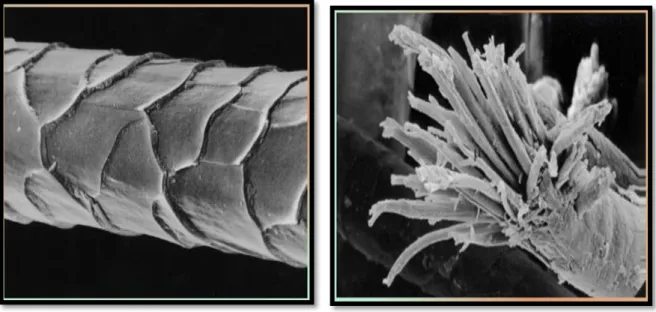
Through microscopic examination of wool fibers, we find that they are made from protein.Wool fibers are also cross-linked through the disulfide bonds present in amino acids cysteine. It was found that wool has two structures. One is Alpha-keratin and the second is beta-keratin that was through X-ray diffraction. The microstructure of wool fibers consists of three basic parts: the cuticle, cortex, and medulla (Figure 2).
The cuticle (epidermis) is a layer of overlapping cells surrounding wool fibers. The cortex is the internal cells form 90% of the wool fiber. There are two basic types of cortical cells; or the cortical, and paracortical, each with a different chemical composition. In superior fibers, these two types of cells are of distinct halves. The cells expand differently when the moisture absorbed, making the fiber curve, this creates a crease in the wool. In rough fibers, ortho and para cortical-chemical cells are more random so there is less curl. Also, fiber crease makes wool an insulator for air. The medulla is a mass of degenerated cells in the central part of the fiber. This layer may disappear or be difficult to see in the fine wool (Rippon et al 1992).
Figure 2: SEM (Scanning Electron Microscopy) of wool fiber showing the cuticle cells (Rippon et al 1992).
Chapter 2
Wool
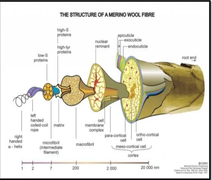
10 4-The Physical structure of wool:
In addition to its chemical complexity, wool also has a very complex physical structure, as shown schematically in (Figure 3). A wool fiber can be considered as a biological composite consisting of regions that are both chemically and physically different. Australian merino wool fibers range in diameter typically from 17 to 25μm. They are composed of two types of cell: the internal cells of the cortex and external cuticle cells that form a sheath around the fiber.
Chapter 2
wool
Cuticle cells (or scales), which overlap like tiles on a roof, make wool unique amongst textile fibers. The complex physical structure of cuticle cells is shown in (Figure 4). An important function of cuticle cells is to anchor wool fibers in the skin of sheep. The exposed edge of each cuticle cell points from the fiber root towards the tip. This gives rise to a larger surface frictional value when a fiber is drawn in the against-scale direction than in the with-scale direction. The frictional difference helps to expel dirt and other contaminants from the fleece, but it is also responsible for wool's property of felting when agitated in water. This characteristic, which is not shared by any other textile fiber, enables fabrics with very dense structures to be produced, such as blankets, felts and overcoat materials. When felting is regarded as undesirable (for example in knitted garments that will be machine-washed), processes are available to remove the frictional difference and make wool shrink resistant. The fiber surface is also largely responsible for the natural softness of wool and its property as one of the smoothest textile fibers. Even after the natural wool grease has been removed by scouring with a detergent, wool fibers are relatively difficult to wet compared with other textile materials. This natural water repellency makes wool fabrics 'shower-proof ' and able to resist water high-S proteins low-S proteins high-tyr proteins root end cortex matrix macrofibrilmeso-cortical cell ortho-cortical cell para-cortical cell membrane complex right handed - helix left handed coiled-coil rope intermediate filament (Microfibril) nuclear remnant epicuticle endocuticle exocuticle cuticle a CSIRO Textile & Fiber Technology Graphics (Roe 1992) based on a drawing (Fraser 1972); CSIRO schematic diagram of wool fiber (Figure 3). This property is the result of a waxy, hydrocarbon coating that is chemically bound to the surface of each scale. The coating survives processes such as dyeing and can only be removed by a severe chemical treatment.
Chapter 2
Wool
12 The cortex of wool comprises approximately 90% of the fiber. It consists of over lapping spindle shaped cells cortical cells, shown schematically in (Figure 4).
Both the cuticle and cortical cells have highly complex substructures, cortical cells are held together by the cell membrane complex (CMC), which also separates cortical cells from those of the cuticle. The CMC is a continuous region, containing relatively lightly cross-linked proteins and waxy lipids that extend throughout the whole fiber. Although it comprises only around 5% of the total fiber mass, it plays an important role in the overall properties of wool. It is a region of relatively low mechanical strength in the fiber composite. When wool worsted fabrics are abraded during prolonged wear, breakdown tends to occur mainly by fracture along the boundaries between cortical cells, resulting in fibrillation. (Figure 4) shows separation of individual cortical cells in a fiber taken from a severely abraded fabric. Because the CMC is only slightly cross-linked, it is also more susceptible to chemical attack. Than other regions of the fiber; for example if strongly alkaline conditions or very high temperatures are used during fabric manufacturing processes. Being the only continuous phase in the fiber, it also provides a channel by which dyes and chemicals
Figure 4: Schematic of a wool fiber showing cuticle and cortical cells (Rippon et al 1992).
Chapter 2
Wool
can diffuse in and out of wool. Fine wool fibers contain two main types of cortical cell (ortho- and para-). In the case of merino wool, these are arranged bilaterally. Coarser types of wool (diameters >25 m) tend to have less distinct segmentation of the two types of cortical cells. The bilateral segmentation of merino wool is associated with the highly desirable natural crimp of the fibers. An interesting feature is that the orthocortex is always orientated towards the outside radius of the crimp. This occurs as a result of the two segments rotating around the fiber in phase with the crimp (Leeder et al., 1984).
The structure of the proteins in wool differs between the various regions of the fiber. Some of the proteins in the microfibrils are helical, like a spring, which gives wool its flexibility, elasticity, resilience and good wrinkle recovery properties. Other proteins, particularly in the matrix that surrounds the microfibrils, have a more amorphous structure and are responsible for wool's advantage over other fibers of absorbing a relatively large amount of water without feeling wet (up to around 30% of the mass of the dry fiber). The matrix proteins are also responsible for wool's property of absorbing and retaining large amounts of dyestuffs. Wool, a fiber that has evolved over thousands of years to insulate and protect sheep, is the most complex and versatile of all textile fibers. It can be used to make products as diverse as cloth for billiard tables to the finest woven and knitted fabrics. The insulating and moisture absorbing properties of the fiber make fine wool products extremely comfortable to wear. The
Figure 5: Diagram showing relationship between ortho/para segmentation and crimp in a merino fiber (Rippon et al 1992).
Chapter 2
Wool
14 3-Chemical structure of wool:
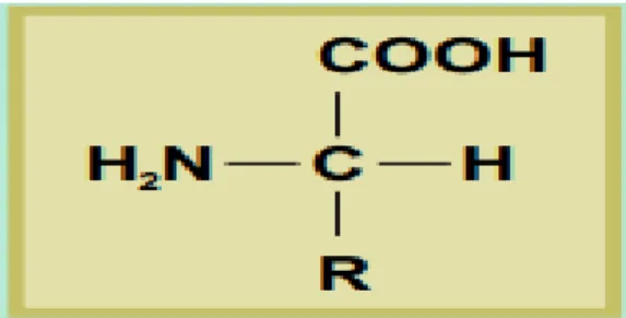
It has been estimated that wool contains more than 170 different proteins. These are not uniformly distributed throughout the fiber; proteins of different structures are located in specific regions. This heterogeneous composition is responsible for the different physical and chemical properties of the various regions of wool. The proteins in wool are composed of amino acids; so called because they contain basic amino (-NH) and acidic carboxyl (- 2 COOH) groups. The general structure of an amino acid is shown in (Figure 6).
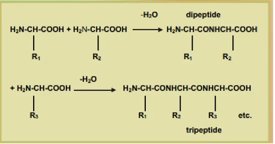
Individual amino acids differ from each other in the nature of the side group, shown as R in Figure 6. Of the 22 naturally-occurring amino acids, wool contains 18. The side groups of amino acids vary in size and can be grouped, according to their chemical properties: hydrocarbon, which is hydrophobic (water-hating); hydrophilic (water-loving); acidic; basic; and amino acids that contain sulphur. In proteins, including wool, the amino acids are joined together to form long polymer chains, as shown in (Figure 7). These compounds can be regarded as polyamides because each structural unit is joined by an amide group. When the polymer chain is a protein however, the amide repeat unit (-NHCHRCO-) is called a peptide group (Rippon et al., 2003).
Chapter 2
Wool
In wool, individual polypeptide chains are joined together to form proteins by a variet y of covalent (chemical bonds), called crosslinks, and non-covalent physical interactions (Figure7).
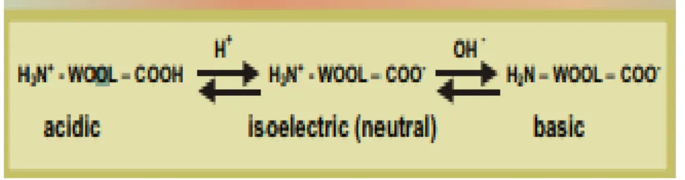
The most important crosslinks are the sulphur-containing disulphide bonds, which are formed during fiber growth by a process called “keratinisation”. These make keratin fibers insoluble in water and more stable to chemical and physical attack than other types of proteins. Disulphide bonds are involved in the chemical reactions that occur in the 'setting' of fabrics during finishing. In this process, disulphide crosslinks are rearranged to give wool fabrics smooth-drying properties so that ironing is not required after laundering. Another type of crosslink is the isopeptide bond, formed between amino acids containing acidic or basic groups. In addition to the chemical crosslinks, some other types of interactions also help to stabilize the fiber under both wet and dry conditions. These arise from interactions between the side groups of the amino acids that constitute wool proteins. Thus, hydrophobic interactions occur between hydrocarbon side groups; and ionic interactions occur between groups that can exchange protons. These ionic interactions or 'salt linkages' between acidic
Chapter 2
Wool
16 properties. This is its ability to absorb and desorb both acids and alkalis, as shown in (Figure .8). The ionic groups also control the dyeing behavior of the fiber, as a result of their interactions with negatively charged dye molecules (Rippon et al., 2003).
2- Keratinase and wool degradation:
They were initially classified as 'proteinases of unknown mechanism' by the nomen culture committee on the international union of biochemistry in 1978 with EC number 3.4.99 in 1983 (Owen et al 1983). In the 1990s, they were defined as a serine proteases due to high sequence homology with alkaline protease, and their inhibition by serine protease inhibitors (Wang et al 1995, Taha et al 1998 ,Bressollier et a1 1999).
Function keratinases are produced only in the presence of keratin-containing substrate. They mainly attack the disulfide (-S-S-) bond of the keratin substrate. Keratinase production in various microorganisms was reported on by a number of researchers. It was found that keratinase in fungi, Streptomyces and bacteria were produced in nearly at alkaline pH and almost thermophilic temperatures. These enzymes have a wide range of substrate specificity such as it can degrade other fibrous protein fibrin, elastin, collagen and other non fibrous protein like casein, bovine serum albumin gelatin etc. (Noval et al 1959, Mukhapadhayay et al 1989, Dozie et al 1994, Lin et al 1995; Letourneau et al 1998, and Bressollier et al 1999).
3- Distribution:
At first (Molyneux et al 1959) attempted to isolate some bacteria that are able to degrade keratin.[ He isolated organisms from the contents of experimentally induced dermoid cysts from mid lateral region of sheep. Examination of wool sample showed degraded wool with
Figure 8: Amphoteric behaviour of sheep wool (Rippon et al 2003).
Chapter 2
Wool
numerous cortical and cyticular cells. He found disruption of wool fiber in both in vivo and in vitro. He showed that the organisms belong to genus Bacillus and the organism was capable of attacking native wool protein. The same year (Noval et al 1959) published another article on enzymatic decomposition of native keratin by Streptomyces fradiae. They showed extracellular enzyme secreted by these bacteria capable of degrading the human hair in its native state.
Keratinolytic protein from keratinophilic fungi were reported by ( Yu et al 1968, Asahi et al 1985, Willams et al 1989, Mukhopadhay et al 1989) reported keratinase production by Streptomyces sp. He isolated an inducible extracellular homogenous enzyme, which shows a 7.5-fold increases in its activity after DEAE cellulose column chromatography. The enzyme-activity was inhibited by reduced glutathione, PMSF and 2-¬Mercaptaethanol. (Williams et al 1990) continued his work on enriched feather degrading culture and characterized the organism to its species level for the first time. The microorganisms were identified as Bacillus licheniformis, purified and characterized keratinase from feather degrading Bacillus licheniformis strain isolated by (Williams et al 1990) with the help of membrane ultra filtration and C-75 gel chromatography. He purified enzyme with 70-fold increased activity. SDS-PAGE analysis revealed that purified keratinase had a molecular weight of 33 kDa. (Dozie et al 1994) reported a thermostable, alkaline-active, keratinolytic proteinase from Chrysosporium keratinophylum which was able to solubilize keratin in lactose-mineral salt medium with DMSO. Optimum pH for the enzyme activity was 9 and optimum temperature was 90 °C. (Wang et al 1999) scaled up the fermentation condition of keratinase to a pilot scale fermenter. They optimized the fermentation condition to a level of 10-fold increase in enzyme product.
5. Catalytic mechanism:
The mechanism by which microorganisms degrade keratin varies, so the product during degradation is not the same. Some fungi reduce the disulfide bonds through the sulfites secreted on the surface of the mycelia and the acidic environment, while Streptomyces through the production of intracellular reductase. However, water-insoluble keratin can only
Chapter 2
Wool
18 cells. Observations of pure white high-temperature actinomycetes revealed that the disulfide bond reduction was performed by a cell-linked redox system. No sulfhydryl groups were detected during keratolysis of S. freundii and B. licheniformis. This may be due to the fact that the cysteine (-SH) produced by the reduction of the cysteine disulfide bond was quickly converted to other product .
Keratinase actually has the activity of disulfide reductase and polypeptide hydrolase. At present, it is generally believed that the degradation process of keratinase is divided into three steps, namely denaturation, hydrolysis and transamination. First, the disulfide reductase acts on the keratin disulfide bond to reduce cysteine (-S-S-) to cysteine (-SH), so that the high-level structure of keratin disintegrates to form degenerative keratin protein. The degenerative keratin protein is gradually hydrolyzed into polypeptides, oligo peptides and free amino acids by the action of polypeptide hydrolase. Finally, ammonia and sulfide are produced by transamination to completely hydrolyze keratin (creativeenzymes.com).
6-Applications:
In agriculture, keratinase produced by microorganisms can degrade keratin into polypeptides and amino acids, which can be used to make organic fertilizers. The organic fertilizer not only solves the problem of energy shortage, but also degrades the sources of pollution and greatly improves the environment.
In the feed industry, the main component of feathers and wool is keratin, which has a crude protein content of more than 80%. The animal has a complete range of essential amino acids, which is a good source of feed protein that can replace or partially replace fish meal. Therefore, the development and utilization of waste feathers have important application prospects. On the one hand, it solves the problem of insufficient protein resources in the current feed industry, as well as can solve the problem of environmental pollution.
Keratinase can also be used in cosmetics and medicine industry. Keratinase is an important invasive factor of dermatophytes. There forem whether keratinase inhibitors or keratinase monoclonal antibodies can inhibit the action of keratinase, weaken the invasiveness of dermatophytes, and achieve the purpose of treating skin fungal infections
Chapter 2
Wool
is an important research direction of keratinase and dermatophyte infection. Keratinase has highly efficient keratin hydrolyzing activity. The use of keratinase to improve the permeability of drugs in keratin can improve the efficacy of skin external medicine.
Chapter 3
Materials and methodes
20
Materials and methods:
1. Materials:
This work was done out at the laboratory of microbiology of the faculty of sciences of nature and life of the university Abdelhamid Ibn Badis of Mostaganem, (Algeria).
Laboratory materials and culture media are presented at the end (see Annex).
1.1. Biological material:
Sheep wool were reported from the farm of Oulad ben sahli ,Mazouna ,Relizane a sample of wool from a living sheep .
Culture media:
The media used during this work is NA nutrient agar and NB nutrient broths (annex 03) were either liquid media or solid media (1% agar p / v). The sterilization of the media is carried out by autoclaving at 121 ° C. for 20 minutes the composition and method of preparation of the media used in our study are reported in detail in the annex.
2. Methods
2.1 Preculture:Firstly, 0.5 g was added in a vial containing 100 ml of nutrient broth (NB) nutrient broth (see Annex) and incubated at 37°C for 24 hours in an incubator to release the microorganisms retained in the wool.
After the preparation of a mineral medium MM (see annex) was divided into 3 vials each containing 120ml of MM too measure optical density
1. The first vial containing 0.5g of wool. This later was previously washed and cleaned from dirt and disinfects it with bleach solution.
2. The second vial containing bacteria and wool; 1ml was taken from the first NB solution and 0.5g of disinfected wool.
3. The third vial containing bacteria ;1ml of NB was put it in vial of MM media
Chapter 3
Materials and methods
2.2 Bacterial culture:
The first bacterial cells in NB (see annex) is performed by decimal dilutions of each sample up to 10-6 then 0.5 ml of each dilution is seeded on selective nutrient agar medium. And incubated at 37°C for 24 to 48 hours.
Figure.9: Delution of the bacterial culture in the MM medium (https://nptel.ac.in/courses/)
2.3 Streaking
A pure strain from a single species of microorganism was isolated. Samples can then be taken from the resulting colonies and a microbiological culture can be grown on a new plate so that the organism can be identified, studied, or tested. The operation is repeated several times until
Chapter 3
Materials and methods
22
3. Identification of purified strains
Identification is based on the determination of morphological and biochemical characters
(Lamouliatte et al 1992). On each of the boxes used for counting, we classified the colonies
into several categories according to their macroscopic appearance. In each category, a colony was chosen randomly to perform the first tests orientation. Each selected colony is purified on nutrient agar medium. For a preliminary identification of these bacteria, we are based on studies: morphological (the characteristic forms of colonies, their arrangement, the presence or absence of spores and their Gram staining, spore coloring). Biochemical study (mobility test, catalase test, respiratory type test and amylase search ...).
3.1. The Bergey's manual of systematic bacteriology
In 1923, David Bergey, professor of bacteriology at the university of pennsylvania, published a classification of bacteria that could be used for the identification of bacterial species, the bergey's Manual of determinative bacterriology, which is currently in its 9th edition
(Bergeys.org). Then came a much more detailed book containing descriptions of all identified
bacterial species, the Bergey's manual of systematic bacteriology. This reference book of bacterial taxonomists relied until recently on the phenotypic classification of bacteria, that is to say, on their apparent characters, and did not provide any details on the natural evolution of bacteria in relation to each other.
This manual, divided into four volumes and 33 sections, grouped together bacteria with a few readily determinable characters (shape, morphology, gram staining, endospores, mobility...). The responses to gram staining played an important role in this classification mainly gram-positive and gram-negative bacterium.
3.2. Phenotypic characterization:
The morphology of colonies and cells the macroscopic study makes it possible to determine the morphology of the colonies obtained on culture media (size, the shape of the colonies...). Microscopic observation is necessary to define the shape of bacterial cells (shells or bacilli). The gram staining of the isolates after 24 hours of culture on NA makes it possible to detect the morphological forms of different bacterial cells. (Carr et al 2002).
Chapter 3
Materials and methodes
3.3. Gram stain test:
The responses to Gram staining play a key role in the classification that distinguishes gram-positive and gram-negative bacteria. An isolated colony, taken from solid medium after 24h culture at 37 ° C, is spread in a droplet of water on the glass slide with a bacterial loop sterilized by the flame of the beak. After drying the blade in air, the smear is fixed by the bunsen burner flame on the back of the glass slide. After cooling, the microorganisms are stained in gentian violet for 1 min. after washing with water; the smear is brought into contact for 1 min with the lugol solution. The liberated iodine will fix the previous dye. The smear is contacted for 1 min with safranin which acts as a contrast dye: the bacteria having a negative reaction to the gram stain appear pink, the bacteria having a positive reaction to the gram stain which have not been discolored remain purple. The smear is washed with distilled water and dried thoroughly. The observation of the smear is performed by optical microscopy (x100 immersion objective).
4. Biochemical study:
4.1. Catalase testCatalase is a respiratory enzyme capable of degrading hydrogen peroxide (H2O2) in water and O2 .
Place a microscope slide inside a petri dish. The use of a petri dish is optional as the slide catalase can be properly performed without it. However, to limit catalase aerosols, which have been shown to carry viable bacterial cells, the use the use of a petri dish is strongly recommended. Using a sterile inoculating loop collect a small amount of organism from a well-isolated 18- to 24-hour colony and place it onto the microscope slide, place 1 drop of 3% H2O2 into the organism on the microscope slide. Do not mix. Immediately cover the petri dish with a lid to limit aerosols and observe for immediate bubble formation (O2 + water = bubbles). Observing for the formation of bubbles against a dark background enhances readability.
Chapter 3
Materials and methodes
24 4.2. Mobility mannitol test:
The fermentation of the sugars is carried out in a liquid medium (oxidation-fermentation: O-F). The growth of the isolates and the shift of the color indicator reflect the fermentation of the sugar. Acidification of the medium changes the indicator, bromothymol blue, which changes from green to yellow. After seeding the O-F + glucose and O-F + mannitol tubes by isolates S1, S2 (Marchal and Bourdon, 1982). The incubation of these tubes was at 37 ° C and the development of a yellow color was observed.
4.3. Amylase test:
This test was carried out by growing the strain on a nutrient agar containing 1% starch (see Annex). After obtaining a good bacterial culture, the agar was coated with a Lugol solution. The hydrolysis of starch is thus evidenced by the appearance of a clear zone around the colony, whereas a negative result results in a brown color around the crop (De Vos et al 2009).
4.4. Nitrate reductase test:
The study of the reduction of nitrates is done by the demonstration of the nitrates formed in acetic or sulfuric medium, which give a pink coloring. Nitrates can also go to the nitrogen stage (N2).Four drops of the 'nit1' and then 'nit 2' reagents are added to a 24- to 48-hour incubation at 37° C in nitrated broth. After agitation, the reading is immediate. Many cases of figures can occur: When the color is yellow or red; the nitrates are reduced, we speak of nitrate reductase positive (NR +).When the medium remains colorless, the zinc powder is added (nitrate reducers), after five minutes the tubes are again observed: If the medium becomes pink, there are still nitrates, so they have not been reduced by the bacteria: nitrate reductase negative (NR-) and if the medium remains colorless, there are no more nitrates, the bacteria reduced them beyond the nitrate stage: positive nitrate reductase (NR +) (Tortora et al 2003).
4.5. Growth at 6.5% NaCl:
The NB medium at a NaCl concentration of 6.5% (see Annex) is inoculated with the four strains and then incubated at 37 ° C. for 24 hours. Following incubation, the
Chapter 3
Materials and methodes
Presence of bacterial growth indicates salt tolerance and lack of growth. It indicates salt sensitivity.
4.6. Gelatinase test:
This test was carried out by seeding the strain on the gelatin-based medium (see Annex 03). After obtaining a good bacterial culture (24 to 72 hours of incubation at 37°C), the gelatin was covered with a gelatin test solution which contains mercuric chloride, concentrated HCl and H2O, is thus evidenced by the appearance of a clear area around the colony. (De Vos et al 2009).
4.7. Protease test:
Casein Agar or skim milk agar is a growth medium used for the detection of hydrolytic microorganisms. The agar is supplemented with skim milk as the casein source. The medium is opaque due to the casein in colloidal suspension. The hydrolysis reaction causes the milk agar, normally the opacity of real milk, to clear around the growth area.
4.8. Lecithinase test:
This test was carried out by seeding strains one on nutrient agar containing the emulsion of the sterile egg yolk (see Annex). After 24 to 72 hours of incubation at 37 ° C., the appearance of a clear zone around the culture proves that the strain possesses lecithinase (De Vos et al 2009).
4.9. Citrate test:
This test was carried out by seeding the strains in an inclined tube medium (see Annex). After 24 to 72 hours of incubation at 37 ° C, this reaction is indicated by the color change of the pH indicator, bromothymol blue becoming blue.
4.10. Seeding on chapman agar medium:
Chapter 3
Materials and methodes
26 No bend (the medium remains red): the colonies are mannitol negative because they do not ferment mannitol, slight alkalinization of the medium by the use of peptones in their energy metabolism.
Turn yellow in the middle: colonies are positive mannitol because they ferment mannitol with acidification of the medium.
4.11. API 20E test:
The analytical profile index or API is a classification of bacteria based on experiments, allowing fast identification. This system is developed for quick identification of clinically relevant bacteria. Because of this, only known bacteria can be identified.
Chapter 4
Results and discussion
5. Bacterial growth:
Optical density of the sample measured at a wavelength of 600 nm. Also seen written as
"O.D. 600," "o.d. 600," "OD600" etc. It is a common method for estimating the concentration
of bacterial or other cells in a liquid. It is measured once a week in a spectrophotometer can be used as a measure of the concentration of bacteria in a suspension. As visible light passes through a cell suspension the light is scattered.
Chapter 4
Results and discussion
29
Results and discussion:
1. Preculture :
The mother suspension is diluted successively in a sterile physiological solution 1 ml of suspension to be diluted in a tube containing 9 ml.
2. Culture media:
After dilution the of preculture the 10-6tube culture was seeded on nutrient agar Figure.11: Dilution of the preculture to up to 10-6
Chapter 4
Results and discussion
3.
Streaking
Isolation a pure strain from a single species of microorganism to see precisely the strains morphology and the macroscopic aspect (figure.13), following tables.
S1
Chapter 4
Results and discussion
30
4. Macroscopic aspect
Macroscopic study includes the different characters of bacterial strains such as diameter, color, shape ... .etc. The results are grouped in the following table.
Bacillus subtilis
Shape Irregular
Color White or yellow
Diameter 0.3µm Elevation Hunchbaked Surface Brillante Margin Serrated Consistency Gluey
Bacillus licheniformus
Shape Rod_shaped Color White Diameter 0.2µm Surface Creamy Border Circular Opacity TranslucentFigure.14: Macroscopic view for S1
Chapter 4
Results and discussion
5. Microscopic aspect:
Microscopic observations of gram stain:
The aim of this study was to screen bacteria of the genus Bacillus that has the sporulated gram-positive bacillus form, gram staining performed according to the technique of ( Dussault 1955) revealed a dominance of 98% of the isolates are short. Gram-positive bacilli, with rounded ends and often arranged in pairs or in chains as well (figure 16).
Microscopic exams also showed two forms of cells (hulls and sticks). The observed sticks are represented in the table below:
Table.1: Gram staining test results and microscopic cell form
Strains
Microscopic Cell form
Gram
S1
Bacillus
positive
S2
Bacillus
positive
Figure.16: Results of gram staining for S1, S2 strains
Chapter 4
Results and discussion
32
1. Catalase test results:
In the presence of molecular oxygen, certain metabolic reactions lead to the formation of oxygenated water. Catalase is an enzyme that degrades oxygenated water into water and oxygen. Bubble formation is observed (Figure.17).
Figure.17: Results catalase test for S1, S2 strains
S1
Chapter 4
Results and discussion
2.Mobility mannitol test result
:
Mannitol fermentation reading both of tubes contains yellow medium, fermentation of mannitol: mannitol +.
Reading mobility:both of strains have : no diffusion around the sting: immobile bacteria.
3. Glucose test result
:
It tests an organism's ability to ferment the sugar glucose for S2 only the aerobic tube has turned yellow then the organism is able to oxidase glucose aerobically and for S1 both tubes are yellow then the organism is capable of fermentation (Figure.19).
Figure.18: Mobility mannitol test result of strains S1, S2
S2
Chapter 4
Results and discussion
34 4. Starch hydrolysis test result:
The starch agar plate is inoculated by a microorganism. Then, it is incubated for 24 – 48 hours. After incubation, iodine is poured in the plate. A clearing of light yellow/gold zone around bacterial growth indicates hydrolyzes or the presence of amylase (Figure.20).
5. Nitrate reductase test result:
After 24h incubation at 37 ° C, the two strains tubes of nitrate broth that contains the bacteria, the growth of isolates Was observed (Figure.21);(Table.2).
Figure.20: Test result of starch hydrolysis of S1, S2 strains
Figure.21: Nitrate reductase test result for the strains S1, S2
S1
S2
Chapter 4
Results and discussion
6. Growth on 6.5 % Nacl result:
A positive salt tolerance test is indicated by growth and/or turbidity in media without indicator. Media containing indicator will produce a color change from purple to yellow (Figure.22), (table.2).
7. Voges Proskauer test result:
The test results VP show that strains S1, S2 are positive the red color is observed (Figure.23), (Table 02).
Chapter 4
Results and discussion
36 8. Protease test result:
The proteolytic (caseinolytic) activity of the strains on the LEA selective medium can show exactly the diameter of the colony; also a lysis zone is noticed for the two strains (figure 24).
9. Lecithinase test result (Egg yolk):
After 24 hours of incubation at 37°C., the growth of the two strains S1, S2 the egg yolk agar medium, which signifies that these strains does not possessed lecithinase, (figure.25).
S1
S2
Figure.24: Protease (casein hydrolysis) test results for S1, S2 strains
Figure.25: Lecithinase test results for S1, S2 strains
Chapter 4
Results and discussion
10.Gelatinase test results :
After 24h incubation at 37 ° C, the gelatinase result of the two strains S1, S2 evidenced by the appearancethe colonis of clear areas around it . (Figure.26).
11.Seeding on chapman medium :
Chapman agar is a selective agar for Staphylococcus the ngative result is observed in the figure below (figure 27).
Figure.26: Gelatinase test results for strains S1, S2
Chapter 4
Results and discussion
38 Wool weight after 11 weeks incubation:
Was mesured after 11 weeks from the progression of the bacteria growth in the tow bottles that contains MM (mineral medium ) .
Wool in bottle 1 became 0. 444g Wool in bottle 3 became 0.376g
Biochemical tests results discussion:
The results of the precedent tests are summarized in this table below: Table.2: Biochemical tests results for the bacterial strains.
Tests Strain 1 Strain 2
Catalase + + Mannitol + + Motility - - Amylase + + Nitrate reductase + - Growth on 6.5% NaCl + + Citrate + + Protease + + Lecithinase - - Chapman - -
Chapter 4
Results and discussion
12. API 20E identification:
Preparation of the inoculum: with the aid of a pipet, take a 24h colony well isolated on agar medium. And realize a bacterial suspension in 5 ml of sterile physiological water inoculation of the gallery: homogenize the bacterial suspension, For CIT, VP, GEL tests, fill tubes and cups and the other tests, fill only the tubes. Recover the ODC, ADH, LDC, H2S, URE tests with 2 drops of paraffin oil. Put the cover of the gallery and incubate it at 37 ° C for 24 hours.
ONPG ADH LDC ODC CIT H2S URE TDA IND VP
+ - - - + - - - + +
GEL GLU MAN INO SOR RHA SAC MEL AMY ARA
+ + + + +/- + +/- +/- + +
ONPG ADH LDC ODC CIT H2S URE TDA IND VP
+ + - - + - - - - +
S1
Chapter 4
Results and discussion
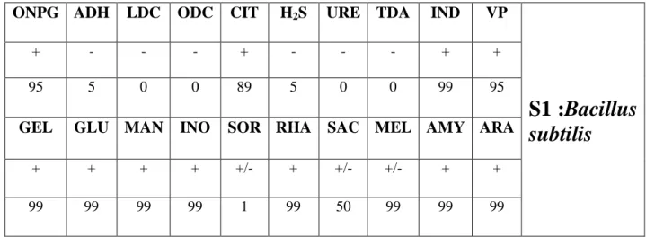
40 Comparing the result found :
Comparing our results with the API identification table by ( Matrice API 20,01.0,011 E Bacillus identification M. Cavalla 31/03/2003).
API 20E Result for strain 1:
Table 3: Bacillus API identification by M.cavalla B.subtilis
ONPG ADH LDC ODC CIT H2S URE TDA IND VP
S1 :Bacillus
subtilis
+ - - - + - - - + +
95 5 0 0 89 5 0 0 99 95
GEL GLU MAN INO SOR RHA SAC MEL AMY ARA
+ + + + +/- + +/- +/- + +
99 99 99 99 1 99 50 99 99 99
API 20E Result for strain 2:
Table 4: Bacillus API identification by M.cavalla B.lichenformis
ONPG ADH LDC ODC CIT H2S URE TDA IND VP
S2 :Bacillus
lichenformis
+ + - - + - - - - +
99 93 0 0 77 3 0 0 2 99
GEL GLU MAN INO SOR RHA SAC MEL AMY ARA
+ + + + +/- + +/- +/- + +
Chapter 4
Results and discussion
Discussion:
Strain 1 (Bacillus subtilis ):
The first strain is rod-shaped bacteria bacillus catalase-positive bacterium, found in soil and the gastrointestinal tract of ruminants and or trash B. subtilis has historically been classified as an obligate aerobe, though evidence exists that it is a facultative anaerobe. It is one of the bacterial champions in secreted enzyme production and used on an industrial scale by biotechnology companies.
Positive results for catalase and gram staining , beta-galactosidase,, utilization of citrate as a source of carbon , gelatinase , degradation of tyrosine acid production from amygdalin,Voges-Proskauer reaction ,indol, ,mannose rhamnose, inositiol, arabinose et melibiose.
Negative results for rarginine dihydrolaselysine decarboxylase, ornithine decarboxylase, hydrolysis of urea, acid production from H2S production, lecithinase and chapman.
Variable results for melibiose, sorbitol,saccharose. Strain 2 (Bacillus lichenformis):
The second strain is a rod-shaped bacterium also; It is a gram-positive, mesophilic bacterium. Its optimal growth temperature is around 50°C, though it can survive at much higher temperatures. The optimal temperature for enzyme secretion is 37 °C
Bacillus lichenformis is a bacterium commonly found in the soil. It is found on bird feathers, especially chest and back plumage and some other keratinous waste, and most often in ground-dwelling birds (like sparrows) and aquatic species (like ducks).
Positive results for catalase, beta-galactosidase,, utilization of citrate as a source of carbon, gelatinase , degradation of tyrosine acid production from amygdalin,Voges-Proskauer reaction, ,mannose rhamnose, inositiol, arabinose et melibiose .
Negative results for arginine dihydrolase lysine decarboxylase, ornithine decarboxylase, ,indol, hydrolysis of urea, acid production from H2S production also lecithinase and chapman
Chapter 4
Results and discussion
42
5.Bacterial growth :
A spectrophotometer (the measurement of optical density) was used to measure this growth. In precise technical conditions.
Table.5: Optical density results over 11 week incubation:
Bottle 1
Bottle 2
Bottle3
Wool
Bac Bac + woolThe
optical
density
at 600
nm
Week 1 0.204 0.143 0.143 Week 2 0.222 0.157 0.155 Week 3 0.235 0.167 0.170 Week 4 0.283 0.191 0.189 Week 5 0.288 0.260 0.212 Week 6 0.302 0.238 0.242 Week 7 0.330 0.217 0.301 Week 8 0.313 0.208 0.320 Week 9 0.319 0.142 0.313 Week 10 0.326 0.126 0.450 Week 11 0.331 0.103 0.486Time per weeks
Optical density at 600nm