HAL Id: inserm-00333359
https://www.hal.inserm.fr/inserm-00333359
Submitted on 23 Oct 2008HAL is a multi-disciplinary open access archive for the deposit and dissemination of sci-entific research documents, whether they are pub-lished or not. The documents may come from teaching and research institutions in France or abroad, or from public or private research centers.
L’archive ouverte pluridisciplinaire HAL, est destinée au dépôt et à la diffusion de documents scientifiques de niveau recherche, publiés ou non, émanant des établissements d’enseignement et de recherche français ou étrangers, des laboratoires publics ou privés.
A key role for stress-induced satellite III transcripts in
the relocalization of splicing factors into nuclear stress
granules.
Alexandra Metz, Johann Soret, Claire Vourc’H, Jamal Tazi, Caroline Jolly
To cite this version:
Alexandra Metz, Johann Soret, Claire Vourc’H, Jamal Tazi, Caroline Jolly. A key role for stress-induced satellite III transcripts in the relocalization of splicing factors into nuclear stress granules.. Journal of Cell Science, Company of Biologists, 2004, 117 (Pt 19), pp.4551-8. �10.1242/jcs.01329�. �inserm-00333359�
Introduction
Cells respond to changes in environmental conditions by expressing a family of highly conserved proteins termed heat shock proteins (HSPs) whose function is to protect cells from the deleterious effects of stress (Lindquist, 1986; Pirkkala et al., 2001). Hsp gene expression is regulated by the family of heat shock transcription factors (HSFs), among which HSF1 is responsible for the stress-induced activation of these genes in mammalian cells (Morimoto, 1998; Pirkkala et al., 2001). This factor, which is present in an inactive monomeric state in non-stressed cells, undergoes trimerization, post-translational modifications and becomes DNA-binding competent following stress exposure (Pirkkala et al., 2001; Sarge et al., 1993). In addition, in primate cells, HSF1 activation is accompanied by its rapid and reversible redistribution from a diffuse nucleocytoplasmic pool to a few nuclear structures termed nuclear stress granules which form by direct binding of the active factor with satellite III (sat III) repeated elements present in the 9q12 heterochromatic region in humans (Jolly et al., 2002; Jolly et al., 1997; Jolly et al., 1999; Sarge et al., 1993). Recently, we and others have shown that within these structures, HSF1 actually activates the transcription by RNA
polymerase II of sat III repeats into stable RNAs which remain associated with the 9q12 locus for a certain time after synthesis (Jolly et al., 2004; Rizzi et al., 2004). Interestingly, certain splicing factors such as the hnRNP HAP and the serine/arginine-rich (SR) proteins 9G8, SF2/ASF and SRp30c have been shown to relocalize upon stress to nuclear stress granules, whereas others, such as small nuclear ribonucleoproteins (snRNPs) or SC35, do not, suggesting a possible mechanism to regulate splicing activity during stress by the sequestration of certain splicing factors within particular subnuclear regions (Chiodi et al., 2000; Denegri et al., 2001; Denegri et al., 2002; Weighardt et al., 1999).
In this study, we wanted to understand the mechanism and role of SR protein targeting to nuclear stress granules. We found that the recruitment of hSF2/ASF and hSRp30c to the granules depends on the initial targeting of HSF1 to the granules and requires the presence of RNA. In addition, we show that the targeting of hSF2/ASF to the granules requires the second RNA-recognition motif of the protein, and that hSF2/ASF and sat III transcripts are present in the same RNP complexes in vivo, supporting the proposal that the targeting and/or retention of this protein into the granules relies on an
Exposure of cells to stressful conditions results in the rapid synthesis of a subset of specialized proteins termed heat shock proteins (HSPs) which function in protecting the cell against damage. The stress-induced activation of hsp genes is controlled by the heat shock transcription factor 1 (HSF1). At the cellular level, one of the most striking effects of stress is the rapid and reversible redistribution of HSF1 into a few nuclear structures termed nuclear stress granules which form primarily on the 9q12 locus in humans. Within these structures, HSF1 binds to satellite III repeated elements and drives the RNA polymerase II-dependent transcription of these sequences into stable RNAs which remain associated with the 9q12 locus for a certain time after synthesis. Other proteins, in particular splicing factors, were also shown to relocalize to the granules upon stress. Here, we investigated the role of stress-induced satellite III transcripts in the relocalization of splicing factors to the granules. We show that the recruitment of the two serine/arginine-rich (SR) proteins
SF2/ASF and SRp30c requires the presence of stress-induced satellite III transcripts. In agreement with these findings, we identified the second RNA-recognition motif (RRM2) of hSF2/ASF as the motif required for the targeting to the granules, and we showed by immunoprecipitation that the endogenous hSF2/ASF protein is present in a complex with satellite III transcripts in stressed cells in vivo. Interestingly, satellite III transcripts also immunoprecipitate together with small nuclear ribonucleoproteins (snRNPs) in vivo whereas the intronless hsp70 transcripts do not, supporting the proposal that these transcripts are subject to splicing. Altogether, these data highlight the central role for satellite III transcripts in the targeting and/or retention of splicing factors into the granules upon stress.
Key words: Heat shock, HSF1, Nuclear stress granules, Satellite III transcripts, SR proteins
Summary
A key role for stress-induced satellite III transcripts in
the relocalization of splicing factors into nuclear
stress granules
Alexandra Metz1, Johann Soret2, Claire Vourc’h1,*, Jamal Tazi2 and Caroline Jolly1,*
1INSERM U309, Institut Albert Bonniot, Domaine de la Merci, 38706 La Tronche CEDEX, France 2Institut de Génétique Moléculaire, CNRS UMR5535, IFR 122, 34293 Montpellier CEDEX 5, France *Authors for correspondence (e-mail: claire.vourch@ujf-grenoble.fr; caroline.jolly@ujf-grenoble.fr)
Accepted 25 May 2004
Journal of Cell Science 117, 4551-4558 Published by The Company of Biologists 2004 doi:10.1242/jcs.01329
4552
interaction with sat III transcripts. Interestingly, sat III transcripts also co-immunoprecipitate with snRNPs in vivo, whereas the intronless hsp70 transcripts do not, supporting the proposal that these transcripts are subject to splicing.
Materials and Methods
Cell culture, transient transfections and drug treatments
HeLa cells were grown in Dulbecco’s modified Eagle’s medium (DMEM) supplemented with 2 mM L-glutamine and 10% fetal bovine serum. Transient transfections were performed with the Polyfect reagent (Qiagen). The transcription inhibitors DRB
(5,6-dichloro-1-β-D-ribofuranosylbenzimidazole) and α-amanitin were used at 50
µg/ml and 2 µg/ml respectively.
Antibodies and expression plasmids
Rat monoclonal anti-HSF1 antibody (Stressgen) was used at 1:300 in immunofluorescence experiments (Cotto et al., 1997). Mouse monoclonal anti-myc antibody (Roche) was used at 1:100 in immunofluorescence experiments. Mouse monoclonal anti-SF2/ASF antibody (clone 103) (Caceres et al., 1997) was purchased from Zymed. Mouse monoclonal anti-Sm antibody (clone Y12) (cat. no. MS-450-P) was purchased from NeoMarkers and used at 1:250 in immunofluorescence experiments.
The DBD+TRIM-myc construct was kindly provided by V. Zimarino (San Raffaele Scientific Institute, Milano, Italy). The SRp30c-GFP construct was provided by S. Stamm (Max-Planck Institute of Neurobiology, Martinsried, Germany). EGFP-fusions (hSF2/ASF, hSF2/ASF∆RS) were generated in the laboratory of J. Tazi (Allemand et al., 2002; Allemand et al., 2001). The other GFP-tagged deletion mutants of hSF2/ASF were generated by PCR using the TA cloning kit (Invitrogen), with the GFP-hSF2/ASF construct as a template. For GFP-hSF2/ASF∆RRM1, the following primers were used: P1 (5′- TATGGTCGCGACGGCTATGATTACGAT-3′)and P2 (5′-TTATGTACGAGAGCGAGATCTGCTATG-3′), nucleotides 366 to 744. For GFP-hSF2/ASF∆RRM2, the fragment corresponding to nucleotides 1 to 213 was obtained with primers P3 (5′ -ATG-TCGGGAGGTGGTGTGATTCGTGGC-3′) and P4 (5′ -GGC-AGTTTCTCCGTTTTCAGACCGCCTGGATGG-3′). The fragment corresponding to nucleotides 585 to 744 was obtained with primers P5 (5′-CGGTCTGAAAACGGAGAAACTGCCTACAT-CCGG-3′) and P2. The final deletion-mutant hSF2/ASF∆RRM2 was obtained by PCR with these two last mentioned fragments as templates and with primers P3 and P2. The RRM2-GFP fragment was generated using the following primers: P6 (5′-AGAGT-GGTTGTCTCTGGACTG-3′) and P7 (5′-CGGGATTCCGCT-CATGAGATC-3′).
Coimmunoprecipitation assays of SF2/ASF-RNA and Sm-RNA complexes
HeLa cells (heat shocked or not) were harvested at the times indicated in the figures (42°C, 1 hour or 42°C, 3 hours followed by 3 hours at 37°C). Cells were lysed on ice for 30 minutes in buffer I [10 mM Tris pH 7.6, 150 mM NaCl, 5 mM MgCl2, 0.5 mM EDTA, 0.4% NP-40, 1 µg/ml leupeptin, 1 µg/ml pepstatin, 1 µM Pefabloc (Roche), 20 U of RNase inhibitor (Sigma)]. Cell extracts were first incubated with 5 µg of mouse IgG antibody and protein G-agarose beads for 2 hours at 4°C. After centrifugation at 7000 g for 2 minutes, the supernatant was incubated for 2 hours at 4°C with 5 µg of mouse anti-SF2/ASF or mouse anti-Sm antibody in buffer I. Protein G agarose beads (Amersham) were then added and samples were incubated overnight at 4°C while shaking gently. After washing in buffer I, immunoprecipitates were resuspended in RNA lysis buffer (140 mM NaCl, 0.5% NP-40, 0.5 mM EDTA, 10 mM Tris pH 7.6, 20 U of RNase inhibitor), extracted with
phenol-chloroform, and precipitated with ethanol. RNAs were assayed by reverse transcription.
Reverse transcription
Reverse transcription (RT) was performed on one-sixth of immunoprecipitated RNA in the presence of α-[32P] dCTP as previously described (Jolly et al., 2004). The sequence of the antisense primer used for sat III transcripts was 5′ TCCATTCCATTCCTG-TACTCGG 3′. The antisense primers used for hsp90α and hsp70 transcripts were the same as previously described (Wang et al., 1999). A sense hsp90αprimer was used as a negative control and a control (input) in which RNAs were ommitted was also performed. After the reaction, not incorporated α-[32P] dCTP was eliminated by centrifugation at 500 g for 2 minutes on Sephadex G50 columns. Samples were then spotted onto Hybond-N membrane, and the signals were quantified with the PhosphorImager System (Molecular Dynamics) and normalized against the input.
Immunofluorescence, RNA FISH and microscopy
Immunofluorescence and fluorescence in situ hybridization (FISH) to detect transcripts were performed in formaldehyde-fixed cells as described (Mathew et al., 1999). Antibody staining was revealed using either rat-TRITC (tetramethylrhodamine isothiocyanate) or anti-mouse-TRITC antibodies (Sigma). DNA was counterstained with 250 ng/ml DAPI (4′,6-diamidino-2-phenylindole·2HCl) diluted in an antifading solution (90% glycerol, 20 mM Tris-HCl pH 8, 2.3% DABCO (1-4 diazabicyclo (2,2,2) octane). Images were acquired on a Zeiss axiophot microscope equipped with a cooled charged-coupled camera (C4880 Hamamatsu), using a 633, NA 1.25 oil immersion objective and an intermediate magnification of 1.25.
Results
HSF1 is the key determinant in splicing factors recruitment to nuclear stress granules
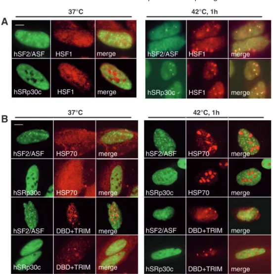
It was previously shown that upon stress, certain SR proteins accumulate into nuclear stress granules (Chiodi et al., 2000; Denegri et al., 2001; Weighardt et al., 1999). We have recently shown that nuclear stress granules correspond to active transcription sites of chromosome 9-sat III repeats by RNA polymerase II, and that these transcriptional events are HSF1-dependent (Jolly et al., 2004). Based on the earlier observation that splicing-factor recruitment to the granules is subsequent to HSF1 targeting (Denegri et al., 2001), we wanted to determine the role of HSF1 in the stress-induced recruitment of SR proteins. To address this, we prevented HSF1 relocalization to nuclear stress granules in two different ways. First, we transiently overexpressed the heat shock protein HSP70, a negative regulator of HSF1 (Shi et al., 1998). Indeed, we have previously shown that HSP70 overexpression efficiently prevents the relocalization of HSF1 to the granules and the subsequent RNA polymerase II-dependent transcription of sat III repeats following heat exposure (Jolly et al., 2004). We also used a mutant of HSF1 that only contains the DNA-binding and trimerization domains (DBD+TRIM) and constitutively forms granules (Jolly et al., 2002). An interesting feature is that it acts as a dominant negative mutant, preventing the stress-induced relocalization of the endogenous HSF1 to the granules and the subsequent sat III transcription (Jolly et al., 2004). We thus investigated the distribution of hSRp30c or hSF2/ASF splicing factors fused to GFP that were transiently expressed, alone or in combination with either HSP70 or the DBD+TRIM Journal of Cell Science 117 (19)
mutant of HSF1. Results are presented in Fig. 1. As shown previously (Chiodi et al., 2000; Denegri et al., 2001; Weighardt et al., 1999), hSF2/ASF and hSRp30c displayed a typical speckled pattern at 37°C, and they both relocated to nuclear stress granules after one hour at 42°C, in addition to a persisting diffuse staining of the nucleoplasm (Fig. 1A). The distribution of these two SR proteins was not significantly altered when co-expressed with HSP70 protein in non-stressed cells (Fig. 1B). By contrast, at 42°C, HSP70 accumulated massively in nucleoli as described previously (Pelham, 1984; Welch and Feramisco, 1984), and hSF2/ASF and hSRp30c no longer relocalized to the granules (Fig. 1B). Likewise, the two proteins were not recruited to the granules formed by the DBD+TRIM mutant, neither at 37°C nor at 42°C. Together, these results demonstrate that the stress-induced targeting of hSF2/ASF and hSRp30c to nuclear stress granules requires the initial targeting of HSF1.
Determinant of SR protein localization to nuclear stress granules
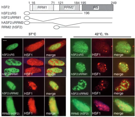
We tried to identify the domain required for the targeting of hSF2/ASF to nuclear stress granules. A typical example of the SR protein family is hSF2/ASF, which has two N-terminal RNA recognition motifs (RRMs) and a C-terminal region that is rich in arginine and serine residues, the so-called RS domain
(Sanford et al., 2003). To establish the role of these domains in the stress-induced targeting of the protein to the granules, we transiently expressed the following deletion mutants of hSF2/ASF fused to GFP and examined their intranuclear distribution: mutant hSF2/ASF∆RS lacking the entire RS domain (Allemand et al., 2001), mutant hSF2/ASF∆RRM1 lacking the first RNA-recognition motif, and mutant hSF2/ASF∆RRM2 lacking the second RNA-recognition motif. As described previously (Allemand et al., 2001), the deletion of the RS domain from hSF2/ASF did not interfere with its localization in speckles at 37°C (Fig. 2). Likewise, the two hSF2/ASF∆RRM1 and hSF2/ASF∆RRM2 mutant proteins displayed a typical speckled pattern at 37°C (Fig. 2). After a 1-hour heat shock, both the hSF2/ASF∆RS and the hSF2/ASF∆RRM1 mutants were still recruited efficiently to the granules (Fig. 2), whereas hSF2/ASF∆RRM2 was no longer recruited to the granules but remained distributed in speckles (Fig. 2). This demonstrates that the motif required for the targeting of hSF2/ASF to the granules resides in the second RRM of the protein. To determine whether the RRM2 of hSF2/ASF by itself is sufficient for the relocalization to nuclear stress granules, we also generated a fusion protein, similar to that described previously (Caceres et al., 1997), between the RRM2 domain of hSF2/ASF and GFP. As shown in Fig. 2, this protein distributed diffusely in the nucleus and cytoplasm at 37°C, and did not localize to the granules following heat shock. Fig. 1. HSF1 is the key determinant
in recruiting hSF2/ASF and hSRp30c to nuclear stress granules.
(A) hSF2/ASF-GFP or hSRp30c-GFP proteins (green) were transiently expressed in HeLa cells and HSF1 (red) was detected by
immunofluorescence. Both proteins display a typical speckled pattern at 37°C, and are targeted to HSF1 granules upon stress. (B) hSF2/ASF-GFP or hSRp30c-hSF2/ASF-GFP (green) were co-expressed in HeLa cells with either the HSP70 protein or with a dominant negative mutant of HSF1
(DBD+TRIM-myc) (red). HSP70 and the myc-tagged HSF1 mutant were subsequently detected by
immunofluorescence. Overexpression of HSP70 does not alter SR protein distribution at 37°C (left panels). By contrast, at 42°C, HSP70
overexpression prevents the targeting of both hSF2/ASF and hSRp30c to the granules (right panel). Likewise, hSF2/ASF and hSRp30c are not recruited to the granules formed by the DBD+TRIM mutant at 37°C or at 42°C. Bars, 5 µm.
4554
Altogether, these findings show that the second RNA-recognition motif is necessary but not sufficient for the stress-induced targeting of hSF2/ASF to the granules.
Recruitment of SR proteins into the granules requires the presence of stress-induced sat III transcripts
Based on this observation, we next asked whether the recruitment of hSF2/ASF and hSRp30c into the granules requires sat III transcription. Since nuclear stress granules correspond to the sites where transcription of sat III repeats from the 9q12 locus takes place (Jolly et al., 2004; Rizzi et al., 2004), we first confirmed that transiently expressed GFP-tagged hSF2/ASF and hSRp30c proteins colocalized in the granules together with the sat III transcripts that had been detected by FISH (Fig. 3A). Given that several splicing factors were shown to interact directly with the C-terminal domain (CTD) of RNA polymerase II (Du and Warren, 1997; Kim et al., 1997; McCracken et al., 1997; Mortillaro et al., 1996; Yuryev et al., 1996), an alternative hypothesis is that splicing-factor targeting and/or retention into the granules merely occurs through interaction with the CTD of RNA polymerase II, that is also concentrated into these structures (Jolly et al., 2004). To address this question, we analyzed the distribution of these proteins in cells treated with the transcription inhibitors DRB or α-amanitin. However, a critical point in this experiment was the time point at which the drug was added to ensure efficiently inhibited transcription of these sequences. We have shown recently that sat III RNAs are stable and that for example the addition of transcription inhibitor after a 1-hour heat shock has no effect
on sat III transcript foci detected by FISH (Jolly et al., 2004). Because these transcripts become visible by RNA FISH in a small fraction of HeLa cells after 30 minutes of heat shock, and are present in virtually every cell after one-hour heat shock treatment (A.M., J.S., C.V., J.T. and C.J., unpublished data), we chose to add the drugs during the period of transcription, i.e. after 40 minutes of heat shock. FISH control experiments confirmed that transcription of sat III, hsp70 and
hsp90α was effectively inhibited following the addition of both inhibitors to heat-treated cells (A.M., J.S., C.V., J.T. and C.J., unpublished data). Interestingly, hSF2/ASF and SRp30c proteins under these conditions were no longer recruited to the granules following heat shock (Fig. 3B), suggesting that the presence of sat III RNA is a prerequisite for the recruitment of these proteins to the stress granules. We also treated cells with RNase A following heat shock, and observed that neither hSF2/ASF nor hSRp30c were present in nuclear stress granules when sat III transcripts had been degraded by RNase A, confirming the need for the presence of sat III transcripts in retaining these proteins (Fig. 3B). By contrast, RNase A and transcription inhibitors had no effect on the localization of RNA polymerase II, showing that physical interaction of SR proteins with RNA polymerase II is not involved in retaining hSF2/ASF and hSRp30c in nuclear stress granules. More support that RNA is required to keep splicing factors in nuclear stress granules comes from the observation that hSF2/ASF and hSRp30c are detected in the granules as long as sat III transcripts are present: we have recently shown that after RNA synthesis, sat III transcripts remained associated with the 9q12 locus in the granules for a few hours (Jolly et al., 2004). For example, cells that were Journal of Cell Science 117 (19)
Fig. 2. The second RNA-recognition motif is required for targeting hSF2/AF to the granules. GFP-tagged deletion mutants of hSF2/ASF (green) were transiently expressed in HeLa cells, and their distribution relative to HSF1 (red) was analyzed by immunofluorescence in non heat-shocked and heat-shocked cells. DeletiOn Of tHe RS dOmain
(HSF2/ASF∆RS) or the first RNA-recognition motif (hSF2/ASF∆RRM1) does not affect the speckled distribution of the protein at 37°C, and does not impede the targeting to nuclear stress granules at 42°C. By contrast, deletion of the second RNA-recognition motif (hSF2/ASF∆RRM2), which doesnot affect the localization of the protein at 37°C, prevents its stress-induced recruitment to the granules. The RRM2 domain alone is however not sufficient for the targeting to the granules. Bar, 5µm.
allowed to recover for three hours after being heat-shocked, had splicing factors that were still concentrated in the granules together with sat III transcripts, whereas HSF1 or RNA polymerase II were no longer detected in these loci. These findings demonstrate the requirement for active RNA transcription and RNA presence for the redistribution of SR proteins into nuclear stress granules.
To investigate the role of sat III transcripts in the targeting of hSF2/ASF to the granules in more detail, we immunoprecipitated endogenous hSF2/ASF-RNA complexes from non heat-shocked or heat-shocked cells, followed by RT to determine whether hSF2/ASF protein and sat III transcripts are present in the same complexes in vivo. Hsp70 and hsp90α transcripts were used as controls in these experiments. As shown in Fig. 3C, a significant level of hsp90αtranscripts was associated with hSF2/ASF at 37°C, indicating that this gene is constitutively expressed (Hickey et al., 1989). A faint signal was also obtained for hsp70 and sat III transcripts at 37°C, in accordance with previous RT results that show a low constitutive expression of both genes (Jolly et al., 2004). In cells that were heat-shocked or allowed to recover after heat shock, all three transcripts – including the intronless hsp70 transcripts – co-immunoprecipitated with hSF2/ASF (Fig. 3C). No significant signal was obtained with the hsp90α sense primer, confirming the specificity of the signals obtained with the other primers. These findings clearly show the in vivo existence of ribonucleoprotein complexes with both sat III transcripts and hSF2/ASF present, supporting the idea that the
relocalization of this protein into the granules depends on a physical interaction with sat III transcripts.
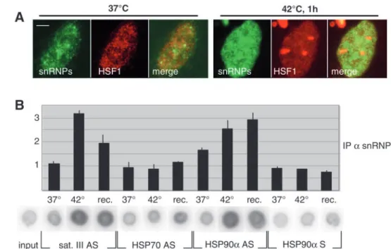
Sat III transcripts are also associated with the spliceosome components snRNPs
Because elevated concentration of hSF2/ASF are found in the granules, the observation that sat III transcripts are complexed in vivo with hSF2/ASF might reflect a sequestration of hSF2/ASF into the granules via interaction with sat III transcripts. An alternative and non-exclusive hypothesis, however, could be that sat III transcripts are spliced. To address this question, we asked whether sat III transcripts are complexed in vivo with other major components of the splicing reaction, the snRNPs. At the cellular level, snRNPs detected with the Y12 anti-Sm antibody which recognizes U1, U2, U4, U5 and U6 were diffusely distributed throughout the nucleoplasm, and also concentrated into the speckles and the Cajal bodies (Nyman et al., 1986) (Fig. 4A). At 42°C, however, snRNPs were not found accumulated into nuclear stress granules, although not excluded from these regions (Fig. 4A). We next performed immunoprecipitation of endogenous snRNPs-RNA complexes from non shocked or heat-shocked cells, followed by reverse transcription. Results are presented in Fig. 4B. A faint level of hsp90α, hsp70 and sat III transcripts were associated with snRNPs at 37°C. In cells exposed to heat shock or allowed to recover after heat shock, both hsp90αand sat III transcripts co-immunoprecipitated with Fig. 3. SR protein targeting to the
granules requires the presence of RNA. (A) Transiently expressed hSF2/ASF-GFP protein (green) was detected in heat-shocked HeLa cells together with sat III transcripts (red) and visualized by RNA FISH. hSF2/ASF colocalizes with sat III transcripts within stress granules. Bar: 5 µm. (B) Whether RNA was required in SR protein targeting to the granules was analyzed by treating the cells either with a transcription inhibitor (α-amanitin or DRB) added twenty minutes before the end of the 1-hour heat shock, or with RNase A added following heat shock. All three treatments prevent the relocalization of hSF2/ASF and hSRp30c but not of HSF1 and RNA polymerase II to nuclear stress granules. (C) Sat III transcripts co-immunoprecipitate with hSF2/ASF protein. The endogenous hSF2/ASF protein was
immunoprecipitated from non heat-shocked or heat-heat-shocked cells with a specific monoclonal antibody (Caceres et al., 1997). Co-immunoprecipitated RNAs were extracted and analyzed by
reverse transcription with antisense primers specific for hsp70, hsp90αand sat III transcripts. Sense primers to hsp90αtranscripts were used as a negative control. The y-axis corresponds to the intensity ratios between signal and input. In non heat-shocked cells kept at 37°C, a strong signal for the constitutively expressed hsp90αtranscripts and faint signals for hsp70 and sat III transcripts are observed. In cells that were heat-shocked for one hour at 42°C or that were allowed to recover for 3 hours at 37°C following heat shock (rec), a strong signal is obtained for all three transcripts, thus showing that they were all present in vivo in a complex with hSF2/ASF. As expected, no significant signal was obtained with the sense primer to hsp90αtranscripts.
4556
snRNPs, thus demonstrating that these transcripts are complexed with snRNPs in vivo. It is worth noting, however, that the intensities of the signals obtained in this experiment were five to ten-fold lower than those obtained in the hSF2/ASF immunoprecipitation experiment, in accordance with the observation that snRNPs are not concentrated with sat III transcripts in the granules. Interestingly, at 42°C, no significant signal was obtained for hsp70 transcripts, which are intronless and supposedly not spliced (Wu and Morimoto, 1985). Altogether, these findings support the idea that at least a portion of sat III transcripts are spliced.
Discussion
We wished to determine the role of stress-induced sat III transcripts in the heat-induced redistribution of splicing factors to nuclear stress granules. We show here, that this relocalization is dependent on HSF1-driven transcription and requires the presence of sat III transcripts within the granules. The hSF2/ASF protein and sat III transcripts are found in vivo in the same ribonucleoprotein complex, and the second RNA-recognition motif of hSF2/ASF is necessary but not sufficient for its targeting to the granules. Altogether these findings demonstrate the pivotal role for the heat-induced sat III transcripts emerging from the 9q12 locus, in targeting and/or retention of splicing factors within the granules. Our data do not allow to discriminate direct or indirect binding of hSF2/ASF to sat III transcripts, and both hypotheses can be envisaged. For instance, the requirement of an RNA-recognition motif for the proper localization into the granules supports the proposal that hSF2/ASF might interact directly with these transcripts and thereby mediate the recruitment into the granules of other proteins such as hnRNP HAP, whose targeting to the granules requires a domain rich in arginine-glutamic acid dipeptides (R/E domain) involved in
protein-protein interactions (Denegri et al., 2001). Alternatively, retention of hSF2/ASF in the granules might involve protein-protein interactions via the RRM2 domain. Indeed, although only the RS domain of hSF/ASF is clearly implicated in protein-protein interactions (Wu and Maniatis, 1993; Amrein et al., 1994; Kohtz et al., 1994; Sanford et al., 2003), it has been proposed that the RRM2 is also involved in such interactions (van Der Houven Van Oordt et al., 2000). In particular, it has been recently shown that this domain contains a conserved heptapeptide motif essential for splicing repression, which probably drives protein-protein interactions (Dauksaite and Akusjarvi, 2004). Another example of RRM-mediated protein-protein interaction is the Y14-protein component of the exon junction complex (EJC) (Shi and Xu, 2003). In addition, the fact that the RRM2 domain of hSF2/ASF lacks canonical RNA recognition motifs known as RNPs, also favors this hypothesis (Birney et al., 1993; Zhu and Krainer, 2000). In this context, our findings at least exclude the possibility that hSF2/ASF is retained in the granules by RNA polymerase II. In fact, our observation that the RRM2 domain in not sufficient by itself for the localization into the granules suggests that the situation may be even more complex. For instance, the targeting and/or retention of hSF2/ASF in the granules probably involves several mechanisms implicating different protein motifs. Further experiments will be needed to dissect these events.
Another interesting point of our study is that, only one of the two RRMs of hSF2/ASF is involved in the targeting to the granules. The usual function of RNA-recognition motifs (RRMs) is to mediate RNA binding and to determine substrate specificity for individual SR proteins, whereas the RS domain is required for protein-protein interactions (for a review, see Sanford et al., 2003). Our observation is in good agreement with previous studies showing that the different RRMs of multi-RRM proteins are implicated in distinct functions, Journal of Cell Science 117 (19)
Fig. 4. Sat III transcripts are associated with Sm proteins. (A) SnRNPs (green) were detected in non heat-schocked and heat-shocked HeLa cells together with HSF1 (red). SnRNPs were not recruited to the stress granules although they were not excluded from these regions. Bar: 5
µm. (B) The endogenous snRNPs proteins were immunoprecipitated from non heat-shocked or heat-shocked cells with a specific monoclonal antibody (clone Y12). Co-immunoprecipitated RNAs were extracted and analyzed by reverse transcription as previously described. In non heat-shocked cells (37°C), a strong signal for the constitutively expressed hsp90α transcripts and faint signals for hsp70 and sat III transcripts were observed. In cells that were heat-shocked for 1 hour at 42°C or that were allowed to recover for 3 hours at 37°C following heat shock (rec), a strong signal is obtained for
hsp90αand sat III transcripts but not for
the intronless hsp70 transcripts, thus showing that hsp90αand sat III transcripts are present in vivo in a complex with snRNPs. As expected, no significant signal was obtained with the sense primer to hsp90αtranscripts.
ranging from subnuclear localization to splice site selection. So are, for example, the tandem RRMs of hnRNPA1 not equivalent (Mayeda et al., 1998). Although both are required for alternative splicing function, each RRM plays distinct roles; RRM2 can function in alternative splicing when duplicated, whereas RRM1 cannot. Another example is the second RRM of SF2/ASF which is an atypical RNA binding motif described as RRM-like (Birney et al., 1993). Splice-site selection by SF2/AFS is determined by the nature of its RRM with RRM2 having a dominant role, as demonstrated by its ability to confer specificity to a heterologous protein (Caceres et al., 1997; Chandler et al., 1997; Mayeda et al., 1999; van Der Houven Van Oordt et al., 2000). Furthermore, the RRM2 of polypyrimidine tract-binding protein-associated splicing factor (PSF) is required for localization to subnuclear speckles (Dye and Patton, 2001). Likewise, proper nucleolar accumulation of nucleolin requires at least two of its five RRMs (Creancier et al., 1993). In this context, our observation that the RRM2 of SF2/ASF determines SF2/ASF targeting into and/or retention in nuclear stress granules could be the signature of SF2/ASF binding-specificity – and perhaps splicing specificity too (see below) – on RNAs transcribed from the 9q12 locus, as previously suggested for other transcripts (van Der Houven Van Oordt et al., 2000).
What could be the significance of this stress-induced accumulation of splicing factors? One hypothesis is that the presence of splicing factors merely reflects the splicing of sat III transcripts, as strongly supported by our findings that sat III and hsp90α transcripts, but not the intronless hsp70 transcripts, are complexed with snRNPs in vivo. In this case, the particular composition of the splicing complexes present in the granules, containing some but not all splicing factors (Chiodi et al., 2000; Denegri et al., 2001; Denegri et al., 2002; Weighardt et al., 1999), might specify a certain type of transcript. Another possibility is that the binding of splicing factors to these transcripts plays a role in their stabilization. Indeed, we have recently shown that sat III transcripts are stable RNAs which remain associated with the 9q12 locus for a certain time after synthesis even throughout mitosis (Jolly et al., 2004), and we show here that hSF2/ASF remains in the granules as long as sat III transcripts are present. Finally, based on the observation that heat shock inhibits splicing activity and modifies the local concentration of hnRNP proteins (Denegri et al., 2001; Weighardt et al., 1999), our findings support a model in which the sequestration of splicing factors within the granules, via association with stable transcripts, could represent a novel mechanism to finely regulate splicing function during stress, as it has been proposed for the non-coding hsr-omega nuclear (hsrw-n) transcripts in Drosophila melanogaster (Prasanth et al., 2000). Indeed, the relative ratio of hnRNPs to SR proteins is known to affect alternative splicing (Mayeda and Krainer, 1992), and the organization of these two types of splicing factors in distinct subnuclear domains might thus provide a way to regulate alternative splicing during stress. Moreover, certain specific non-coding nuclear RNAs produced in trinucleotide repeat disorders or following adenovirus-2 infection can titrate hnRNPs and SR proteins and thereby affect processing of other pre-mRNAs (Himmelspach et al., 1995; Timchenko et al., 1999). One can thus imagine a similar role for the stress-induced sat III.
We are grateful to E. Millou for technical assistance, to V. Zimarino and M. Vujanac for providing the DBD+TRIM-myc construct, to S. Stamm for the SRp30c-GFP construct, and to C. Cochet and O. Filhol for providing access to radioactivity facilities. We also wish to thank T. Gautier and B. Gilquin for helpful discussions. This work was supported by the Association pour la Recherche sur le Cancer (contract numbers 5786 and 3449), the Région Rhône-Alpes, and the Ministère de la Recherche.
References
Allemand, E., Gattoni, R., Bourbon, H. M., Stevenin, J., Caceres, J. F., Soret, J. and Tazi, J. (2001). Distinctive features of Drosophila alternative
splicing factor RS domain: implication for specific phosphorylation, shuttling, and splicing activation. Mol. Cell. Biol. 21, 1345-1359.
Allemand, E., Dokudovskaya, S., Bordonne, R. and Tazi, J. (2002). A
conserved Drosophila transportin-serine/arginine-rich (SR) protein permits nuclear import of Drosophila SR protein splicing factors and their antagonist repressor splicing factor 1. Mol. Biol. Cell 13, 2436-2447.
Amrein, H., Hedley, M. L. and Maniatis, T. (1994). The role of specific
protein-RNA and protein-protein interactions in positive and negative control of pre-mRNA splicing by Transformer 2. Cell 76, 735-746.
Birney, E., Kumar, S. and Krainer, A. R. (1993). Analysis of the
RNA-recognition motif and RS and RGG domains: conservation in metazoan pre-mRNA splicing factors. Nucleic Acids Res. 21, 5803-5816.
Caceres, J. F., Misteli, T., Screaton, G. R., Spector, D. L. and Krainer, A. R. (1997). Role of the modular domains of SR proteins in subnuclear
localization and alternative splicing specificity. J. Cell Biol. 138, 225-238.
Chandler, S. D., Mayeda, A., Yeakley, J. M., Krainer, A. R. and Fu, X. D.
(1997). RNA splicing specificity determined by the coordinated action of RNA recognition motifs in SR proteins. Proc. Natl. Acad. Sci. USA 94, 3596-3601.
Chiodi, I., Biggiogera, M., Denegri, M., Corioni, M., Weighardt, F., Cobianchi, F., Riva, S. and Biamonti, G. (2000). Structure and dynamics
of hnRNP-labelled nuclear bodies induced by stress treatments. J. Cell Sci.
113, 4043-4053.
Cotto, J., Fox, S. and Morimoto, R. (1997). HSF1 granules: a novel
stress-induced nuclear compartment of human cells. J. Cell Sci. 110, 2925-2934.
Creancier, L., Prats, H., Zanibellato, C., Amalric, F. and Bugler, B. (1993).
Determination of the functional domains involved in nucleolar targeting of nucleolin. Mol. Biol. Cell 4, 1239-1250.
Dauksaite, V. and Akusjarvi, G. (2004). The second RNA binding domain
of human splicing factor ASF/SF2 is the critical domain controlling
adenovirus E1A alternative 5′splice site selection. Biochem. J. 381,
343-350.
Denegri, M., Chiodi, I., Corioni, M., Cobianchi, F., Riva, S. and Biamonti, G. (2001). Stress-induced Nuclear Bodies Are Sites of Accumulation of
Pre-mRNA Processing Factors. Mol. Biol. Cell 12, 3502-3514.
Denegri, M., Moralli, D., Rocchi, M., Biggiogera, M., Raimondi, E., Cobianchi, F., de Carli, L., Riva, S. and Biamonti, G. (2002). Human
chromosomes 9, 12, and 15 contain the nucleation sites of stress-induced nuclear bodies. Mol. Biol. Cell 13, 2069-2079.
Du, L. and Warren, S. L. (1997). A functional interaction between the
carboxy-terminal domain of RNA polymerase II and pre-mRNA splicing. J. Cell Biol. 136, 5-18.
Dye, B. T. and Patton, J. G. (2001). An RNA recognition motif (RRM) is
required for the localization of PTB- associated splicing factor (PSF) to subnuclear speckles. Exp. Cell Res. 263, 131-144.
Hickey, E., Brandon, S. E., Smale, G., Lloyd, D. and Weber, L. A. (1989).
Sequence and regulation of a gene encoding a human 89-kilodalton heat shock protein. Mol. Cell. Biol. 9, 2615-2626.
Himmelspach, M., Cavaloc, Y., Chebli, K., Stevenin, J. and Gattoni, R. (1995). Titration of serine/arginine (SR) splicing factors during
adenoviral infection modulates E1A pre-mRNA alternative splicing. RNA 1, 794-806.
Jolly, C., Morimoto, R., Robert-Nicoud, M. and Vourc’h, C. (1997). HSF1
transcription factor concentrates in nuclear foci during heat shock: relationship with transcription sites. J. Cell Sci. 110, 2935-2941.
Jolly, C., Usson, Y. and Morimoto, R. I. (1999). Rapid and reversible
relocalization of heat shock factor 1 within seconds to nuclear stress granules. Proc. Natl. Acad. Sci. USA 96, 6769-6774.
Jolly, C., Konecny, L., Grady, D. L., Kutskova, Y. A., Cotto, J. J., Morimoto, R. I. and Vourc’h, C. (2002). In vivo binding of active heat
4558
shock transcription factor 1 to human chromosome 9 heterochromatin during stress. J. Cell Biol. 156, 775-781.
Jolly, C., Metz, A., Govin, J., Vigneron, M., Turner, B. M., Khochbin, S. and Vourc’h, C. (2004). Stress-induced transcription of satellite III repeats.
J. Cell Biol. 164, 25-33.
Kim, E., Du, L., Bregman, D. B. and Warren, S. L. (1997). Splicing factors
associate with hyperphosphorylated RNA polymerase II in the absence of pre-mRNA. J. Cell Biol. 136, 19-28.
Kohtz, J. D., Jamison, S. F., Will, C. L., Zuo, P., Luhrmann, R., Garcia-Blanco, M. A. and Manley, J. L. (1994). Protein-protein interactions and
5′-splice-site recognition in mammalian mRNA precursors. Nature 368,
119-124.
Lindquist, S. (1986). The heat-shock response. Annu. Rev. Biochem. 55,
1151-1191.
Mathew, S., Behm, F., Dalton, J. and Raimondi, S. (1999). Comparison of
cytogenetics, Southern blotting, and fluorescence in situ hybridization as methods for detecting MLL gene rearrangements in children with acute leukemia and with 11q23 abnormalities. Leukemia 13, 1713-1720.
Mayeda, A. and Krainer, A. R. (1992). Regulation of alternative pre-mRNA
splicing by hnRNP A1 and splicing factor SF2. Cell 68, 365-375.
Mayeda, A., Munroe, S. H., Xu, R. M. and Krainer, A. R. (1998). Distinct
functions of the closely related tandem RNA-recognition motifs of hnRNP A1. RNA 4, 1111-1123.
Mayeda, A., Screaton, G. R., Chandler, S. D., Fu, X. D. and Krainer, A. R. (1999). Substrate specificities of SR proteins in constitutive splicing are
determined by their RNA recognition motifs and composite pre-mRNA exonic elements. Mol. Cell. Biol. 19, 1853-1863.
McCracken, S., Fong, N., Yankulov, K., Ballantyne, S., Pan, G., Greenblatt, J., Patterson, S. D., Wickens, M. and Bentley, D. L. (1997).
The C-terminal domain of RNA polymerase II couples mRNA processing to transcription. Nature 385, 357-361.
Morimoto, R. I. (1998). Regulation of the heat shock transcriptional response:
cross talk between a family of heat shock factors, molecular chaperones, and negative regulators. Genes Dev. 12, 3788-3796.
Mortillaro, M. J., Blencowe, B. J., Wei, X., Nakayasu, H., Du, L., Warren, S. L., Sharp, P. A. and Berezney, R. (1996). A hyperphosphorylated form
of the large subunit of RNA polymerase II is associated with splicing complexes and the nuclear matrix. Proc. Natl. Acad. Sci. USA 93, 8253-8257.
Nyman, U., Hallman, H., Hadlaczky, G., Pettersson, I., Sharp, G. and Ringertz, N. R. (1986). Intranuclear localization of snRNP antigens. J. Cell
Biol. 102, 137-144.
Pelham, H. R. (1984). Hsp70 accelerates the recovery of nucleolar
morphology after heat shock. EMBO J. 3, 3095-3100.
Pirkkala, L., Nykanen, P. and Sistonen, L. (2001). Roles of the heat shock
transcription factors in regulation of the heat shock response and beyond. FASEB J. 15, 1118-1131.
Prasanth, K. V., Rajendra, T. K., Lal, A. K. and Lakhotia, S. C. (2000).
Omega speckles - a novel class of nuclear speckles containing hnRNPs associated with noncoding hsr-omega RNA in Drosophila. J. Cell Sci. 113, 3485-3497.
Rizzi, N., Denegri, M., Chiodi, I., Corioni, M., Valgardsdottir, R., Cobianchi, F., Riva, S. and Biamonti, G. (2004). Transcriptional activation
of a constitutive heterochromatic domain of the human genome in response to heat shock. Mol. Biol. Cell 15, 543-551.
Sanford, J. R., Longman, D. and Caceres, J. F. (2003). Multiple roles of the
SR protein family in splicing regulation. Prog. Mol. Subcell. Biol. 31, 33-58.
Sarge, K. D., Murphy, S. P. and Morimoto, R. I. (1993). Activation of heat
shock gene transcription by heat shock factor 1 involves oligomerization, acquisition of DNA-binding activity, and nuclear localization and can occur in the absence of stress. Mol. Cell. Biol. 13, 1392-1407.
Shi, H. and Xu, R. M. (2003). Crystal structure of the Drosophila Mago
nashi-Y14 complex. Genes Dev. 17, 971-976.
Shi, Y., Mosser, D. D. and Morimoto, R. I. (1998). Molecular chaperones as
HSF1-specific transcriptional repressors. Genes Dev. 12, 654-666.
Timchenko, N. A., Welm, A. L., Lu, X. and Timchenko, L. T. (1999). CUG
repeat binding protein (CUGBP1) interacts with the 5′region of C/EBPbeta
mRNA and regulates translation of C/EBPbeta isoforms. Nucleic Acids Res.
27, 4517-4525.
van Der Houven van Oordt, W., Newton, K., Screaton, G. R. and Caceres, J. F. (2000). Role of SR protein modular domains in alternative splicing
specificity in vivo. Nucleic Acids Res. 28, 4822-4831.
Wang, S. M., Khandekar, J. D., Kaul, K. L., Winchester, D. J. and Morimoto, R. I. (1999). A method for the quantitative analysis of human
heat shock gene expression using a multiplex RT-PCR assay. Cell Stress Chaperones 4, 153-161.
Weighardt, F., Cobianchi, F., Cartegni, L., Chiodi, I., Villa, A., Riva, S. and Biamonti, G. (1999). A novel hnRNP protein (HAP/SAF-B) enters a
subset of hnRNP complexes and relocates in nuclear granules in response to heat shock. J. Cell Sci. 112, 1465-1476.
Welch, W. J. and Feramisco, J. R. (1984). Nuclear and nucleolar localization
of the 72,000-dalton heat shock protein in heat-shocked mammalian cells. J. Biol. Chem. 259, 4501-4513.
Wu, B. J. and Morimoto, R. I. (1985). Transcription of the human hsp70
gene is induced by serum stimulation. Proc. Natl. Acad. Sci. USA 82, 6070-6074.
Wu, J. Y. and Maniatis, T. (1993). Specific interactions between proteins
implicated in splice site selection and regulated alternative splicing. Cell 75, 1061-1070.
Yuryev, A., Patturajan, M., Litingtung, Y., Joshi, R. V., Gentile, C., Gebara, M. and Corden, J. L. (1996). The C-terminal domain of the
largest subunit of RNA polymerase II interacts with a novel set of serine/arginine-rich proteins. Proc. Natl. Acad. Sci. USA 93, 6975-6980.
Zhu, J. and Krainer, A. R. (2000). Pre-mRNA splicing in the absence of an
SR protein RS domain. Genes Dev. 14, 3166-3178.