HAL Id: hal-01372362
https://hal-univ-rennes1.archives-ouvertes.fr/hal-01372362
Submitted on 13 Oct 2016HAL is a multi-disciplinary open access archive for the deposit and dissemination of sci-entific research documents, whether they are pub-lished or not. The documents may come from teaching and research institutions in France or abroad, or from public or private research centers.
L’archive ouverte pluridisciplinaire HAL, est destinée au dépôt et à la diffusion de documents scientifiques de niveau recherche, publiés ou non, émanant des établissements d’enseignement et de recherche français ou étrangers, des laboratoires publics ou privés.
with Activation of the Ventricular Intrinsic Preference
Algorithm: What Is the Mechanism?
R.P. Martins, V. Galand, N. Behar, P. Mabo, J.-C. Daubert, Christophe
Leclercq
To cite this version:
R.P. Martins, V. Galand, N. Behar, P. Mabo, J.-C. Daubert, et al.. Atrial Fibrillation in a Dual-Chamber ICD Recipient with Activation of the Ventricular Intrinsic Preference Algorithm: What Is the Mechanism?. Pacing and Clinical Electrophysiology, Wiley, 2016, 39 (8), pp.880–882. �10.1111/pace.12900�. �hal-01372362�
Atrial fibrillation in a dual chamber ICD recipient with activation of the
ventricular intrinsic preference algorithm: what is the mechanism?
Raphael P. Martins, M.D., Ph.D.1,2,3, Vincent Galand, M.D.1,2,3, Nathalie Behar, M.D.1,2,3,
Philippe Mabo, M.D, Ph.D.1,2,3, Jean-Claude Daubert, M.D.1,2,3, Christophe Leclercq, M.D.,
Ph.D.1,2,3
Working group address:
1 CHU Rennes, Service de Cardiologie et Maladies Vasculaires, Rennes, F-35000, France
2 Université de Rennes 1, LTSI, Rennes, F-35000, France
3 INSERM, U1099, Rennes, F-35000, France
Corresponding author:
Raphael P. Martins, Service de Cardiologie et Maladies Vasculaires, CHU de Rennes, 2 rue
Henri Le Guilloux, 35000 Rennes, France. Tel: + 33 299 282 507, Fax: +33 299 282 529,
email: raphael.martins@chu-rennes.fr
Word count: 1161
Type: Pacemaker/ICD problem
2
Case presentation
A 72-year-old man with ischemic cardiomyopathy and sinus node dysfunction was implanted
with a dual chamber implantable cardioverter defibrillator (ICD) (St Jude Medical Ellipse DR
2277-36, St. Paul, MN, USA) for secondary prevention. The programmed parameters were as
follows: DDDR 50-130 bpm, paced atrioventricular (AV) delay (AVD) = 150 ms, atrial
pacing threshold = 1 V, atrial detection = 2.5 mV, ventricular pacing threshold = 0.75 V,
ventricular detection = 12 mV, post-ventricular atrial refractory period (PVARP) = 275 ms. The “Ventricular Intrinsic Preference” (VIP) mode was programmed with the following
settings, VIP extension: 200ms, search interval: 30sec, search cycles: 3 beats. Ten months
after implantation, patient was asymptomatic. Atrial and ventricular pacing burdens were
73% and <1%, respectively. No VT episodes occurred during follow-up, but device
interrogation revealed 24 episodes of atrial fibrillation (AF), as shown in Figure 1. What is
the mechanism of AF initiation? How could you reprogram the device to avoid these AF
episodes?
Commentary
Many studies demonstrated that right ventricular (RV) pacing can induce dyssynchronous left
ventricular contraction and precipitate left ventricular dysfunction, resulting in adverse
outcomes such as AF or heart failure.[1] Few strategies can reduce RV pacing burden in dual
chamber systems, i.e. programming DDD pacing with a fixed long AV delay, AV hysteresis
or specific AAI-DDD mode switch algorithms.
All device brands developed their own mode switch algorithm, most of them based on an
AAI pacing mode while monitoring AV conduction. The pacemaker switches to DDD pacing
conduction resumes. One of the major drawbacks of such algorithms is that long AV delays
are permitted, leading to AV dyssynchrony, diastolic mitral regurgitation, or benign
arrhythmias such as AV nodal reentrant tachycardia as previously described.[2]
Non-conducted atrial sensing/pacing events may also occur without mode switching to DDD
mode, resulting in short-long-short ventricular sequences. Such short-long-short cycles have
been described to infrequently trigger malignant ventricular arrhythmias in patients with
cardiomyopathies. [3-4]
The VIP algorithm functioning is different since non-conducted atrial events are not tolerated
permitted and the algorithm works like a periodic AV-delay hysteresis. Consequently, every
single atrial event will be followed by a ventricular sensed or paced beat and short-long-short
ventricular cycles cannot happen. Three parameters have to be programmed, namely the “VIP extension”, the “search interval” and the “search cycles”. Periodically (“search interval”,
from 30 sec to 30 min), the device extends the programmed AV interval by the “VIP extension” (from 50 to 200 ms) for a determined number of beats (“search cycles”, 1, 2 or 3
consecutive beats). When an intrinsic R wave is sensed by the device within the AV delay +
VIP extension, spontaneous ventricular activations occurs and the device functions as a single
chamber atrial pacemaker. When no intrinsic R wave is sensed within the AV delay + VIP extension, the AV delay shortens after the programmed “search cycles” to its original
programmed value. Consequently, the device is designed to sustain long AV delays, which
may favor retrograde VA conduction and initiate a pacemaker-mediated tachycardia (PMT),
as previously described. [5]
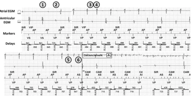
Initially, as shown in figure 2, the atrium is paced since the rate response mode is activated
(SIR = Sensor indicated rate), with intrinsic ventricular activation occurring after a long AV
delay (mark 1, Figure 2). The third ventricular beat is paced since no intrinsic R wave was
4
after this paced beat, a retrograde atrial conduction occurs, detected by the device but not
taken into account, since it occurs in the PVARP. An AV delay is not triggered after this
atrial event and the device continues to inadvertently and ineffectively pace the atrium. After the third “VP” with long AV delay (= programmed AV delay + VIP extension), the AV delay
returns to its original programmed value (i.e. 150 ms, mark 3, Figure 2), still responsible for a
clear retrograde atrial conduction (mark 4, Figure 2). At this time, a “pseudo-PMT” is
triggered, the ventricular pacing favoring retrograde VA conduction, which constantly occurs
during the PVARP, and the device continues to inadvertently and ineffectively pace the
atrium (since the atrial pacing spike occurs during the absolute refractory period of the
atrium) which perpetuate the phenomenon. Progressively, the atrial pacing rate slowly
decreases (up to 707 ms), and at one given time (mark 5, figure 2), the atrial pacing spike
captures the atria, probably during the vulnerable window, which consequently triggers AF (mark 6, Figure 2), activating the device mode switching (“CAM”).
In our case, we describe for the first time the occurrence of episodes of AF initiated by the
VIP algorithm activation. Long AV delay favored retrograde VA conduction, initially
occurring during the PVARP, and not triggering an AV delay. Consequently, the device
continued to inadvertently pace the atrium without capturing it, the atrium being in absolute
refractory period. Once the pacing spike occurred in the vulnerable window of the refractory
period and captured the atrium, an episode of AF occurred. In our patient, reprogramming a
shorter VIP extension was sufficient to avoid retrograde VA conduction, while maintaining
spontaneous ventricular activation without significant increase in ventricular pacing burden.
One may assume that shortening the PVARP could have avoided AF episodes, since the
phenomenon would have generated a PMT, detected and stopped by the device after the ninth “AS-VP” at maximal tracking rate.
To note, if one looks closely at the ventricular EGMs during the “pseudo-PMT”, there is
variation in the “Vp” complexes morphology: VP#4 and Vp#12 and Vp#13 (first two
complexes of the bottom strip of Figure 1) appear to be fully paced while Vp#5-11 appear to
be different and may represent varying levels of fusion. Therefore, it cannot be excluded that
this may represent a form of atrio-ventricular nodal reentrant tachycardia (AVNRT, slow-fast
or slow-intermediate) that would have persisted even in the absence of ventricular pacing.
However, spontaneous AVNRT had never been observed in this patient, and this hypothesis,
although theoretically possible, is unlikely.
In summary, we describe here a new pro-arrhythmic effect of the VIP algorithm, generating
AF in a patient with no history of atrial arrhythmias. Clinicians should be aware of such
potential events, since the reprogramming of the device may prevent new episodes and avoid
the initiation of otherwise unnecessary anticoagulation.
References
1. Tops LF1, Schalij MJ, Bax JJ. The effects of right ventricular apical pacing on ventricular function and dyssynchrony implications for therapy. J Am Coll Cardiol. 2009;54:764-76.
2. Frisch DR1, Kenia AS, Walinsky P, Balog J. Managed ventricular pacing facilitating atrioventricular nodal reentrant tachycardia. Pacing Clin Electrophysiol. 2014;37:1568-71
3. Vavasis C1, Slotwiner DJ, Goldner BG, Cheung JW. Frequent recurrent polymorphic ventricular tachycardia during sleep due to managed ventricular pacing. Pacing Clin Electrophysiol. 2010;33:641-4.
4. Mansour F1, Khairy P. Electrical storm due to managed ventricular pacing. Heart Rhythm. 2012;9:842-3.
5.Barold SS1, Stroobandt RX. Pacemaker-mediated tachycardia initiated by an atrioventricular search algorithm to minimize right ventricular pacing. J Electrocardiol. 2012;45:336-9.
6
Figure legends:
Figure 1: Initiation of an AF episode. From top to bottom: atrial EGM, ventricular EGM,
markers and delays (AA, VV, and AV delays, respectively).
Ap: Atrial pacing; As: Atrial sensing; CAM: Automatic mode switching; EGM:
Electrograms; SIR: Sensor indicated rate; VIP: ventricular intrinsic preference; Vp: