Automation Of Single-Cell Techniques
In Neural Tissue
by
Joseph D. Steinmeyer
B.S. University of Michigan (2008) S.M Massachusetts Institute of Technology (2010)Submitted to the Department of Electrical Engineering and Computer in partial fulfillment of the requirements for the degree of
MASSACHUSETTS INSTfrE OF TECHNOLOGY
JUN
10
2014
LIBRARIES
Science Doctor of Philosophyat the
MASSACHUSETTS INSTITUTE OF TECHNOLOGY
June 2014
@Massachusetts Institute of Technology, 2014. All rights reserved.
Author ....
Signature redacted
...
...
.
. . . .
Department of Electrical gineeri'ng and Computer Science May 12, 2014
Signature redacted
Certified by ...
Accepted by ...
Meh met Fatih Yanik Assistant Professor of Electrical Engineering Thesis Supervisor
Signature
redacted
/
/Q
J
Leslie A. Kolodziejski Prof sor of Electrical Engineering Chair of the Committee on Graduate StudiesAutomation Of Single-Cell Techniques In Neural Tissue
byJoseph D. Steinmeyer
Submitted to the Department of Electrical Engineering and Computer Science on May 12, 2014, in partial fulfillment of the requirements for the degree of
Doctor of Philosophy
Abstract
The highly heterogeneous nature of cells in the context of native tissue environ-ments necessitates the development of tools and techniques that can manipulate and analyze samples with single-cell resolution. While the past decades have seen significant progress in analyzing individual cells in tissue, both electrically and morphologically, the ability to genetically manipulate and biochemically analyze such cells in a high-throughput manner has seen only limited advances, and therefore a significant technological gap in accessing cells with single-cell specificity in tissue remains. We present a system design and workflow that fills in this gal) in technology through the implementation of precision automation and redesign of standard biological techniques, resulting in greatly improved throughput while maintaining single-cell accuracy and precision. This thesis comprises three parts: First we discuss the design and implementation of an expandable
comlputer-cont
rolled automation system enabling the rapid maneu-vering and targeting of inicropipettes within tissue environments as well as a methodology for cleaning and reuse of these lnicropipettes to enable significant gains in throughput. Second we apply this automation to transfecting neu-rons in brain slices with DNA and RNA for subsequent analysis with greater throughput than previous methods. Third, we apply our automation to col-lecting the contents of single neurons embedded in relevant tissue environments for molecular analysis. The work presented greatly improves the throughput of traditional single-cell methods of transfection and cell-sampling by between one and two orders of magnitude and fills in a gap in the workflow of the rapidly expanding field of single-cell analysis.Thesis Supervisor: Mehmet Fatih Yanik
Acknowledgements
The past years of work would not have been possible without the support of family, friends, and colleagues. I would like to thank my Ph.D. supervisor Dr. Mehmet Fatih Yanik for his support throughout my graduate studies here at MIT. I'm also grateful to Dr. Dennis Freeman and Dr. Jongyoon Han for serv-ing on my thesis committee. My many lab mates have been amazserv-ing in help-ing me learn and carry out research includhelp-ing Matthew Angel, Chris Rohde, Mark Scott, Thomas Dieffenbach, Chrysanthi Samara, Paul Tilberg, Zachary Wissner-Gross, Cody Gilleland, Jaqueline Wolfrum, Steve Sheridan, Yao Zhou, Alexei Finski, Mohammed Azimi, Peter Eimon, and others. Others at MIT who've helped me in countless ways include Professor Joel Voldman, Profes-sor Leslie Kaelbling, ProfesProfes-sor Cardinal Warde, ProfesProfes-sor Marc Baldo, Adam Hartz, Shawna Young, Julian Greene, and the entire OEOP community, Ron Roscoe, Janice Balzer, Al McGurl, Lourenco Peres, John Sweeney, Cheryl Ch-eney. Finally for my family, including Libby Steinneyer and my parents Mark and Mary Ann Steinmeyer, as well as Rachel Greenhaus, Carol Kaye, and the Chesapeake and Ohio Railway who've been the core of my support, I'm very much grateful.
Not that it was beautiful, but that, in the end, there was a certain sense of order there; something worth learning... -Anne Sexton
Contents
1 The Study of Single Cells: Background and Motivation 11
1.1 Single Cells in History . .. . . . .. . .. . .. . . .. . . .. 13
1.2 Manipulation and Analysis of Single Cells in Biology . . . . 13
1.2.1 Cell Lines as a Means of Biochemical Amplification . . . 14
1.2.2 Single Cells In Tissue Environments . . . . 17
1.3 Single-Cell Manipulation in Tissue Environments . . . . 19
1.4 Single-Cell Analysis in Tissue Environments . . . . 24
1.5 Remaining Need and Overview of Work . . . . 27
1.5.1 Proposed Solution . . . . 28
2 Rapid and Scalable Automation for Glass Micropipettes 30 2.1 Glass 1\licropipettes: Background . . . . 31
2.2 Front-Loading of Reagents . . . . 32
2.2.1 Limited Diffusion of Large Molecules . . . . 33
2.2.2 Implications for Use and Reuse . . . . 33
2.3 Cleaning and Rinsing of Micropipettes . . . . 34
2.3.1 Sterilization . . . . 35
2.3.2 Rinsing MIcropipette Tips . . . . 35
2.4 System Automation . . . . 35
2.4.1 System Characterization . . . . 37
2.4.2 Human-Control and Software . . . . 38
2.5 Expanding Automation for Single-Cell Sampling . . . . 40
2.5.1 Well Format Modifications . . . . 41
2.6 D iscussion . . . . 41
3 Automating Single-Cell Transfection in Tissue Environments 45 3.1 Single-Cell Electroporation: Background . . . . 45
3.1.1 Limitations with Single-Cell . . . .
3.2 Single-Cell Electroporation using Front-Loaded Micropipettes . . 3.2.1 Repeated SCE of Multiple Vectors . . . .
3.2.2 System Throughput . . . . 3.2.3 Softw are . . . . 3.2.4 Long-Term Operation . . . .
3.3 Multicolor Combinatorial Labeling of Single Cells . . . . 3.3.1 Combinatorial Genetic Modification of Single Cells within
B rain Slices . . . . 3.4 D iscussion . . . .
4 Automation of Single-Cell Sampling
4.1 Sample Deposition . . . .
4.2 Sample Collection from Single Cells . . . .
4.2.1 SDS Injection . . . . 4.3 Micropipette Tip Geometry Considerations . . . .
4.4 Cell Sampling . . . . 4.4.1 Sampling Timing . . . . 4.5 Benefits and Concerns with High Concentration SDS . . . . 4.6 Recovery Rate Analysis . . . .
4.6.1 Quantitative Analysis . . . . 4.7 Cross-Contamination Analysis . . . . 4.8 D iscussion . . . . 5 Continuing Development
5.1 Further RNA Sampling Analysis . . . . 5.2 Expandability and Merging Other Techniques . . 5.2.1 Protein Analysis at the Single Cell Level . 5.2.2 Microfluidic Adaptation . . . . 5.3 Summary of Work . . . .
Appendices
A System Hardware and Automation Methods A.1 Automating Interfacing with Micropipettes . . .
A.2 Diffusion Measurements and Simulations . . . .
A.3 System Images . . . .
49 49 49 51 52 52 54 57 61 63 66 67 68 69 69 74 75 79 80 80 83 87 87 88 88 89 89 91 92 92 95 97
B General Analytics for Experiments 101
B .1 Im aging . . . 101
B.1.1 Image Handling ... 102
B.1.2 Immunohistochemistry ... 102
B.1.3 Dendritic Spine Density Analysis Statistics . . . 102
B.1.4 Gel Analysis for Quantification . . . 103
B.2 Tissue Harvest and Maintenance . . . 103
B.3 DNA and RNA Preparation . . . 104
B.3.1 Manufacture of synthetic mRNA . . . 105
B.3.2 SCE of mRNA . . . 105
B.3.3 Bulk Transfection of Tissue Slices with mRNA . . . 105
B.4 Electrical Characterization of Micropipettes . . . 106
B.5 Detergents for Release of Material . . . 107
B.6 Additional SCE Information . . . 109
B.7 Cell-type Variability in SCE Efficiency . . . 111
B.8 Additional SCS Analytical Information . . . 113
B.8.1 Semi-Quantitative Analysis of Recovered Material . . . . 113
B.8.2 RT-PCR Analysis . . . 115
B.8.3 Cell Sampling Real-Time Analysis . . . 117
13.9 O il D eposition . . . 117
B.10 Solutions and Formulas . . . 122
List of Figures
1.1 Problems with Cell Population Averaging . . . . 17
1.2 Further Problems with Cell Population Averaging . . . . 18
2.1 Limited Tip Diffusion . . . . 34
2.2 Basic Automated Manipulator System . . . . 36
2.3 Drift and Repeatability of Single Micropipette Due to MP-285 and Long-Travel Stage . . . . 39
2.4 Dual Pipette Automation Version 1 . . . . 40
2.5 Dual Pipette Automation Version 2 . . . . 42
2.6 Two-Point Drift . . . . 43
3.1 Electroporation-mediated Uptake of Molecules . . . . 47
3.2 Electroporation Styles . . . . 48
3.3 Limited Tip Diffusion . . . . 50
3.4 SCE with No Cross-Contamination . . . . 51
3.5 Transfection Cycle Timing: Rapid Transformation of Single Cells 52 3.6 Graphical User Interface (GUI) for Control . . . . 53
3.7 Quadruple Labeling . . . . 55
3.8 Quadruple Labeling . . . . 55
3.9 Triple-Transfection Efficiency . . . . 56
3.10 Initial Co-Transfection Results . . . . 58
3.11 Comparative Analysis of Kalirin Results . . . . 60
4.1 Deposition Into Water Verification . . . . 64
4.2 Deposition Into Water Verification . . . . 67
4.3 Micropipette Geometries . . . . 70
4.4 Dual Micropipettes for Single Cell Sampling . . . . 70
4.6 Single-Cell Sampling Control Software . . . . 72
4.7 Cell Sam pling . . . . 73
4.8 Quantified Cell Sampling . . . . 74
4.9 Averaged Quantified Cell Sampling . . . . 75
4.10 Effect of SDS on Ribonuclease Inhibition . . . . 77
4.11 SD S Sensitivity . . . . 78
4.12 Pre-Bleach Resolved . . . . 81
4.13 Pre-Bleach Resolved . . . . 82
4.14 Cross Contamination Analysis . . . . 84
A.1 Primary Control GUI (Detailed) . . . . 94
A.2 Flowchart of SCE System Operation . . . . 95
A.3 Calibration Curve for Fluorescence Intensity vs. Concentration . 96 A.4 SCS System Image I . . . . 97
A.5 SCS System Image 2 . . . . 98
A.6 SDS Sample Heater . . . . 99
A.7 Dual Pipette Automation Version 2 . . . 100
B.1 Electroporation-mediated Uptake of Molecules . . . 106
B.2 Effect of Non-ionic Detergents on RT-PCR Efficiency at Small V olum es . . . 108
B.3 Distribution of Fluorescence Emission Strength in Electroporated C ells . . . 110
B.4 Different Fluorophores Do Not Affect Measured Dendritic Spine D ensity Count . . . 110
B.5 Transfection of Single Cells in Acute Slices with Fluorescent Dyes 111 B.6 Sequential Transfection of Cells with Plasmids . . . 112
B.7 Housekeeping Genes Primer Curves . . . 113
B.8 PCR Standard Curve for P-Actin mRNA . . . 114
B.9 EGFP and niCherry Selective Primer Verification . . . 117
B.10 Multiple Quantified Cell Sampling . . . 119
B.1 1 Protrusion Outgrowth . . . 119
B.12 Sample Droplet in Oil . . . 120
List of Tables
1.1 Single Cell Manipulative Techniques in Tissue . . . 1.2 Single Cell Analytical Techniques in Tissue . . . .
4.1 Cross Contamination Analysis of Cell Samples . .
Single-Cell Electroporation Efficiencies . . . . Efficiency of SCE of Plasmids for Several Neuronal Primer Sequences for Initial Sample Verification . Primer Sequences for Initial Verification . . . . First-Strand Synthesis Recipe . . . . Non-Nested Single-Step PCR Formula . . . . Nested PCR Formula (Step 1) . . . . Nested PCR Formula (Step 2) . . . . Standard Cell Lysis Buffer . . . . Organotypic Slice Culture Formula . . . . Rat Ringer's Solution . . . .
. . . 109 Subtypes . .
111
. . . 116 . . . 118 . . . 122 . . . 122 . . . 123 . . . 123 . . . 124 . . . 124 . . . 124 20 25 85 B.1 B.2 B.3 B.4 B.5 B.6 B.7 B.8 B.9 B.10 13.11Chapter 1
The Study of Single Cells:
Background and Motivation
A significant trend in recent years in the many the sub-fields of the life sciences has been the focus on the biology of individual cells as actually analyzed at the single-cell levell. Most of what is considered as "common knowledge" in regards to cell biology today, was not generated by analyzing individual cells directly, but rather by analyzing populations of cells and extrapolating downwards to the single-cell level[l]. This is not to say knowledge gained so far in biology is a waste or without benefit; such a viewpoint couldn't be further from the truth in
fact. Throughout the twentieth century, there have been many questions that haven't required the ability to analyze individual cells in order to find answers. However as the life sciences have advanced, new questions have appeared and many of these involve variability at the actual level of single cells. In the case of tumor biology and neuroscience, for example, cell-to-cell variability is at the core of new phenomena just being discovered so it is no longer sufficient to extrapolate information about individual cells from many; direct access to the individual cell is now needed.
This shift in focus has been catalyzed by recent improvements in manip-ulative and analytic techniques. Historically, the study of cells, regardless of environment or type, has suffered from one major disconnect: the mismatch between the scale of cell parameters, and the scale at which researchers can manipulate and analyze parameters using instruments. This mismatch has at
various points encompassed all physical phenomena, including mechanical, elec-trical, and chemical, (and continues to do so in subsets of these parameters), and improvements in minimizing or eliminating the difference between what should be measured and what can be measured has been the core of many, if not all, biotechnological advances. While certain parameters of cells can now readily be manipulated and analyzed at sufficiently low-enough levels, there are still many areas where this measurement/parameter mismatch continues to re-quire researchers to either make assumptions regarding phenomena, or avoid the pursuit of particular lines of inquiry until the mismatch can be overcome.
One of these mismatches recently overcome is single-cell DNA and RNA sequencing [2]. In the past few years, there's been an explosion in the amount of research on sequencing the DNA and RNA transcripts of individual cells. In particular, the ability to analyze copy numbers of low-expression level mRNA species from single cells, something achievable with any regularity and ease only in the past few years, has revealed the potentially questionable nature of previously held assumptions in certain areas of cell biology as well as revealed completely new insights into a far more complex set of heterogeneous cell states than previously thought to exist[3]. The importance importance and promise of these advances are such that the prominent life science journal Nature named single-cell sequencing as the "Method of the Year: 2013" [4].
Single-cell transcript analysis needs to be framed in the context of it, being as yet one more improvement in a general trend towards complete direct anal-ysis of cells, a gradual trend that has been going on for centuries, and which has advanced rapidly in the last several decades. From a systems engineer-ing perspective, many of the advances in molecular and cellular biology from polymerase chain reaction (PCR), to patch clamping, to microscopy, have been matters of achieving amplification of sufficiently high fidelity to enable manip-ulation and study of cellular signals. This introduction chapter covers a brief history of single-cell analysis from this engineering perspective, from the ear-liest work with cells up to the state-of-the-art as of 2014. We then look at a few of the outstanding issues that remain, and show how the bulk of the work in this thesis, specifically the automated platform for single-cell manipulation and analysis that we developed, can be used to fill in some of these technolog-ical gaps and enable previously unachievable capabilities in certain analyttechnolog-ical dimensions.
1.1
Single Cells in History
For centuries, the study of life was restricted only to what was able to seen, and more generally sensed, by the unaided human. The modern concept of all life being comprised of cells is a relatively recent idea, being traced back to the discovery of cells in the eighteenth century. The first attempts at single-cell anal-ysis coincided directly with the initial observations of cells by Robert Hooke and Antonie van Leeuwehoek using crude microscopes of their own construction [5]. In one letter to Robert Hooke, (who made the first recorded observations of cells in cork and who is widely credited with coining the term "cell") Leeuwen-hoek emphasizes the marked difference between different cells and what could be considered among the first single cell analysis[6].
Examining this water.. .I found floating therein divers earthy par-ticles, and some green streaks, spirally wound serpent-wise... and I judge that some of these little creatures were above a thousand times smaller than the smallest ones I have ever yet seen, upon the rind of cheese, in wheaten flour, mould, and the like.
What is commonly held to be the birth of cell doctrine in Theodor Schwann's publication of his 1838 work Microscopic Investigations on the Accordance in the Structurc and Growth of Plants and Animals, stressed both the variability of cell types in organisms and various settings as well as unifying principles that exist across all cell types[7]. Cell-to-cell variability was therefore recognized early on, and as work was carried out in the following centuries on higher-level organisms, more specializations of cell types were found and classified. Researchers discovered that, there was no general type of "tree cell", for example, but rather mixes of many different cell types within individual organisms even within specific organs and tissues. The realization of cell heterogeneity was especially important in the development of of modern neuroscience at the end of the nineteenth century where single-cell optical analysis was integral in Ramon
Santiago Cajal's development of neural doctrine[8].
1.2
Manipulation and Analysis of Single Cells in
Biology
The ability to carry out experimental analysis on single cells requires two major capabilities: the ability to manipulate and the ability to analyze with cellular or
sub-cellular levels of precision and accuracy. The primary difficulty in analyzing the input/output characteristics of a cell is the mismatch of scales between the parameters of a single cell and those capable of being measured and manipulated by researchers. For example:
" A standard mammalian cell has dimensions of maybe 10 pm, far smaller than can be viewed by the naked eye, and therefore a microscope is needed to close the gap.
* The electrical properties of a cell are on the order of millivolts and exist spatially over distances as small as several nanometers2. Mechanical de-vices with fine precision and accuracy as well as high-fidelity electronics are therefore needed to manipulate and analyze these properties.
" The quantity of biomolecules generated by a single cell are far below what can be detected by an unaided human and therefore require amplification techniques. These techniques include enzymatic methods, chain reactions, etc...
This thesis focuses primarily on rapid, targeted single-cell perturbation and analysis of biomolecular parameters (gene expression primarily), and will there-fore focus only on that subfield of scaling for single-cell analysis. Optical tech-niques and electrical techtech-niques as applied to single cells comprise fascinating fields of research in their own right, and for the readers that are interested, there are a number of good reviews on the state of the art can serve as a start-ing point[9, 10].
1.2.1
Cell Lines as a Means of Biochemical Amplification
The quantity of biological material produced by a single cell is extremely small. For proteins, total amount of material in a cell may be as low or lower than 50 pg, with individual polypeptide species making up tiny fractions of that number [11, 12]. In the case of RNA, total quantity in a mammalian cell is estimated to be approximately 10 pg, of which only several percent is mRNA [13, 14]. In the case of DNA, the total amount of most sequences in a cell will be two. For most of the history of biology, these amounts of starting material were far below what was capable of being analyzed.
2
The relatively simple solution to this problem was to avoid work on a single instance of a cell and instead the use many identical cells at the same time in order to be able to respond to and produce signals at then obtainable levels of precision and accuracy. This has been the approach for working with most prokaryotic cell types for the last century, where the fundamental unit of analysis in terms of experimental readouts became the clonal population rather than the individual cell. This approach has much justification, since bacteria have amazingly low error rates in replication and transcription, being on the order of ow as 1 x 10-li errors per base pair copied, and as far as cell types go daughter cells of an E. coli division are about as close to copies as can be achieved[15]. From a systems engineering point of view, the bacteria population
themselves
act as high-fidelity amplifiers, and utilization of them in such a way proved valuable in early biological work. Consequently, much cell biology, and much of what we know of cells is extrapolated backwards from population-based experiments.Cell-line amplification has also been applied to higher order cells including many mammalian cell lines. The first expandable human cell line by George Otto Gey in 1951 ( the famous HeLa cell) was followed up with numerous other well-established lines, and all together these cell lines have provided what have been viewed as scaled versions of single mammalian cells. By culturing a flask of HeLa Cells, a researcher could obtain ig to mug of molecular species, and the middle decades of the twentieth century saw the development of manipulative and analytical techniques that could work at those levels.
lost,
if not all modern molecular biology analytical techniques, including Northern, Southern, Western, (and Eastern) blots, Polymerase Chain Reaction (PCR), as well as many "bulk" transfection techniques such as electroporation, lipofection, and viral transfection were all developed in the context of cell line cultures and their associated amounts of material.(In)Homogenous Cell Populations
In spite of its benefits, there were/are several key limitations to cell line culture. First is that a cell culture is not a collection of exactly identical cells. Even in the case of extremely low mutation rates in E. coli replication, mutations are inevitable (and in fact necessary for many techniques), so it is impossible to assume that all cells will share identical genomes. Compound this with epigenetic variation heavily present in eukaryotic cells, variations in cell cycle
at any given point in time, as well as the now well-documented stochasticity of mRNA and protein levels in cells, and it becomes nearly impossible to claim that any two cells in a cell line culture are "identical" at any given point in time [16, 17, 18, 19]. Researchers have even come to realize recently that within so-called "homogenous" cell populations, the variability in cell signals and states between two "identical" cells can vary more than that between two averaged populations exposed to different stimuli [20, 21].
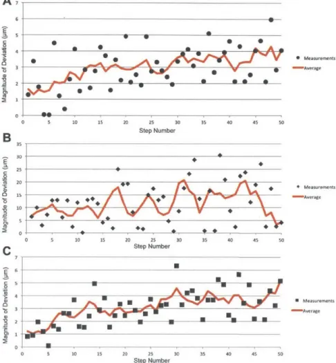
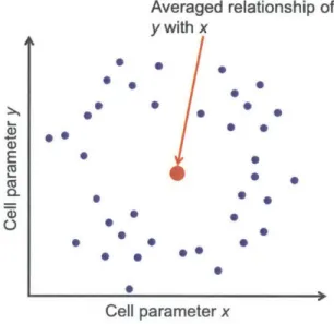
Cell cultures and cell lines therefore need to be viewed as biochemical ampli-fiers with potentially lower fidelity than previously held. This shouldn't suggest that they are useless as a research tool. If the amplitude of the signal be-ing measured exceeds any cell-to-cell variability or "noise" within a cell-culture amplifier, then such a platform is sufficient, but as the amplitude of the signal approaches the cell-to-cell variability for a response, the validity of any readouts generated should be called into question. If a cell culture contains a vast distri-bution of cell parameters of an unknown nature, in analyzing the population, these trends will be lost. This resulting "averaging effect" can have significant implications, masking trends found both within a population or between popu-lations. Two key issues with the averaging effect are highlighted in Figures 1.1 and 1.2. Many of these issues have in fact been confirmed in cell populations as single-cell analysis techniques have come into popular usage in recent years [20, 21, 4].
(In)Validity of Cell Cultures
Cell culture-based biology and the utilization of cell lines as models has undoubt-edly yielded important experimental results for biology and helped to develop knowledge about cellular processes, however in addition to intra-culture vari-ability, there is also a significant concern about the overall validity of cell lines and in vitro culture in general as tools for answering biological questions. HeLa Cells are far from a *"typical" cell found in the human body 3. Even the culture of primary cells, cells extracted directly from tissue and then cultured in vitro, exhibit vast differences in many parameters. While true for all cell types, this is particularly the case in neurons. A neuron in its native tissue environment in the central or peripheral nervous system will be surrounded by astrocytes and microgila as well as various classes of other neurons and, if in a vertebrate, sur-rounded by myelinating oligodendrocytes (or Schwann Cells if in the peripheral
3
E O) 0 - -average of~ 0" 0 average of y 0
Cell samples Cell samples
Figure 1.1: Two problems resulting from cell population averaging are demonstrated here. On the left, a collection of cells and their measured value of parameter y are plotted along with their average. At its most basic level, averaging of cells will obscure the distribution. In a situation where the distribution is Gaussian or somethign similar, this won't be a problem necessarily, however it can become a particular issue as shown in the right plot. Here the average obtained from bulk analysis corresponds to a value which no individual cell itself exhbits. This can lead to innacurate assessment of response and analysis.
nervous system) [22, 8]. All of these cells project onto the neuron, maintaining or modulating all aspects of its environment. When removed from this envi-ronmuent, the neuron should be expected to act and respond differently which is problematic in dissecting how native neurons function[23].
1.2.2
Single Cells In Tissue Environments
The goal of biology is the study of life, and therefore the model systems in which work is carried out should be as close as possible to reality while still allowing for the ability to manipulate and analyze the system. Cell culture in vitro and established cell lines, obviously make sacrifices in this regard, being significantly deviated from real conditions, but they can still have their use, the validity of which is based upon questions. For example, it is perfectly reasonable to study the question of cellular response to changes in osinolarity in a cell culture platform since one would expect that many general principles will still be present assuming the cells are healthy. It is less relevant to study native neural networks of the brain in dissociated primary neurons; the latter will not model the former very well. In other areas of biology, the behavior of individual tumor cells within a cultured piece of tissue have been found to behave far differently than tumor cells cultured in vitro, calling into question of
E
L)
M
Averaged relationship of
y with x
*
0,
0
.
0
E
0
0 Ce . 00
0
0 )
0 0Cell parameter x
Figure 1.2: In the event of merging a cell population for analysis, the creation of "false states" can occur. Here, the distribution of two cellular parameters (mRNA, protein level expression, etc...) are plotted in a two-dimensional plane. It is obvious to the reader that a circular-distribution exists in the x - y plane, however to a researcher
analyzing this collection of cells only at the population level, this pattern will be obscured, resulting not only in a loss of distribution (similar to that shown in the right plot of Figure 1.1), but also in the creation of a non-existent pairing between x and y values.
study and screening of treatments using the latter format[24].
There is consequently a lot of motivation to work with cells when when in their native tissue environments, either in vivo or in slice culture format. In working in these environments, however, the researcher can no longer rely upon high (cell-culture amount) levels of starting material for experiments. Instead, due to the heterogeneity of cells in tissue environments, the biochemical manip-ulation and analysis of cellular state requires direct access to the biochemical state of the individual cells. To varying degrees methods and technologies have been developed in the last two decades towards this end and a brief overview of the benefits and shortcomings of the major groups of single-cell biochemical manipulation and analysis techniques for tissue environments are detailed in the Sections 1.3 and 1.4.
1.3
Single-Cell Manipulation in Tissue
Environ-ments
Table 1.1 details available tissue-conpatile techniques for obtaining some de-gree of single-cell biochemical modulation. The limitations are discussed in sections below.
Transgenics
One common means of achieving manipulation with single-cell resolution is through the use of transgenic lines. 1Many incarnations of this technique have been developed, with the vast majority being primarily in mouse until relatively recently[25]. An extreme implementation of single-cell manipulation in tissue is the so-called "brainbow" series of research carried out by The Carp Lab and associates at Harvard University beginning in 2006, where cells in nervous tis-sue were combinatorially labeled with a variety of fluorescent species enabling previously unachieved densities of cell imaging and analysis [26, 27]. Transgenic lines can be used to target expression to specific cell types, for example confin-ing the labelconfin-ing or expression of transcripts to subclasses of cells usconfin-ing cell-type specific promoters. There are several major limitations in tra nsgenic targeting from the point of view of many research studies. First, transgenics are a before-the-fact method, where the targeting is implemented during development and the production and crossing of specific specimen lines. Second, the inherent nature of transgenic technology precludes its use in higher-level tissue work, for
Technique Single-Cell Precision Single-Cell Accuracy Throughput Wild-Type Tissue Transgenics Yes No High No Viral Transfection Yes No High Yes Biolistic Transfection Yes No High Yes Bulk Electroporation Yes No High Yes Single-Cell Microinjection Yes Yes Low Yes Single-Cell Electroporation Yes Yes Low Yes Optoporation No Yes Limited Yes Table 1.1: Single-cell precision refers the ability to effectively target individual cells when surrounded by many cells, but not the ability to taret a specific cell. Single-cell accuracy refers to the ability to target a specific cell. In general most techniques can be tuned to yield single-cell precision, often times through sparse, stochastic labeling in the case of bulk techniques, however only manual techniques provide full-function single-cell accuracy.
example in human tissue. Researchers have succeeded in culturing post-mortem brain tissue and recent research has found evidence of unique structures or cell subclasses found only in the brains of higher order mammals, including humans [28, 29, 30]. Research published in the last few years on many topics of neu-ral development lend support to seveneu-ral unique features, and if research is to be carried out on single cells in relevant human tissue culture environments, whether post-mortem neural tissue or culture tumor tissue, a technique should need to be able deal with wild-type tissue, ruling out transgenics[31, 32].
Biolistic and other Bulk Transfection Techniques
While transgenics cannot be used to target a given cell in wild-type tissue other techniques can do so4. Biolistic transfection is one such method that allows the transfection of individual cells in a tissue environment[33, 34]. In biolis-tic transfection a device, often termed a "gene gun," fires small gold parbiolis-ticles coated with the reagent to be transfected at the tissue sample. Particles and the material they carry then become lodged in the cells and will subsequently be processed or incorporated. Depending on the settings, very sparse to very dense can be achieved. Which cells get transfected is left up to chance however since the gene gun provides only the most basic targeting capabilities, possibly providing slight regional accuracy and precision.
Biolistic transfection can be termed a "bulk" transfection methodology in that it is applied to the greater majority of a given tissue and relies upon pa-rameters and probability to determine how many and which specific cells get transfected. Several other bulk transfection methods exist including bulk tissue electroporation and bulk lipofection[35, 36]. While relying on different physio-logical properties for operation, both techniques provide means of transfecting pre-defined densities of cells while lacking cell precision and accuracy. Viral Transfection is another possibility for transfecting cells within tissue. In the case of organotypic culture the protocol for transfection is a simple as placing a viral solution above and allowing to diffuse into volume of the tissue. By varying the concentration of virus applied differing densities of transfection can be achieved, however which specific cells get transfected is still largely up to chance. This can be a major limitation in studying a neural circuit for example, where the researcher would want to target a specific neuron that has already
4
Several of these techniques are actually how you generate transgenic lines in the first place, using bulk electroporation in utero
been identified via another method viral for neural tracing [37, 38]. Some degree of cell selection can be implemented with these bulk techniques by using vectors that possess cell-type selective expression controls. These can yield more com-plex patterns, where expression of a vector is the product of both semi-random transfection (from the bulk method) and cell-type-specific expression (from the vector) [391. This does not overcome all limitations, however. Labeling cells based off of presumed sub-type categories is inherently limited by knowledge of those subtypes. For years, attempts have been made to subdivide cell types based off of presence/absence of molecular markers, morphological data, electro-physiological responses, and location[40]. Consequently there are experimental situations where the capability to arbitrarily transfect single cells "on the fly" would be desirable.
Single-Cell Microinjection
The techniques described so far can be relatively high-throughput, allowing many cells to be transfected at once, but which exact cells get transfected is still largely up to chance. There also exist a series of techniques that provide single-cell precision and accuracy. Unfortunately these techniques are also of-ten of a much lower throughput than the bulk techniques above. Single-cell microinjection is one such technique where a glass micropipette with tip dimen-sions usually < 0.1 im is loaded with a reagent and then used to pierce a cell of interest and introduice the reagent[41, 42, 43]. While this technique is often used in monolayer culture, it does have the drawback of easily getting clogged in tissue environments due to the extracellular matrix and small size of the tip opening. Additionally, the mechanical puncturing of the cell can be violent to the cell resulting in damage or death[41].
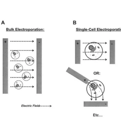
Single-Cell Electroporation
Single-Cell Electroporation (SCE) is a variant of standard electroporation where the electroporating field is only applied in the vicinity of single cell [44, 45]. In most modern (last decade) implementations, a micropipette is used as a hy-brid electrode and delivery device [46, 47]. Single-cell electroporation (SCE) has emerged as a versatile means for transfecting cells due to its potential for high efficiency[48, 49], its ability to transfect a variety of agents includ-ing dyes[50], plasinids[51], and RNAi reagents[52], and its tissue and in vivo compatibility[53, 51, 54]. The fundamental operation of the technique relies
upon loading a micropipette with a transfection reagent, which may contain a mixture of multiple agents such as plasmids, in an ionic solution, and then positioning the tip opening on or near the cell of interest before applying an electrical signal to electroporate the membrane of the targeted cell. The mi-cropipette therefore serves as both a highly-focused electrode and a sample delivery device.
Optoporation/Optical-Mediated Expression
Optoporation is another method that can achieve single-cell specificity in tissue environments approach[55]. Optoporation is similar to single-cell electropo-ration in many ways relying on targeted environmental change to induce the formation of pores in a cell membrane. Modern optics are more than capa-ble of achieving sub-cellular resolution, and multi-photon systems can provide 3-dimensional accuracy and precision [56]. While SCE utilizes an electrode to deliver its reagents and electric field, optoporation needs only to introduce light to the defined region in order to porate the target cell, minimizing the potential for damage. However, reagents to be transfected still need to be introduced to the region, and in a tissue culture environment, this is very difficult to do without directing deposition using a micropipette. In a cellular monolayer environment, the entire culture solution containing the reagents could be used, using light as the selection mechanism, however this
can
waste large amounts of expensive reagents and can be difficult to implement in tissue due to limited diffusion of large macromolecules through the intercellular space and because of the rate of degradation, particular of RNA in extracellular solution (See Fig. 4.10)[57]. It use in complex tissue environments is therefore limited when compared to other single cell techniques.Conclusion of Techniques
It should be apparent that there are two general classes of transfection tech-niques for cells in tissue environments: So-called "bulk" techtech-niques that are high throughput compatible but lack single-cell accuracy or precision, and a set of techniques that can indeed target single cells with high precision, but do so at a low throughput. Little middle-ground exists.
1.4
Single-Cell Analysis in Tissue Environments
In addition to manipulating single cells in tissue through transfection, there is also a need to biochemically analyze those single cells. Fortunately analytical techniques have been developed with sufficient sensitivity to analyze individual cells for many biochemical parameters including mRNA, DNA, protein, and metabolite content[58]. However, there are still distinct limitations in carrying out these analyses on cells from tissue environments. The current state of the tissue-compatible single-cell sampling methods are shown in Table 1.2. While the following discussion can apply to all single-cell analytics, because of the content of this thesis, we focus primarily on RNA-based analysis.
Dissociation-based Collection of Single Cells
The simplest way in which to achieve single-cell biochemical analysis of cells from tissue is to mechanically or enzymatically dissociate the tissue and sort the cells using either Fluorescence Activated Cell Sorting (FACS) or a similar technol-ogy [59, 60, 61]. Such an approach is extremely amenable to high-throughput deployment and has been used in many recent publications in a variety of tissue environents[62, 63]. From an analytical standpoint, however, there are two major issues with this approach. The first is that dissociation of cells takes a long time and is potentially very harsh on the cells. By the time cells are extracted and sorted, they may have started to negatively respond to the dis-sociation environment/chemicals. Second, when using disdis-sociation and FACS sorting there is no current way to associate morphological information about the cell with its biochemical analysis since the cell identities are mixed and conse-quently lost during the sorting process. Loss of this information is particularly critical in cases where tissue and cell type heterogeneity is of importance. For example in studying neurons, morphological characteristics, and location, can be as critical in identifying cell subtypes as biochemical markers [40]. Losing one class of data can greatly limit experimental usefulness.
FISH FISSEQ
An alternative to breaking tissue apart and analyzing cells separately is to lock the tissue in place chemically through fixation, and analyze cells using markers and fluorescent labels. This methodology finds its roots in
immunohistochem-Technique Dissociation and FACS Sorting FISH Variants and FISSEQ Manual Micropipette Aspiration Laser-Capture Micro-dissection Optically-Targeted Techniques Single-Cell Precision Single-Cell Accuracy Context* Throughput Yes No No High Yes Yes Yes High Yes Yes Yes Low Yes Yes Yes High Yes Yes Yes Low Live Tissue Partial(dissociated) No Yes No Yes Table 1.2: Single-cell precision refers the ability to effectively sample individual cells when surrounded by many cells, but not the ability to sample a specific cell identified in tissue. Single-cell accuracy refers to the ability to sample a specific cell identified in tissue. In general most techniques can be tuned to yield single-cell precision, but most high-throughput techniques require either fixed tissue, loss of single-cell accuracy or both. Manual Micropipette aspiration can provide both single-cell accuracy and precision in live tissue but suffers from low throughput. *Context refers to the ability to match morphological information about a particular cell with its biomolecular analysis. Dissociating cells prior to RNA-seq, for example, mixes the cells and therefore lacks this capability.
ical staining and in the case of nucleic acid identification goes back to early fluorescent in situ hybridization (FISH) work from the 1980s[64]. More mod-ern implementations have built upon this approach with work in the previous decade developing into single-molecule FISH as well as Fluorescent in Situ Se-quencing (FISSEQ) and associated technologies[65, 66, 67, 68]. In this class of approaches, fixed tissue is analyzed for the presence of tags that bind to specific sequences (RNA or DNA), and using fluorescence imaging, allow the detection and even quantification of subspecies at a single-cell level. The obvious benefit of this approach is that it enables analysis while cells are still in the context of their tissue environment, and so is an improvement over dissociation and FACS in that regard. There are limitations in the spatial resolution with cur-rent implementations of FISSEQ as well as analytical limitations that are not as good as RNA-seq and similar techniques. Furthermore, this class of techniques does require tissue fixation, which has the limitations discussed in Section 1.4 above[66, 68].
Laser-capture microdissection
Laser Capture Micro-dissection (LCM) is a unique technology that provides a way to extract single cells from tissue environments and then transfer them into high-throughput single-cell analytical devices[69]. At its core LCM relies upon a high-intensity laser to cut out a particular cell from its environment and when coupled with a vibratome and rinsing/deposition tool can provide high-throughput collection of single cell contents coupled to original morphological data taken pre-extraction. LCM, however is generally carried out almost exclu-sively in pre-fixed tissue [70]. There have been more recent implementations of this technique where live cells can be removed, however to the author's knowl-edge this was done in cultured cells rather than tissue environments[71, 72]. If LCI can be adapted for use in tissue environments, it will still face the hurdle of content extraction since in live tissue, assuming off-target radiation effects can be minimized, the ability to collect material will still be dependent upon a mechanical means of isolation or which there is no clear solution.
Optical Techniques
The last several years have seen a series of optically-mediated analytical tech-niques that provide single-cell specificity in live tissue. At the forefront of this is the Transcriptoine-in vivo Analysis (TIVA) Tag methodology[73]. In this
tech-nique, tissue is globally exposed to specialized RNA tags that are universally taken up by all cells. Upon excitation with light, the tags activate and anneal to all poly-A RNAs in the cell. Targeted illumination of a single cell can therefore tag only that particular cell's poly-A mRNA. Following local tissue extraction, the presence of a biotin label on the tags allows a biotin-strepdavidin pulldown to be carried out, thus collecting the RNA content of the targeted cell. This technique potentially fills several gaps in the field by allowing a means to rapidly target a single-cell for collection using a minimally-evasive method. No fixation is used during the collection process, with only a lysing and disruption step followed by RNA-pulldown. This technique requires regional destruction of the tissue for collection so analyzing adjacent cells is out of the question, however, and it is not readily apparent how such an approach could be varied to allow the handling of multiple samples at once.
Manual Micropipette Aspiration
A simple, yet reliable means of collecting single-cell content from tissue environ-ments is via imicropipette aspiration as originally shown in a series of papers in 1992 [74, 75]. In this method, by achieving a low-leakage patch seal on a cell and then applying negative pressure, the content, or at least a portion of it, can be extracted and then deposited for downstream analysis, at which point it can be used to analyze most biomolecules including DNA, RNA, protein, and metabo-lites [76]. This technique has high spatial accuracy and precision and can also be done on live tissue without the need for fixation. However, this technique is inherently low-throughput. The cytoplasm of a cell is a gelatinous material, not easily aspirated with a micropipette and prone to clogging[77]. When col-lected into a pipette tip. it is very difficult to free it from the glass. As a result, following aspiration, the tip of the glass micropipette is broken off for collection [78, 79, 80]. Destruction of the tip obviously prohibits reuse of the micropipette for additional samplings, and a one-cell-one-micropipette paradigm is enforced resulting in low throughput, labor-intensive operation.
1.5
Remaining Need and Overview of Work
In surveying the state of the field in the previous two sections and Tables 1.1 and 1.2, it is readily apparent that two general classes of techniques exist for single-cell manipulation and single-single-cell analysis in tissue environments. On one side,
there are a collection of techniques that are high-throughput compatible, but which lack one or more features such as cellular selectivity or a the inability to work in live tissue. On the other side, there exist single-cell techniques that have sufficient specificity and targeting capabilities, but which are low-throughput and therefore incompatible for high or even medium-throughput use. Very little middle-ground in between these two groups.
1.5.1
Proposed Solution
This thesis introduces several novel techniques and workflows that attempt to fill in the gap between the sets of manipulation and analysis techniques avail-able for the single-cell researcher. By taking the principle operations of a set of traditionally manual, low-throughput single-cell techniques (single-cell electro-poration and micropipette-mediated single-cell aspiration), and redesigning to overcome features that make these two useful techniques low-throughput, sig-nificant gains in throughput can be achieved while no sacrifice is made in terms of live or wild-type tissue compatibility and single-cell precision and accuracy.
The content of this thesis is as follows:
" Chapter 2 details the initial and on-going development of a software and hardware package that enables the quick shuttling of a micropipette into and out of a solution bath containing tissue samples as well as a plate as-sembly for deposition, sampling, cleaning, and rinsing of tips. It will also discuss some the development of a cleaning procedure for nicropipettes, however details of its deployment in both a single-cell electroporation set-ting and a single-cell harvesset-ting setset-ting will be discussed in more detail in Chapters 3 and 4, respectively.
" Chapter 3 discusses in detail the integration of single-cell electroporation (SCE) into the automated micropipette platform introduced in Chapter 2. High efficiency of SCE as well as its ability to be rapidly switched to transfect different reagents demonstrates an unprecedented flexibility in manipulating individual cells.
" Chapter 4 details the on-going work to use the system for rapid single-cell sampling (SCS) in tissue environments for downstream analysis using conventional single-cell analytics. While analysis of mRNA transcripts is the major readout analyzed, we also discuss the benefits and possibility of
using the approach demonstrated to analyze proteins and other biological macromolecules.
e
Chapter 5 briefly details on-going work with integrating the platform with electrical recording and SCE and SCS and summarizes the overall contribution of the work to the greater research field.Chapter 2
Rapid and Scalable
Automation for Glass
Micropipettes
This chapter covers
the
development of a novel technique for using conventional pulled glass micropipettes to interface with single-cells in a way that allows reuse and cyclic operation. In addition we discuss the flexible collection of automation hardware and software we developed that greatly increases the throughput of micropipette
usage in transfecting and biochemically sampling cells in tissue en-vironment. This increase in speed drastically reduces cost-per-operation in bothtransfecting
cells using single-cell electroporation (SCE) and collecting cellular material using single-cell sampling (SCS), which will be discussed separately in Chapters 3 and 4, respectively.I
'The system automation and operation workflows developed in this work were carried out over the course of five years in parallel with the single-cell electroporation (SCE) and single-cell sampling (SCS) work discussed in Chapters 3 and 4, respectively. For the sake of ordering in this thesis, automation from all aspects of system development will be covered in this chapter and particular implementations will be covered in later chapters. For an overview and early details from the work on automation, please see the author's Master's thesis[81].
2.1
Glass Micropipettes: Background
Glass micropipettes have been a fundamental tool in interfacing with individ-ual cells for a large portion of the twentieth century2. Glass micropipettes are interesting in that they were among the first devices with micron-level features that could be reliably and characteristically manufactured by humans, predat-ing even microelectronic fabrication techniques by several decades. By takpredat-ing advantage of properties of glass cylinders as they are heated and pulled, macro-scopic features (inner-to-outer diameter ratio) can be reliably transformed into microscopic features. Because a glass micropipette is a confining device, isolat-ing its interior environment from the outside electrically, chemically, and too a lesser degree optically and thermally, these scaled physical parameters pro-vide a means of scaling environmental isolation that could otherwise only be implemented macroscopically. For example, placing a metallic electrode into the large end of a micropipette filled with an ionic solution yields an electrode with spatial resolution at or below one micron. Such a tool was fundamental in allowing early electrophysiological recordings of neurons[83]. Further advances in this same technique yielded patch clamping, which allowed interfacing with individual membrane-bound ion channels of cells [84, 85]. Micropipettes also allow a means of obtaining high reagent resolution both for the release or col-lection of material at the cellular level. For example, researchers can "spritz" chemicals onto specific neural synapses because of the spatial resolution afforded by nicropipettes[86]. Merging both of these techniques allows single-cell elec-troporation (SCE), the transfection of reagents into large cells with minimal off-target transfection. Alternatively, the molecular content from a single cell can be extracted andI isolated from cells nearby, and this forms the basis of the work in Chapter 4 of this thesis [74].
In surveying how glass micropipettes are utilized in the biology, it becomes readily apparent that the vast majority of uses are short-term in nature. In almost all cases the lifetime of a micropipette starts when it is pulled, and ends a short time later after it has been used for the delivery of one type or reagent, or the sampling of one specific cell. From an economic and experimental standpoint, this approach makes sense. Micropipettes easily clog and often one "use" is all that can be obtained from them using standard techniques.
2A summary of the development and refinement of glass micropipettes can be found first
few several chapters of Advanced Micropipette Techniques for Cell Physiology by Flaming and
Furthermore, the extremely low cost of glass micropipettes (< 1 USD), leaves little motivation to reuse them if one is not concerned with time.
This disposable usage of micropipettes does have a major disadvantage, how-ever, in that it requires large amounts of time to carry out a series of experi-ments. Micropipettes are delicate and care must be taken in creating and setting them up for experiments. In addition, variability in protocols for creation of mi-cropipettes often requires multiple attempts, where a researcher will set up the tip, carefully position it, and only then discover that it lacks the correct parame-ters for use, at which point another micropipette is inserted. This entire process can take tens of minutes, and is labor-intensive. Regardless, the experimental benefit of micropipettes are unmatched, providing a unique means of interfacing with cells both in vitro and in vivo for the purpose of measuring or manipu-lating the electrical, chemical, and mechanical state of a cell while minimizing disturbances to the surrounding region. Despite the age of the technology, it is tried and proven and a, the devices used to generate micropipettes are standard components of most neuroscience labs.
We initially sought to take the glass micropipette, a device currently limited to a manual, low-throughput deployment, and increase the throughput with which it, can be used while maintaining all the benefits of its functionality. The core of this work focuses on three developments:
" Front-Loading of Reagents to provide a quick means of loading and
releasing arbitrary reagents to cells and small volumes
" Cleaning and Rinsing of the Micropipette to overcome the need to
dispose between uses
" Automation of all non-user-critical steps to improve throughput and
minimize support labor for the researcher
2.2
Front-Loading of Reagents
Traditionally mnicropipettes are " rear-loaded" with the solution or reagent de-sired by the researcher. This process involves manually filling the micropipette from its non-pulled end and must be done prior to an experiment. While there are times when front-loading is used for injecting materials with limited and/or expensive supply such as proteins, we have not been able to find cases in the
literature where researchers have reused micropipettes between different front-loading cycles[42, 43].
2.2.1
Limited Diffusion of Large Molecules
We began our work by identifying the front-loading of micropipettes as a part of a solution to their manual usage. By loading a micropipette with a generic conducting solution (a basic media which is compatible with all samples), we wanted to see if small volumes could be front-loaded into the tip and then used to carry out tasks such as deposition. In support of this implementation is that in a given micropipette experiment such as single-cell electroporation (see Chapter 3), the vast majority of solution used for loading will not be used. Transfection of a single cell using SCE, for example will require on the order of tens of pL of solution, when the tip will be loaded with on the order of pL of solution, a difference of four or five orders of magnitude. Due to the high hydraulic resistance of the micropipette tip, front-loading of a micropipette was hypothesized to provide a means of volume loading with much finer resolution than possible
3
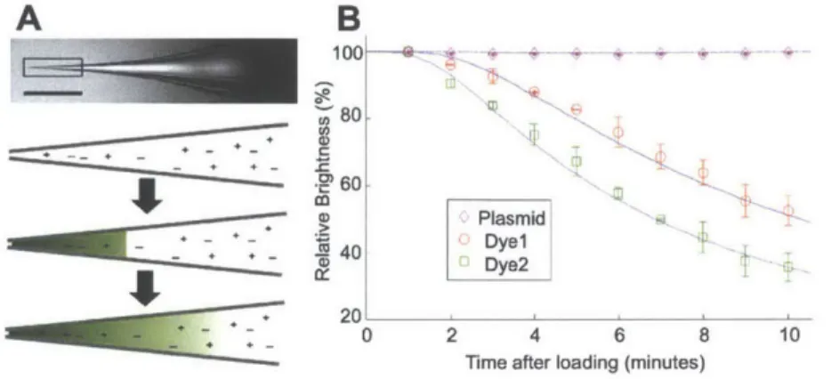
Front-loading into a micropipette already loaded with generic solution could raise the question of diffusion or loss of small front-loaded quantities into the greater bulk of the micropipette. As simulations and experiments showed, however, the rate of how fast this happens for relevant biomolecules is slow enough as to allow experiments to be carried out. Figure 2.1 shows that small molecules such as dyes with molecular weights of several hundred Daltons (Da) will relatively quickly diffuse into the greater volume of the micropipette. Larger molecules such as plasmids, with molecular mass on the order of I MDa or more remain relatively stationary at the tip of the micropipette, however, providing support for a front-loading prior to usage.
2.2.2
Implications for Use and Reuse
The successful demonstration of front-loading in micropipettes allowed us to then investigate means of reusing micropipettes. Because we could preload a micropipette with a generic solution, and then quickly (10s of seconds) introduce a sufficient quantity of material to the tip to carry out experiments, we then speculated that if we could remove that material with similar speed., multiple 3Details of these intial experiments are included in the Author's Master's Thesis and in Section A.2 of this thesis[81].
A
B
100
- --6 04
*~0
Pasmid .40 De D2020
2 4 6 8 10Time after loading (minutes)
Figure 2.1: A: In a micropipette filled with saline (scale bar I mm), a front-loaded reagent diffuses over time, decreasing concentration of the sample at the tip (drawings are not to scale). (b) Approximately 2 nL of three different fluorescent molecules, Alexa Fluor 594 (Dye) and 488 (Dye2) hydrazide salts and SYBR-Green-labeled pEGFP-NI (Plasmid), were front-loaded into micropipettes and their fluorescence monitored at 1 minute intervals over ten minutes and compared to simulations (continuous lines). Each data point is the mean ± s.d. of three independent experiments. Concentration was correlated with brightness using standard curves discussed in Figure A.3 and Section A.2
iterations of different materials could be loaded and unloaded into the same micropipette.
2.3
Cleaning and Rinsing of Micropipettes
Optical analysis of loading and reloading cycles with micropipettes using dif-ferent colored dyes verified that the micropipettes could be reused (data not shown see M.S. thesis), however this did not guarantee that samples were iso-lated from each other in time. A chemical means of cleaning the micropipette , in conjunction with simple ejection of the previous volume,was then proposed as a way of more effectively isolating separate samples from one another. In addition, because the micropipettes were to be used in a biological tissue envi-ronment, early experiments showed that extracellular debris would quickly clog a micropipette tip, preventing the expulsion and reuse of tips.
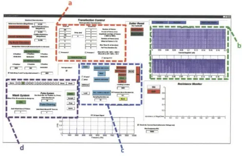
![Figure 2.2: All of the automation and experiments detailed in this thesis were built upon the basic layout shown above, first detailed here[81]](https://thumb-eu.123doks.com/thumbv2/123doknet/14135308.469590/36.918.191.702.210.476/figure-automation-experiments-detailed-thesis-built-layout-detailed.webp)