Myocardial injury and its prevention in the perioperative setting
M. Zaugg
1 2*, M. C. Schaub
2and P. FoeÈx
31
Institute of Anaesthesiology, University Hospital ZuÈrich, Switzerland.
2Institute of Pharmacology and
Toxicology, University of ZuÈrich, Switzerland.
3Nuf®eld Department of Anaesthetics, The John Radcliffe
Hospital, Oxford, UK
*Corresponding author: Institute of Anaesthesiology, University Hospital ZuÈrich, RaÈmistrasse 100, CH-8091 ZuÈrich, Switzerland. E-mail: [email protected]
Br J Anaesth 2004; 93: 21±33
Keywords: anaesthetic techniques, inhalation; complications, myocardial ischaemia; heart, cardioprotection; hibernation; stunning
In the UK, there are ~20 000 deaths within 30 days of surgery every year, 9000 of which have a cardiac cause.93
The number of major cardiac complications is likely to be in the region of 150 000 per annum. As 60% of patients who die within 30 days of surgery suffer from coronary artery disease94 it is reasonable to assume that the majority of
cardiac complications of anaesthesia and surgery results from myocardial ischaemia leading to myocardial injury (Fig. 1).
Myocardial responses to ischaemia
Acute myocardial ischaemia
The acute occlusion or progressive constriction of a coronary artery causes reduction or abolition of systolic shortening and thickening of the ischaemic wall.81
Ischaemic segments also demonstrate paradoxical wall motion (termed post-systolic shortening or post-systolic thickening). These functional changes relate directly to the severity of the reduction in coronary blood ¯ow. As post-systolic shortening and thickening occur after aortic valve closure, they do not contribute to ejection and result in an internal shift of blood in the ventricle, and may impair relaxation. Acute or progressive ischaemia of the left ventricle also cause an increase in chamber stiffness in the ischaemic and in remote non-ischaemic segments.81 This
generalized increase in myocardial chamber stiffness con-tributes to an elevation of the left ventricular end-diastolic pressure, especially in the presence of volume loading.81
The increase in end-diastolic pressure contributes to a vicious circle as it further impairs coronary blood ¯ow by increasing diastolic wall tension. The mechanisms of the
increase in ventricular chamber stiffness of remote, well-perfused myocardium have not been elucidated. However, ventricular stiffening does not depend on loading conditions and is likely to result from the release of mediators. Mechanisms of myocardial ischaemia
The time-course of the effects of ischaemia on cardiac tissue is well known. There is a marked reduction of contractile function resulting from decreased ATP production a few seconds after the onset of ischaemia. Leakage of potassium ions is responsible for the alterations of ST-segments. Within minutes, an intracellular acidosis develops associ-ated with an increase in myoplasmic Ca2+and the beginning
of cell swelling. Later, cellular lesions become irreversible. The ultrastructure of the cells becomes altered and macromolecules (CK-MB, troponins) are released. An increased concentration of cytosolic and mitochondrial Ca2+plays a central role in the damage to the cells and their
membranes104(Fig. 2).
Myocardial ischaemia occurs in the presence of ®xed or dynamic coronary artery stenoses and, in the case of the right ventricle, in response to afterload mismatch. The main causes of ischaemia with ®xed coronary stenoses include tachycardia, excessive left ventricular ®lling, and hypox-aemia. Tachycardia, systolic hypertension and b-adrenergic stimulation increase the oxygen requirements and may decrease oxygen delivery. Causes of ischaemia in the presence of dynamic stenoses include those described above and, in addition, activation of sympathetic and parasympa-thetic systems. Moreover, several endothelium-derived mediators may enhance vasoconstriction.
In the normal heart, the role of the autonomic nervous system is overshadowed by local metabolic coronary vasoregulation. Normally, activation of the sympathetic
nervous system at the level of a1- and b1-adrenoceptors
increases blood pressure, heart rate, and contractility. As a result, myocardial oxygen consumption increases and local vasoregulation decreases coronary vascular resistance. The direct effect of a1-adrenoceptor stimulation,
vasoconstric-tion, is minimal under normal circumstances but may be exaggerated in the presence of coronary artery disease, extreme exercise, or haemorrhagic shock. Activation of the parasympathetic system causes bradycardia and hypoten-sion, thereby reducing myocardial oxygen requirements. Local regulation increases coronary vascular resistance and the direct coronary vasodilatation caused by acetylcholine is masked.
Endothelins act on vascular smooth muscle and are extremely powerful vasoconstrictors. However, they exert complex physiological effects. Through activation of endothelin B (ETB)-receptors, endothelial nitric oxide
synthase is activated and nitric oxide is released causing cGMP-mediated vasodilatation. Activation of ETB
-recep-tors also increases the activity of cyclo-oxygenase leading to the release of prostaglandin I2 (PGI2). The latter causes
vasodilatation and minimizes smooth muscle cell prolifer-ation. When the endothelium is normal, there is a delicate balance between vasoconstriction and vasodilatation in response to endothelins. The vasodilatory role of the endothelium becomes more apparent when it is damaged by atheroma, hypercholersterolaemia, hypertensive heart disease, and after reperfusion. An imbalance of mediators
can then develop and facilitate vasoconstriction. There are several important mechanisms involved in this vasocon-striction. Norepinephrine causes a1-adrenoceptor-mediated
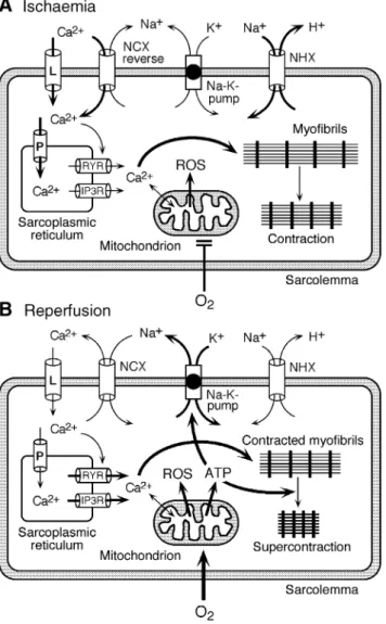
Fig 2 Changes and consequences of cation ¯uxes during ischaemia-reperfusion. (A) Cessation of oxygen supply in ischaemia leads to a loss of ATP production and an increase of reactive oxygen species (ROS) in the mitochondria. Reduced activity of the ATP consuming Na+-K+-pump
lowers the outside±inside sodium gradient, Na+ accumulates in the
myocyte and the resting membrane potential is lowered. With the development of acidosis, the Na+-H+-exchanger (NHX) further increases
intracellular Na+. Under these conditions the Na+-Ca2+-exchanger (NCX)
operates in the reverse mode, letting Ca2+into the cell. Ca2+also enters
through the sarcolemmal L-type voltage-gated Ca2+-channel (L) as the
resting membrane potential is low. The increased Ca2+is taken up into
the sarcoplasmic reticulum (SR) by the SR Ca2+-pump SERCA2 (P) and
released from there via two types of release channels, the ryanodine receptor channel (RYR) and the IP3 receptor channel (IP3R), leading to contraction. (B) Reoxygenation during reperfusion restores ATP production with a further boost of ROS. Reactivation of the Na+-K+
-pump by ATP slowly restores the sodium gradient leading to normal cation ¯uxes with the NCX eventually extruding the excess of cytosolic Ca2+. During the early reperfusion phase when the intracellular Ca2+
level is still high, myocardial contracture (supercontraction of myocytes) may develop. When contracture affects the entire heart as it may occur after global ischaemia, it has been termed the `stone heart' phenomenon.104 157
Fig 1 Schematic characterization of cardiac `stunning' and `hibernation'. In stunning normal blood ¯ow and energy metabolism are accompanied by reduced contractility, designated as `¯ow-contractility mismatch'. The impaired contractile function seems to be caused primarily by increased intracellular Ca2+, activation of Ca2+-dependent non-lysosomal cysteine
proteinases (calpains), and degradation of sarcomere-associated proteins including troponin-I. Hibernation is characterized by reduced contractility accompanied by reduced oxygen consumption as a consequence of reduced blood ¯ow. Partially damaged cardiomyocytes can be rescued to full function after stunning as well as after hibernation, provided normal blood ¯ow is restored after the latter within the critical time period before irreversible cell damage has occurred. Despite their distinct de®nition, stunning and hibernation may merely represent intermediary states in a continuum extending from unimpaired functional myocytes to necrosis.157
vasoconstriction. When the endothelium is damaged, acetylcholine causes muscarinic receptor mediated vaso-constriction,79 instead of endothelium-dependent
vaso-dilatation. Thus, with endothelial damage both sympathetic and parasympathetic stimulation may cause coronary vasoconstriction. As sympathetic overactivity is part of the perioperative stress response, exaggerated vasoconstriction may be expected to contribute to peri-operative myocardial ischaemia and cardiac damage. Similarly, the effects of endothelins are altered when the endothelium is damaged and activation of nitric oxide-synthase and cyclo-oxygenase does not occur. Consequently, only effects of endothelins in these circum-stances are those on endothelin A (ETA)- and ETB-receptors
in vascular smooth muscle resulting in vasoconstriction and smooth muscle proliferation.
Myocardial stunning (¯ow-contractility mismatch)
The term myocardial stunning was coined by Braunwald and Kloner14 in 1982 to describe a reduction in functionafter a brief period of ischaemia followed by reperfusion. The impairment of function could last for several hours or days at a time when coronary blood ¯ow was normal and there was no obvious cellular damage. In clinical practice, interventions such as transluminal coronary angioplasty, coronary artery bypass graft surgery, and thrombolysis after myocardial infarction are human models of ischaemia-reperfusion phenomena. During the perioperative period, a high proportion of adult patients suffer from episodes of myocardial ischaemia, most of which are silent but can be prolonged. Importantly, silent myocardial ischaemia is supposed to be a common cause of myocardial stunning and is a predictor of adverse cardiac outcome.
Mechanisms of myocardial stunning
Myocardial ischaemia followed by reperfusion causes reversible or irreversible damage depending on its duration (Figures 1 and 2). In stunning, ischaemic damage is, in principle, reversible.67 Three main mechanisms are
in-volved in the establishment of stunned myocardium: formation of free oxygen radicals, accumulation of intra-cellular Ca2+, and degradation of contractile proteins. Many
studies have shown that during ischaemia, but more importantly during reperfusion, considerable production of free oxygen radicals occurs.99 Free radicals do not have a
single target, but adversely affect many components of the cell including sarcolemmal and subcellular membranes of organelles. The role of free radicals in stunning is con®rmed by the improved post-ischaemic functional recovery in the presence of superoxide dismutase.138 Production of free
radicals involves xanthine oxidase, oxidation of catechol-amines, uncoupling of mitochondrial respiration, and activation of neutrophils.
During ischaemia and the early phase of reperfusion, there is an increase in the concentration of intracellular
Ca2+. Although disappearing in the late phase of
reperfusion, Ca2+ overload can decrease the sensitivity of
contractile proteins to Ca2+, thus diminishing the
developed force.9 Ca2+ overload my result from altered
characteristics of the Na+/Ca2+ antiport and from altered
Ca2+¯uxes at the level of the sarcoplasmic reticulum. Such
alterations in Ca2+ handling may be attributable to
ischaemia-induced intracellular acidosis. During the early phase of reperfusion, the H+/Na+ antiport is maximally
stimulated. While the acidosis is progressively corrected, there is an increase in intracellular Na+leading to a further
increase in Ca2+.
Furthermore there is some evidence that translocation of heat-shock proteins (Hsp-27, aB-crystalline) with covalent binding to myo®brils, together with degradation of con-tractile proteins such as troponin I, as evidenced in a transgenic mouse model overexpressing troponin I frag-ments,88 may be, at least in part, involved in the
pathogenesis of myocardial stunning. Also, an increase in coronary vascular resistance and a reduction in vasodilator response were previously reported during reperfusion and may represent some sort of vascular counterparts of stunning in endothelial cells (`microvascular stunning').10
However, not all studies con®rm this phenomenon.32
Myocardial hibernation
The concept of myocardial hibernation was put forward by Rahimtoola in 1985.110 111In the hibernating myocardium,
ventricular function is diminished as a consequence of insuf®cient coronary blood ¯ow (Fig. 1). However, this reduction is not necessarily permanent: an improved balance of supply and demand may augment myocardial function.7 78The issue of myocardial hibernation is
clinic-ally important because the risk of adverse cardiac outcome in cardiac and non-cardiac surgery increases with a reduction of the ejection fraction. If coronary revasculariza-tion increases the ejecrevasculariza-tion fracrevasculariza-tion the risk of adverse cardiac outcome is likely to be reduced. The likelihood of improved function after coronary reperfusion can be predicted by the result of a dobutamine stress echocardio-gram. If dobutamine worsens ventricular function (revers-ible ischaemia) coronary revascularization is likely to improve cardiac function.1
Mechanisms of myocardial hibernation
In hibernating myocardium, cardiac metabolism is down-regulated. It has been proposed that abolition of contractility of hibernating cardiac tissue is attributable to chronic stunning caused by multiple episodes of severe ischaemia followed by repetitive reperfusion. Other experimental models suggest that hibernation occurs as a result of chronic low-¯ow states. In either case, hibernating myocardium should be salvageable by restitution of an adequate coronary blood ¯ow.
Myocardial preconditioning
In 1986, Reimer and colleagues112 reported that a brief
period of ischaemia decreased the rate of ATP depletion during a further period of ischaemia. Murry and col-leagues89reported that brief periods of ischaemia made the
heart more resistant to infarction during a subsequent acute coronary occlusion, reducing the infarct size by 70±80%. This phenomenon, termed myocardial preconditioning is the most powerful means of achieving cardiac protection. Protection conferred by ischaemic and (most) pharmaco-logical preconditioning protocols exhibits two windows: an acute memory phase that develops within minutes of the ischaemic stimulus and lasts only between 1 and 3 h (classic or early preconditioning), and a longer, more delayed, phase (late preconditioning) starting after 12±24 h and persisting for 2±4 days.156Delayed protection by preconditioning is
thought to be primarily attributable to alterations in gene expression. Myocardial preconditioning has been docu-mented in all animal species in which it has been studied and in human cardiac tissue. Whether preconditioning facilitates the recovery of function in stunned myocardium is still debated.19 101 However, delayed preconditioning, in
con-trast to early preconditioning, always confers protection against stunning.
Preconditioning can result from successive episodes of angina or silent myocardial ischaemia. Indeed, infarct size is known to be smaller, if it has been preceded by angina.69 91 100 Preconditioning also reduces the risk of
ischaemia-induced ventricular tachycardia and ventricular ®brillation.145 In an almost experimental situation,
pre-conditioning can occur during coronary angioplasty where several temporary occlusions are applied and the effects of a longer period of occlusion are minimized. During coronary angioplasty, sequential occlusions cause less angina, smaller ST-segment changes and lesser lactate production than the ®rst 90 second occlusion.28 Also, delayed
preconditioning can be observed after prolonged nitro-glycerin administration in patients undergoing angioplasty 24 h later. Although ischaemic preconditioning, or agents that mimic preconditioning, elicit cardiac protection in patients undergoing cardiac surgery,51 the clinical use of
preconditioning in cardiac surgery is controversial as some studies show bene®ts48 148 while others do not,59 72 102 or
even demonstrate deleterious effects. Mechanisms of myocardial preconditioning
Ischaemic stimuli cause the release of stress mediators such as adenosine, bradykinin, norepinephrine, and opioids. The mechanisms of preconditioning involve several types of triggers and mediators156(Fig. 3). Amongst them, adenosine
A1- and A3-receptors, bradykinin2-receptors, d1-opioid
receptors and a1-adrenoceptors play an important role.
Via G-proteins, phospholipase C (PLC) and protein kinase C (PKC), these receptors act on mitochondrial and sarcolemmal KATP channels and Ca2+ channels.152 153 As
many mediators are involved in preconditioning, and many
substances are capable of preventing it, a `summation hypothesis' has been proposed by Downey and col-leagues.31 36 In order for ischaemic preconditioning or
pharmacological preconditioning to occur, it is necessary to reach an activation threshold. This threshold represents the sum of the activity of several mediators. The role of lactate as a triggering mechanism is controversial, even though lactate can open KATPchannels and increase the expression
of heat-shock proteins.4 65 Ischaemic preconditioning is
caused by temporary occlusions, generally lasting 5 min, followed by reperfusion. A comparison of various numbers of cycles of ischaemia-reperfusion shows that protection is best with 1±4 cycles. With six or more cycles protection is reduced und ultimately lost.47 While one cycle may be
enough to trigger the release of protective substances, many more cycles may be unable to enhance further release of triggers. A high number of short ischaemic periods may start to cause cumulative ischaemic damage, thus decreasing the ef®cacy of preconditioning.47
Preconditioning reduces cardiomyocyte necrosis in vivo and in vitro. Apoptosis is known to increase with ischaemia.
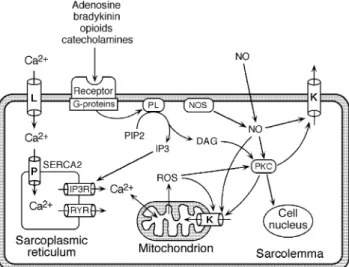
Fig 3 Main signalling pathways in ischaemic preconditioning. Stimulation of G-protein coupled receptors by primary messengers activates phospholipases (PL), which in turn produce two second messengers originating from phosphatidylinositol bisphosphate (PIP2), namely inositol trisphosphate (IP3) and diacylglycerol (DAG). The former releases Ca2+from the sarcoplasmic reticulum (SR) via the IP3
receptor channel (IP3R), the latter activates different PKC isoforms (PKC). PKC isoforms translocate to their appropriate target sites, activating the sarcolemmal and mitochondrial ATP-dependent potassium channels (K) and initiating distinct gene expression in the cell nucleus. Nitric oxide originating from either the endogenous NO-synthase (NOS) or from extracellular sources may also activate PKC and the potassium channels directly or via its reactive nitrogen oxide products (not shown in this ®gure). The same mechanism holds true for the reactive oxygen species (ROS) that are produced in the mitochondria under stress and increased Ca2+. Ca2+ enters the myocyte via the L-type voltage-gated
Ca2+-channel (L) and is taken up into the SR by the Ca2+-pump
(SERCA2). Ca2+-release from the SR for contraction primarily occurs via
In ischaemia-reperfusion, both ischaemia and reperfusion contribute equally to apoptosis.76 By contrast, necrosis
occurs primarily during reoxygenation. Preconditioning effectively reduces necrosis and apoptosis. Opening of KATP channels together with modulation of Ca2+
homeo-stasis may explain why inhalation anaesthetics inhibit apoptosis in cardiomyocytes.3 151
Role of adenosine
The role of adenosine in preconditioning has been well documented. Adenosine A1-receptor activation plays an
important role. These receptors are coupled with KATP
channels68via Gi-proteins. Activation of adenosine
recep-tors decreases the production of reactive oxygen species and attenuates myocardial stunning.92 The role of adenosine
receptors in preconditioning is con®rmed by the observation that adenosine A1-receptor antagonists can block KATP
channels, thereby preventing ischaemic preconditioning. In addition, preconditioning can be mimicked by adenosine A1-receptor agonists.
Role of bradykinin
Bradykinin is an in¯ammatory stress mediator and a vasodilator. In some experimental models, an infusion of bradykinin has been shown to reduce ischaemic injury,15 36
while bradykinin receptor antagonists negated the protec-tion conferred by ischaemic precondiprotec-tioning.15 36
Role of opioids
Morphine and fentanyl have been shown to precondition the myocardium.58 120 152 Conversely, d-opioids receptor
antagonists prevent ischaemic preconditioning.84 122 123
Role of adrenergic receptors
Both a- and b-adrenoceptors are involved in precondition-ing. While preconditioning is induced primarily by b1
-adrenoceptors,34 b
2-adrenoceptor may play a role via
activation of L-type calcium channels. Brief episodes of ischaemia cause the release of norepinephrine in the myocardium, while exogenous a1-adrenoceptor agonists
may cause pharmacological preconditioning.132
Role of free oxygen radicals and nitric oxide
Free radicals cause myocardial damage during ischaemia-reperfusion. However, treatment with small amounts of free radicals before an ischaemic insult can reduce infarct size in vitro,6 an effect that is abolished by free radical
scavengers. Nitric oxide-cGMP signalling is also important.25 33 92 152 Inhalation anaesthetics may modulate
the activity of various isoenzymes of nitric oxide synthase (nNOS, eNOS, iNOS) as they are heterogeneously distributed in the myocardium.
Calcium ions
A preischaemic increase in Ca2+ represents a second
messenger in the development of ischaemic
precondition-ing,85even though Ca2+overload is a major contributor to
cell damage. Short-time administration of increased Ca2+
concentrations to myocardial tissue is an effective pre-conditioning stimulus, which can be inhibited by adminis-tration of Ca2+channel blockers.141
Protein kinase C (PKC)
PKC transfers g-phosphoryls from ATP to hydroxyl groups of serine/threonine residues in proteins. This phosphoryl-ation controls the function of many cellular effectors. PKC plays an important role in ischaemic and pharmacological preconditioning.50 113 134 139 PKC activators can induce,
while PKC inhibitors prevent preconditioning.150
Inhala-tion anaesthetics may directly activate PKC. Importantly, preconditioning-associated isoform translocation of PKC to subcellular targets is highly dependent on species, age, and on the type of preconditioning stimulus.
Role of ATP-dependent potassium channels (KATPchannels)
Ultimately, KATPchannels hold the central role in ischaemic
and pharmacological preconditioning153(Figures 2 and 3).
Sarcolemmal KATP channels were described by Noma in
1983.96These channels open when ATP levels fall, allowing
potassium ef¯ux so causing membrane hyperpolarization and reducing the action potential duration. These changes decrease the open probability of voltage-gated Ca2+
chan-nels. The resulting reduction in Ca2+ concentration
pre-serves ATP levels and reduces coronary vascular tone.118
The increase in extracellular potassium also facilitates coronary vasodilatation and increases blood ¯ow to the ischaemic region.5K
ATPchannels mediate the response to
hypoxia and the hyperaemic response to brief coronary occlusions. However, reduction of action potential duration does not correlate with the reduction in infarct size. Surface KATPchannels were initially thought to mediate
precondi-tioning. More recent evidence indicates that mitochondrial KATP channels play a pivotal role in mediating cardiac
preconditioning.35 153 Opening of mitochondrial K ATP
channels may optimize mitochondrial energy production, decrease mitochondrial Ca2+ overload, and prevent
opening of mitochondrial permeability transition pores (Fig. 4).30 43 83 157 Numerous studies have con®rmed the
important role of these channels. The role of KATPchannels
in human preconditioning is evidenced by the observation that ischaemic preconditioning does not occur in patients taking sulfonylureas, as these agents block KATP
channels.13 21
Other bene®cial effects associated with cardiac preconditioning
Glycogen depletion and lactate accumulation during ischaemic preconditioning periods play a role in myocardial protection. Indeed, transient exposure to lactate improves contractile recovery in rat heart.29 Entrapment of
neutro-phils and platelets in the coronary vasculature occurs in ischaemia. The protective effects of preconditioning also
extend to the endothelium of the coronary vasculature and to the adhesion properties of platelets and leucocytes to these vessels. Ischaemic preconditioning reduces ICAM-1 pro-duction and thus neutrophil entrapment.116In turn, reduced
neutrophil and platelet entrapment is associated with enhanced post-ischaemic function.44 45
Late preconditioning
Late preconditioning is mediated by inducible nitric oxide synthase129 146 and can be elicited by nitric oxide
donors.46 130 It can also be triggered by heat stress,
lipopolysaccharides (LPS), or monophosphoryl lipid A; these are known to trigger delayed endogenous protective mechanisms against myocardial ischaemia-reperfusion injury appearing after 24 h and lasting for several days.11
Alterations of gene expression of protective and anti-protective proteins along with KATPchannel opening have
been proposed as the main mechanisms for this delayed protection. The endocannabinoid system is involved in the protection conferred by LPS, in relation with nitric oxide production.74 Two endocannabinoids act through
inter-action with G-protein coupled membrane receptors, namely CB1- and CB2-receptors, which are present throughout the
body.103 Endocannabinoids have been implicated in the
in¯ammatory response. In isolated rat hearts, endocannabi-noids acting through CB2-receptors and nitric oxide, play a
role in the protection conferred by heat stress against myocardial ischaemia.52Hemin is an activator of the potent
antioxidant enzyme heme oxygenase-1 and may play a role in delayed protection.149 Enhanced expression of heme
oxygenase-1 has been observed during the recovery phase of porcine myocardial stunning.125Indeed, in experimental
studies, a signi®cant attenuation of stunning, as evidenced by enhanced recovery of wall thickening was observed in animals pretreated for 1 week with hemin.143 Whether
inhalation anaesthetics are capable of elicting late pre-conditioning is not yet clear.
Remote preconditioning
Ischaemic preconditioning can be generated by short episodes of myocardial, limb, or visceral ischaemia. In the heart itself, preconditioning can develop in areas remote from the preconditioning ischaemic stimulus (for review see156). This remote preconditioning may involve the
release of adenosine, bradykinin, norepinephrine, and activation of KATP channels. Systemic effects of localized
ischaemic preconditioning have been reported. This raises the issue that regional ischaemia of non-vital tissues might protect remote vital organs.37In volunteers, transient remote
ischaemia of one limb induced remote ischaemic precondi-tioning of the opposite limb as evidenced by preservation of endothelial function (estimated as extent of vasodilator response).66 Cytokines and other metabolic mediators are
likely to play a role. The autonomic system may also be involved as well as modulation of platelet, endothelium, and leucocyte function.
Protective effects of anaesthetics against
ischaemia
Anaesthetics and myocardial stunning
Most inhalation anaesthetics and high dose opioids confer protection, increasing the rate of myocardial recovery after reperfusion.58 115 In 1988, Warltier and colleagues142
demonstrated that administration of halothane and iso¯ur-ane before ischaemia improved the speed of recovery of function after a brief (15 min) period of ischaemia. After 5 h, the functional recovery in the presence of inhalation anaesthesia was 100% vs 50% only in the controls.142
Since then, these observations have been con®rmed repeat-edly.22 134However, some studies of isolated heart
prepar-ations showed no protection by inhalation anaesthetics.97 117
More recently, sevo¯urane and des¯urane were shown to confer cardiac protection.108 109 119 133 135 It is likely that
protection by inhalation anaesthetics is attributable to pharmacological preconditioning of the heart, as in many studies the inhalational anaesthetic was given before ischaemia and reperfusion. Indeed, in some studies the administration of the inhalation anaesthetic was discon-tinued before ischaemia-reperfusion and resulted in reduced infarct size.18 24 Nonetheless, protective effects of
inhal-ation anesthetics were also reported if inhalinhal-ation anaes-thetics were administered exclusively during the reperfusion phase.
Intravenous anaesthetics appear to confer less protec-tion.23 27 144 Fentanyl and propofol appear to be
equiva-lent.114 In the isolated heart, as opposed to the intact
instrumented heart, propofol has been shown to reduce infarct size and cellular damage.70 71 82 Propofol-induced
protection was not abolished by block of KATP channels.
While the effects of opioids on infarct size have been well demonstrated, there are only a few studies of their effects on myocardial stunning.114 144 In the isolated heart, high
concentrations of fentanyl have been shown to offer signi®cant protection.58 Protection was mediated by
d-opioid receptors, adenosine A1-receptors, PKC, and
KATP channels.56 57 The role of d-opioid receptors is
supported by the abolition of their protective effect by naloxone.20 122 131As wash-out of opioids does not prevent
their effect, they must act as preconditioning agents.2 75It is
further possible that opioids act bene®cially via a reduction in adhesion and migration of neutrophils.128 140
Anaesthetics and cardiac preconditioning
Many anaesthetic agents have been shown to reduce infarct size in experimental models. Not all anaesthetics have the same ef®cacy. There is greater reduction of infarct size by halothane, en¯urane, and iso¯urane in comparison with pentobarbital, ketamine-xylazine, or propofol anaesthesia in rabbits. Dogs anaesthetized with barbiturates exhibit larger infarcts than their conscious counterparts.53 This may be
explained by the observation that barbiturates competitively antagonize adenosine A1-receptors, a pivotal signalling
pathway in cardiac preconditioning. Indeed, adenosine
receptor antagonists decrease anaesthesia-induced precon-ditioning.24 113 The same is true of PKC antagonists.134
Albeit not proven, it may be speculated that pharmaco-logical preconditioning by inhalation anaesthetics may be of smaller magnitude than ischaemic preconditioning.18
Pharmacological preconditioning by inhalation anaes-thetics appears to be primarily mediated by stimulation of adenosine receptors113 and activation of K
ATP
chan-nels.62 113Protective effects by inhalation anaesthetics also
occur in the presence of cardioplegic protection. Ischaemic and anaesthetic-induced preconditioning are not additive suggesting the same end-effector,12 identi®ed in many
studies as KATP channels. Nonetheless, sevo¯urane can
potentiate late ischaemic preconditioning in an in vivo rabbit model.87 The signalling components involved in cardiac
protection by inhalational anaesthetics, are similar to ischaemic preconditioning, but show distinct differences.26
Both sarcolemmal and mitochondrial KATP channels may
mediate anaesthesia-induced preconditioning as demon-strated in des¯urane-mediated preconditioning.135 Yet,
mitochondrial KATPchannels may play the more important
role. Halothane partially blocks sarcolemmal KATP
chan-nels,113 while iso¯urane does not. Anaesthesia-induced
preconditioning is clearly species dependent. Halothane preconditions in rabbit but not rat or human. Iso¯urane preconditions in rabbit and human, but not rat (for review see155 156).
Anaesthetics may also modulate the effects of ischaemic preconditioning. Several anaesthetic agents have direct effects on KATPchannels (barbiturates) or have prominent
physiological effects that are induced by KATP channels
(iso¯urane, halothane).17Accordingly, ischaemic
precondi-tioning is abolished by glibenclamide under ketamine-xylazine anaesthesia but not pentobarbital anaesthesia. In a comparison of the effects of ischaemic preconditioning under pentobarbital, iso¯urane, and ketamine-xylazine anaesthesia, infarct size was not different in the absence of preconditioning, but the magnitude of infarct size limitation by ischaemic preconditioning was different depending upon the basal anaesthesia.38In the presence of
halothane anaesthesia, nicorandil given before ischaemia did not demonstrate protective effects, whereas ischaemic preconditioning did reduce infarct size. Yet, a KATPchannel
blocker prevented the combined effect of ischaemia and nicorandil.90 The complexity of modulatory effects of
anaesthetics on cardiac preconditioning has been substan-tiated in a cellular model of simulated ischaemia. Modulatory effects of anaesthetics were demonstrated by the inhibition of diazoxide-induced mitochondrial KATP
channel opening by R-ketamine, thiopental and pentobarbi-tal. Conversely, urethane, 2,2,2-trichloroethanol (a main metabolite of a-chloralose) and fentanyl potentiated the channel-opening effect of diazoxide. This potentiation could be blocked by chelerythrine, a speci®c PKC inhibitor. By contrast, S-ketamine, propofol, xylazine, midazolam and etomidate do not affect mitochondrial KATP channel
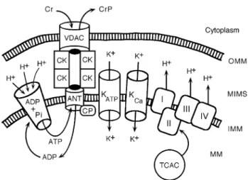
Fig 4 Functional connections between the mitochondrial permeability transition pore (mPTP) and the oxidative energy production during ischaemia-reperfusion and pharmacological preconditioning (PC). The selective adenine nucleotide translocator (ANT) at the inner mitochondrial membrane (IMM) regulates ATP supply to the cytoplasm in exchange for ADP, which will be regenerated to ATP in the mitochondrial matrix (MM) by the ATP synthase (ADP + Pi). The synthase is driven by the proton gradient across the IMM. The high proton concentration in the intermembrane space (MIMS) is maintained by the respiratory chain complexes (I±IV), which are energetically fuelled by the tricarboxylic acid cycle (TCAC) in the MM. On its way out of the MM, ATP passes through the ANT and enters a channel formed by an octameric complex of the mitochondrial creatine kinase (CK) where its gamma-phosphoryl is transferred to creatine (Cr) to produce creatine phosphate (CrP), which leaves the mitochondrion through the voltage-dependent anion channel (VDAC or porin) in the outer membrane (OMM) into the cytoplasm. Wherever energy is required, transphosphorylation from CrP to local ADP yields ATP for immediate use. The Cr±CK system serves as energy shuttle between the production centre and the place of consumption. The VDAC allows solutes to pass up to a molecular weight of 5000 Da. The nucleotide conductivity of ANT is controlled by cyclophilin-D (CP) at its inner opening. Binding of Ca2+ (which is increased during ischaemia-reperfusion) to CP induces
ANT to form a non-selective channel for solutes up to a molecular weight of 1500 Da. Cyclosporin-A can bind to CP and prevents channel opening, while atractyloside binds to ANT itself and favours channel opening. During ischaemia cessation of ATP production produces a decrease of diffuse K+ in¯ux. Consequently, the MM shrinks somewhat at the
expense of an increase of the MIMS leading to destabilization of the complex between ANT, CK and VDAC. On reperfusion additional ROS and Ca2+trigger opening of the ANT channel, which then seems to join
directly to the VDAC forming a non-selective mega-pore, the mPTP. This leads to the collapse of the IMM potential, to massive MM swelling and disruption of the OMM. As K+acts as the main MM volume regulator,
activation of K+ in¯ux represents the most powerful mechanism to
prevent mitochondrial destabilization and therewith irreversible destruction and cell death. Both ischaemic and pharmacological PC activate the mitochondrial ATP-dependent potassium channels (mK-ATP) affording myocyte protection against ischaemia-reperfusion injury. In addition, a large conductance Ca2+-activated potassium channel (K-Ca)
known to exist in the surface membrane of vascular smooth muscle cells was also found in the MIM. This channel is regulated by physiological variations of cytosolic Ca2+, and when selectively activated, it also
activity.152These observations illustrate the complex
inter-ference of anaesthetics with ischaemic preconditioning and stress the concept of anaesthetics acting as modulators of cardiac preconditioning.
To date, there are few data on the possibility of inhalation anaesthetics conferring late preconditioning, whereas delayed protection by opioids is well established.121 Kehl
and colleagues60examined the effect of iso¯urane
adminis-tration 24 h before a 60 min coronary occlusion followed by 3 h reperfusion in a canine model. While iso¯urane exerted early protection, there was no late protection. By contrast, delayed preconditioning was observed in a rabbit model.137
Finally, improved collateral blood ¯ow may also play a role in the bene®cial effects elicited by anaesthetics. Indeed, sevo¯urane increases collateral ¯ow, an effect not reversed by glibenclamide.64 In addition, halothane, iso¯urane, and
sevo¯urane reduce the number of neutrophils45 73 and
platelets44 sequestered in the coronary vasculature after
ischaemia. This may contribute to the observed bene®cial effects. Inhalation anaesthetics further suppress the post-ischaemic expression of CD11b45 and thus decrease
neutrophil adhesion to the endothelium.86However,
sevo-¯urane does not reduce the expression of glycoprotein IIb/ IIIa, a platelet adhesion molecule involved in the platelet± endothelium interaction.44
Iso¯urane
In the absence of ischaemia, iso¯urane causes opening of KATPchannels, an effect blocked by sulfonylureas.61This
results in a reduction in infarct size in experimental animals.63 Iso¯urane also decreases infarct size in an
in vitro model of human myocardium.113 Sarcolemmal
and mitochondrial KATP channels appear to be
invol-ved.61 106 113 133 136Iso¯urane increases the open probability
of the sarcoplasmic KATP channel for a given ATP
concentration.39In a cellular model, iso¯urane signi®cantly
enhanced the diazoxide-mediated activation of mitochon-drial KATPchannels. This effect was completely blocked by
chelerythrine (a PKC inhibitor). Pretreatment with inhal-ation anaesthetics potentiated the diazoxide-mediated pro-tection against ischaemia. Cardiopropro-tection was unaffected by the sarcoplasmic KATPchannel blocker HMR-1098, but
sensitive to modulation of nitric oxide and adenosine-Gi signalling pathways.153Administration of iso¯urane before
aortic cross-clamping in patients undergoing coronary artery bypass surgery causes cardiac index to be higher after cardiopulmonary bypass with less changes in ST-segments than in the control group. However, there were no differences in terms of arrhythmias.42Thus, iso¯urane may
offer some additional protection to cardioplegia. These ®ndings are consistent with the observation of lower (albeit not statistically signi®cant) perioperative levels of CK-MB and troponin reported by Belhomme8 when iso¯urane is
used. Moreover, iso¯urane was found to increase 5¢-nucleotidase activity in atrial tissue indicating increased PKC activity.8
Sevo¯urane
Sevo¯urane reduces infarct size in dogs via opening of KATP
channels.133Preservation of myocardial blood ¯ow through
collateral circulation, observed with sevo¯urane, is inde-pendent of KATPchannels. In sepsis, ultrastructural changes
in the myocardium have been documented and sevo¯urane protected cardiac output in septic (caecal ligation and perforation) rats.124 Recently, the ®rst clinical
double-blinded multicentre study has shown sevo¯urane to protect myocardium and kidney in patients undergoing coronary artery bypass grafting.54This study also visualized for the
®rst time PKC translocation (predominantly isoforms d and e) to subcellular targets such as the sarcolemma, mitochon-dria, intercalated disks, and nuclei in response to sevo-¯urane. Moreover, the observed renoprotective effect of sevo¯urane raises the intriguing possibility that systemic-ally administered sevo¯urane may confer multiorgan protection in high-risk patients.
Des¯urane
In isolated human atrial trabeculae, des¯urane improved the recovery of isometric contraction after a 30 min period of anoxia. The preconditioning effect of des¯urane was abolished by glibenclamide, 5-hydroxydecanoate (5-HD), DPX (an adenosine receptor blocker), phentolamine, and propranolol.41 These observations suggest that
precondi-tioning by des¯urane is mediated by mitochondrial KATP
channels,41 adenosine A
1-receptors, and a- and
b-adreno-ceptors. In contrast, selective block of sarcolemmal KATP
channels did not reduce des¯urane-induced precondition-ing, while it abolished anoxia-induced preconditioning. Des¯urane increases sympathetic activity in volunteers and releases catecholamines from myocardial stores in rat and human myocardium.40 Preconditioning by des¯urane may
thus be, at least partly, elicited by stimulation of the a/b-adrenoceptor pathways.77
Pharmacological interventions by
nonanaesthetic agents currently used for the
prevention of perioperative ischaemia
Several classes of drugs have been proposed in order to reduce the risk of ischaemic complications of anaesthesia and surgery. However, based on current clinical data, only beta-blockers, a2-adrenoceptor agonists, and possibly
statins may have the potential to affect perioperative cardiovascular outcome.
Nitroglycerin
While nitroglycerin is used successfully in the treatment of myocardial ischaemia, there is no evidence that its prophy-lactic administration before anaesthesia and surgery decreases the risk of perioperative cardiac complications.127
Calcium channel blockers
Though effective in the management of ischaemic heart disease, Ca2+ channel blockers have never been shown to
offer any protection against perioperative cardiac complic-ations of anaesthesia and surgery.126 127 This absence of
protection seems surprising in view of the strong antioxidant effect of certain calcium channel blockers.147
Adenosine modulators
These compounds facilitate the release of the coronary vasodilator adenosine by the ischaemic myocardium, thereby improving collateral blood ¯ow toward the com-promised area. Though promising results were obtained,80
development of the only agent tested in clinical trials, acadesine, was stopped.
a2-Adrenoceptor agonists
There is renewed interest in the use of clonidine, dexmedetomidine, and (temporarily, as it is not currently being further developed for clinical use) mivazerol; these drugs reduce the level of sympathetic activity and make the circulation more stable. Clonidine decreases the risk of perioperative myocardial ischaemia95 and a recent
meta-analysis has also shown a reduction in the risk of adverse outcome.127Mivazerol has been tested in a large multicentre
trial and was shown to decrease the incidence of cardiac complications in vascular surgical patients but not in non-vascular surgical patients,98 yet its development was
stopped. Nicorandil
This drug is both a nitrate and a KATPchannel opener. It is
effective in the management of ischaemic heart disease and its associated dysrhythmias. It may prove useful in the perioperative prevention of cardiac complications.55 In
clinical practice, nicorandil, a KATP channel opener and
nitrate, is widely used in the treatment of angina. Nicorandil induces myocardial preconditioning. In isolated human heart muscle nicorandil conferred cardioprotection (im-proved recovery of function in a hypoxia-reoxygenation model). This effect was abolished by ischaemic precondi-tioning.16In a rabbit model early treatment with nicorandil
(pre-ischaemia) decreased infarct size, while nicorandil administration after ischaemia was ineffective. Ischaemic preconditioning reduced infarct size and the combination of ischaemic preconditioning and nicorandil showed ef®cacy intermediate between ischaemic preconditioning and before administration of nicorandil.49The effect of nicorandil was
blocked by 5-hydroxydecanoate, a KATP channel blocker.
Thus, nicorandil appears to protect by opening KATP
channels, and to interact with ischaemic preconditioning. By contrast, nicorandil appears to offer additional protection when administered with iso¯urane, in terms of functional recovery of the stunned myocardium.105
Statins
In a case-controlled study, Poldermans and colleagues107
evaluated the effects of statins on perioperative mortality in patients undergoing major vascular surgery. In statin-treated patients, the risk for perioperative mortality was ~20% of that observed in non-statin-treated patients. The authors
concluded that perioperative statin use may reduce peri-operative mortality in high-risk vascular patients.
b-Blockers
These drugs, at present, occupy centre stage for cardiac prophylaxis because several studies have shown a reduction in the incidence of cardiac complications of anaesthesia and surgery in patients deliberately given beta-blockers prophylactically.154The bene®cial effects of perioperative
b-blocker administration are discussed in detail in another article in this issue.
Conclusions
Myocardial ischaemic injury is a potential perioperative threat. Ischaemia induces a palette of myocardial states with distinct pathophysiological backgrounds ranging from the paradoxically bene®cial effects of preconditioning on one side to the complete loss of cellular integrity and to cell death on the other side. Preconditioning mimicking agents such as inhalation anaesthetics and opioids induce a pronounced protective cardiac phenotype and thus may decrease, along with b-blockers, a2-adrenoceptor agonists,
and anti-in¯ammatory/preconditioning-mimicking statins, the deleterious effects of myocardial ischaemia in peri-operative medicine.
Acknowledgements
This work was supported by the Swiss National Science Foundation Grant 3200-063417.00 and Grant 3200B0-103980/1, the Swiss Heart Foundation, a Grant from the Swiss University Conference, and a Grant from the Hartmann±MuÈller Foundation, ZuÈrich, Switzerland.
References
1 Afridi I, Kleiman NS, Raizner AE, Zoghbi WA. Dobutamine echocardiography in myocardial hibernation. Optimal dose and accuracy in predicting recovery of ventricular function after coronary angioplasty. Circulation 1995; 91: 663±70
2 Aitchison KA, Baxter GF, Awan MM, et al. Opposing effects on infarction of delta and kappa opioid receptor activation in the isolated rat heart: implications for ischaemic preconditioning. Basic Res Cardiol 2000; 95: 1±10
3 Akao M, Ohler A, O'Rourke B, Marban E. Mitochondrial ATP-sensitive potassium channels inhibit apoptosis induced by oxidative stress in cardiac cells. Circ Res 2001; 88: 1267±75 4 Aresta F, Gerstenblith G, Weiss RG. Repeated, transient lactate
exposure does not `precondition' rat myocardium. Can J Physiol Pharmacol 1997; 75: 1262±6
5 Ashcroft SJ, Ashcroft FM. Properties and functions of ATP-sensitive K-channels. Cell Signal 1990; 2: 197±214
6 Baines CP, Goto M, Downey JM. Oxygen radicals released during ischemic preconditioning contribute to cardioprotection in the rabbit myocardium. J Mol Cell Cardiol 1997; 29: 207±16 7 Bax JJ, Visser FC, Poldermans D, et al. Time course of functional
recovery of stunned and hibernating segments after surgical revascularization. Circulation 2001; 104: I314±8
preconditioning by iso¯urane in coronary artery bypass graft surgery. Circulation 1999; 100: II340±4
9 Bolli R. Mechanism of myocardial `stunning'. Circulation 1990; 82: 723±38
10 Bolli R, Triana JF, Jeroudi MO. Prolonged impairment of coronary vasodilation after reversible ischemia. Evidence for microvascular `stunning'. Circ Res 1990; 67: 332±43
11 Bolli R. The late phase of preconditioning. Circ Res 2000; 87: 972±83
12 Boutros A, Wang J, Capuano C. Iso¯urane and halothane increase adenosine triphosphate preservation, but do not provide additive recovery of function after ischemia, in preconditioned rat hearts. Anesthesiology 1997; 86: 109±17 13 Brady PA, Terzic A. The sulfonylurea controversy: more
questions from the heart. J Am Coll Cardiol 1998; 31: 950±6 14 Braunwald E, Kloner RA. The stunned myocardium: prolonged,
postischemic ventricular dysfunction. Circulation 1982; 66: 1146±9
15 Brew EC, Mitchell MB, Rehring TF, et al. Role of bradykinin in cardiac functional protection after global ischemia-reperfusion in rat heart. Am J Physiol 1995; 269: H1370±8
16 Carr CS, Yellon DM. Ischaemic preconditioning may abolish the protection afforded by ATP-sensitive potassium channel openers in isolated human atrial muscle. Basic Res Cardiol 1997; 92: 252± 60
17 Cason BA, Gordon HJ, Avery EGt, Hickey RF. The role of ATP sensitive potassium channels in myocardial protection. J Card Surg 1995; 10: 441±4
18 Cason BA, Gamperl AK, Slocum RE, Hickey RF. Anesthetic-induced preconditioning: previous administration of iso¯urane decreases myocardial infarct size in rabbits. Anesthesiology 1997; 87: 1182±90
19 Cave AC, Collis CS, Downey JM, Hearse DJ. Improved functional recovery by ischaemic preconditioning is not mediated by adenosine in the globally ischaemic isolated rat heart. Cardiovasc Res 1993; 27: 663±8
20 Chien GL, Mohtadi K, Wolff RA, Van Winkle DM. Naloxone blockade of myocardial ischemic preconditioning does not require central nervous system participation. Basic Res Cardiol 1999; 94: 136±43
21 Cleveland JC Jr, Meldrum DR, Cain BS, Banerjee A, Harken AH. Oral sulfonylurea hypoglycemic agents prevent ischemic preconditioning in human myocardium. Two paradoxes revisited. Circulation 1997; 96: 29±32
22 Coetzee A, Moolman J. Halothane and the reperfusion injury in the intact animal model. Anesth Analg 1993; 76: 734±44 23 Coetzee A. Comparison of the effects of propofol and halothane
on acute myocardial ischaemia and myocardial reperfusion injury. S Afr Med J 1996; 86 (Suppl. 2): C85±90
24 Cope DK, Impastato WK, Cohen MV, Downey JM. Volatile anesthetics protect the ischemic rabbit myocardium from infarction. Anesthesiology 1997; 86: 699±709
25 Csonka C, Szilvassy Z, Fulop F, et al. Classic preconditioning decreases the harmful accumulation of nitric oxide during ischemia and reperfusion in rat hearts. Circulation 1999; 100: 2260±6
26 Da Silva R, Grampp T, Pasch T, Schaub MC, Zaugg M. Differential activation of mitogen-activated protein kinases in ischemic and anesthetic preconditioning. Anesthesiology 2004; 100; 59±69 27 De Hert SG, Cromheecke S, ten Broecke PW, et al. Effects of
propofol, des¯urane, and sevo¯urane on recovery of myocardial function after coronary surgery in elderly high-risk patients. Anesthesiology 2003; 99: 314±23
28 Deutsch E, Berger M, Kussmaul WG, et al. Adaptation to
ischemia during percutaneous transluminal coronary angioplasty. Clinical, hemodynamic, and metabolic features. Circulation 1990; 82: 2044±51
29 Doenst T, Guthrie PH, Chemnitius JM, Zech R, Taegtmeyer H. Fasting, lactate, and insulin improve ischemia tolerance in rat heart: a comparison with ischemic preconditioning. Am J Physiol 1996; 270: H1607±15
30 Dolder M, Wendt S, Wallimann T. Mitochondrial creatine kinase in contact sites: interaction with porin and adenine nucleotide translocase, role in permeability transition and sensitivity to oxidative damage. Biol Signals Recept 2001; 10: 93±111 31 Downey JM, Cohen MV. Signal transduction in ischemic
preconditioning. Adv Exp Med Biol 1997; 430: 39±55
32 Duncker DJ, McFalls EO, Krams R, Verdouw PD. Pressure-maximal coronary ¯ow relationship in regionally stunned porcine myocardium. Am J Physiol 1992; 262: H1744±51
33 Ferdinandy P, Szilvassy Z, Balogh N, et al. Nitric oxide is involved in active preconditioning in isolated working rat hearts. Ann N Y Acad Sci 1996; 793: 489±93
34 Frances C, Nazeyrollas P, Prevost A, et al. Role of b1- and b2
-adrenoceptor subtypes in preconditioning against myocardial dysfunction after ischemia and reperfusion. J Cardiovasc Pharmacol 2003; 41: 396±405
35 Garlid KD, Paucek P, Yarov Yarovoy V, et al. Cardioprotective effect of diazoxide and its interaction with mitochondrial ATP-sensitive K+channels. Possible mechanism of cardioprotection.
Circ Res 1997; 81: 1072±82
36 Goto M, Liu Y, Yang XM, et al. Role of bradykinin in protection of ischemic preconditioning in rabbit hearts. Circ Res 1995; 77: 611±21
37 Gunaydin B, Cakici I, Soncul H, et al. Does remote organ ischaemia trigger cardiac preconditioning during coronary artery surgery? Pharmacol Res 2000; 41: 493±6
38 Haessler R, Kuzume K, Chien GL, et al. Anaesthetics alter the magnitude of infarct limitation by ischaemic preconditioning. Cardiovasc Res 1994; 28: 1574±80
39 Han J, Kim E, Ho WK, Earm YE. Effects of volatile anesthetic iso¯urane on ATP-sensitive K+ channels in rabbit ventricular
myocytes. Biochem Biophys Res Commun 1996; 229: 852±6 40 Hanouz JL, Massetti M, Guesne G, et al. In vitro effects of
des¯urane, sevo¯urane, iso¯urane, and halothane in isolated human right atria. Anesthesiology 2000; 92: 116±24
41 Hanouz JL, Yvon A, Massetti M, et al. Mechanisms of des¯urane-induced preconditioning in isolated human right atria in vitro. Anesthesiology 2002; 97: 33±41
42 Haroun Bizri S, Khoury SS, Chehab IR, Kassas CM, Baraka A. Does iso¯urane optimize myocardial protection during cardiopulmonary bypass? J Cardiothorac Vasc Anesth 2001; 15: 418±21
43 Hausenloy DJ, Maddock HL, Baxter GF, Yellon DM. Inhibiting mitochondrial permeability transition pore opening: a new paradigm for myocardial preconditioning? Cardiovasc Res 2002; 55: 534±43
44 Heindl B, Conzen PF, Becker BF. The volatile anesthetic sevo¯urane mitigates cardiodepressive effects of platelets in reperfused hearts. Basic Res Cardiol 1999; 94: 102±11
45 Heindl B, Reichle FM, Zahler S, Conzen PF, Becker BF. Sevo¯urane and iso¯urane protect the reperfused guinea pig heart by reducing postischemic adhesion of polymorphonuclear neutrophils. Anesthesiology 1999; 91: 521±30
46 Hill M, Takano H, Tang XL, et al. Nitroglycerin induces late preconditioning against myocardial infarction in conscious rabbits despite development of nitrate tolerance. Circulation 2001; 104: 694±9
47 Iliodromitis EK, Kremastinos DT, Katritsis DG, Papadopoulos CC, Hearse DJ. Multiple cycles of preconditioning cause loss of protection in open-chest rabbits. J Mol Cell Cardiol 1997; 29: 915±20
48 Illes RW, Wright JK, Inners McBride K, Yang CJ, Tristan A. Ischemic preconditioning improves preservation with crystalloid cardioplegia. Ann Thorac Surg 1994; 58: 1481±5
49 Imagawa J, Baxter GF, Yellon DM. Myocardial protection afforded by nicorandil and ischaemic preconditioning in a rabbit infarct model in vivo. J Cardiovasc Pharmacol 1998; 31: 74±9
50 Ismaeil MS, Tkachenko I, Hickey RF, Cason BA. Colchicine inhibits iso¯urane-induced preconditioning. Anesthesiology 1999; 91: 1816±22
51 Jenkins DP, Pugsley WB, Alkhulai® AM, et al. Ischaemic preconditioning reduces troponin T release in patients undergoing coronary artery bypass surgery. Heart 1997; 77: 314±18
52 Joyeux M, Arnaud C, Godin-Ribuot D, et al. Endocannabinoids are implicated in the infarct size-reducing effect conferred by heat stress preconditioning in isolated rat hearts. Cardiovasc Res 2002; 55: 619±25
53 Jugdutt BI. Different relations between infarct size and occluded bed size in barbiturate-anesthetized versus conscious dogs. J Am Coll Cardiol 1985; 6: 1035±46
54 Julier K, da Silva R, Garcia C, et al. Preconditioning by sevo¯urane decreases biochemical markers for myocardial and renal dysfunction in coronary artery bypass graft surgery: a double-blinded, placebo-controlled, multicenter study. Anesthesiology 2003; 98: 1315±27
55 Kaneko T, Saito Y, Hikawa Y, Yasuda K, Makita K. Dose-dependent prophylactic effect of nicorandil, an ATP-sensitive potassium channel opener, on intra-operative myocardial ischaemia in patients undergoing major abdominal surgery. Br J Anaesth 2001; 86: 332±7
56 Kato R, FoeÈx P. Fentanyl reduces infarction but not stunning via d-opioid receptors and protein kinase C in rats. Br J Anaesth 2000; 84: 608±14
57 Kato R, Ross S, FoeÈx P. Fentanyl protects the heart against ischaemic injury via opioid receptors, adenosine A1 receptors
and KATPchannel linked mechanisms in rats. Br J Anaesth 2000;
84: 204±14
58 Kato R, FoeÈx P. Myocardial protection by anesthetic agents against ischemia-reperfusion injury: an update for anesthesiologists. Can J Anaesth 2002; 49: 777±91
59 Kaukoranta PK, Lepojarvi MP, Ylitalo KV, Kiviluoma KT, Peuhkurinen KJ. Normothermic retrograde blood cardioplegia with or without preceding ischemic preconditioning. Ann Thorac Surg 1997; 63: 1268±74
60 Kehl F, Pagel P-S, Krolikowski J-G, et al. Iso¯urane does not produce a second window of preconditioning against myocardial infarction in vivo. Anesth Analg 2002; 95: 1162±8
61 Kersten JR, Lowe D, Hettrick DA, et al. Glyburide, a KATP
channel antagonist, attenuates the cardioprotective effects of iso¯urane in stunned myocardium. Anesth Analg 1996; 83: 27±33 62 Kersten JR, Schmeling TJ, Hettrick DA, et al. Mechanism of myocardial protection by iso¯urane. Role of adenosine triphosphate-regulated potassium KATPchannels. Anesthesiology
1996; 85: 794±807
63 Kersten JR, Schmeling TJ, Pagel PS, Gross GJ, Warltier DC. Iso¯urane mimics ischemic preconditioning via activation of KATP
channels: reduction of myocardial infarct size with an acute memory phase. Anesthesiology 1997; 87: 361±70
64 Kersten JR, Schmeling T, Tessmer J, et al. Sevo¯urane selectively
increases coronary collateral blood ¯ow independent of KATP
channels in vivo. Anesthesiology 1999; 90: 246±56
65 Keung EC, Li Q. Lactate activates ATP-sensitive potassium channels in guinea pig ventricular myocytes. J Clin Invest 1991; 88: 1772±7
66 Kharbanda RK, Mortensen UM, White PA, et al. Transient limb ischemia induces remote ischemic preconditioning in vivo. Circulation 2002; 106: 2881±3
67 Kim SJ, Depre C, Vatner SF. Novel mechanisms mediating stunned myocardium. Heart Fail Rev 2003; 8: 143±53
68 Kirsch GE, Codina J, Birnbaumer L, Brown AM. Coupling of ATP-sensitive K+channels to A1 receptors by G proteins in rat
ventricular myocytes. Am J Physiol 1990; 259: H820±6
69 Kloner RA, Shook T, Przyklenk K, et al. Previous angina alters in-hospital outcome in TIMI 4. A clinical correlate to preconditioning? Circulation 1995; 91: 37±45
70 Ko SH, Yu CW, Lee SK, et al. Propofol attenuates ischemia-reperfusion injury in the isolated rat heart. Anesth Analg 1997; 85: 719±24
71 Kokita N, Hara A, Abiko Y, et al. Propofol improves functional and metabolic recovery in ischemic reperfused isolated rat hearts. Anesth Analg 1998; 86: 252±8
72 Kolocassides KG, Galinanes M, Hearse DJ. Ischemic preconditioning, cardioplegia or both? J Mol Cell Cardiol 1994; 26: 1411±14
73 Kowalski C, Zahler S, Becker BF, et al. Halothane, iso¯urane, and sevo¯urane reduce postischemic adhesion of neutrophils in the coronary system. Anesthesiology 1997; 86: 188±95
74 Lagneux C, Lamontagne D. Involvement of cannabinoids in the cardioprotection induced by lipopolysaccharide. Br J Pharmacolol 2001; 132: 793±6
75 Liang BT, Gross GJ. Direct preconditioning of cardiac myocytes via opioid receptors and KATP channels. Circ Res 1999; 84: 1396±400
76 Liu H, McPherson BC, Yao Z. Preconditioning attenuates apoptosis and necrosis: role of protein kinase Ce and -d isoforms. Am J Physiol Heart Circ Physiol 2001; 281: H404±10 77 Lochner A, Genade S, Tromp E, Podzuweit T, Moolman JA.
Ischemic preconditioning and the beta-adrenergic signal transduction pathway. Circulation 1999; 100: 958±66
78 Louie HW, Laks H, Milgalter E, et al. Ischemic cardiomyopathy. Criteria for coronary revascularization and cardiac transplantation. Circulation 1991; 84: III290±5
79 Ludmer PL, Selwyn AP, Shook TL, et al. Paradoxical vasoconstriction induced by acetylcholine in atherosclerotic coronary arteries. N Engl J Med 1986; 315: 1046±51
80 Mangano DT. Effects of acadesine on myocardial infarction, stroke, and death following surgery. A meta-analysis of the 5 international randomized trials. The Multicenter Study of Perioperative Ischemia (McSPI) Research Group. JAMA 1997; 277: 325±32
81 Marsch SC, Wanigasekera VA, Ryder WA, Wong LS, FoeÈx P. Graded myocardial ischemia is associated with a decrease in diastolic distensibility of the remote nonischemic myocardium in the anesthetized dog. J Am Coll Cardiol 1993; 22: 899±906 82 Mathur S, Farhangkhgoee P, Karmazyn M. Cardioprotective
effects of propofol and sevo¯urane in ischemic and reperfused rat hearts: role of KATP channels and interaction with the
sodium-hydrogen exchange inhibitor HOE 642 (cariporide). Anesthesiology 1999; 91: 1349±60
83 Mayer B, Oberbauer R. Mitochondrial regulation of apoptosis. News Physiol Sci 2003; 18: 89±94
ischemic preconditioning through protein kinase C activation in rabbits. Mol Cell Biochem 1998; 186: 3±12
85 Miyawaki H, Ashraf M. Ca2+ as a mediator of ischemic
preconditioning. Circ Res 1997; 80: 790±9
86 Mobert J, Zahler S, Becker BF, Conzen PF. Inhibition of neutrophil activation by volatile anesthetics decreases adhesion to cultured human endothelial cells. Anesthesiology 1999; 90: 1372±81
87 Mullenheim J, Ebel D, Bauer M, et al. Sevo¯urane confers additional cardioprotection after ischemic late preconditioning in rabbits. Anesthesiology 2003; 99: 624±31
88 Murphy AM, Kogler H, Georgakopoulos D, et al. Transgenic mouse model of stunned myocardium. Science 2000; 287: 488±91
89 Murry CE, Jennings RB, Reimer KA. Preconditioning with ischemia: a delay of lethal cell injury in ischemic myocardium. Circulation 1986; 74: 1124±36
90 Nakae I, Takaoka A, Mitsunami K, et al. Cardioprotective effects of nicorandil in rabbits anaesthetized with halothane: potentiation of ischaemic preconditioning via KATP channels.
Clin Exp Pharmacol Physiol 2000; 27: 810±17
91 Napoli C, Liguori A, Chiariello M, et al. New-onset angina preceding acute myocardial infarction is associated with improved contractile recovery after thrombolysis. Eur Heart J 1998; 19: 411±19
92 Narayan P, Mentzer RM Jr, Lasley RD. Adenosine A1 receptor activation reduces reactive oxygen species and attenuates stunning in ventricular myocytes. J Mol Cell Cardiol 2001; 33: 121±9
93 NCEPOD. Extremes of Age. The 1999 Report of the National Con®dential Enquiry into Perioperative Deaths, 1999; www.ncepod.org.uk
94 NCEPOD. Then and Now: The 2000 Report of the National Con®dential Enquiry into Postoperative Deaths, 2001; www.ncepod.org.uk
95 Nishina K, Mikawa K, Uesugi T, et al. Ef®cacy of clonidine for prevention of perioperative myocardial ischemia: a critical appraisal and meta-analysis of the literature. Anesthesiology 2002; 96: 323±9
96 Noma A. ATP-regulated K+channels in cardiac muscle. Nature
1983; 305: 147±8
97 Oguchi T, Kashimoto S, Yamaguchi T, Nakamura T, Kumazawa T. Comparative effects of halothane, en¯urane, iso¯urane and sevo¯urane on function and metabolism in the ischaemic rat heart. Br J Anaesth 1995; 74: 569±75
98 Oliver MF, Goldman L, Julian DG, Holme I. Effect of mivazerol on perioperative cardiac complications during non-cardiac surgery in patients with coronary heart disease: the European Mivazerol Trial (EMIT). Anesthesiology 1999; 91: 951±61
99 O'Neill CA, Fu LW, Halliwell B, Longhurst JC. Hydroxyl radical production during myocardial ischemia and reperfusion in cats. Am J Physiol 1996; 271: H660±7
100 Ottani F, Galvani M, Ferrini D, Nicolini FA. Clinical relevance of prodromal angina before acute myocardial infarction. Int J Cardiol 1999; 68 (Suppl. 1): S103±8
101 Ovize M, Przyklenk K, Hale SL, Kloner RA. Preconditioning does not attenuate myocardial stunning. Circulation 1992; 85: 2247±54 102 Perrault LP, Menasche P, Bel A, et al. Ischemic preconditioning in cardiac surgery: a word of caution. J Thorac Cardiovasc Surg 1996; 112: 1378±86
103 Piomelli D, Giuffrida A, Calignano A, Rodriguez de Fonseca F. The endocannabinoid system as a target for therapeutic drugs. Trends Pharmacol Sci 2000; 21: 218±24
104 Piper HM, Meuter K, Schafer C. Cellular mechanisms of ischemia-reperfusion injury. Ann Thorac Surg 2003; 75: S644±8 105 Piriou V, Ross S, Pigott D, Evans R, FoeÈx P. Bene®cial effect of
concomitant administration of iso¯urane and nicorandil. Br J Anaesth 1997; 79: 68±77
106 Piriou V, Chiari P, Knezynski S, et al. Prevention of iso¯urane-induced preconditioning by 5-hydroxydecanoate and gadolinium: possible involvement of mitochondrial adenosine triphosphate-sensitive potassium and stretch-activated channels. Anesthesiology 2000; 93: 756±64
107 Poldermans D, Bax JJ, Kertai MD, et al. Statins are associated with a reduced incidence of perioperative mortality in patients undergoing major noncardiac vascular surgery. Circulation 2003; 107: 1848±51
108 Preckel B, Schlack W, Comfere T, et al. Effects of en¯urane, iso¯urane, sevo¯urane and des¯urane on reperfusion injury after regional myocardial ischaemia in the rabbit heart in vivo. Br J Anaesth 1998; 81: 905±12
109 Preckel B, Thamer V, Schlack W. Bene®cial effects of sevo¯urane and des¯urane against myocardial reperfusion injury after cardioplegic arrest. Can J Anaesth 1999; 46: 1076±81
110 Rahimtoola SH. A perspective on the three large multicenter randomized clinical trials of coronary bypass surgery for chronic stable angina. Circulation 1985; 72: V123±35
111 Rahimtoola SH. The hibernating myocardium. Am Heart J 1989; 117: 211±21
112 Reimer KA, Murry CE, Yamasawa I, Hill ML, Jennings RB. Four brief periods of myocardial ischemia cause no cumulative ATP loss or necrosis. Am J Physiol 1986; 251: H1306±15
113 Roscoe AK, Christensen JD, Lynch C, 3rd. Iso¯urane, but not halothane, induces protection of human myocardium via adenosine A1 receptors and adenosine triphosphate-sensitive potassium channels. Anesthesiology 2000; 92: 1692±701 114 Ross S, Munoz H, Piriou V, Ryder WA, FoeÈx P. A comparison of
the effects of fentanyl and propofol on left ventricular contractility during myocardial stunning. Acta Anaesthesiol Scand 1998; 42: 23±31
115 Ross S, FoeÈx P. Protective effects of anaesthetics in reversible and irreversible ischaemia-reperfusion injury. Br J Anaesth 1999; 82: 622±32
116 Rubino A, Yellon DM. Ischaemic preconditioning of the vasculature: an overlooked phenomenon for protecting the heart? Trends Pharmacol Sci 2000; 21: 225±30
117 Sahlman L, Waagstein L, Haljamae H, Ricksten SE. Protective effects of halothane but not iso¯urane against global ischaemic injury in the isolated working rat heart. Acta Anaesthesiol Scand 1995; 39: 312±16
118 Samaha FF, Heineman FW, Ince C, Fleming J, Balaban RS. ATP-sensitive potassium channel is essential to maintain basal coronary vascular tone in vivo. Am J Physiol 1992; 262: C1220±7 119 Schlack W, Preckel B, Stunneck D, Thamer V. Effects of halothane, en¯urane, iso¯urane, sevo¯urane and des¯urane on myocardial reperfusion injury in the isolated rat heart. Br J Anaesth 1998; 81: 913±19
120 Schultz JE, Hsu AK, Gross GJ. Morphine mimics the cardioprotective effect of ischemic preconditioning via a glibenclamide-sensitive mechanism in the rat heart. Circ Res 1996; 78: 1100±4
121 Schultz JE, Gross GJ. Opioids and cardioprotection. Pharmacol Ther 2001; 89: 123±37
122 Schultz JJ, Hsu AK, Gross GJ. Ischemic preconditioning is mediated by a peripheral opioid receptor mechanism in the intact rat heart. J Mol Cell Cardiol 1997; 29: 1355±62