The effect of a maxillary lip bumper on tooth positions
8
0
0
Texte intégral
(2) 26 While the picture of the effect of a lip bumper on the mandibular dentition is relatively clear, nothing is known about the effects of such an appliance when used in the maxilla. A lip bumper in the maxilla could be a good alternative for increasing the arch perimeter in the interceptive treatment of subjects with a Class III tendency. In such cases with a retrognathic and small maxilla, there is often an obvious space deficiency. Extraction of maxillary teeth is an unfavourable solution because it exaggerates the discrepancy in size between the maxillary and mandibular dental arches. The possibilities of proclining the incisors or transversally expanding the dental arch are limited for reasons of stability. Distalization of the molars would be a possible way to gain space, but cannot be carried out with headgear because of the risk of increasing the maxillary retrognathism through the orthopaedic effect. This risk would be less with a lip bumper which, simultaneously to the holding or distalization of the molars, could bring about a slight proclination of the incisors and a transverse development of the dental arch. The present study was undertaken in order to evaluate the effects of a lip bumper on the maxillary dentition. Subjects and methods Seven boys and 15 girls participated in the study. Their ages varied between 9 years 3 months and 13 years 7 months (median age 10 years 6 months). The children were treated with a lip bumper in the maxilla for 10–14 months (median 12 months). In addition to the lip bumper, nine of the children also had a Goshgarian transpalatal arch (TPA) anchored to the first permanent molars. Five children wore the TPA throughout the period of treatment with the lip bumper and the remaining four children for 1.5–9 months of this period. No other appliance was used in the maxilla during this period. The children were treated at the Department of Orthodontics, University of Bern. The lip bumper was inserted in an attempt to gain space in the maxilla. A headgear for distalization of the maxillary molars in order to gain space was contra-indicated in these children because of. R . H Ä S L E R A N D B. I N G E RVA L L. Figure 1. Type of lip bumper used for the treatment.. their Class III or tendency to Class III skeletal intermaxillary relation. The type of lip bumper used is shown in Figure 1. It was made of 1.1-mm stainless steel and had custom-made acrylic shields in the labial fold opposite the anterior teeth (on each side in the region between the canine and the central incisor). The shield covered the gingiva 2–3 mm above the gingival margin and reached 6–7 mm occlusal to the gingival margin. The lip bumper was anchored in buccal tubes on the maxillary first permanent molars and was adjusted to lie 2–3 mm away from the labial surfaces of the incisors and canines, and from the buccal surfaces of the premolars. The children were instructed to wear their lip bumper day and night, and to remove it only for meals or for tooth brushing. Control visits were scheduled every second month, at which time the position of the lip bumper was checked and adjusted if necessary. The lip bumper was used passively, i.e. it was not adjusted for active expansion. The effects of the lip bumper were documented by measurements on dental casts and profile cephalograms made immediately before and after treatment. The recording on the cast included measurement of the width of the maxillary dental arch at the first permanent molars, premolars and canines. The measuring points are shown in Figure 2. When the premolars or permanent canines were not erupted, the corresponding points on the deciduous teeth were used. No measurement was made when a deciduous tooth was replaced by its successor during the period of observation. The length of the dental arch was measured from a line connecting the tip of the mesiobuccal cusp of the.
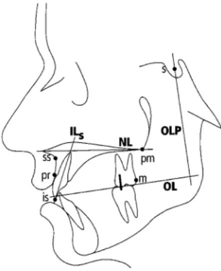
(3) E F F E C T O F A M A X I L L A RY L I P BU M P E R. 27. Figure 2 Measuring points used in the recording of the dental arch dimensions. The figure also shows the median changes in widths and arch length during the period of observation.. right and left first molars to the mid-point of the incisal edge of the two central incisors. The mean of the measurement to the right and left incisor was used as the variable for arch length. All measurements were made with electronic dial calipers to the next tenth of a millimetre. The results of the measurements of the dental arch dimensions were compared with the annual changes of the same dimensions in the untreated group of Moyers et al. (1976). For this comparison, their sample was matched with the present individuals with regard to sex and age. This matching was undertaken separately for each variable. The reference points and lines used in the cephalometric analysis are shown in Figure 3. The point m was located on the distal surface of the first molar band. Before radiography a metal. Figure 3 Reference points and lines used in the measurements on the cephalograms.. rod was inserted in the buccal tubes of the right and left first permanent molar bands, respectively. The length of the straight metal rod, which extended vertically gingivally and occlusally mesial to the mesial opening of the buccal tubes, was 15 mm. The metal rod was used to measure the inclination of the first molars in relation to OLP. The design of the rod on the right and left sides was different so that a differentiation could be made. In the cephalometric analysis the change in position of point m, as well as in the inclination of the molar on the two sides was averaged. The dimensions measured on the cephalograms were reduced to zero magnification. The changes of the distances ss–pm, pr–pm, and is–pm, as well as of the angle ILs/NL in the treated group were compared with the annual changes of the same variables in the untreated sample of Bahtia and Leighton (1993). Their sample was matched with the present individuals with regard to sex and age. This was carried out individually for each variable. Analysis of antero-posterior linear changes was performed with the method of Pancherz (1982). A coordinate system, consisting of the occlusal line (OL) and a perpendicular to this line through the point sella (OLP), was drawn on a tracing of the pre-treatment cephalogram. The co-ordinate system was transferred to the post-treatment cephalogram by superimposing on structures of the anterior cranial base as described by Björk (1968). All variables recorded on the casts or cephalograms were measured twice with new markings on the casts or new tracings. The mean of the two measurements was used in the analysis..
(4) 28. R . H Ä S L E R A N D B. I N G E RVA L L. Errors of the method and statistical methods used The errors of the method were calculated from the duplicate measurements made before and after treatment. Systematic differences between the duplicate measurements were tested with Wilcoxon’s matched pairs, signed ranks test. The accidental errors of the method (si) were calculated with the formula si = √Σd2/2n, where d is the difference between two measurements and n the number of recordings. Differences between distributions were tested with Mann–Whitney’s U-test and between paired observations with Wilcoxon’s matched pairs, signed ranks test. The number of duplicate determinations of the variables measured on the casts varied between 18 and 44. No systematic differences were found for these variables. The accidental errors varied from 0.16 to 0.41 mm. The number of duplicate determinations of the cephalometric variables was 36. One systematic difference was found. The angle ILs/NL was, on average, 0.40 degrees larger at the second than at the first measurement (0.01 < P < 0.05). The accidental errors for the measurement of distances on the cephalograms varied between 0.20 and 0.31 mm. The errors for the measurement of the molar inclination and for the angle ILs/NL were 0.71 and 0.81 degrees, respectively. Because the analysis. of the results of the treatment was based on replicated measurements, the errors were reduced by a factor of 0.7. Results The changes of the dimensions of the maxillary dental arch during treatment are given in Table 1. The variation in number of observations in Table 1 and in Figure 4 is due to the fact that the widths at the premolars/deciduous molars and at the canines could not be measured in all subjects due to the varying stage of development of the dentition. There was no difference in the changes between cases having and not having had a TPA during treatment. Therefore, no differentiation with regard to the use of a TPA was made. The change in width between the first permanent molars during the treatment varied widely from a decrease of 2 mm to an increase of 7.5 mm. The median change during treatment was small and not significant, and nor was any significant difference found in relation to the reference sample. The widths between the second premolars or the second deciduous molars, as well as between the first premolars, increased significantly during treatment and developed significantly differently to the corresponding dimensions in the reference sample. The change in width in the individual cases treated with the lip bumper is shown in Figure 4. All subjects of the treatment group had an increase of the dimensions mentioned. The widths between. Table 1 Median and range (in mm) of changes in the dimensions of the maxillary dental arch during treatment. The table also gives the median annual changes in the matched reference sample (Moyers et al., 1976). The varying number of observations is due to varying development of the dentition. Width between. n. Median. Range. Median in reference sample. Significance of difference in test-reference. First molars Second premolars Second deciduous molars First premolars First deciduous molars Canines Deciduous canines Arch length. 22 6 7 11 3 6 3 22. 0.3 2.2* 1.5* 2.2* 0.9 0.8 1.1 1.9**. –2.0–7.5 0.1–4.6 0.2–3.3 0.6–4.7 0.8–1.0 –0.6–1.7 0.9–2.6 0.1–4.0. 0.5 –0.1 0.2 –0.1 0 –0.3 0 –0.3. NS * * ** NS NS NS ***. *0.01 < P < 0.05; **0.001 < P < 0.01; ***P < 0.001; NS, non-significant..
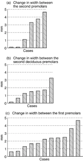
(5) 29. E F F E C T O F A M A X I L L A RY L I P BU M P E R. Figure 4 Change in width in the individual cases between the second premolars (a), between the second deciduous molars (b), and between the first premolars (c) during the treatment.. Figure 5. the first deciduous molars and between the canines also showed a numerical increase, but the number of observations was too small to allow statistical analysis. The length of the dental arch increased significantly during treatment and also when compared with the reference sample. All subjects showed an increase in arch length (Figure 5). For the reference sample, in contrast, the arch length decreased in 20 cases (up to 0.7 mm). The changes of the variables measured on the profile cephalogram are given in Table 2. There was no significant difference in the change of first molar position between patients who had or had not worn a TPA. Therefore, no differentiation of the sample with regard to the use of a TPA was undertaken. During the period of treatment the maxilla (point ss) and the maxillary incisors (point is) moved anteriorly by 1.0 and 1.5 mm (median), respectively. Only one patient showed a distal movement of the maxilla or incisors. The anterior movement of the molars was less and not significant. The movement of the molars varied from an anterior movement of 1.5 mm to a posterior movement of 2.8 mm. The next largest posterior movements were 1.4 and 0.65 mm. The crowns of the first molars tipped posteriorly by 5.8 degrees (median). The molars tipped anteriorly in only one case. The maxilla increased in length (distances ss–pm, pr–pm, is–pm) by 1.0–1.3 mm (median) and the incisors proclined 1.4 degrees. The proclination of the incisors was, however, not significant and none of these changes were significant compared with the changes in the reference material.. Change in arch length in the individual cases during treatment..
(6) 30. R . H Ä S L E R A N D B. I N G E RVA L L. Table 2 Median and range (in mm and degrees) of changes in antero-posterior position of points ss, is, and m, as well as dimensions of the maxilla and inclination of the maxillary central incisors and maxillary molars during treatment. The table also gives the median annual changes in maxillary dimensions and in the inclination of the incisors in the matched reference sample (Bahtia and Leighton, 1993) n = 18. Median. Antero-posterior position of points ss is m Inclination of first molars (degree) Distance ss–pm Distance pr–pm Distance is–pm ILs/NL (degree). Range. 1.0** 1.5** 0.4. –0.3–1.8 –1.7–3.8 –2.8–1.5. –5.8** 1.0** 1.2** 1.3** 1.4. –18.2–3.8 –0.3–3.4 –0.3–3.6 –0.3–4.0 –2.8–7.7. Median in reference sample. Significance of difference in test-reference. 0.8 1.0 1.1 –0.1. NS NS NS NS. A positive sign means anterior movement or change in inclination in an anterior direction. NS, not significant. **0.001 > P > 0.01.. Discussion For this study, a lip bumper with vestibular shields was chosen. The force from the lip on a bumper with shields has in the mandible been found to be greater than on a wire lip bumper (Hodge et al., 1997) and this may also be assumed to be true for the maxilla. The difference in force is thought to be due to the larger surface area of contact between the lip and the appliance when shields are used. The upper lip is much weaker than the lower. The mean pressure at rest from the lower lip on the lower incisors amounts to 9–12 g/cm2 against 2–5 g/cm2 from the upper lip on the upper incisors (Thüer et al., 1985; Thüer and Ingervall, 1986, 1990). Therefore, a bumper with shields is necessary if the distally-directed force from the lip bumper on the molars is to be of any appreciable magnitude. The changes of most of the variables during the period of treatment were compared with the changes of the same dimensions in samples of children followed for the study of normal growth and development. These samples (Moyers et al., 1976; Bahtia and Leighton, 1993) comprise children with normal occlusion and varying types of malocclusions. It cannot be taken for granted that the changes with growth and development of these children are quite comparable with those of the children of the present study, who had a Class. III or a tendency to Class III intermaxillary skeletal relationship. Furthermore, the children of the reference samples were from different populations than those of this investigation. The data in the reference samples were collected several decades ago. It is therefore possible that secular changes may influence a comparison with the present results. A control group of children with the same characteristics as the group of treated children would have been preferable for the comparison. The collection of such material was, however, impossible for ethical reasons and also because of the scarcity of children with Class III morphology. When comparing the treated children and the reference samples the limitations mentioned should be kept in mind. The median increase in width between the first permanent molars during treatment was negligible. This may be due to the fact that the lip bumper was used passively, i.e. a change in width between the first molars was hindered by the rigid lip bumper and that, in many cases, the inter-molar width was controlled by a TPA. In one subject, however, the width between the first molars was purposely expanded 7.5 mm. In the premolar area, on the other hand, there was a considerable widening of the dental arch, which was significant when compared with the reference sample. There was also an increase in inter-canine width, which, however, was not.
(7) E F F E C T O F A M A X I L L A RY L I P BU M P E R. significant. The number of inter-canine width observations was, however, small. The increase in maxillary inter-premolar widths achieved by the lip bumper treatment was much the same as the increase in mandibular inter-premolar widths achieved by the use of a lower lip bumper (Osborn et al., 1991; Nevant et al., 1991; Werner et al., 1994; Grossen and Ingervall, 1995; Davidovitch et al., 1997; O’Donnell et al., 1998). In contrast to the situation in the mandible, there is possibly more than one explanation for the increase in arch width from a lip bumper used in the maxilla. One explanation, which would hold true for both the maxilla and the mandible, is that the lip bumper changes the oral environment by holding the lips and cheeks away from the dental arches, thus altering the equilibrium between the forces from the circumoral soft tissues and from the tongue acting on the teeth. The effect of the lip bumper would then be similar to that of the vestibular shields of a Fränkel appliance (Fränkel, 1974). The other explanation is that a maxillary lip bumper increases the growth in the mid-palatal suture. This has been shown to be the case with the use of vestibular shields in growing rabbits (Kalogirou et al., 1996). In that animal experiment, however, the shields were extended to create tension in the buccinator insertions. The authors suggested that the increased sutural growth was due to relief of the buccal pressure and continued tongue pressure against the dento-alveolar bone, leading to separation of the adjoining bone and sutural growth as a passive filling process. In the present study, the increase in width between the first molars, as well as between the second premolars/second deciduous molars and between the first premolars/first deciduous molars, and between the canines was the same in subjects with and without a TPA during treatment. A TPA holds the two maxillary halves together, thereby decreasing the possibility of mid-palatal sutural growth expressing itself. Therefore, the explanation for the increase in maxillary dental arch width produced by the lip bumper treatment is most likely the change in equilibrium of the forces acting on the surfaces of the teeth. The growth in length of the maxilla was not affected by the lip bumper treatment as the. 31 distance ss–pm increased similarly in the treated group and the reference sample. The same is true for the distances pr–pm and is–pm. In relation to the reference line OLP, the maxilla (point ss) in the treated group moved 1 mm (median) anteriorly during the period of observation. Unfortunately, the literature contains no such measurement for untreated samples. The median anterior movement of is was 1.5 mm, i.e. somewhat more than for point ss. This may be due to eruption of the incisor and/or to a slight increase in its inclination, which changed more in the treated group than in the reference sample. The anterior movement of the first molar (point m) was only half that of the maxilla (point ss) and signifies a slight holding effect (median about half a millimetre) from the lip bumper on the molar. In single cases the molars may move distally but this rarely exceeds 1 mm. The small effect of the lip bumper on the molars in terms of holding or distalization may be due to the small force produced by the upper lip but, as mentioned in the introduction, in many studies a similar small effect was also found in the mandible. In a previous study of the effect of a lip bumper in the mandible (Grossen and Ingervall, 1995), the state of development and eruption of the second molars was found not to influence the effect of the bumper on the first molars. A similar analysis could not be carried out in the present study because we refrained from taking additional radiograms, and because one or both second molars were only erupted in four cases as judged from the dental casts. The increase in arch length from molar holding/ distalization and from incisor eruption/proclination was limited, and quite comparable with that found with the use of a lip bumper in the mandible (Osborn et al., 1991; Grossen and Ingervall, 1995; Davidovitch et al., 1997; O’Donnell et al., 1998). The main effect of a maxillary lip bumper seems to be a widening of the dental arch across the premolars. This is, of course, beneficial, but it is not the ultimate solution to the space deficiency problem in a retrognathic maxilla. On the other hand, no negative effects of the use of a maxillary lip bumper were found. It is an open question whether the expansive effect of a lip bumper and the proclination of the.
(8) 32 incisors are stable in the long term. The results of Soo and Moore (1991) indicated an adaptation of the lower lip to the tooth position achieved with lower lip bumper treatment. In their study, the pressure from the lower lip both at rest and during speech first increased (at 1 month), but then (at 8 months) decreased below baseline. These observations are at variance with the results of recent studies. O’Donnell et al. (1998) found no decrease of the pressure from a lower lip bumper on the first molars after one year of uninterrupted use. Ingervall and Thüer (1998) found the pressure from the lower lip on the lower incisors to be the same after 8 months of lower lip bumper treatment as at the start. The lip had not adapted to the changed position of the incisors, nor had it reacted to the extension by the lip bumper. Therefore, the conclusion of Houston and Edler (1990) may be correct, namely, ‘with a few exceptions, the initial position of the lower incisors provides the best guide to their position of stability’. Address for correspondence Professor Bengt Ingervall Klinik für Kieferorthopädie Freiburgstrasse 7 CH-3010 Bern, Switzerland References Bahtia S N, Leighton B C 1993 A manual of facial growth. Oxford University Press, Oxford Bjerregard J, Bundgaard A M, Melsen B 1980 The effect of the mandibular lip bumper and maxillary bite plate on tooth movement, occlusion and space conditions in the lower dental arch. European Journal of Orthodontics 2: 257–265 Björk A 1968 The use of metallic implants in the study of facial growth in children: method and application. American Journal of Physical Anthropology 29: 243–254 Cetlin N M, Ten Hoeve A 1983 Nonextraction treatment. Journal of Clinical Orthodontics 17: 396–413 Davidovitch M, McInnis D, Lindauer S J 1997 The effects of lip bumper therapy in the mixed dentition. American Journal of Orthodontics and Dentofacial Orthopedics 111: 52–58. R . H Ä S L E R A N D B. I N G E RVA L L. Hodge J J, Nanda R S, Ghosh J, Smith D 1997 Forces produced by lip bumpers on mandibular molars. American Journal of Orthodontics and Dentofacial Orthopedics 111: 613–622 Houston W J B, Edler R 1990 Long-term stability of the lower labial segment relative to the A–Pog line. European Journal of Orthodontics 12: 302–310 Ingervall B, Thüer U 1998 No effect of lip bumper therapy on the pressure from the lower lip on the lower incisors. European Journal of Orthodontics 20: 525–534 Kalogirou K, Ahlgren J, Klinge B 1996 Effects of buccal shields on the maxillary dentoalveolar structures and the midpalatal suture—histologic and biometric studies in rabbits. American Journal of Orthodontics and Dentofacial Orthopedics 109: 521–530 Moyers R E, van der Linden F, Riolo M, McNamara J Jr 1976 Standards of human occlusal development, Monograph No 5, Craniofacial Growth Series. Center for Human Growth and Development, University of Michigan, Ann Arbor, Michigan Nevant C T, Buschang P H, Alexander R G, Steffen J M 1991 Lip bumper therapy for gaining arch length. American Journal of Orthodontics and Dentofacial Orthopedics 100: 330–336 O’Donnell S, Nanda R S, Ghosh J 1998 Perioral forces and dental changes resulting from mandibular lip bumper treatment. American Journal of Orthodontics and Dentofacial Orthopedics 113: 247–255 Osborn W S, Nanda R S, Currier G F 1991 Mandibular arch perimeter changes with lip bumper treatment. American Journal of Orthodontics and Dentofacial Orthopedics 99: 527–532 Pancherz H 1982 The mechanism of Class II correction in Herbst appliance treatment. A cephalometric investigation. American Journal of Orthodontics 82: 104–113 Soo N D, Moore R N 1991 A technique for measurement of intraoral lip pressures with lip bumper therapy. American Journal of Orthodontics and Dentofacial Orthopedics 99: 409–417 Thüer U, Ingervall B 1986 Pressure from the lips on the teeth and malocclusion. American Journal of Orthodontics and Dentofacial Orthopedics 90: 234–242 Thüer U, Ingervall B 1990 Effect of muscle exercise with an oral screen on lip function. European Journal of Orthodontics 12: 198–208. Fränkel R 1974 Decrowding during eruption under the screening influence of vestibular shields. American Journal of Orthodontics 65: 372–406. Thüer U, Janson T, Ingervall B 1985 Application in children of a new method for the measurement of forces from the lips on the teeth. European Journal of Orthodontics 7: 63–78. Grossen J, Ingervall B 1995 The effect of a lip bumper on lower dental arch dimensions and tooth positions. European Journal of Orthodontics 17: 129–134. Werner S P, Shivapuja P K, Harris E F 1994 Skeletodental changes in the adolescent accruing from use of the lip bumper. Angle Orthodontist 64: 13–22.
(9)
Figure

Documents relatifs