Epidemiol. Inject. (1990). 105, 335-353 3 3 5 Printed in Great Britain
Sex differentials in susceptibility to lymphatic filariasis and
implications for maternal child immunity
L. BRABIN*
UNDP/ World Bank/ WHO Special Programme for Research and Training in Tropical Diseases, World Health Organization, Geneva, Switzerland
(Accepted 18 May 1990)
SUMMARY
This paper reviews epidemiological data to see if there are sex differentials in prevalence, density and clinical pathology due to lymphatic filariasis. Of 53 studies from Africa, South East Asia, the Indian Subcontinent and The Americas, 43 showed a lower mean prevalence of infection in females than in males. Prevalence is consistently lower in women of reproductive age and this is statistically significant in 16 of 32 studies classified by age and sex. Density of infection is also lower in the reproductive age but may be higher in children and in older women. Clinical disease is also lower in women and pathology has a later age of onset and rise to peak prevalence than in males. The paper assesses the evidence that lower rates of infection and clinical pathology are due to less exposure of females to infective vectors. It seems unlikely that exposure alone could account for these differences which are observed for both bancroftian and brugian filariasis, irrespective of periodicity. Several investigators have suggested that females have increased resistance to infection and this is supported by serological studies showing high antibody positivity to adult worm antigens in females. The review concludes that the association with the reproductive years suggests a pregnancy-associated mechanism. This has important implications for maternal fetal interactions and maternal filarial infection may influence the development of immunity in children.
INTRODUCTION
For most tropical diseases the importance of sex differentials in susceptibility to infection has largely remained unexplored. In general males are more susceptible to parasitic infections than females [1, 2], although in some diseases like malaria, susceptibility increases during pregnancy [3,4]. For helminthic infections opinion has been divided on whether sex differentials are related to lower exposure to infective vectors or to immunological factors. This review has the objective of, firstly, analysing the epidemiological data on differences between males and
* Requests for reprints: Dr L. Brabin, Liverpool School of Tropical Medicine, Pembroke Place, Liverpool L3 5QA.
336 L. BRABIN
females in microfilarial prevalence, mean microfilarial density and lymphatic pathology; and secondly, assessing the relative contribution of exposure and host immunity to the observed patterns of infection. The analysis indicates that there is compelling evidence that in the reproductive age, women experience a lower burden of infection than men. The consistency of this pattern argues against differences in exposure as the main cause and suggests that host immunity -possibly related to pregnancy-associated mechanisms - may act to reduce microfilarial densities. The paper concludes that sex differentials in susceptibility to lymphatic filariasis have important implications for maternal-fetal interactions.
PROBLEMS IN ASSESSING EPIDEMIOLOGICAL DATA ON LYMPHATIC FILARIASIS
Filariasis is a complex disease with major distinctions between Wuchereria and
Brugia infections in the natural host range, microfilarial periodicity and vector
susceptibility. Signs and symptoms of filariasis differ from one endemic area to another although there is an overlapping of clinical syndromes which makes comparison possible. The quality of epidemiological studies varies and data are often not reported by both age and sex. Prevalence data are reported more often, and are more likely to be grouped by age and sex than microfilarial densities, which are frequently quoted as mean figures. Parameters for clinical disease are often poorly defined and in mass surveys, clinical examinations may be cursory. Entomological and sociological studies may not be available to support explanations for observed sex differences. Finally, DEC (diethylcarbamazine) has been in widespread use for over 30 years and there are few recent studies where its usage does not complicate the interpretation of the epidemiological data. Pre-DEC studies are available but rely on relatively insensitive blood slide assessment. Microfilarial concentration filtration techniques are more sensitive but have been largely used to evaluate control programmes.
DIFFERENCES IN PREVALENCE OF INFECTION
Area differences Africa
Table 1 lists prevalence figures for 15 studies conducted in various regions of Africa. In 13 of these prevalence was higher in males than females, comparing the two sexes at all ages. Table 2 compares prevalence in women of reproductive age with men of a similar age range. Many studies classify younger women of childbearing age (15-20 years) in the 10-19 years group and so, for comparability, the most widely used category is 20-39 years. Only 7 of the 15 studied in Table 1 were classified by both age and sex. In all but one (Comores) [16], prevalence was lower in women.
Indian Subcontinent
The same comparisons are shown for the Indian Subcontinent. Prevalence in women at all ages was lower for 12 of the 14 areas (Table 3), while in the
Sex differences in lymphatic filariasis
337
Table 1. Prevalence (all ages) of microfilaremia o/W. bancrofti in Africa by sexMales Females Area (reference)
Gambia [5]
Gambia and Casamance [6]: Coastal village Inland village Swamp Casamance Liberia [7] Liberian savanna [8] Liberian savanna [9] Nigeria Igwun basin [10] North Cameroun [11] South Tanzania [12] Coastal Tanzania [13] Kenya, two villages [14] Kenya coast [15] Comores, Sabah [16] i Number examined 283 89 119 97 130 864 546 338 402 362 2167 905 875 2759 668 Prevalence (%) 35 51 31 40 52 19 22 23 22 22 26 23 23 17 34 Number examined 320 70 80 97 124 1104 709 394 326 264 1562 1098 919 1807 758 Prevalence (%) 38 46 14 32 49 10 12 21 16 10 19 20 21 13 36
Table 2. Prevalence (%) of microfilaremia in men and in women of reproductive age in Africa Male Female Age range-years (reference) Gambia 21-40 [5] Liberia 20-39 [9] 20-39 [7] Tanzania 16-45 [12] 20-39 [13] Kenya 20-39 [14]° 20-39 [14f Comores 20-39 [16] No.f 70 73 214 1126 184 68 90 117 A 1 Prevalence 54-3 370 31-3 290 391 26-5 400 461 No. 118 102 336 907 324 87 140 171 A Prevalence 51-7 32-3 14-9 18-5 26-2 21-8 33-6 46-8 P value* <0-75 <0-75 < 0001 < 0001 < 0 0 1 <0-5 < 0 5
* x2 test for significance of difference between males and females.
t Number examined
" Mambrui village. * Jaribuni village.
reproductive years, prevalence in women was always lower (Table 4). The study in Ceylon was a large-scale assessment of the anti-filariasis campaign [27]. South East Asia
Of 22 investigations in South East Asia, all but 5 showed a lower prevalence in females of all ages (Table 5), while in the reproductive years (Table 6), only one study showed a lower prevalence in men (Tonga) [41].
338 L. BRABIN
Table 3. Prevalence (all ages) of microfilaremia on the Indian Subcontinent by sex Males Females Area (reference) India: Madras [17] Calcutta [18] Andra Pradesh [19] Pondicherry [20] Cochin State [21] Ernakulam Mattancherri Bhagalpur town [22] Bhagalpur rural [23] Assam [24] Bokakht* Chabua Patna, Bihar [25]
Formerly East Pakistan [26]: Akcha Madarganj Formerly Ceylon [27]f Number examined 4069 535 5747 10675 3901 1825 1008 820 1122 824 6357 322 433 6838 Prevalence (%) 16 16 20 7 9 16 14 5 5 9 20 16 17 4 Number examined 3333 415 5553 8449 3406 1093 932 522 1091 915 3128 280 398 6950 Prevalence (%) 14 10 17 6 5 12 11 5 4 7 16 16 15 3 * B. malayi the predominant species - in other areas W. bancrofti is predominant. t Study followed widespread control measures.
Table 4. Prevalence (%) of microfilaremia in men and in women of reproductive
age in the Indian subcontinent
Male Female © — o _ ^ (reference) India: 21-40 [17] 15-44 [18] 15-49 [19] 21-40 [20] 2 1 ^ 0 [22] 21-40 [23] 21—40 [24]" 21-40 [24]" Formerly E. Pakistan: 20-39* [26]c 20-39 [26]" Formerly Ceylon 15-34"[27] Xo.t 1461 308 2919 4125 279 289 410 268 87 133 136145 Prevalence 161 19-5 200 7-0 7-2 16-3 6-8 13-8 27-6 210 4-7 No. 1225 171 2248 3638 180 202 355 306 87 113 146192 Prevalence 13-9 117 15-6 5-8 5-6 14-7 5-9 8-2 230 13-3 4 0 P-value* <0-25 < 0 0 5 < 0001 < 0 0 5 <0-5 <0-25 < 0-75 < 005 <0-5 <0-25 < 0001 * x2 test for significance of difference between males and females.
t Number examined.
" Bokakhat area. * Chabua area.
c
Are a (reference ) Tabl e 5 . Prevalence (all ages) of microfilaremia in South East Asia by sex Mal e Femal e Filtratio n techniqu e (+ / — ) Specie s Indonesia : Flore s [28 ] Flore s [29 ] Sulawes i [30 ] Borne o [31 ] W . Kalimanta n [32 ] Celebe s [33 ] Jakart a [34 ] Jakart a [35 ] Malaysia : Saba h [36 ] Papu a Ne w Guinea : Sepi k [37 ] Coo k Islands : [38 ] Tahiti : [39 ] Frenc h Oceania : [40 ] Tonga : [41 J Samoa : [42 ] Philippines : Sama r [43 ] Luzo n [44 ] Japan : Katsiyam a [45 ] Ohno'[45 ] Kore a mainland : [46 ] Vietna m : [47 ] China : [48 ] B. timori B. timori B. malayi B. malayi B. malayi B. malayi W. bancrofti W. bancrofti B. malayi (SP) * W. bancrofti W. bancrofti (SP ) W. bancrofti (SP ) W. bancrofti (SP ) W. bancrofti (SP ) W. bancrofti (SP ) W. bancrofti W. bancrofti W. bancrofti W. bancrofti B. malayi B. malayi^ W. bancrofti Numbe r examine d 9 8 10 4 26 2 16 3 19 2 193 1 10 1 17 0 303 0 4 1 16 5 339 0 145 3 15 0 276 6 356 5 12 4 52 6 46 3 27 8 531 2 822 4 Prevalenc e (% ) 3 1 29 3 8 4 0 2 7 2 9 17 3 2 10 6 8 4 1 38 3 5 6 6 2 3 7 3 7 6 2 16 3 10 Numbe r examine d 10 3 9 6 22 3 12 6 18 1 172 7 9 5 18 6 243 4 3 8 21 2 311 1 112 7 14 6 237 8 365 7 16 5 40 5 32 5 27 9 724 9 840 3 Prevalenc e (% ) 2 1 19 2 6 25 24 2 1 16 3 1 7 6 8 27 31 2 7 71 15 4 17 5 2 10 3 10 * S P (su b periodic) . t Predominan t species . C O C O C O
340 L. BRABIN
Table 6. Prevalence (%) of microfilaremia in men and women of reproductive age
in 8. East Asia Males Females Ase ranee-years (reference) Indonesia: 20-39 [28] 20-39 [30] 15-50 [31] 2 1 ^ 0 [32] 20-39 [33] 15-50 [35] Malaysia: 15-44 [36] Papua New Guinea:
21-40 [37] Tahiti: 20-39 [39] French Oceania: 21-40 [40] Tonga: 21-50 [41] Samoa: 20-39 [42] Philippines: 20-39 [43] No.t 33 72 103 74 604 103 1273 18 1068 507 64 967 747 A prevalence 30-3 58-3 45-6 37-8 391 35-9 13-5 77-8 49-8 48-3 68-0 33-6 111 No. 33 78 70 62 530 116 1097 14 1033 365 65 815 1000 A prevalence 18-2 30-8 271 22-6 23-2 310 9-2 57-1 35-3 34-8 740 17-7 5-7 P-value* < 0-25 < 0001 < 0025 < 0 1 0 < 0001 <0-5 <001 <0-25 < 0001 < 0001 <0-75 < 0001 < 0001
* x2 test for significance of difference between males and females, t Number examined.
The Americas
In Trinidad, (Table 7), prevalence was lower in females but was the same for both sexes in Brazil.
Prevalence in relation to age
In most of these studies, prevalence in girls was similar to, or higher, than that in boys, and a number of authors mention that a difference in prevalence is only evident from about age 15 onwards. In the post-reproductive period, prevalence may again be high in women.
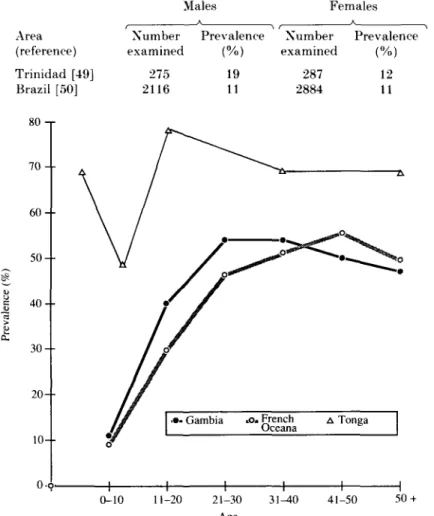
Males
The pattern of infection in males is characterized by an increase with age to about 21 years, after which, prevalence plateaus. Fig. 1 shows prevalence figures for males plotted by age from studies in The Gambia [5], French Oceania [40] and Tonga [41]. The shape of the curve is similar in each area, but in Tonga, prevalence was higher because filtration techniques were used which would detect more infections. Piessens [51] has suggested that this plateau effect, which has been observed in bancroftian, malayan and timorian filariasis, reflects that the number of younger, previously amicrofilaremic individuals who develop patent micro-filaremia is roughly equal to the number of older subjects who become amicrofilaremic due to a belated development of host resistance. There are some
Sex differences in lymphatic filariasis
341
Table 7. Prevalence (all ages) of microfilaremia ofW. bancrofti in The Americas,
by sex Males Females Area (reference) Trinidad [49] Brazil [50] Number examined 275 2116 Prevalence (%) 19 11 Number examined 287 2884 Prevalence (%) 12 11 80 -r 7 0 6 0 5 0 4 0 3 0 2 0 - 10--0-o. 0-10 11-20 21-30 31-40 Age 41-50 50 +
Fig. 1. Prevalence (%) of microfilaraemia in males by age in Gambia. French Oceania and Tongo. [5, 40, 41]
communities in which there is no stabilization with age, and prevalence rates continue to rise. Most notable in this regard is Papua New Guinea [37] where 100% prevalence in males older than 40 years has been reported. Since hyper-reactive malarious splenomegaly is also endemic in this region of Papua New Guinea, this may alter the host immune response to filariasis.
Females
Among females, prevalence rates often do not show the same pattern as in males: in French Oceania [40] they continue to rise; in Liberia [7] they plateau at a later age than in males and in Papua New Guinea, rise to a plateau of 82 % prevalence in women over 40 years. As noted, much of the variation relates to the reproductive years. If women are less exposed, or have lighter infections, the question arises whether lower prevalence partly relates to the insensitivity of
342 L. BBABIN
35 -r
50-59 60 +
Fig. 2. Geometric mean microfilarial densities by age and sex in Sada, Comores (from Bruhnes, 1975) [16]. Periodic W. bancrofti, sample size 1426.
blood slide techniques to detect low density parasitemias. This would seem to be the case in the Tongan study where 68 % and 74 % respectively of men and women aged 21-50 were positive by filtration (Table 6) but only 3 3 % of males and 26% of females by blood slide. There is evidence from a small number of investigations using both sensitive and insensitive methods that microfilarial densities decrease in women of reproductive age. This suggests that the main difference between the sexes is that women of reproductive age have lighter infections and difficulties in detecting these by blood slide is reflected in their lower prevalence figures.
DIFFERENCES IN MICROFILARIAL DENSITIES BY AGE
Figs. 2-5 show a reduction in microfilarial densities in women of reproductive years in the Comores [16] and Papua New Guinea [37] (periodic bancroftian filariasis), in Sabah [36] periodic B. malayi) and French Oceania [40] (sub-periodic bancroftian filariasis). In Sabah (Fig. 4) the range of microfilarial counts is also shown, and these are not as wide in females of reproductive years as in males of the same age. The reduction in densities in the Comores is particularly interesting because, contrary to other studies, prevalence at all ages, and in the reproductive years, was higher in females (Tables 1 and 2). In Papua New Guinea, filtration techniques were used so densities should not be under-represented in women. Three studies [33, 52, 53] used probit analysis to plot median microfilarial densities (MfD-50) by sex and age. These are shown in Table 8 and the same trend
Sex differences in lymphatic filariasis
343 2-5 -, E S o in 0-5-c 1-4 5-9 10-14 15-29 Age 30-44Fig. 3. Mean log10 (microfllarial count + 1) by age and sex in Papua New Guinea
(from Knight et al; 1979) [58], Periodic Bancroftian filariasis, sample size 233.
to lower microfilarial densities in women aged 20-39 years is evident. Other studies show a similar pattern to the above (Andra Pradesh [19], Ceylon [27], Mattancheri in Cochin [21]), in which microfilarial densities in both sexes are similar for the youngest and oldest age groups, but differ in the middle years.
Summary
These epidemiological studies suggest that density of infection is lower in women of reproductive age under widely different conditions of exposure. Difficulties in detecting low density infections may in some cases explain the lower prevalence of infection which is observed virtually everywhere.
FACTORS AFFECTING DENSITY OF INFECTION IX WOMEN
Area specific factors South East Asia
Although exposure must be an important factor influencing the epidemiology of lymphatic filariasis, a number of investigators consider that immunity to infection is enhanced in women. This divergence of opinion is seen clearly in discussion of the data from American Samoa, where sub-periodic W. bancrofti is endemic. Murray [42] was in no doubt that women had better immunity than men. He 12 H Y O 105
344 L. BRABIN 10 15 20 25 30 35 40 45 50 55 + Range Male Female 1-8 1-33 1-208 1-172 1-303 1-89 1-72 1-62 1-185 1-75 1-84 1-80 1-157 1-29 1-195 1-36 1-12 1-52 1-92 1^19 1-21 1-131 1-121 3-29 Fig. 4. Median and range of microfilarial counts (B. malayi. sub-periodic) in Sabah.
by age and sex (from Barclay. 1969). [36]
states that clothing was of no significance because males and females dressed alike. Lower rates in women were not due to differences in occupational exposure because mosquitoes caught in the centre of the village had a much higher infection rate than those collected at the edge or further away. He considered that transmission took place in the village and women were more exposed than men. A completely contrary view was taken by McCarthy and Fitzgerald [54] who stated that, while women were covered from neck to knee, men frequently worked bare from the waist up. They considered that transmission took place in the bush tracks and plantations which were frequented most by males, but no entomological data was presented. In the Cooke Islands where W. bancrofti is also sub-periodic, transmission was said to take place in and around the villages [55].
Indian Subcontinent
A similar disagreement in interpretation of sex differences was apparent in studies from East Pakistan where bancroftian filariasis is transmitted by Culex
Sex differences in lymphatic filariasis
140 -r 1 2 0 - • 100 - •345
E o 60 -• 40 -+ 2 0 -0 ••- Male •o«Female •+- -t- -t- - I 55 + 0-14 15-24 25-34 35^4 45-54 AgeFig. 5. Mean microfilarial densities by age and sex in French Oceania (from Rosen, 1954) [40]. Xon-periodic W. bancrofti, sample size 2580.
Table 8. Median microfilarial density (MfD-50) of B. malayi in Indonesia and of W. bancrofti in two areas of Haiti
Haiti - Leogane Suburbs Indonesia — Celebes Haiti - Limbe Citv
Age 0-9 10-19 20-29 30-39 3; 40 Male 3-7 (55) 5-9 (107) 8-2(101) 9-1 (87) 5-0 (42)* Female 3-5 (55) 4-7 (92) 4 1 (64) \ 4-3(3-7)/ 5-8 (28)* Male 8-3(192) 12-7 (160) 10-2(158) 12-4 (86) + Female 13-6(185) 15-2 (260) 4-5 (222)} 22-1 (94) + Male 18-7 (27) 10-7 (27) 28-8 (23) 13-8(21) Female 11-8(27) 13-5(31) 10-8 (33) 27-0 (32) Number examined in parenthesis.
* 40-49 years.
+ 40-59 Wars.
pipiens fatigans. Barry and colleagues [56] found no difference in the microfilarial
rate in the first 19 years, but a divergence thereafter. They explained this by the cloistering of females at home and more open clothing on males. Yet another study in the same area, and at approximately the same time, noted that in the rural areas (and the sample of Barry and colleagues included 190 rural villages), men, women and children all worked in the fields [26].
346 L. BRABIN
Africa
Jordan [57] stated that in many parts of Africa, the night-biting Anopheles
gambiae and funestus were the main vectors and it was unlikely that one sex would
be more exposed than the other. Ripert and colleagues [11] confirmed that in North Cameroun A. gambiae females were caught mostly in homes at the beginning of the rainy season. Fadzean [6] insisted, nonetheless, that Gambian men were more exposed as they sat outside in the evening, while women were protected by the smoke from the fires as they cooked. In the Comores, Brunhes [16] agreed that transmission took place in the huts at night, but said that men were more nocturnally mobile, visiting their polygamous wives in neighbouring villages. On the Kenyan coast, socio-economic differences between Arabs and Bantu farmers were reflected in their infection rates [14]. However, in both socio-economic groups, microfilarial densities were lower in women of reproductive age
Serological parameters to estimate exposure
Grove and colleagues [44] considered that immunological parameters may provide the best index of exposure to infection with W. bancrofti. In The Philippines they found prevalence, microfilarial densities and antibodies to B.
malayi adult worm antigen to be lower in females at all ages except less than 10
years. Their study suggests that, in this area, exposure was less in all females older than 10 years. On the contrary, in Papua New Guinea, although prevalence was lower in females aged 15-44 years than in males, antibody seropositivity rates to adult antigens were higher in women (15-29 years: 86-7 % v. 71-4 % ; 30-44 years: 85-2% v. 63-6%) [58]. The authors considered that higher levels of filaria antibodies indicated a different type of immune response. Similar findings were presented in a study in Haiti [52] and it was suggested that female hormones might influence the expression of parasitemia in bancroftian filariasis.
Summary
From the evidence presented, it would seem that exposure alone cannot account for the consistently lower prevalence and density of infection in women from so many different areas and cultural conditions, exposed to both W. bancrofti and B.
malayi infections. Although more serological studies are needed, there is much to
support the hypothesis that immunological factors, which may be hormonal and pregnancy-mediated, could explain the lower burden of infection in women of reproductive years.
DIFFERENCES IN CLINICAL MANIFESTATIONS
At a community level, there seems to be a relationship between a high prevalence of infection and lymphatic disease. At the same time, patients with elephantiasis are likely to be amicrofilaremic and a correlation between severity of lymphatic lesions and host response, either cellular or humoral, to parasite antigens, has been described [59, 69]. If host responses are enhanced during the reproductive years, clinical disease might be expected to increase. However, there
Sex differences in lymphatic filariasis 347
Table 9. Prevalence of clinical manifestations of filariasis in El Kashish village,
Egypt by sex and age
Males Females Age range (years) 0-9 10-19 20-29 30-39 40-49 50 + Total r No. examined 17 38 14 13 6 14 102 % 6 24 79 69 83 86 46 i No. examined 30 15 13 12 8 11 89 % 13 20 15 42 50 36 25
seems little evidence that this occurs, and overall, clinical manifestations are lower in females than males.
Clinical disease associated with W. bancrofti infections
Table 9 shows the age and sex distribution of clinical manifestations of filariasis in a village in Egypt [61]. Pathology was lower in women from age 20 onwards and whereas peak prevalence of clinical disease was reach in women over 60 years, it occurred earlier in men (50-59 years). As in most areas where W. bancrofti is endemic, difference in clinical disease was due largely to the frequency of hydrocele in men. Although female genitalia were not examined, it is unlikely that many cases would have been observed since external genital involvement, for unknown reasons, is rare. Nonetheless, examination of females by male field-workers is culturally unacceptable in some areas (eg Pakistan) [26], and makes comparison between the sexes difficult. It is considered that more cases of hydrocele are detected in men than lymphodema in women [62] but hydroceles may form more easily than limb lymphodema because of the smaller increase in lymphatic pressure involved. When elephantiasis rates are reported to be higher in women, the differences are often not statistically significant because of the small number of patients.
In Igwun, Nigeria, 49 females (68-l%) and 61 males (55-5%) showed signs of lymphatic filariasis, but the authors state that their sample was not random [10]. Another unusual feature of this study was that all clinical signs were associated with microfilaremia. In the Sepik region of Papua New Guinea, 32% of males and 26% of females had a history of acute lymphangitis, but 37% (16) females suffered from obstructive disease compared to 25% (14) men [37]. Peak prevalence occurred at age > 40 years in 10 of 11 women and at age 31-40 in 6 of 7 men. In the Cooke Islands, filarial fevers were significantly more frequent in males (45 %
v. 21-3%; P < 0024) [38].
Clinical disease associated with B. malayi infections
\nB. malayi areas, the predominant lesion is elephantiasis, especially of the legs
and below the knee. Lower elephantiasis rates have been observed for sub-periodic
B. malayi in women in Malaysia [36, 63]. For periodic B. malayi, in West
348 L. BRABIN
elephantiasis [32]. Similar results were seen in S. Sulawesi and the disease appeared at an earlier age in males [30]. In West Flores, elephantiasis rates were similar but total filarial disease was higher in males (62% v. 48%) [29].
Summary
Clinical disease is generally lower in females and age of onset, as well as peak prevalence, is likely to occur at an older age. This may be the result of lower prevalence and density of infection in the reproductive years, which delays the onset of clinical pathology.
IMPLICATIONS FOR MATERNAL-FETAL INTERACTIONS Increased resistance a gender or a pregnancy-associated mechanism ?
Lower microfilarial loads in the reproductive years suggests a pregnancy-enhanced immunity. Experimental data in jirds have shown non-pregnant females to be less susceptible to infection than male animals [64]. Yet in a later study, older female jirds (retired breeders) were found to be more susceptible than young female jirds [65]. It was suggested that resistance to B. pahangi infections derived from the absence of testicular lymphatics as a preferential anatomical site for the development of adult filariae. It was found that significant microfilaremias occurred only in animals harbouring considerable worm burdens in the testes. The reason for increased susceptibility in old age was, however, not clear since a study by Wesley [66] had found that resistance was androgen dependent, and resistance in females attributed to the lack of androgen rather than the presence of estrogen. Declining estrogen levels in old female jirds would be unlikely to increase susceptibility.
The data reviewed above, while substantiating the experimental evidence of increased resistance in females, favour the interpretation that hormonal changes during pregnancy may depress parasitemia. This would explain the higher parasitemias seen in post-reproductive jirds, and does not conflict with the conclusion that androgen increases susceptibility in males.
Effects of maternal microfilaremia on development of childhood immunity A mechanism which reduces the burden of infection in the child-bearing years would be beneficial if either:
(a) morbidity from filarial infection increased during pregnancy or
(b) host immunity of the mother during pregnancy influenced the development of immunity in her offspring.
Table 10 summarizes some results of animal experimental and clinical studies of prenatal exposure to filarial antigens, and one experimental study of filarial infection during pregnancy and lactation [67-75]. Experimental data suggest that immune response in the offspring is mediated by the presence of infection in the mother, and that filarial infection is more severe during pregnancy and lactation in animals recently infected. Acute infections in human filariasis would be rare in women living for many years in endemic areas, although acute infection may occur in migrants, whose immune response is known to differ from residents of endemic areas [76]. Two studies [41, 77] indicate that some children have higher
Sex differences in lymphatic filariasis
349
Table 10. Summary of experimental and clinical studies on maternal-fetal
interactions Author (Model/Sample) Chatterjee et al. [67] (cotton rats) Schrater et al. [68]. (gerbils)
Haque and Capron [69] (rats) Klei et al. [70] (jirds) Dutta et al. [71] (hospital) Dissanavke et al. [72] Weil et al. [73] Argarwal et al. [74] (hospital) Rao et al. [75] (hospital) Results Experimental studies
Infection in pregnancy and lactation associated with higher microfilarial densities, extended patency period, higher recovery rate of adult worms Gender associated (female) resistance
to infection eliminated when mother was microfilaremic during pregnancy. Transplacental transfer of Dipetalonema
microfilariae resulting in impaired spleen cell response to T cell mitogens and filarial antigens. Progeny
developed heavy microfilarial loads. Progeny of microfilaremic mothers
developed microfilaremia and had smaller lymphatic inflammatory lesions. Clinical studies
Filaria-specific maternal antibodies crossed placenta and persisted to six months
Antifilarial antibodies of isotypes which do not cross placenta (IgM and IgE) found in cord sera.
Microfilariae occasionally observed in cord blood.
than average microfilarial densities but it is not known whether this is due to host factors or exposure. Ottesen and colleagues [78] found that significant clustering of patients with filariasis was most compatible with genetic transmission of disease susceptibility. The alternative hypothesis, that susceptibility was environmentally determined, was also compatible, but estimated to be 1-9 times less likely than the genetic hypothesis. Familial predisposition was not associated with HLA antigen specificities. Maternal infection, resulting in a predisposition to infection in offspring, as indicated by the experimental data [70], would be another possibility.
CONCLUSIONS
The observation that microfilarial densities are lower in women of reproductive age may be important for a clearer understanding of the epidemiology of filarial infection and disease, particularly as it relates to pregnancy and to the development of immunity and predisposition to infection in children born to women living in endemic areas. The mechanisms which result in greater resistance to infection are not understood, but hormonal factors associated with pregnancy may restrict the fertility of adult worms or render the host more refractory to reinfection by the parasite. It would seem important to address the question of
350 L. BRABIN
how increased resistance can be achieved, seemingly without an increase in lymphatic pathology. One of the major problems facing vaccine development in filariasis is how to stimulate an immune response which alters the balance between host and parasite without producing deleterious responses in terms of pathology. An investigation of immune changes in pregnancy may give some insight into this subtle interaction.
REFERENCES
1. Bundy DAP. Gender-dependent patterns of infection and disease. Parasitol Today 1988; 4: 186-9.
2. Goble FC. Kanopka EA. Sex as a factor in infectious diseases. Trans New York Acad Sci 1973; 35: 325-46.
3. McGregor IA. Current concepts concerning man's resistance to infection with malaria. Bull Soc Path Exot Fil 1983; 76: 433-5.
4. Brabin BJ. An analysis of malaria in pregnancy in Africa. Bull WHO 1983; 61: 1005-16. 5. McGregor IA, Hawking F, Smith DA. The control of filariasis with hetrazan. A field trial
in a rural village (Keneba) in the Gambia. Br Med J 1952; 2: 908-11.
6. McFadzean JA. Filariasis in Gambia and Casamance, West Africa. Trans R Soc Trop Med Hyg 1954; 267-73.
7. Zielke E, Chlebowsky HO. Studies on Bancroftian filariasis in Liberia, West Africa. 1. Distribution and prevalence in the Nord-Western savanna area. Tropenmed Parasitol 1979; 30: 91-6.
8. Zielke E, Chleblowsky HO. Studies of Bancroftian filariasis in Liberia, West Africa. II. Changes in microfilaraemia in a rural population some years after first examination. Tropenmed Parasitol 1979; 30: 153-6.
9. Chleblowsky HO, Zielke E. Studies of Bancroftian filariasis in Liberia, West Africa. IV. Notes on side effects observed during a diethylcarbamazine treatment campaign in a rural area endemic for Wuchereria bancrofti and onchocerca volvulus. Tropenmed Parasitol 1980; 31: 339-44.
10. Udonsi JK. Bancroftian filariasis in the Igwun Basin, Nigeria. Acta Trop 1988; 45: 171-9. 11. Ripert C, Eono P, Eono D, Tribouley J, Appriou M, Issoufa H. Etude epidemiologique de
la bancroftose dans la vallee du Logone (Nord Cameroun). Med Trop 1982; 42: 59-66. 12. Jordan P. Microfilarial density and infection rates of Wuchereria bancrofti and
Acantho-cheilonema perstans in the southern province of Tanganyika territory. Anns Trop Med Parasitol 1955; 49: 42-53.
13. McMahon JE, Magayuka SA, Kolstrup N, Mosha FW, Bushrod FM, Abaru DE. Studies on the transmission and prevalence of Bancroftian filariasis in four coastal villages of Tanzania. Ann Soc Trop Med Hyg 1981; 75: 415-31.
14. Wijers DJB, Kinyanjui DH. Bancroftian filariasis in Kenya, II. Clinical and parasitological investigations in Mambrui, a small coastal town, and Jaribuni, a rural area more inland (Coast Province). Ann Trop Med Parasitol 1977; 71: 333-45.
15. Nelson GS, Heisch RB, Furlong M. Studies in filariasis in East Africa. II. Filarial infections in man, animals and mosquitoes on the Kenya coast. Trans R Soc Trop Med Hyg 1962; 56: 202-17.
16. Brunhes J. La filariose de bancroft dans la sous-region Malgache (Comores-Madagasear-Reunion). Memoires Orstom 1975; No 81. Orstom: Paris.
17. Krishnaswami AK. Filariasis in Mangalore (South India). Ind J Malariol 1955; 9: 1-16. 18. Bhattacharya NC, Gubler JD. A survey for Bancroftian filariasis in the Calcutta area. Ind
J Med Res 1973; 61; 8-11.
19. Rao CK, Datta KK, Sundaram RM, et al. Epidemiological studies in bancroftian filariasis in East Godivari District (Andra Pradesh): baseline filariometric indices. Ind J Med Res 1980; 71: 712-20.
20. Nair CP. Filariasis in centrally administered areas. Part I. Filaria survey of Pondicherry settlement. Ind J Malariol 1960; 14: 233-52.
21. Singh J, Raghavan NGS. Krishnaswami AK. Filariasis in Travancore-Cochin State. Ernakulam and Mattancheri. Ind J Malariol 1956; 10: 219-38.
Sex differences in lymphatic filariasis 351
22. Varma BK, Dass NL, Sinha VP. Filariasis in the rural population around Bhagalpur Town.Part I. Ind J Malariol 1961; 15 . 285-92.
23. Varma BK, Dass NL, Sinha VP. Filariasis in the rural population around Bhagalpur Town. Part II. Ind J Malariol 1961; 15: 293-9.
24. Basu PC. Filariasis in Assam State. Ind J Malariol 1975; 11: 293-308.
25. Kant L, Sen SK, Puri BS. Filariasis in Patna (Bihar). Ind J Malariol 1966; 10: 199-217. 26. Wolfe MS, Aslamkhan M. Bancroftian filariasis in two villages in Dinajpur District, East
Pakistan. I. Infections in man. Am J Trop Med Hyg 1972; 21: 22-9.
27. Abdulcader MHM, Padley R. Filariasis records in Ceylon. Ind J Malariol 1960; 14: 521-35. 28. Dennis DT, Partano F, Purnomo SA, Sulianti Saroso J. Timor filariasis: Epidemiologic
and clinical features in a defined community. Am J Trop Med Hyg 1976; 25: 797-802. 29. Partano F, Purnomo SA, Pribadi W, Soewarta A. Epidemiology and clinical features of
Brugia Timori in a newlv established village, Karakuak, West flores. Indonesia. Am J Trop Med Hyg 1978; 27: 910^5.
30. Partano F, Hudojo, Oemyati S, et al. Malayan filariasis in Mangolembo, South Sulawesi, Indonesia. SE Asia J Trop Med Pub Hlth 1972; 3: 537^17.
31. Sajidiman H, Desowitz RS, Darwis F. Studies on filariasis in the Pacific. 5. Brugia malayi filariasis in treated and untreated populations of South Borneo. SE Asia J Trop Med Pub Hlth 1975; 6: 190-4.
32. Partano F, Djakaria, Oemijata S, et al. Filariasis in West Kalimantan (Borneo), Indonesia. SE Asia J Trop Med Pub Hlth 1977; 8: 459-63.
33. Partano F, Oemijati S, Joesoef A, et al. Malayan filariasis in Central Sulawesi (Celebes), Indonesia. SE Asia J Trop Med Pub Hlth 1977; 8: 452-8.
34. Lie Kian Joe, Hujodo, Wijono, Amaliah S. Wuchereria bancrofti infection in Djakarta, Indonesia. A study of some factors influencing its transmission. Ind J Malariol 1960; 14: 339-51.
35. Oemijati S, Desowitz RS, Partano F, Pant CP, Mechfudin H, Sajidiman H. Studies on filariasis in the Pacific. 4. The application of the membrane filtration concentration technique to a survey of Wuchereria bancrofti filariasis in Kepu District, Jakarta, Indonesia. SE Asia J t r o p Med Pub Hlth 1975; 6: 186-94.
36. Barclay R. Filariasis in Sabah, East Malaysia. Ann Trop Med Parasitol 1969; 63: 473-88. 37. Kazura JW, Spark R, Forsyth K, Brown G, Heywood P, Peters P, Alpers, M. Parasitologic and clinical features of Bancroftian filariasis in a community in East Sepik Province, Papua New Guinea. Am J Trop Med Hyg 1984; 33: 1119-23.
38. Weller PF, Ottesen EA, Heck L, Tingika Tere, Neva FA. Endemic filariasis on a Pacific Island. I. Clinical, epidemiological and parasitological aspects. Am J Trop Med Hyg 1983; 31: 942-52.
39. Beye HK, Kessel J F , Heuls J, Thoovis G, Bamdridge B. Nouvelles recherches sur l'importance, les manifestations cliniques, et la lutte contre la filariose a Tahiti, Oceanic Francaise. Bull Soc Path Exot Fil 1953; 1: 144-63.
40. Rosen L. Observations on the epidemiology of human filariasis in French Oceania. Am J Hyg 1955; 61: 219-48.
41. Desowitz RS, Hitchcock JC. Hyperendemic Bancroftian filariasis in the Kingdom of Tonga: the application of the membrane filter concentration technique to an age-stratified blood survey. Am J Trop Med Hyg 1974; 23: 877-9.
42. Murray WD. Filariasis studies in American Samoa, Naval Med Bull 1948; 48: 327^41. 43. Wenceslao J MA, Oban E, Cabrera BD. Eastern Samar. The fourth endemic focus for
Malayan filariasis in the Republic of the Philippines. SE Asia J Trop Med Pub Hlth 1972; 3: 552-61.
44. Grove DI, Valeza FS, Cabrera BJ. Bancroftian filariasis in a Philippine village: clinical, parasitological, immunological and social aspects. Bull WHO 1978; 56: 975-84.
45. Morishita K. An epidemiological aspect of filariasis in central Japan. Ind J Malariol 1960; 14: 363-74.
46. Dong-Chan Kim. Brugian filariasis in Korea. In: Mak JW, Yong HS, eds. WHO Regional Seminar on Control of Brugian filariasis. Geneva. 1985; 17-25.
47. Ngugen Duy Toan. Filariasis and filariasis control in Vietnam. In: Mak JW, Yong HS, eds. WHO Regional Seminar on Control of Brugian filariasis. Geneva. 1985: 27-30.
352 L. BRABIN
48. Fan PC. Wang YC. Liu JC, Hsu J. Filariasis on Kinmen (Quemoy) Islands. Republic of China. T. Parasitological investigations. SE Asia J Trop Med Pub Hlth 1974; 5: 211-22. 49. Nathan MB. Beckles G, Tikasingh ES, Hamilton PJS. Monteil S. Parasitological and
clinical studies of Wuchereria bancrofti and Mansonella Ozzardi in Coastal North Trinidad. West Indies. West Ind Med J 1982; 31: 168-76.
50. Causey OR, Deane MP, Da Costa O, Deane LM. Studies on incidence and transmission of filaria, Wuchereria bancrofti, in Belem. Brazil. Am J Hyg 1945; 41: 143-9.
51. Piessens WF, Partano F. Host-vector-parasite relationships in human filariasis. In: Weinstein L. Fields BN. eds. Seminars in infectious disease, vol. III. New York: Thieme-Stratton Inc., 1984: 131-52.
52. Raccurt CP. Mojon M. Hodges WH. Parasitological. serological. and clinical studies of Wuchereria bancrofti in Limbe, Haiti. Am J Trop Med Hyg 1984; 33: 1124-9.
53. Raccurt CP. Lowrie RC Jr. Katz SP. Duverseau YT. Epidemiology of Wuchereria bancrofti in Leogane. Haiti. Trans R Soc Trop Med Hyg 1988: 82: 721-5. *
54. McCarthy DD, Fitzgerald N. Habit, habitat and hyperfiliation in the epidemiology of filariasis in Western Samoa. Trans R Soc Trop Med Hyg 1956; 50: 58-65.
55. Maxwell JP. Filariasis in China. Philippine J Sc 1921 ; 19: 257-323.
56. Barry C. Ahmed A. Quadir Khan A. Endemic filariasis in Thakurgaon. East Pakistan. Am J Trop Med Hyg 1971; 20: 592-7.
57. Jordan P. Epidemiology of Wuchereria bancrofti in Africa. Ind J Malariol 1960: 14: 353-62.
58. Knight R, McAdam KPWJ, Matola YG, Kirkham V. Bancroftian filariasis and other parasitic infections in the Middle Fly River region of Western Papua New Guinea. 1. Clinical, parasitological and serological studies. Ann Trop Med Parasitol 1979: 73: 563-76. 59. Grove DI. Davis RS. Serological diagnosis of bancroftian and malayan filariasis. Am J Trop
Med Hyg 1978; 27: 508-13.
60. Piessens WF, McGreevy PB, Ratiwayanto S, et al. Immune responses in human infections with Brugia malayi: correlation of cellular and humoral reactions to microfilarial antigens with clinical status. Am J Trop Med Hyg 1980: 29: 563-70.
61. Feinsod FM. Faris R, Gad A, et al. Clinical manifestations of Wuchereria bancroftian filariasis in an endemic village in the Nile Delta. Ann Soc Beige Med Trop 1987 : 67: 259-65. 62. Harinath BC. Discussion. In: Ciba Foundation Symposium, 127. Filariasis. Chichester:
Wiley 1987: 177.
63. Wilson T. Filariasis in Malaya - a general review. Trans R Soc Trop Med Hyg 1961 : 55: 107-29.
64. Ash LR. Preferential susceptibility of male jirds (Meriones unguiculatus) to infection with Brugia Pahangi. J Parasitol 1971: 57: 777-80.
65. Devereux D, Ash LR. Increased susceptibility to infection with Brugia Pahangi in aged female jirds (Meriones unguiculatus). J Parasitol 1978: 64: 115—18.
66. Wesley IV. The effect of hormones on the preferential susceptibility of the male Mongolian jird (Meriones unguiculatus) to infection with Brugia Pahangi. PhD. Dissertation. University of California. Los Angeles School of Public Health. 1973.
67. Chatterjee RK, Singh DP, Misra LS. Dipetalonema vitae in Mastomys natalensis: effect of pregnancy and lactation on establishment and course of infection. Trop Med Parasitol 1988: 39: 29-30.
68. Schrater AF, Spielman A, Piessens WF. Predisposition to Brugia malayi microfilaremia in progeny of infected gerbils. Am J Trop Med Hyg 1983; 32: 1306-08.
69. Haque A. Capron A. Transplacental transfer of rodent microfilariae induces anti-gen-specific tolerance in rats. Nature, 1982; 299: 361-3.
70. Klei TR, Blanchard DP, Coleman SW. Development of Brugia pahangi infections and lymphatic lesions in male offspring of female jirds with homologous infections. Trans R Soc Trop Med Hyg 1986; 80: 214-6.
71. Dutta SN. Diesfeld HJ, Kirsten C. Der immunfloureszenz-serologische nachweis dia-plazentar von mutter auf kind iibertrangener serum-antikorper gegen D. vitae als antigen in einem Wucheria brancrofti - endemie gebiet in Indien. Tropenmed Parasitol 1976: 27: 479-82.
72. Dissanayke S, De Silva LVK. Ismail MM. IgM antibody to filarial antigens in human cord blood: possibility of transplacental infection. Trans R Soc Trop Med Hyg 1980; 74: 542-4.
Sex differences in lymphatic filariasis 353
73. Weil GJ. Hussain R. Kumaraswami V. Tripathy SP, Phillips KS. Ottesen EA. Prenatal allergic sensitization to helminth antigens in offspring of parasite-infected mothers. J Clin Invest 1983: 71: 1124-9.74. Agarwal M, Prasad GBKS. Harinath BC, Bhata BD. Transplacental transfer of fllarial infection. Ind Pediatr 1986; 23: 169-74.
75. Rao KR. Venkatatanaryana M, Xaidu YD, Xarasinham MVVL, Viswanatham R, Rao. CK. Transplacental transmission in Bancroftian filariasis. Ind J Med Res 1984; 79: 495-6. 76. Piessens WF. Wadee AA, Kurniawan L. Regulation of immune responses in lymphatic filariasis. In Ciba Foundation Symposium 127. Filariasis. Chichester: Wiley. 1987; 164-79. 77. Wilson T. Ramachandran CP. Brugia infections in man and animals: long-term observations on microfilaraemia and estimates of the efficiency of transmission from mosquito vector to definitive host. Ann Trop Med Parasitol 1971: 65: 525-46.
78. Ottesen EA. Mendell XR, MacQueen JM, Weller PF, Amos DB, Ward FE. Familial predisposition to filarial infection — not linked to HLA-A or -B locus specificities. Acta Trop 1981: 38: 205-16.

![Table 5. Prevalence (all ages) of microfilaremia in South East Asia by sex Male Female Filtration technique (+ / —) Species Indonesia: Flores [28] Flores [29] Sulawesi [30] Borneo [31] W](https://thumb-eu.123doks.com/thumbv2/123doknet/14893679.650405/5.785.143.664.238.967/prevalence-microfilaremia-female-filtration-technique-species-indonesia-sulawesi.webp)
![Table 6. Prevalence (%) of microfilaremia in men and women of reproductive age in 8. East Asia Males Females Ase ranee-years (reference) Indonesia: 20-39 [28] 20-39 [30] 15-50 [31] 2 1 ^ 0 [32] 20-39 [33] 15-50 [35] Malaysia: 15-44 [36]](https://thumb-eu.123doks.com/thumbv2/123doknet/14893679.650405/6.785.147.623.200.574/table-prevalence-microfilaremia-reproductive-females-reference-indonesia-malaysia.webp)
![Fig. 2. Geometric mean microfilarial densities by age and sex in Sada, Comores (from Bruhnes, 1975) [16]](https://thumb-eu.123doks.com/thumbv2/123doknet/14893679.650405/8.785.174.583.116.548/fig-geometric-mean-microfilarial-densities-sada-comores-bruhnes.webp)
![Fig. 3. Mean log 10 (microfllarial count + 1) by age and sex in Papua New Guinea (from Knight et al; 1979) [58], Periodic Bancroftian filariasis, sample size 233.](https://thumb-eu.123doks.com/thumbv2/123doknet/14893679.650405/9.785.197.549.128.581/microfllarial-papua-guinea-knight-periodic-bancroftian-filariasis-sample.webp)
![Fig. 5. Mean microfilarial densities by age and sex in French Oceania (from Rosen, 1954) [40]](https://thumb-eu.123doks.com/thumbv2/123doknet/14893679.650405/11.785.208.550.114.567/fig-mean-microfilarial-densities-age-french-oceania-rosen.webp)
![Table 9 shows the age and sex distribution of clinical manifestations of filariasis in a village in Egypt [61]](https://thumb-eu.123doks.com/thumbv2/123doknet/14893679.650405/13.785.253.521.200.383/table-shows-distribution-clinical-manifestations-filariasis-village-egypt.webp)