602
BRIEF REPORTS
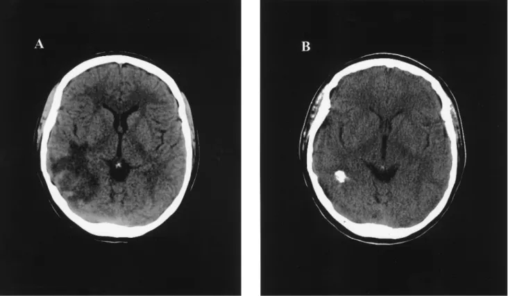
Figure 1. A, CT scan before empirical therapy for toxoplasmic encephalitis in an HIV-infected patient. B, CT scan 2.5 years later, after immune restoration with highly active antiretroviral therapy and 8 months without maintenance therapy for toxoplasmic encephalitis.
Discontinuation of Secondary Prophylaxis for Toxoplasmic Encephalitis in Human
Immunodeficiency Virus Infection After Immune Restoration with Highly Active Antiretroviral Therapy
Toxoplasma gondii is one of the leading causes of neurological
morbidity and mortality in patients with advanced HIV infec-tion. Effective therapies for primary and secondary prevention of toxoplasmic encephalitis in HIV infection have been estab-lished [1, 2]. With the introduction of highly active antiretroviral therapy (HAART), discontinuation of primary and secondary prophylaxis for opportunistic infections in patients with HIV infection and good response to HAART have become a focus of interest. We describe an HIV-infected patient with toxo-plasmic encephalitis and excellent response to HAART who discontinued secondary prophylaxis for toxoplasmosis.
A 26-year-old woman with HIV infection presented to our
Reprints or correspondence: Dr. Heiner C. Bucher, Medizinische Univer-sita¨ts-Poliklinik, Kantonsspital Basel, CH-4031 Basel, Switzerland (bucher @ubaclu.unibas.ch).
Clinical Infectious Diseases 2000; 30:602–3
q 2000 by the Infectious Diseases Society of America. All rights reserved. 1058-4838/2000/3003-0036$03.00
clinic with headache in November 1996. She had acquired HIV infection in 1986 by needle exchange, and, in the following years, did not seek regular medical assistance. She had not received any prophylaxis for opportunistic infections or treat-ment with antiretroviral drugs. Physical examination showed no neurological deficits. A CT scan revealed a 2.8-cm diameter solitary hypodense cerebral lesion with perifocal edema on the right parietotemporal hemisphere (figure 1A); this lesion was suspected to be toxoplasmic encephalitis. IgG antibodies to T.
gondii were positive at 34 U/mL, her CD4 cell count was 29/
mm3, and the plasma viral load was 302,000 copies/mL. The patient was empirically treated with pyrimethamine, sulfadia-zine, and leucovorin.
Four weeks later, a control CT scan showed regression of both the cerebral lesion and the perifocal edema. Because of a severe rash, sulfadiazine was changed to clindamycin, and after 6 weeks, the patient received maintenance therapy. Antiretro-viral therapy with lamivudine, stavudine, and indinavir was then started. The response was excellent, and in the following 18 months, the CD4 cell count progressively increased to11100/
mm3, and her plasma viral load was permanently suppressed (as measured by an ultrasensitive assay [Amplicor; Roche Di-agnostic Systems, Rotkreuz, Switzerland]).
CID 2000;30 (March) Brief Reports 603
therapy for toxoplasmic encephalitis be discontinued to reduce her pill load of 20 tablets a day. At this time, a CT scan showed a 1.7-cm diameter cerebral lesion with calcification that was identical to that on a previous CT scan at a 12-month follow-up. Twelve months after discontinuation of secondary prophy-laxis for T. gondii infection, the patient had no clinical signs of relapse, and her CT scan remained unchanged (figure 1B).
Data from observational studies on the discontinuation of primary prophylaxis for Pneumocystis carinii pneumonia in pa-tients with persistent CD4 lymphocyte counts 1200/mm3and antibodies to T. gondii have shown that no relapses of P. carinii pneumonia and T. gondii infection occurred [3, 4]. However, the number of patients with antibodies to T. gondii was too small to allow for firm conclusions as to whether discontinu-ation of primary prophylaxis for toxoplasmic encephalitis is safe.
Discontinuation of maintenance therapy for patients with established toxoplasmic encephalitis is not recommended [5]. Relapse rates after discontinuation of maintenance therapy, as reported from the pre-HAART era, may be>50% [2]. However, secondary prophylaxis for toxoplasmic encephalitis is all but satisfactory. Rates of breakthrough relapses associated with sul-fadiazine and pyrimethamine, the most effective drug combi-nation, are 10%–40%, and rates of drug-limiting toxicity as-sociated with this combination are 30%–43% [2].
Because of side effects, high pill load, and drug interactions, discontinuation of maintenance therapy for toxoplasmosis after immune restoration with HAART is likely to improve the
qual-ity of life in patients with AIDS and to increase adherence to antiretroviral therapy. Therefore, studies evaluating whether secondary prophylaxis for toxoplasmic encephalitis may be dis-continued for selected patients with excellent response to HAART seem timely.
Anne-Catherine Guex,1Alexander J. Radziwill,2
and Heiner C. Bucher1 1Medizinische Universita¨ts-Poliklinik and2Neurologische
Universita¨tsklinik, Kantonsspital Basel, Basel, Switzerland
References
1. Bucher HC, Griffith L, Guyatt GH, Opravil M. Meta-analysis of prophylactic treatment against Pneumocystis carinii pneumonia and toxoplasma en-cephalitis in HIV-infected patients. J Acquir Immune Defic Syndr Hum Retrovirol 1997; 15:104–14.
2. Katlama C, De Wit S, O’Doherty E, Van Glabeke M, Clumeck N. Pyrime-thamine/clindamycin vs. pyrimethamine/sulfadiazine as acute and long-term therapy for toxoplasmic encephalitis in patients with AIDS. Clin Infect Dis 1996; 22:268–75.
3. Furrer H, Egger M, Opravil M, et al. Discontinuation of primary prophylaxis against Pneumocystis carinii pneumonia in HIV-1–infected adults treated with combination antiretroviral therapy. N Engl J Med 1999; 340:1301–6. 4. Weverling GJ, Mocroft A, Ledergerber B, et al. Discontinuation of
Pneu-mocystis carinii pneumonia prophylaxis after start of highly active
anti-retroviral therapy in HIV-1 infection. Lancet 1999; 353:1293–8. 5. USPHS/IDSA Prevention of Opportunistic Infections Working Group. 1997
USPHS/IDSA guidelines for the prevention of opportunistic infections in persons infected with human immunodeficiency virus. MMWR Morb Mor-tal Wkly Rep 1997; 46(RR-12):1–46.
Intestinal Involvement by Nontuberculous Mycobacteria After Heart Transplantation
The prevalence of disease due to nontuberculous mycobac-teria (NTM) in the United States is 1.8 cases per 100,000 pop-ulation [1]. In the general poppop-ulation, 5%–10% of mycobacteria causing infections are NTM, whereas in transplant recipients NTM cause 25%–40% of all mycobacterial infections [2]. We describe a case of infection with Mycobacterium
avium/Myco-bacterium intracellulare in a heart transplant recipient and
re-view the literature on disease due to NTM [3–12].
A 56-year-old man underwent orthotopic heart transplan-tation in June 1990 for ischemic cardiomyopathy. In November 1992, he was admitted to the hospital because of fever (tem-perature, 397C) during treatment with cyclosporine, azathio-prine, and deflazacort. Physical examination disclosed a small epitrochlear node and a tender periumbilical mass. His
eryth-Reprints or correspondence: Dr. Miguel Yebra, Medicina Interna I, Clı´n-ica Puerta de Hierro, San Martı´n de Porres, 4, Madrid 28035, Spain (myebraf @nexo.es).
Clinical Infectious Diseases 2000; 30:603–5
q 2000 by the Infectious Diseases Society of America. All rights reserved. 1058-4838/2000/3003-0037$03.00
rocyte sedimentation rate was 92 mm/h. Blood cultures and serological tests for Brucella, Salmonella, Yersinia, cytomega-lovirus, and HIV were negative. Ziehl-Neelsen staining of urine and sputum samples were negative, as were cultures of these samples on Lo¨wenstein-Jensen medium. Purified protein deriv-ative skin testing was also negderiv-ative. A CT scan of the chest and abdomen revealed enlarged retroperitoneal and mesenteric lymph nodes.
Biopsy of the epitrochlear node revealed granulomas with scarce acid-fast bacilli, and culture of the node on Lowe¨nstein-Jensen medium was negative. Laparotomy showed mesenteric and retroperitoneal lymphadenopathies. Histopathologic ex-amination of the nodes demonstrated noncaseating granulo-matous adenitis, whereas Ziehl-Neelsen staining of the nodes and culture of the nodes on Lo¨wenstein-Jensen medium were negative. Azathioprine therapy was discontinued, and the cy-closporine dose was reduced. Subsequently, the fever subsided, the erythrocyte sedimentation rate normalized, and lymphad-enopathies regressed.
The patient remained asymptomatic until December 1993, when he presented because of abdominal pain and a 5-kg weight loss. Gastrointestinal examination revealed a distorted duo-denum with duodenal and jejunal mucosal thickening. An