Different molecular assemblies in two new phosphoric triamides
with the same C(O)NHP(O)(NH)
2skeleton: crystallographic study
and Hirshfeld surface analysis
Anahid Saneei1
·
Mehrdad Pourayoubi1·
Titus A. Jenny2·
Aurelien Crochet3·
Katharina M. Fromm3·
Ekaterina S. Shchegravina4Abstract Different molecular assemblies were compared in two new structures [4-CH3-C6H4C(O)NH]P(O)[NH]2 (-CH2)3, 1, and [4-CH3-C6H4C(O)NH]P(O)[NHC6H3(3,4-CH3)2]2, 2, belonging to the families of “cyclic phosphoric triamide” and “phosphoric triamide”, respectively. The differences in the hydrogen bond motifs were discussed (by single crystal X-ray diffraction) as a result of three factors: (1) action of two N atoms with a non-planar environment in 1as an H-bond acceptor, (2) different orientations of three N–H bond vectors in two molecules and (3) different conformations of C=O and P=O groups. These differences lead to more complicated hydrogen bond pattern of 1, with respect to that of 2, as structure 1 may be considered as a model of four-acceptor–three-donor versus a two-acceptor–
three-donor system in 2. The main discrepancies of 1 and 2, monitored by the Hirshfeld surface analysis, are related to the contribution portions of O···H/H···O contacts, in which compound 1 not only involves the greater existence of classical hydrogen bonds but also contains the further C– H···O weak interactions in its crystal packing with respect to compound 2. Instead, in 2, the shortage of O···H/H···O contacts has been partially compensated by the C···H/H···C interactions, due to the presence of more unsaturated car-bon acceptors. The differences in assemblies are also reflected in the solid-state IR spectra, especially for the N– H vibration frequencies. The new compounds were further studied by 1D NMR experiments (1H, 13C,31P), 2D NMR techniques [HMQC and HMBC (H–C correlation), HSQC (N–H correlation)], high-resolution ESI–MS, EI–MS spectrometry and IR spectroscopy.
Keywords Phosphoric triamide · Molecular assembly · Hydrogen bonding pattern · Hirshfeld surface analysis · NMR spectroscopy
Introduction
Supramolecular chemistry is a highly interdisciplinary field of science that has brought together investigators from many disciplines including chemistry, physics and biology (Desir-aju2010; Resnati et al.2015). The engineering of molecular crystals as well as the construction of supramolecular assemblies are both concerned with intermolecular interac-tions and hence are conceptually connected (Braga and Grepioni 1997). There is no doubt that the intermolecular contacts, especially hydrogen bonds, play a fundamental role in crystal engineering and stabilization of crystalline solids (Prins et al.2001; Steiner2002; Metrangolo and Resnati2008)
CCDC 1417623 and 1045702 contain the supplementary
crystallographic data for 1 and 2. These data can be obtained free of charge viahttp://www.ccdc.cam.ac.uk/conts/retrieving.html, or from the Cambridge Crystallographic Data Centre, 12 Union Road, Cam-bridge CB2 1EZ, UK; fax: (+44) 1223-336-033, or e-mail: [email protected].
& Mehrdad Pourayoubi [email protected]
1 Department of Chemistry, Faculty of Sciences, Ferdowsi University of Mashhad, P.O. Box: 9177948974, Mashhad, Iran
2 Department of Chemistry, University of Fribourg, Rte du Muse´e 9, 1700 Fribourg, Switzerland
3 Fribourg Centre for Nanomaterial’s, FriMat, University of Fribourg, Chemin du Muse´e 3, 1700 Fribourg, Switzerland 4 Department of Organic Chemistry, UNN Lobachevsky State
University, Gagarin av., 23, Nizhny Novgorod, Russia
Published in "Chemical Papers 71 (10): 1809–1823, 2017"
which should be cited to refer to this work.
and thus it is crucial to distinguish different types of interac-tions in any design strategy (Resnati et al.2015).
In recent years, phosphoramides have attracted attention in the viewpoint of crystal engineering (Pourayoubi et al.2014). The reasons for such interests are not only because they may be considered as small molecular models of some biologically active macromolecules such as phosphorus–nitrogen com-pounds like tetrapeptide-based phosphoramides (Wu and Hu
2016), but also because there are many examples of biologi-cally active phosphoramide small molecules (Upadhyay,
2012). On the other hand, the basic skeleton in the phospho-ramide compounds, P(=O)(N), is similar to the group occurring in peptides, (C=O)(N), and some similarity was found on the properties (Yizhak et al.2013) with differences arising from the tetrahedral environment in phosphorus and the planar environment in carbon, and also the greater elec-tron-donor capability of P=O with respect to C=O (Toghraee et al.2011). Moreover, the study of intermolecular interac-tions received by a phosphoramide small molecule is interesting, as they are usually similar to the case where such a molecule is placed in a biological environment such as an enzyme’s active site (Carletti et al.2013).
For the study of intermolecular interactions, apart from a traditional numerical analysis based on donor–acceptor distances for the characteristic contacts, the investigation can also be done using Hirshfeld surface analysis which is a new tool for graphical studying all interactions in a crystal combined with detailed quantitative information (McKinnon et al. 2004,2007). This method develops the study of intermolecular interactions to all of the contacts in the structure, including the weak ones (Michalik et al.
2014). Such an analysis is more precise, as the cumulative effect of weak interactions in crystal stabilization may sometimes have more important role than the classical hydrogen bonds which merely consider the interactions between the best hydrogen donor and hydrogen acceptor sites in the crystal. Additionally, this analysis is particu-larly useful in studying how different substituents and functionalities can affect the crystal packing and molecular assembly behaviour (Martin et al. 2015).
Herein, we give a comparative study of molecular assemblies and intermolecular arrangements in two new compounds belonging to the “cyclic phosphoric triamide” and “phosphoric triamide” families. Different hydrogen-bonded graph-set motifs and the complete superstructures of compounds are discussed, and the details of coopera-tion/competition of segments of molecules involved in intermolecular contacts are analysed with X-ray crystal-lography combined with 3D-Hirshfeld surface maps and 2D-fingerprint plots. The newly synthesized compounds are also studied by 1H, 13C,31P,1H–13C HMQC, 1H–13C HMBC and 1H–15N HSQC NMR, ESI–MS, EI–MS spec-trometry and IR spectroscopy.
Experimental
X-Ray measurementsData collection processes were performed for the structures 1(using Mo-Kα radiation, λ = 0.71073 A˚) and 2 (using Cu-Kα radiation, λ = 1.54186 A˚) with a STOE IPDS II diffractometer and a graphite monochromator. The struc-tures were solved with SIR92 (Altomare et al.1994) (for 1) and SUPERFLIP (Palatinus and Chapuis2007) (for 2) and refined using full matrix least squares on F2 with the SHELXL2014 (Sheldrick2015). All hydrogen atoms were included in the refinement at geometrically fixed positions and refined with a riding model (Sheldrick 2008). The molecular graphics were generated by MERCURY (Macrae et al. 2008) and DIAMOND (Brandenburg and Putz1999) for Windows.
Hirshfeld surface analysis
Historically, the Hirshfeld surface (HS) emerged from an effort to define the space occupied by a molecule in a crystal for the purpose of partitioning crystalline electron density into molecular fragments (Spackman and Byrom
1997). Such a surface was named in honour of F. L. Hir-shfeld, who introduced the “stockholder partitioning” scheme (Hirshfeld 1977). Afterwards, it was realized that the Hirshfeld surfaces possessed a number of attributes that make them attractive for identification of intermolecular interactions in the context of crystal packing (McKinnon et al.2004). In a Hirshfeld surface, the parameters deand di describe the distances from a point on the surface to the nearest nucleus outside and inside the surface, respectively. The dnormvalue is the sum of the normalised quantities of diand deby considering the van der Waals radius of atoms involved. This value graphically highlights the regions of the surface involved in a specific type of intermolecular contact by a coloured scheme: red regions represent con-tacts shorter than the sum of van der Waals radii; white regions represent intermolecular distances close to van der Waals contacts and blue regions represent contacts longer than the sum of van der Waals radii (Spackman and Jay-atilaka2009). The fingerprint plots (FPs) are introduced as the two-dimensional representations of the information provided by the generated HSs. The FPs are plotted on an XY-grid formed by de, dipairs (X= diand Y= de), where the frequencies of occurrence of interactions (the number of points with a given de, di pair) are represented by dif-ferent colours. Moreover, the complementary regions are visible in the FPs where one molecule acts as a donor (de[ di) and the other as an acceptor (de\ di) (Spackman and McKinnon 2002).
Spectroscopic measurements
IR spectra of 1 and 2 were recorded using a Buck 500 scientific spectrometer as KBr pellets. 1H, 13C, and 31P NMR spectra were recorded on a Bruker Avance 300 and a Bruker Avance III 500 spectrometer. The chemical shifts were determined relative to TMS for 1H and 13C, and relative to 85% H3PO4 for 31P, as an internal or external standard, respectively.1H–13C HMQC and1H–13C HMBC were carried out on a Bruker Avance III 500 spectrometer, and 1H–15N HSQC experiments were carried out on Agi-lent DDR2 400 spectrometer. The mass spectra were recorded with a MS model CH7A Varian (EI, 70 eV) and a MS model 5973 Network Mass Selective Detector (EI, 20 eV). High-resolution mass spectra were recorded on a Bruker FTMS 4.7 T BioAPEX II spectrometer.
Chemicals
4-Methylbenzamide, 1,3-propanediamine, 3,4-dimethy-laniline, phosphorus pentachloride, phosphorus pentoxide, acetonitrile and methanol were commercially available and chloroform was dried with P2O5and distilled prior to use. General procedure for the synthesis of compounds 1 and 2
4-CH3-C6H4C(O)NHP(O)Cl2 was prepared according to the method used for the analogous compound 4-NO2 -C6H4C(O)NHP(O)Cl2 by using 4-CH3-C6H4C(O)NH2 instead of 4-NO2-C6H4C(O)NH2 (Pourayoubi and Sab-baghi2009). To synthesize compounds 1 and 2 (the related chemical structures are shown in Table 1), a solution of
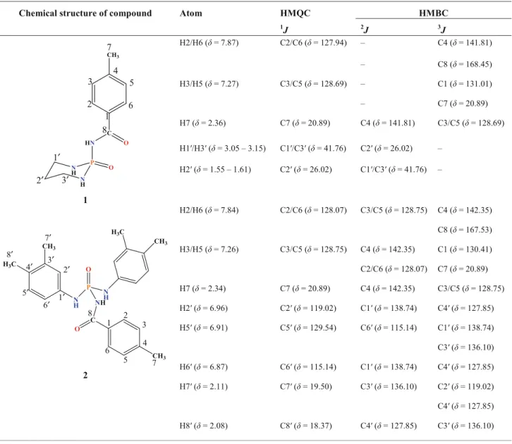
Table 1 2D1H–13C HMQC and HMBC correlations for 1 and 2
Chemical structure of compound Atom HMQC HMBC
1J 2J 3J N H P N H HN C O O CH3 1 2 3 4 5 6 7 1′ 2′ 3′ 8 1 H2/H6 (δ = 7.87) C2/C6 (δ = 127.94) – C4 (δ = 141.81) – C8 (δ = 168.45) H3/H5 (δ = 7.27) C3/C5 (δ = 128.69) – C1 (δ = 131.01) – C7 (δ = 20.89) H7 (δ = 2.36) C7 (δ = 20.89) C4 (δ = 141.81) C3/C5 (δ = 128.69) H1′/H3′ (δ = 3.05 – 3.15) C1′/C3′ (δ = 41.76) C2′ (δ = 26.02) – H2′ (δ = 1.55 – 1.61) C2′ (δ = 26.02) C1′/C3′ (δ = 41.76) – N H P O N H NH C O CH3 H3C CH3 CH3 H3C 1 2 3 4 5 6 7 1′ 2′ 3′ 7′ 8′ 4′ 5′ 6′ 8 2 H2/H6 (δ = 7.84) C2/C6 (δ = 128.07) C3/C5 (δ = 128.75) C4 (δ = 142.35) C8 (δ = 167.53) H3/H5 (δ = 7.26) C3/C5 (δ = 128.75) C4 (δ = 142.35) C1 (δ = 130.41) C2/C6 (δ = 128.07) C7 (δ = 20.89) H7 (δ = 2.34) C7 (δ = 20.89) C4 (δ = 142.35) C3/C5 (δ = 128.75) H2′ (δ = 6.96) C2′ (δ = 119.02) C1′ (δ = 138.74) C4′ (δ = 127.85) H5′ (δ = 6.91) C5′ (δ = 129.54) C6′ (δ = 115.14) C1′ (δ = 138.74) C3′ (δ = 136.10) H6′ (δ = 6.87) C6′ (δ = 115.14) C1′ (δ = 138.74) C4′ (δ = 127.85) H7′ (δ = 2.11) C7′ (δ = 19.50) C3′ (δ = 136.10) C2′ (δ = 119.02) C4′ (δ = 127.85) H8′ (δ = 2.08) C8′ (δ = 18.37) C4′ (δ = 127.85) C3′ (δ = 136.10)
2 mmol diamine (1,3-propanediamine) for 1 and 4 mmol amine (3,4-dimethylaniline) for 2 in dry CHCl3(5 mL) was added dropwise to a stirred solution of 1 mmol 4-CH3 -C6H4C(O)NHP(O)Cl2in dry CHCl3(20 mL) at 0°C. After 4 h, the solvent was removed in vacuum and the solid was washed with H2O. Suitable single crystals for X-ray crys-tallography were obtained at room temperature from a mixture of CH3OH/CHCl3(1:1) for 1 and CH3OH/CH3CN (3:1) for 2 (after a few days).
Spectroscopic data
4-Methyl-N-(2-oxido-1,3,2-diazaphosphinan-2-yl) benzamide, 1
M.p. 218 °C. ESI–MS = 254.10508. (calcd. for C11H 17-N3O2P+254.10529).31P{1H} NMR (121.49 MHz, DMSO-d6, 85% H3PO4): δ = 3.52 (s). 1H NMR (500.13 MHz, DMSO-d6, TMS):δ = 9.10 (very br. s, 1H, CONH), 7.87 (apparent d, J = 8.2 Hz, 2H, H2/H6), 7.27 (apparent d, J= 7.9 Hz, 2H, H3/H5), 4.49 (m, 2H, NH), 3.15–3.05 (m, 4H, H1′/H3′), 2.36 (s, 3H, H7), 1.61–1.55 (m, 2H, H2′).13C NMR (125.76 MHz, DMSO-d6, TMS):δ = 168.45 (s, C8), 141.81 (s, C4), 131.01 (d,3JPC= 7.4 Hz, C1), 128.69 (s, C3/C5), 127.94 (s, C2/C6), 41.76 (d, 2JPC= 3.5 Hz, C1′/ C3′), 26.02 (d,3JPC= 6.2 Hz, C2′), 20.89 (s, C7).15N–1H corr. (DMSO-d6) {–283.16, 4.52} (CH2NHPO) (CONHPO correlation peak was not detected). IR (KBr, cm−1): 3318, 3156, 2930, 2874, 1660 (C=O), 1460, 1271, 1219, 1194, 1095, 998, 822, 742. MS (70 eV, EI): m/z (%)= 30 (84), 43 (73), 56 (87), 72 (88), 91 (90), 117 (87), 119 (95), 135 (100), 136 (88), 155 (22), 181 (23), 251 (88), 252 (84), 253 (13). N-(Bis((3,4-dimethylphenyl)amino)phosphoryl)-4-methylbenzamide, 2
M.p. 248 °C. ESI–MS: 422.19962 (calcd. for C24H29N 3-O2P+422.19919).31P{1H} NMR (121.49 MHz, DMSO-d6, 85% H3PO4): δ = −4.48 (s). Mixture of rotamers (Major rotamer): 1H NMR (500.13 MHz, DMSO-d6, TMS): δ = 9.73 (d,2 JPH= 8.3 Hz, 1H, CONH), 7.84 (apparent d, J= 8.3 Hz, 2H, H2/H6), 7.55 (d,2JPH= 9.5 Hz, 2H, NH), 7.26 (apparent d, J = 7.9 Hz, 2H, H3/H5), 6.96 (d, J= 1.9 Hz, 2H, H2′), 6.91 (d, J = 8.2 Hz, 2H, H5′), 6.87 (dd, J = 8.1, 2.1 Hz, 2H, H6′), 2.34 (s, 3H, H7), 2.11 (s, 6H, H7′), 2.08 (s, 6H, H8′). 13C NMR (125.76 MHz, DMSO-d6, TMS): δ = 167.53 (s, C8), 142.35 (s, C4), 138.74 (s, C1′), 136.10 (s, C3′), 130.41 (d,3JPC= 8.8 Hz, C1), 129.54 (s, C5′), 128.75 (s, C3/C5), 128.07 (s, C2/C6), 127.85 (s, C4′), 119.02 (d,3JPC= 7.4 Hz, C2′), 115.14 (d, 3 JPC= 7.4 Hz, C6′), 20.89 (s, C7), 19.50 (s, C7′), 18.37 (s, C8′). 15N–1H corr. (DMSO-d6) {–258.09, 9.78}
(CONHPO), {−248.80, 7.64} (CArNHPO) (Minor rotamer): 1H NMR (500.13 MHz, DMSO-d 6, TMS): δ = 9.54 (d, 2 JPH= 8.7 Hz, CONH), 7.81 (apparent d, J = 8.3 Hz, H2/ H6), 7.41 (broad, NH), 7.23 (apparent d, J= 8.0 Hz, H3/ H5), 6.96 (d, H2′), 6.91 (d, J = 7.4 Hz, H5′), 6.84 (dd, H6′), 2.33 (s, 3H, H7), 2.13 (s, 6H, H7′), 2.09 (s, 6H, H8′).13C NMR (125.76 MHz, DMSO-d6, TMS):δ = 167.33 (s, C8), 142.00 (s, C4), 139.28 (s, C1′), 135.84 (s, C3′), 130.68 (d, 3 JPC = 8.6 Hz, C1), 129.95 (s, C5′), 128.67 (s, C3/C5), 128.01 (s, C2/C6), 127.13 (s, C4′), 118.61 (d, 3 JPC= 7.6 Hz, C2′), 114.75 (d,3JPC= 7.4 Hz, C6′), 20.86 (s, C7), 19.39 (s, C7′), 18.44 (s, C8′). 15N–1H corr. (DMSO-d6) {–258.60, 9.77} (CONHPO), {−249.55, 7.61} (CArNHPO). IR (KBr, cm−1): 3280, 3097, 2919, 1646 (C=O), 1615, 1511, 1442, 1393, 1359, 1279, 1225, 1165, 1116, 1016, 969, 923, 863, 817, 748, 685. MS (20 eV, EI): m/z (%)= 41 (47), 51 (40), 63 (34), 77 (43), 91 (63), 106 (89), 117 (64), 119 (32), 121 (100), 129 (3), 139 (6), 152 (10), 167 (5), 181 (9), 196 (3), 211 (9), 223 (3), 231 (3), 243 (13), 257 (1), 271 (3), 286 (3), 301 (12), 303 (3), 304 (9), 421 (8). Experimental NMR, IR and Mass spectra of compounds 1 and 2 are given in the Electronic Supple-mentary Information (ESI).
Results and discussion
Mass spectrometric analysisThe high-resolution ESI–MS characterization was carried out for two compounds, which very much supported the presented structures.
The EI mass spectra of compounds 1 and 2 were also recorded, which show the molecular ion peaks [M]+at m/ z 253 and 421, respectively. Compound 1 presents the [M −1]+fragment peak which can be attributed to loss of an H atom. The peak at m/z 117 in the mass spectra of two compounds is assigned to the 4-CH3-C6H4CN+ radical-cation which was formed by the removing of amidophos-phoric acid from the parent ion, as was reported for analogous [C6H5C(O)NH]P(O)[OR]2 phosphoramides by the removing of the esters of phosphoric acid (Mizrahi and Modro 1982). The observation of this fragment indicates that the parent radical-cation rapidly undergoes a migration of phosphorous from nitrogen to oxygen. From this resulting isomer a McLafferty fragmentation would pro-duce the C8H7N+ radical cation and neutral amidophosphoric acid. Typical for the McLafferty frag-mentation is the fact that the charge could appear alternatively on either fragment, i.e. the amidophosphoric acid part could also be charged and hence observed. Typ-ically, the signal related to amidophosphoric acid radical-cation was detected in the mass spectrum of 2 (m/z 304,
(HO)P(O)[NHC6H3-3,4-(CH3)2]2+) after loss of 4-CH3-C6H4CN. It is concluded that the pathway involving elimination of amidophosphoric acid molecule is preferred in 2, due to a considerable lower intensity of the ami-dophosphoric acid radical-cation compared to the 4-methyl-phenyl cyanide radical-cation in compound 2.
For compound 2, the peaks at m/z of 303 (assigned to the ion (O)P(O)[NHC6H3-3,4-(CH3)2]2+) and at 286 ([M −C8H9NO]+) are also revealed. The base peaks for com-pounds 1 and 2 are observed, respectively, at m/z of 135 (related to the 4-CH3-C6H4C(O)NH2+ ion) which results through a classical onium reaction (Bauerschmidt et al.
1992) and 121 (attributed to the 3,4-(CH3)2-C6H3NH2+ fragment ion).
The other important peak in these compounds appears at m/z 119, which is evidence for a bond scission of the amide bond in the NH–C(O) section generating the CH3-C6H4C (O)+ fragment, as reported for analogous compounds (Gholivand et al.2008).
IR and NMR study
Stretching frequencies of the N–H units occur at 3156 and 3318 cm−1for 1 and 3097 and 3280 cm−1 for 2. In both compounds, the lower stretching frequencies are related to NCP–H, engaged in hydrogen bonding interactions with the P=O group (with greater hydrogen bond strengths with respect to the NP–H···O=C hydrogen bond), as demonstrated by a comparison of IR spectra in various compounds and supported by quantum chemical calculations (Pourayoubi et al.2013). The reason for the higher NP–H stretching fre-quency of 1 with respect to that of 2 is related to the involvement of NP–H units in weaker hydrogen bonds as will be discussed in the X-ray crystallography section.
The phosphorus chemical shifts for 1 and 2 are observed at 3.52 and−4.48 ppm, respectively. The negative value for 2may be attributed to the magnetic anisotropy of aromatic rings (of two NHC6H3-3,4-(CH3)2 groups) on the phos-phorus atom existing in the cone-shaped shielding zone. Such negative values of phosphorus chemical shifts were also found in the31P NMR of analogous compounds, which typically include NHC6H4-4-CH3group bonded to P atom, for example: [4-F-C6H4C(O)NH]P(O)[NHC6H4-4-CH3]2 (−4.61 ppm) (Tarahhomi et al. 2011) and [CF3C(O)NH]P (O)[NHC6H4-4-CH3]2 (−5.38 ppm) (Gholivand et al.
2009).
Table 1lists the1H and 13C NMR assignments of both compounds, with the assignments achieved by 2D experi-ments. In the 1HNMR spectrum of 1 the broad signal at 9.10 ppm is related to the corresponding C(O)NHP (O) proton. For 2, a similar proton is revealed as a doublet signal at 9.73 ppm with2JPNH= 8.3 Hz. Compound 2 also shows a set of minor signals attributed to the other rotamer
in solution, whereby we bring here the minor signal for NH at 9.54 ppm with2JPNH= 8.7 Hz. The two rotameric forms interconvert slowly on the NMR time scale.
The multiplet signal at 4.49 ppm for 1 corresponds to two chemically equivalent NP–H protons in the P(O) [NH]2(CH2)3 segment. For 2, the fluxionality would explain why not only two signals at 7.41 ppm and 7.55 ppm are observed for the NP–H protons of the P(O)[NHC6H3 -3,4-(CH3)2]2 segment, but also why the first signal is so broad. The full assignments of major rotamer are also represented in Table1.
The aromatic protons of the 4-CH3-C6H4C(O) segment are revealed as two doublet signals in 1 and as two sets of two doublets in 2, in the range of 7.23–7.87 ppm.
In the 13C NMR spectrum of 1, the doublet signal at 131.01 ppm (3JPC= 7.4 Hz) is related to the C1 atom of 4-CH3-C6H4segment. For the major and minor rotameric forms of 2, the signal of similar C atom is also split as doublets at 130.41 (3JPC = 8.8 Hz) and 130.68 ppm ( 3-JPC= 8.6 Hz), respectively.
Furthermore, the carbon atoms of [NH]2(CH2)3part in 1 show both2JPC(at 41.76 ppm) and3JPC(at 26.02 ppm) in the observed doublet signals. For 2, the signals of NHC 6-H3(3,4-CH3)2 part include only doublets at 119.02 and 115.14 ppm for major rotamer and at 118.61 and 114.75 ppm for minor rotamer related to three-bond sep-aration phosphorus-carbon coupling (3JPC), besides the singlets for the other carbon atoms.
Moreover, the data of 2D 1H–13C HMQC and 1H–13C HMBC techniques were gathered in Table 1 in order to provide information about the interaction between the protons and the carbon atoms, which are directly attached to each other (via HMQC) and also for assignment of the carbon atoms not-bonded to the hydrogen atom (via HMBC) in compounds 1 and 2. The latter spectrum shows the connectivities between proton and carbon atoms with 2
J and3J relations.
Additionally, in the 1H–15N HSQC NMR spectra of 1 and 2 the following cross-peaks could be found: 4.52/– 283.16 ppm for NP–H of 1 and 7.64/−248.80 ppm and 7.61/ −249.55 ppm for NP–H in two rotameric forms of 2. The cross-peak of NCP–H in is absent 1, while two rotameric forms of 2 manifest the peaks at 9.78/−258.09 ppm and 9.77/−258.60 ppm. The disappearance of cross-peak noted for 1 may be due to the hydrogen bond formation in solution.
X-ray crystallography investigation General structural features
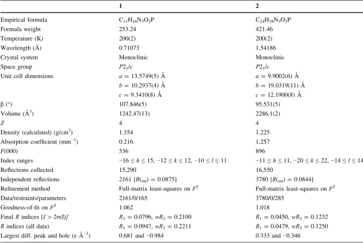
Both compounds 1 and 2 crystallize in the monoclinic crystal system, within the P21/c space group. The
crystallographic data and refinement parameters are pre-sented in Table 2. The asymmetric units of the two compounds contain one molecule each (Figs. 1, 2). The bond angles at the P atoms are in agreement with a distorted tetrahedral configuration with calculatedτ4 geometry index values (Yang et al.2007) of 0.94 (for 1) and 0.92 (for 2). In both structures, the O=P–NCP angles are smaller than the two O=P–NPangles, Table3(in our discussion, the NCPis used for the nitrogen atom within the C(O)NHP(O) segment and the NP is representation of two other nitrogen atoms attached to the phosphorous atom). The P=O and C=O bond lengths [respectively, 1.464(3) A˚ and 1.235(6) A˚ for 1 and 1.4734(11) A˚ and 1.228(2) A˚ for 2] are in the standard range for phosphoric triamides (Toghraee et al. 2011). The six-membered P1/N1/C3/C2/C1/N2 ring of 1 adopts a nearly half-boat conformation on the basis of puckering parameters calculated according to Cremer and Pople (1975) [Q= 0.500(6), θ = 155.1(6)°, Φ = 13.9(14)°]. The out of plane displacements calculated for the atoms of the ring are as follows: P1,−0.066(2); N1, 0.100(5); C3, −0.219(6); C2, 0.304(6); C1,−0.270(6) and N2, 0.151(5).
With respect to the nitrogen atoms, the main differences in 1 and 2 are related to the geometries at the N atoms bound to P,
calculated by the bond-angle sums at the N atoms (Σ). Structure 1includes two NPatoms which are located in the non-planar environment [Σ = 343(3)° and 350(3)°]. The criteria for dis-tinguishing between planar and non-planar geometries are the
Table 2 Crystal data and structure refinement for 1 and 2
1 2
Empirical formula C11H16N3O2P C24H28N3O2P
Formula weight 253.24 421.46
Temperature (K) 200(2) 200(2)
Wavelength (A˚ ) 0.71073 1.54186
Crystal system Monoclinic Monoclinic
Space group P21/c P21/c
Unit cell dimensions a= 13.5749(5) A˚ a= 9.9002(6) A˚
b= 10.2937(4) A˚ b= 19.0319(11) A˚ c= 9.3410(8) A˚ c= 12.1900(8) A˚ β (°) 107.846(5) 95.531(5) Volume (A˚3) 1242.47(13) 2286.1(2) Z 4 4 Density (calculated) (g/cm3) 1.354 1.225 Absorption coefficient (mm−1) 0.216 1.257 F(000) 536 896 Index ranges −16 ≤ h ≤ 15, −12 ≤ k ≤ 12, −10 ≤ l ≤ 11 −11 ≤ h ≤ 11, −20 ≤ k ≤ 22, −14 ≤ l ≤ 14 Reflections collected 15,290 16,550
Independent reflections 2161 [R(int)= 0.0875] 3780 [R(int)= 0.0844] Refinement method Full-matrix least-squares on F2 Full-matrix least-squares on F2
Data/restraints/parameters 2161/0/165 3780/0/285
Goodness-of-fit on F2 1.062 1.018
Final R indices [I[ 2σ(I)] R1= 0.0796, wR2= 0.2100 R1= 0.0450, wR2= 0.1232 R indices (all data) R1= 0.0947, wR2= 0.2211 R1= 0.0479, wR2= 0.1250 Largest diff. peak and hole (e A˚−3) 0.681 and−0.984 0.333 and−0.346
Fig. 1 Displacement ellipsoid plot (50% probability) is shown for 1 with atom numbering scheme. H atoms are drawn as spheres of arbitrary radii
same as previously proposed: N(planar) and N(pyramidal) refer to the cases withΣ ≥ 352.5° and Σ ≤ 339.0°, respectively, and the intermediate entries are the cases withΣ in the range 339.0°–352.5° (Allen and Bruno,2010). In 2, one NPatom is in a non-planar environment [Σ = 350(2)°] and the other NPatom indicates the planar environment [Σ = 360(2)°]. In both structures, the NCPatom is practically planar.
The nitrogen atom in a planar environment shows low Lewis base characteristic and does not take part in
hydrogen bonding as an acceptor, while there is a possi-bility (however small) that a nitrogen atom with a tendency to pyramidality acts in hydrogen bonding pattern as an acceptor. This knowledge is based on a survey on the Cambridge Structural Database (CSD, Groom et al.2016) for the structures with a P(O)NXY segment. There is no structure with an N atom as an acceptor in the C(O)NHP (O)-based phosphoric triamides in the CSD, and a few examples are related to the other families of phosphorus– nitrogen compounds (Pourayoubi et al.2014).
As will be discussed in the section of hydrogen bonding patterns, the number of H-acceptor sites in 1 develops to four and the structure of 2 only includes two acceptors in its hydrogen bonding pattern (Fig.3). On the other hand, in 1the number of H-acceptor sites is higher than the number of H-donor sites, leading to a bifurcated hydrogen bond (N–H)(···O)(···N). In 2, with a larger number of H-donor sites than H-acceptor sites, a double-acceptor site in the (N–H···)(N–H···)O=C entity is found.
Other differences between the two structures are related to the directions of N–H bond vectors which are illus-trated in Fig. 4. With respect to the plane crossing the three N atoms in 1 and 2, two NP–H units in the cyclic compound 1 are located nearly in the same side of the P=O group (one NP–H units has the syn-orientation and the other unit has a gauche-orientation) and in the acyclic compound 2, two NP–H units adopt an anti-orientation relative to the P=O group. The direction of NCP–H bond vector versus P=O is different from what was noted for NP–H bond vector with respect to the P=O (i.e. anti in 1 and syn in 2). The results of such different conformations are reflected in the various H-bonding patterns and diversity of graph-sets created which will be explained in more detail in the next section.
Hydrogen bonding pattern
In the crystal structure of 1, the N3–H3C unit takes part in a normal two-centred hydrogen bond, while surprisingly both N1–H1C and N2–H2C units are involved in the bifurcated intermolecular N–H(···O)(···N) entities. This is a novel feature of this structure and there are no C(O) NHP(O)(NH)2-based phosphoric triamides in the CSD including such three-centred hydrogen bonds. The reasons for the existence of such interactions are due to the presence of nitrogen atoms in a non-planar environment and also their role as an H-acceptor. Within these two N– H units noted, N2–H2C is near to three acceptors, but as will be discussed in the section of Hirshfeld analysis the third neighbour separation (H2C···O1) is a week inter-molecular contact.
The sites involving in the hydrogen bonds of 1 are three N–H units as donors (N1–H1C, N2–H2C and N3–H3C)
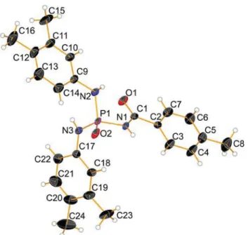
Fig. 2 Displacement ellipsoid plot (50% probability) is shown for 2 with atom numbering scheme. H atoms are drawn as spheres of arbitrary radii
Table 3 Selected bond distances (A˚ ) and angles (°) for 1 and 2 1 P1=O1 1.464 (3) P1–N1 1.627 (4) C4=O2 1.235 (6) P1–N2 1.632 (4) C4–N3 1.360 (6) P1–N3 1.685 (4) O1=P1–N1 114.9 (2) N1–P1–N2 106.6 (2) O1=P1–N2 113.2 (2) N1–P1–N3 104.8 (2) O1=P1–N3 109.89 (19) N2–P1–N3 106.7 (2) O1=P1–N3–C4 −45.8 (4) C4–N3–P1–N1 −169.9 (4) P1–N3–C4=O2 −13.9 (6) C4–N3–P1–N2 77.2 (4) 2 P1=O2 1.4734 (11) P1–N1 1.6824 (12) C1=O1 1.228 (2) P1–N2 1.6392 (14) C1–N1 1.3659 (18) P1–N3 1.6410 (13) O2=P1–N1 108.08 (6) N1–P1–N2 104.29 (7) O2=P1–N2 116.11 (7) N1–P1–N3 108.66 (7) O2=P1–N3 113.90 (7) N2–P1–N3 105.21 (7) O2=P1–N1–C1 −177.11 (12) C1–N1–P1–N2 −52.99 (13) P1–N1–C1=O1 11.7 (2) C1–N1–P1–N3 58.83 (14)
and four sites as acceptors including two O atoms (P1=O1 and C4=O2) as well as two N atoms with relatively high deviation from planarity (N1 and N2). Considering the contacts involving these sites, a 2D grid network is built parallel to the bc plane (Fig. 5). The basic framework of two-dimensional arrangement is a result of cooperation between three types of hydrogen bonds (N1–H1C···N2i, N2–H2C···O2ii and N3–H3C···O1iii; symmetry codes: (i)−x + 1, y + 1/2, −z + 3/2, (ii) –x + 1, −y, −z + 2 and (iii) x,−y + 1/2, z − 1/2, Table4). The other N1–H1C···O2i, N2–H2C···O1iiand N2–H2C···N2iihydrogen bonds do not extend the dimensionality of the hydrogen bond pattern; however, the 3D superstructure is a result of some weak interactions which will be discussed in the section of Hir-shfeld surface analysis.
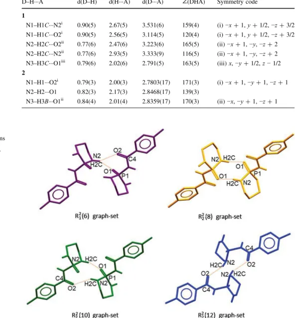
The important ring motifs, based on the hydrogen bonds noted, are shown in Fig. 6 which include R2
1ð Þ, R6 22ð Þ,8 R22ð Þ and R10
2
2ð Þ. Moreover, some high-order cyclic12 motifs can be distinguished in the structure as well as different linear hydrogen-bonded paths.
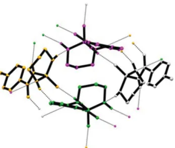
For an example of a bigger ring motif, we bring here the R4
4ð Þ graph-set which is the basic building unit of the 2D16 arrangement. This motif is built from hydrogen bonding interactions between four adjacent molecules via N1– H1C···N2i and N3–H3C···O1iii hydrogen bonds (Fig. 7).
The N2–H2C···O2ii hydrogen bond then connects the adjacent tetramers to each other.
An example of a hydrogen-bond chain graph-set in the structure 1 is shown in Fig. 8, which includes an assembly of five molecules through the N2–H2C···O1 and N3–H3C···O1 hydrogen bonds forming a zigzag C3
4ð Þ14 chain.
In addition to the hydrogen bonds discussed, the struc-ture of 1 may be rationalized by the weak cohesion from the C–H···O (C3–H3A···O2, C···O= 3.265 A˚) and C–H···π-electron ring (C2–H2A···π, H···Cg1 = 2.651 A˚, Cg1= centroid of C5/C6/C7/C8/C9/C10 ring) interactions. The hydrogen bond pattern of 2 is relatively simple, as the nitrogen atoms do not participate as acceptors. So, the
Fig. 3 A general representation for cyclic phosphoric triamide 1 (a) and phosphoric triamide 2 (b) showing the acceptor sites as specified with “A”. The hydrocarbon segments attached to the carbonyl group and secondary N atoms are shown with R′, R1and R2. The CH2CH2CH2linker (R2) is given as curved dashed line
Fig. 4 The directionality of the NP–H and NCP–H bond vectors versus P=O group relative to the plane involving three N atoms (a) for structure 1 and (b) for structure 2. Only the C–C(O)NHP(O)(NH)2 segment in both structures are shown for clarity. The mean planes are shown as red and orange colours
Fig. 5 A view of the two-dimensional array of 1 built from NCP– H···O=P, NP–H···O=C and NP–H···NPhydrogen bonds. The hydrogen bonds are shown as dotted lines and H atoms not involved in hydrogen bonds have been omitted for clarity
structure of 2 is a model of “two-acceptor–three-donor” systems. In the solid state, the molecules of 2 are hydrogen-bonded to each other making a 1D arrangement along the a axis, through the NCP–H···O=P and NP–H···O=C hydrogen bonds, Fig.9, forming alternative R22ð8Þ and R22ð12Þ graph-sets arranged through a chain path. This arrangement also includes S11ð6Þ graph-set motif made by NP–H···O=C intramolecular hydrogen bond.
By considering intermolecular C–H···π-electron ring interactions (C8–H8B···π, H···Cg1 = 3.421 A˚, Cg1 = cen-troid of C17/C18/C19/C20/C21/C22 ring and C23– H23C···π, H···Cg2 = 3.412 A˚, Cg2 = centroid of C2/C3/ C4/C5/C6/C7 ring), a 2D network parallel to (011) plane is formed by the connection of 1D hydrogen-bonded chains.
Study of intermolecular interactions by Hirshfeld surface analysis
For a better understanding of the packing maps and variety of interactions, it was decided to use a graphical tool for identification and understanding of intermolecular inter-actions. For this purpose, we used Hirshfeld surface (HS) analysis which is a useful method for discerning the close contacts around a component (molecule/ion).
Molecular Hirshfeld surfaces (dnorm and shape index) and 2D fingerprint plots of structures 1 and 2 were gen-erated by the CrystalExplorer 3.1 computer program (Wolff et al.2012). The HSs of 1 and 2 with the numerical labels for different contacts are given in Fig. 10a, b. Table 5 lists all of the contacts including the classical
Table 4 Hydrogen bond
geometries for 1 and 2 D–H···A d(D–H) d(H···A) d(D···A) ∠(DHA) Symmetry code 1
N1–H1C···N2i 0.90(5) 2.67(5) 3.531(6) 159(4) (i)−x + 1, y + 1/2, −z + 3/2 N1–H1C···O2i 0.90(5) 2.56(5) 3.114(5) 120(4) (i)−x + 1, y + 1/2, −z + 3/2 N2–H2C···O2ii 0.77(6) 2.47(6) 3.223(6) 165(5) (ii)−x + 1, −y, −z + 2 N2–H2C···N2ii 0.77(6) 2.93(5) 3.333(9) 116(5) (ii)−x + 1, −y, −z + 2 N3–H3C···O1iii 0.79(6) 2.02(6) 2.791(5) 163(5) (iii) x,−y + 1/2, z − 1/2 2
N1–H1···O2i 0.79(3) 2.00(3) 2.7803(17) 171(3) (i)−x + 1, −y + 1, −z + 1 N2–H2···O1 0.82(3) 2.17(3) 2.8468(17) 139(3)
N3–H3B···O1ii 0.84(4) 2.01(4) 2.8359(17) 170(3) (ii)−x, −y + 1, −z + 1
Fig. 6 Different graph-set motifs in structure 1; H atoms bonded to the C atoms have been omitted for the sake of clarity
hydrogen bonds and the weak interactions, with the bold text used for the classical hydrogen bond.
The label 1 is devoted to the most important interaction in both structures (Fig. 10a, b), namely between NCP–H unit and the oxygen atom of P=O group, appearing as the large red spots. The NCP–H···O=P interactions have the shortest N···O distances in two structures, as was noted earlier (Table 4). In the crystal structures of 1 and 2, there are two other NP–H units which take part in some
additional weak intermolecular interactions, especially NP– H···O=C in both structures and NP–H···NPin 1.
As mentioned in the section of X-ray crystallography, the N2–H2C unit of the NH(CH2)3NH segment in 1 is near to three H-acceptor atoms proposing the existence of N2– H2C···O2=C4, N2–H2C···N2 and N2–H2C···O1=P1 con-tacts. The two former interactions are seen as relatively large and small red spots, respectively (labels 2 and 7 in Fig. 10a). The possible N2–H2C···O1=P1 interaction has the H···O distance near the corresponding sum of the van der Waals radii and thus appears as white area. The N1– H1C unit of the NH(CH2)3NH segment also behaves as a double H-atom donor and takes part in N1–H1C···N2 (label 5, Fig.10a) and N1–H1C···O2=C4 (label 6, Fig.10a) weak intermolecular interactions. In contrast to 1, only one of the NP–H units in 2 participates in intermolecular hydrogen bonding (with C=O group) and revealed as a large red area in Fig.10b (labelled 2).
There are also two kinds of weak C–H···O intermolec-ular interactions in compound 1: CAr–H···O=P (label 3 in Fig. 10a), and CAliph–H···O=C (label 4 in Fig. 10a). The CAr–H···O=P contact also exists in structure 2 (label 3 in Fig.10b). All discussed C–H···O close contacts are viewed as small red areas in the related Hirshfeld surface maps.
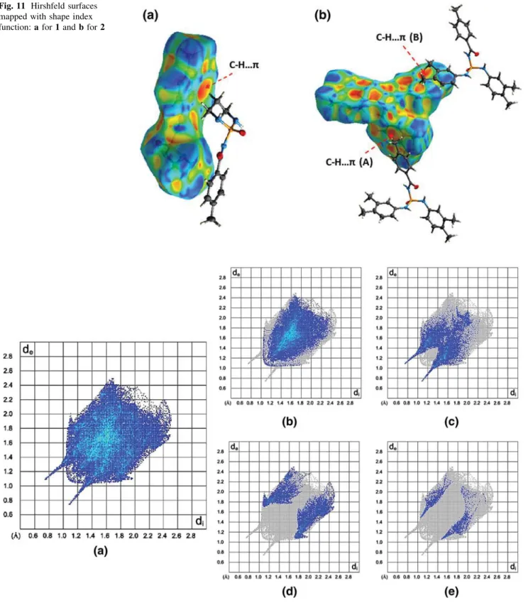
Figure 11a, b displays the Hirshfeld surfaces mapped with shape index functions for 1 and 2. Shape index is a feature of Hirshfeld surface analysis that allows for iden-tification of complementarity between molecules in the crystal packing so that features on the shape index surface that have an identical pattern but opposite colours indicate areas of intermolecular complementarity such as aromatic stacking interactions, e.g. C–H···π and π···π interactions. The red π-hole in Fig. 11a is related to the electron-rich aromatic system and indicates the presence of C–H···π
Fig. 7 Cyclic R4
4ð16Þ tetramer motif in the crystal structure of 1, formed via N1–H1C···N2i and N3–H3C···O1iii hydrogen bonds. H atoms not involved in hydrogen bonding have been omitted for clarity. The symmetry-related molecules are shown with different colours [symmetry codes: (i) –x+ 1, y + 1/2, –z + 3/2, (iii) x, –y + 1/ 2, z− 1/2]
Fig. 8 The C3
4ð14Þ chain graph-set in the crystal structure of 1, formed via N2–H2C···O1ii(red dashed line) and N3–H3C···O1iii(blue dashed line) hydrogen bonds. H atoms bonded to the C atoms have been omitted for the sake of clarity [symmetry codes: (ii) –x+ 1, –y, – z+ 2, (iii) x, –y + 1/2, z − 1/2]
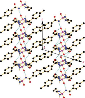
Fig. 9 A crystal packing diagram for structure 2, formed via N1– H1···O2iand N3–H3B···O1ii hydrogen bonds (dashed lines). The H atoms bonded to the C atoms have been omitted for clarity [symmetry codes: (i) –x+ 1, –y + 1, –z + 1, (ii) –x, –y + 1, –z + 1]
interaction, with the C–H from the CH2 part. The three electron-rich π systems are present in the structure 2 and two different C–H···π interactions (as red π-holes) marked (A) and (B) are found in the crystal, as shown in Fig.11b. In (A), one of the 3,4-(CH3)2-C6H3aromatic rings acts as a source ofπ-electrons and in (B), 4-CH3-C6H4ring provides such electrons for aforementioned interactions. For the interactions (A) and (B), the C–H units of methyl groups in 4-CH3-C6H4 and 3,4-(CH3)2-C6H3 groups behave as a donor, respectively.
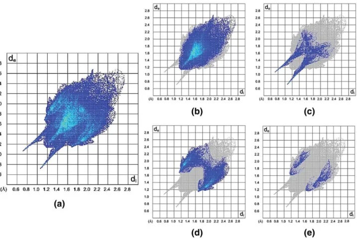
The full fingerprint plots (FPs) of compounds 1 and 2 are illustrated in Figs. 12a and 13a, respectively. These plots represent the total interactions which can be divided into the different interactions to show contribution portions of each contact in the total Hirshfeld surfaces. For two
Fig. 10 Front and back views of the Hirshfeld surfaces for molecules 1 and 2 are shown in aand b, respectively. Labels on HSs are explained in Table5
Table 5 Classical hydrogen bonds (as bold text) and other inter-molecular interactions with distances shorter than the sum of van der Waals radii for 1 and 2
Structure D–H···A Label Figures
1 N3–H3C···O1=P1 1 10a (large red spot) N2–H2C···O2=C4 2 10a (large red spot) C6–H6···O1=P1 3 10a
C3–H3A···O2=C4 4 10a N1–H1C···N2 5 10a N1–H1C···O2=C4 6 10a
N2–H2C···N2 7 10a
2 N1–H1···O2=P1 1 10b (large red spot) N3–H3B···O1=C1 2 10b (large red spot) C3–H3A···O2=P1 3 10b
structures, the H···H interactions (manifested in the middle area of the scattered points in the 2D fingerprint plots) make up the majority of the Hirshfeld surfaces (54.3 and 59.9%, Figs. 12b, 13b) relative to other contacts, as a
consequence of the higher number of hydrogen atoms in these structures.
Fingerprint plots of Figs. 12c and 13c show the O···H interactions in structures 1 and 2, with two sharp H-bond spikes in the regions of bottom right (de\ di, O···H) and
Fig. 11 Hirshfeld surfaces mapped with shape index function: a for 1 and b for 2
top left (de[ di, H···O) of the related plots, providing the closest contacts with minimum di+ devalues of≈1.8 A˚ for both structures. The O···H interactions (23.1% for 1 and
9.7% for 2) include both N–H···O and C–H···O contacts. As would be expected greater of O···H/H···O interactions are observed for 1, with all of N–H units involved in the superstructure. The fewer number of unsaturated carbon atoms in 1 is also reflected in smaller C···H/H···C contacts in 1 (17.9%), as compared to 2 (25.5%), Figs.12d and13d. The N···H contacts, showed in Figs. 12e and 13e, only make up 4.5% for 1 and 3% for 2, but are significant because the nitrogen atom bonded to phosphorus has a low Lewis base characteristic and the existence of N···H interactions is a highlight of these structures, with significantly more value for the structure 1 which includes two N atoms with remarkable deviance from planarity. A comparison of Figs. 12e and 13e (for N···H contacts) indicates that the shortest de+ di(shown as blue points on related FPs) for 1 is near 2.6 A˚ and for 2 is near 2.8 A˚, in which the shortest N···H contact in 1 is related to the weak intermolecular NP– H···NPi interaction (N1–H1C···N2i, Tables4,5).
In addition to the interactions discussed above, there are additional short contacts such as C···C and O···C that comprise only a small fraction of the Hirshfeld surfaces (Fig.14) and are not shown in related fingerprint plots.
Finally, a visual inspection of FPs in Figs. 12 and 13
shows that the upper deand divalues on the full FP of 1 are
Fig. 13 Fingerprint plots for 2. Close contacts are as follows: a all, b H···H, c O···H/H···O, d C···H/H···C and e N···H/H···N
Fig. 14 Relative contributions of various intermolecular contacts for structures 1 (inside circle) and 2 (outside circle)
more compact than those in the full FP of 2 (de\2.5 A˚ and di\ 2.5 A˚ in 1, de\ 2.7 A˚ and di\ 2.7 A˚ in 2), which indicates more efficient packing in 1.
Conclusions
Different molecular assemblies were compared in two new structures belonging to the families of “cyclic phosphoric triamide” (1) and “phosphoric triamide” (2). Both struc-tures include similar C(O)NHP(O)(NH)2skeleton and the differences observed in the hydrogen bond maps are driven by the following factors: (1) differences in the geometries of the nitrogen atoms bonded to the P=O group, where the N atoms with the non-planar environment in cyclic phos-phoric triamide cooperate as an H-bond acceptor, but none of the N atoms in the acyclic phosphoric triamide do not take part as an acceptor; (2) different orientations of P=O versus C=O; and (3) different orientations of N–H units bond vectors. So, structures 1 and 2 are considered as models of “four-acceptor– three-donor” and “two-accep-tor– three-donor”. The more complicated hydrogen bond pattern of 1 and the simple hydrogen bond pattern of 2 were discussed. The details of intermolecular interactions of 1 and 2 were studied by the Hirshfeld surface analysis. The main discrepancies monitored by such analysis are related to the contribution portions of O···H/H···O contacts, in which the compound 1 not only involves the greater existence of classical hydrogen bonds but also contains the further C–H···O weak interactions. In the structure of 2, the shortage of O···H/H···O contacts has been partially com-pensated by the C···H/H···C interactions, due to the greater presence of unsaturated carbon acceptors. In summary, the N atoms of diazaphosphorinane with non-planar environ-ment lead to the engaging of more acceptors in the hydrogen bonding pattern, which results in the created diverse hydrogen-bonded motifs as well as more com-pacted and higher density of 1 with respect to 2 that these observations were evidenced by fingerprint plot analysis.
Acknowledgements AS and MP would like to thank the Ferdowsi University of Mashhad for financial supporting this study (Grant No. 28386/3-2013/10/02). Crystallography experiments have been sup-ported by FriMat, University of Fribourg.
References
Allen FH, Bruno IJ (2010) Bond lengths in organic and metal-organic compounds revisited: X–H bond lengths from neutron diffraction data. Acta Crystallogr B 66:380–386. doi:10.1107/ S0108768110012048
Altomare A, Cascarano G, Giacovazzo C, Guagliardi A, Burla MC, Polidori G, Camalli M (1994) SIR92—a program for automatic
solution of crystal structures by direct methods. J Appl Crystal-logr 27:435. doi:10.1107/S002188989400021X
Bauerschmidt S, Hanebeck W, Schulz KP, Gasteiger J (1992) Elucidation of reactions in the mass spectrometer. Anal Chim Acta 265:169–182. doi:10.1016/0003-2670(92)85023-Y
Braga D, Grepioni F (1997) From alkynols to alkynol complexes. A molecular assembly study. Organometallics 16:4910–4919. doi:10.1021/om970331t
Brandenburg K, Putz H (1999) DIAMOND. Crystal impact. GbR Bonn, Germany
Carletti E, Colletier J-P, Schopfer LM, Santoni G, Masson P, Lockridge O, Nachon F, Weik M (2013) Inhibition pathways of the potent organophosphate CBDP with cholinesterases revealed by X-ray crystallographic snapshots and mass spectrometry. Chem Res Toxicol 26:280–289. doi:10.1021/tx3004505
Cremer D, Pople JA (1975) General definition of ring puckering coordinates. J Am Chem Soc 97:1354–1358. doi:10.1021/ ja00839a011
Desiraju G (2010) Crystal engineering: a brief overview. J Chem Sci 122:667–675. doi:10.1007/s12039-010-0055-2
Gholivand K, Madani Alizadehgan A, Mojahed F, Soleimani P (2008) Crystal structures and mass spectral fragmentation studies of some new carbacylamidophosphate compounds. Polyhedron 27:1639–1649. doi:10.1016/j.poly.2008.01.023
Gholivand K, Shariatinia Z, Mashhadi SM, Daeepour F, Farshidnasab N, Mahzouni HR, Taheri N, Amiri S, Ansar S (2009) Structural diversity in phosphoramidate’s chemistry: syntheses, spectro-scopic and X-ray crystallography studies. Polyhedron 28:307– 321. doi:10.1016/j.poly.2008.10.057
Groom CR, Bruno IJ, Lightfoot MP, Ward SC (2016) The Cambridge Structural Database. Acta Crystallogr B 72:171–179. doi:10. 1107/S2052520616003954
Hirshfeld FL (1977) Bonded-atom fragments for describing molecular charge densities. Theor Chim Acta 44:129–138. doi:10.1007/ BF00549096
Macrae CF, Bruno IJ, Chisholm JA, Edgington PR, McCabe P, Pidcock E, Rodriguez-Monge L, Taylor R, van de Streek J, Wood PA (2008) Mercury CSD 2.0—new features for the visualization and investigation of crystal structures. J Appl Crystallogr 41:466–470. doi:10.1107/S0021889807067908
Martin AD, Britton J, Easun TL, Blake AJ, Lewis W, Schro¨der M (2015) Hirshfeld surface investigation of structure-directing interactions within dipicolinic acid derivatives. Cryst Growth Des 15:1697–1706. doi:10.1021/cg5016934
McKinnon JJ, Spackman MA, Mitchell AS (2004) Novel tools for visualizing and exploring intermolecular interactions in molec-ular crystals. Acta Crystallogr B 60:627–668. doi:10.1107/ S0108768104020300
McKinnon JJ, Jayatilaka D, Spackman MA (2007) Towards quanti-tative analysis of intermolecular interactions with Hirshfeld surfaces. Chem Comm 3814–3816. doi:10.1039/B704980C
Metrangolo P, Resnati G (2008) Halogen versus hydrogen. Science 321:918–919. doi:10.1126/science.1162215
Michalik S, Maƚecki JG, Mƚynarczyk N (2014) Synthesis of [Re2Cl4(O)2(μ-O)(3,5-lut)4] and investigation of its structure via X-ray and spectroscopic measurements and DFT calcula-tions. Chem Pap 68:689–696. doi:10.2478/s11696-013-0493-7
Mizrahi V, Modro TA (1982) Phosphoric carboxylic imides. 1. Preparation and fragmentation behavior of dialkylphosphoryl (and phosphinyl) acetyl (and benzoyl) imides and related systems. J Org Chem 47:3533–3539. doi:10.1021/jo00139a030
Palatinus L, Chapuis G (2007) SUPERFLIP—a computer program for the solution of crystal structures by charge flipping in arbitrary dimensions. J Appl Crystallogr 40:786–790. doi:10.1107/ S0021889807029238
Pourayoubi M, Sabbaghi F (2009) Synthesis, spectroscopic charac-terization and crystal structure of a new acetyl phosphorylamidate P(O)[NHC(O)C6H4(4-NO2)][N(CH(CH3)2) (CH2C6H5)]2. J Chem Crystallogr 39:874–880. doi:10.1007/
s10870-009-9582-4
Pourayoubi M, Izadyar M, Elahi B, Parvez M (2013) Combination of X-ray crystallography and theoretical study to evaluate the effect of N–H···O=P versus N–H···O=C hydrogen bonds on the N–H stretching frequencies. J Mol Struct 1034:354–362. doi:10.1016/ j.molstruc.2012.10.055
Pourayoubi M, Toghraee M, Zhu J, Dusˇek M, Bereciartua PJ, Eigner V (2014) Database analysis of hydrogen bond patterns in phosphoric triamides completed with seven new compounds: a crystallographic and 15N NMR study. CrystEngComm 16:10870–10887. doi:10.1039/C4CE01793E
Prins LJ, Reinhoudt DN, Timmerman P (2001) Noncovalent synthesis using hydrogen bonding. Angew Chem Int Ed 40:2382–2426. doi:10.1002/1521-3773(20010702)40:13\2382:AID-ANIE2382 [3.0.CO;2-G
Resnati G, Boldyreva E, Bombicz P, Kawano M (2015) Supramolec-ular interactions in the solid state. IUCrJ 2:675–690. doi:10. 1107/S2052252515014608
Sheldrick GM (2008) A short history of SHELX. Acta Crystallogr A 64:112–122. doi:10.1107/S0108767307043930
Sheldrick GM (2015) Crystal structure refinement with SHELXL. Acta Crystallogr C 71:3–8. doi:10.1107/S2053229614024218
Spackman MA, Byrom PG (1997) A novel definition of a molecule in a crystal. Chem Phys Lett 267:215–220. doi: 10.1016/S0009-2614(97)00100-0
Spackman MA, Jayatilaka D (2009) Hirshfeld surface analysis. CrystEngComm 11:19–32. doi:10.1039/b818330a
Spackman MA, McKinnon JJ (2002) Fingerprinting intermolecular interactions in molecular crystals. CrystEngComm 4:378–392. doi:10.1039/b203191b
Steiner T (2002) The hydrogen bond in the solid state. Angew Chem Int Ed 41:48–76. doi:10.1002/1521-3773(20020104)41:1\48: AID-ANIE48[3.0.CO;2-U
Tarahhomi A, Pourayoubi M, Rheingold AL, Golen JA (2011) Different orientations of C=O versus P=O in P(O)NHC(O) skele-ton: the first study on an aliphatic diazaphosphorinane with a gauche orientation. Struct Chem 22:201–210. doi:10.1007/ s11224-010-9682-y
Toghraee M, Pourayoubi M, Divjakovic V (2011) Study on H-bond patterns in phosphoric triamides having a P(O)NHC(O) skeleton, a gauche orientation of P(O) vs C(O) in new compounds. Polyhedron 30:1680–1690. doi:10.1016/j.poly.2011.03.045
Upadhyay LSB (2012) Urease inhibitors: a review. IJBT 11:381–388 Wolff SK, Grimwood DJ, McKinnon JJ, Turner MJ, Jayatilaka D, Spackman MA (2012) CrystalExplorer (Version 3.1), University of Western Australia
Wu X, Hu L (2016) Design and synthesis of peptide conjugates of phosphoramide mustard as prodrugs activated by prostate-specific antigen. Bioorg Med Chem 24:2697–2706. doi:10. 1016/j.bmc.2016.04.035
Yang L, Powell DR, Houser RP (2007) Structural variation in copper (I) complexes with pyridylmethylamide ligands: structural analysis with a new four-coordinate geometry index,τ4. Dalton Trans 955–964. doi:10.1039/B617136B
Yizhak RV, Znovjyak KO, Ovchynnikov VA, Sliva TY, Konovalova IS, Medviediev VV, Shishkin OV, Amirkhanov VM (2013) Synthesis and crystal structures of new dioxouranium(VI) complexes based on carbacylamidophosphates (CAPh). Investi-gation of extraction properties of some CAPh ligands in respect of dioxouranium(VI) nitrate. Polyhedron 62:293–299. doi:10. 1016/j.poly.2013.06.043