HAL Id: tel-03143998
https://tel.archives-ouvertes.fr/tel-03143998
Submitted on 17 Feb 2021
HAL is a multi-disciplinary open access archive for the deposit and dissemination of sci-entific research documents, whether they are pub-lished or not. The documents may come from teaching and research institutions in France or abroad, or from public or private research centers.
L’archive ouverte pluridisciplinaire HAL, est destinée au dépôt et à la diffusion de documents scientifiques de niveau recherche, publiés ou non, émanant des établissements d’enseignement et de recherche français ou étrangers, des laboratoires publics ou privés.
Serotonin and emotional behaviours : developmental role
of the 5-HT7 receptor
Jimmy Olusakin
To cite this version:
Jimmy Olusakin. Serotonin and emotional behaviours : developmental role of the 5-HT7 receptor. Neurons and Cognition [q-bio.NC]. Sorbonne Université, 2019. English. �NNT : 2019SORUS287�. �tel-03143998�
Sorbonne Université
Ecole Doctorale Cerveau, Cognition et Comportement
Institut du Fer a Moulin, INSERM UMR1270
Team: Development of Neuronal Circuits
Serotonin and emotional behaviours: developmental role of the 5-HT7
receptor.
par Jimmy OLUSAKIN
Thèse de doctorat de la Sorbonne Université
Dirigée par Patricia Gaspar
Présentée et soutenue publiquement le 26 Septembre 2019
Devant le jury composé de :
Dr. Stephanie Daumas
President
Dr. Mark S. Ansorge
Rapporteur
Dr. Carine Becamel
Rapporteur
Dr. Massimo Pasqualetti
Examinateur
Dr. Fekrije Selimi
Examinatrice
Acknowledgement
Maya Angelou once said “people live in direct relation to the heroes and sheroes they have, always and in all ways”. My supervisor, a sheroe, whom I like to call « The Boss » has been a real mentor to me. She made my PhD journey smooth, her door is always open and at any time very easy to talk to, even at times when I come with ridiculous questions, she explains calmly with no judgements. For this and many more, I say a big thank you! The team members she put together, past and present, Audrey, Anne, Mariano, Sophie, Alex, and Aude have all been very welcoming, easy to discuss with and very supportive. I’d particularly want to appreciate Anne, for being readily available to discuss and criticize my project even after she left the team. A special thank you goes to Mariano for being very instrumental in designing my project, without you, I probably wouldn’t have been able to get this far. Thanks for being a friend and colleague.
To Susan Sara, another Sheroe, you were the reason I decided to pursue a PhD. You motivated me and have continued to do so over the course of the entire journey. I say thank you and God bless.
I’d like to specially thank the ENP and the Labex Biopsy for giving me the opportunity to be part of a prestigious PhD program. You guys made my Neuroscience dream a reality by awarding me the fellowship to come to Paris for this awesome training. To Laura, Yvette and Laetitia, you guys are the best! Settling-in in Paris was made very comfortable by you guys. I doubt anyone would have done a better job, from administration to welfare to housing, you guys made my transition here in Paris smooth and easy. You guys rock!
To my 2014-2018 ENP family, it still feels like yesterday when we all gathered together for our orientation. Over the course of these 4+ years, some of you guys have become real family to me. Soham, thanks for always being there for me, you helped me through some very difficult times and I appreciate everything. Ana, Pietro, Melisa and Sana, you guys have been my adopted siblings and friends. Thanks for not being judgmental, and accepting me, love you guys. Sebastien, Cristina, Arturo, Kasia, Alesandra, Bence, Moses and Yemisi thanks for our friendship over the years. You guys are a lovely addition to my friend circle.
My Parisian life would not have been interesting without friends like Jean-Marc, Theo, Audrey, Melanie, Fanny, and Delfina. Thank you guys!
A very big appreciation also goes to the entire IFM. I appreciate that we are all a one big happy family in this institute. The teams create a very conducive working environment where everyone is ready to help and make constructive criticism. I wouldn’t have asked for a better institute to work in Paris.
To my siblings, thank you for all the motivation and encouragement. I wish you all the very best life has to offer. Tosin, you have been my rock, from the very beginning of this journey. I go to bed knowing you always have my back. I know I can always count on your love and support. Thank you for everything! To Dideolu, my superman, you will always be my heart and even though it almost killed me not being there when you were born, all I do is for you my boy. I hope someday when you old enough to read this thesis, you will be proud of daddy.
To my parent, I cannot begin to write how grateful I am to you guys for everything you’ve done for me. I only hope I continue to make you happy and proud. Thank you for all the encouragement, motivation and prayers. You are the best!
Finally, all this work and journey has only been made possible through the strength, blessings and Grace from the Almighty. I appreciate the life YOU have given me to sail through it all.
Abstract
Antidepressants that block the serotonin transporter, (Slc6a4/SERT), selective serotonin reuptake inhibitors (SSRIs) improve mood in adults but have paradoxical long-term effects when administered during developmental periods, increasing the risk to develop anxiety and depression. The basis for this developmental effect is not known. In a previous study, we identified a subpopulation of layer 5–6 pyramidal neurons of the prefrontal cortex (PFC) that transiently expressed Slc6a4/SERT during an early postnatal period in mice (P0– P10). These PFC-SERT+ neurons establish glutamatergic synapses with subcortical targets, including the dorsal raphe nucleus (DRN). PFC-to-DRN circuits develop postnatally, coinciding with the period of PFC Slc6a4/SERT expression. Genetic ablation of SERT or early-life exposure to the SSRI, fluoxetine (from P2 to P14), increases the number of functional PFC glutamate synapses (hyperinnervation) on both 5-HT and GABA neurons in the DRN which also causes anxiety/ depressive-like symptoms. From our transcriptomic profiling of these PFC-SERT+ neurons at a developmental age of P7, we identified the Htr7 gene which could be mediating the developmental effects of SERT ablation. In this thesis, using anatomical and behavioural assays, I showed that pharmacological blockade of the 5-HT7 receptor using specific antagonist was sufficient to prevent the hyperinnervation to the DRN and the anxiety/depressive-like symptoms. In addition, we showed that the 5-HT7 knockout mice have no morphological and behavioural deficits. Furthermore, overexpression of 5-HT7 in the developing PFC, was sufficient to reproduce the anxiety/depressive-like symptoms observed in the absence of PFC-SERT. In conclusion activation of the PFC 5-HT7 receptor during development, could be responsible for the hyperinnervation of glutamatergic synapses to DRN and therefore precipitates the depressive-like behaviours.
Résumé
Les antidépresseurs, en bloquant le transporteur de sérotonine (Slc6a4 / SERT), améliorent l'humeur chez l'adulte, mais ont des effets paradoxaux à long terme sur le développement, augmentant ainsi le risque de développer l'anxiété et la dépression chez l’adulte. Les mécanismes à l’origine de cet effet sur le développement ne sont pas connus. Dans une étude précédente, nous avions identifié une sous-population de neurones pyramidaux de la couche corticale préfrontal 5 (PFC) qui exprimait de manière transitoire le transporteur SERT au cours de la période postnatale précoce chez la souris (P0-P10). Ces neurones PFC-SERT+ établissent des synapses glutamatergiques avec des cibles sous-corticales, y compris le noyau du raphé dorsal (DRN). L'ablation génétique de SERT ou une exposition précoce à la fluoxétine provoque une hyper-innervation synaptique dans le DRN, ainsi que des symptômes d'anxiété / dépression. Nous avons identifié le gène Htr7 qui pourrait médier les effets de SERT sur le développement. En utilisant des tests anatomiques et comportementaux, j’ai pu montrer que le blocage pharmacologique du récepteur 5-HT7 était suffisant pour prévenir une hyper-innervation du DRN et l’apparition de symptômes d’anxiété / dépression. De plus, nous avons montré que les souris knock-out 5-HT7 ne présentaient aucun déficit morphologique et comportemental. En outre, la surexpression de 5-HT7 dans le PFC en développement était suffisante pour reproduire les symptômes d'anxiété / dépression observés en l'absence d’expression de SERT par les neurones du PFC. En conclusion, l'activation du récepteur 5-HT7 au cours du développement dans le PFC peut être responsable de l'hyper-innervation synaptique dans le DRN et favorise ainsi l’apparition de comportements dépressifs.
FOREWORD
This thesis, contains three studies that will be discussed as separate chapters, preceded by a general introduction and conclusion.
.
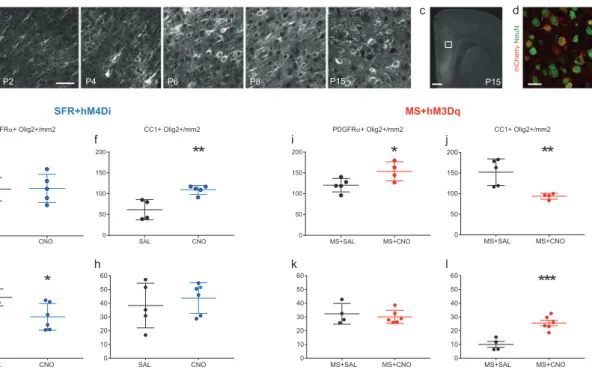
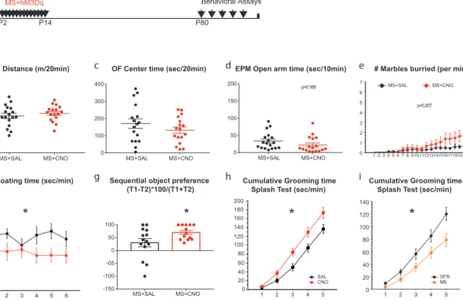
The first section, which has been published (article included), describes our findings on the precocious oligodendrocyte differentiation in the medial prefrontal (mPFC) of developing mice exposed to chronic maternal separation. Using an unbiased mRNA expression profiling in the mPFC of these mice, we demonstrated an increased expression of myelin-related genes and decreased immediate early genes. In adulthood, these mice showed a depletion of oligodendrocyte progenitor pool. My contribution to the
experiments in this study was the following:
-Injections of DREADD constructs into the mPFC of P1 mice -Western blot experiments
The second section, which has been published (article included), describes our findings on a subpopulation of prefrontal cortex (PFC) pyramidal neurons that transiently express the serotonin transporter (5HTT) during development. We demonstrated that developmental expression of 5HTT in this subpopulation of PFC neurons control synaptic maturation of PFC-to-DRN circuits, and remodeling of these circuits in early life controls behavioural responses to stress in adulthood. My contribution to
the experiments in this study was the following:
-In situ experiments showing the temporal expression of 5HTT mRNA in the PFC
-Pharmacological experiment showing that these 5HTT positive PFC neurons uptake 5-HT -Behavioural experiments and processing tissue for immunohistochemistry.
The third section describes a possible mechanism of PFC 5HTT+ neuron mode of action. From our transcriptomic data, we identified one 5-HT receptor, Htr7, which was strongly expressed in these neurons early during postnatal life. We observed a decrease in the expression of the Htr7 in the layer V neurons in the PFC of the 5HTT KO mice compared to the control mice. This observation led us to hypothesize that over-activation of the 5-HTR7 by 5-HT could be one of the mechanisms involved in morphological and behavioural deficits observed in both the 5HTT KO and in mice exposed to serotonin specific reuptake inhibitors.
I was the main investigator in this study, exploring two specific aims:
-Is over-activation of the Htr7 responsible for the hyper-innervation phenotype? -Is Htr7 implicated in the developmental effects of SSRIs on anxiety and depression?
Table of contents
INTRODUCTION ... 8
1.0 Development of emotional neural circuits ... 8
1,1 Critical periods in development ... 8
1.2 Critical periods in Limbic system development ... 9
2.0 Developmental role of Serotonin (5-HT) ... 12
2.1 Development of central 5-HT systems ... 12
2.2 The serotonin transporter: cellular target of SSRIs during development ... 13
3.0 5-HT receptors ... 14
4.0 5-HT regulated emotional and cognitive behaviours ... 17
4.1 Human association to depression phenotypes ... 17
4.2 Animal model-based association to depression phenotypes ... 20
4.3 5-HT regulation of PFC circuits ... 24
5 The 5-HT7 Receptor... 27
5.1 Gene structure and alternative splicing ... 27
5.2 Intracellular signaling of 5-HT7 ... 29
5.3 Tissue localization of the 5-HTR7 in the brain ... 31
5.4 Effect of 5-HT7 signalling on neuronal morphology and functions ... 35
5.5 Effects of 5-HT7 on other biological processes ... 35
CHAPTER I: Early life stress impairs postnatal oligodendendrogenesis and adult behavior through activity dependent mechanisms ... 41
CHAPTER II: SSRIs target prefrontal to raphe circuits during development modulating synaptic connectivity and emotional behavior ... 42
CHAPTER III: Serotonin and emotional behaviours: developmental role of the 5-HT7 receptor ... 43
Materials and Methods ... 44
Animals ... 44
Brain tissue processing ... 44
Western Blot ... 45
Cell Cultures ... 45
RT-qPCRs ... 46
Drug Administration (5-HT7 receptor agonist and antagonist) ... 46
Viral injections in the PFC ... 47
Array tomography ... 48
Behavioural studies ... 50
Statistical analyses ... 52
RESULTS ... 53
The 5-HT7 receptor (Htr7) is expressed in the PFC during early postnatal development ... 53
Pharmacological blockade of 5-HT7 receptor during the postnatal periods interacts with developmental fluoxetine effects. . 56
Emotional phenotype of the Htr7-KO mice; effects of PN-FLX in Htr7-/- and Htr7+/- mice ... 62
Overexpressing Htr7 in the developing PFC causes anxiety- and depressive-like behaviours in adulthood ... 67
Activation of PFC 5-HTR7 regulates PFC-DRN circuitry ... 70
DISCUSSION ... 73
Pharmacological manipulation of 5-HT7 during development alleviates anxiety- and depressive-like behaviours caused by
postnatal SSRI administration. ... 75
Anatomical effects of 5-HT7 receptor stimulation ... 77
Future perspectives ... 78
INTRODUCTION
1.0 Development of emotional neural circuits
1,1 Critical periods in development
Adaptation of an organism to its environment occurs through several mechanisms beginning during the prenatal period and lasting through the neonatal and early adolescent period. Environmental signals, through processes such as activity-dependent plasticity, interact with the unfolding genetic blueprint of the central nervous system, giving rise to a stable, individual phenotype (Ladd et al., 2000; Sroufe, 1997; Rutter et al., 1997). This in turn governs one’s perception of, and responsiveness to, salient features of the environment. This process of adaptation can be viewed as “fine tuning” or “environmental programming” of neural circuitry (Ladd et al., 2000).
Early in development, and across species, the presence of sensitive/critical periods for experience-dependent plasticity has been demonstrated. These sensitive periods critically shape functional connectivity between neurons and therefore regulate the wiring of the brain. A sensitive period may be defined as a definite portion of an animal’s life devoted to the shaping of neural connections. A relationship also exists between the critical period and brain weight; assuming that brain weight is a rough measure of brain complexity, then it follows that the more complex the brain, the longer the critical period. It therefore follows that this “window” where heightened plasticity resulting from extrinsic (environmental) or intrinsic (genetic) factors may shift developmental pathways and confer risk of mental or emotional disorders. (Berardi et al., 2000; Gingrich et al., 2017).
It is generally accepted that the functional maturation of cortical circuits particularly for the sensory systems, is characterized by developmental time windows, during which the influence of sensory experience on synaptic reorganization is predominantly strong (Fox, 1994; Katz and Shatz, 1996; Yuste and Sur, 1999; Kral et al., 2001). It has been documented that sensory functions reach maturity at the end of critical periods. Classical studies have indicated that the effects of sensory deprivation or alterations in sensory input are only apparent when the manipulation of the sensory input is made during the critical period and similar deprivations and/or alterations outside these periods (and in mature animals) have little or no effects.
1.2 Critical periods in Limbic system development
The most extensively studied critical period is that of the visual system. Inputs from both eyes first converge and compete for space in the central brain targets, both functionally and structurally. As postnatal development progresses into the critical window, several mechanisms, such as the induction of GABA neuron development, brain derived neurotrophic factors, and others, allows for molecular cascades leading towards dendritic spine motility, pruning and axonal retraction all leading to formation of ocular dominance columns (Hensch, 2005; Suri et al., 2015). Ocular dominance, is defined as the relative anatomical or physiological strength of connections from either eye to individual cells in the primary visual cortex. Sensory experience is essential for modifying ocular dominance maps during the critical period. Monocular deprivation (closure of one eye) during a critical period - postnatal day 13 to 30 in mice (Gordon and Stryker, 1996) causes amblyopia (a permanent loss of visual acuity through the closed eye) despite there being no damage to the retina or its target (Wiesel and Hubel, 1963; Berarchi et al., 2000).
Adult emotional characteristics emerges from an array of interactions between multiple limbic systems whose underlying neuronal circuit properties infer their function. As complex as the interconnectivity of the limbic circuitry are, their formation and connectivity is predominantly laid down during embryonic and postnatal development even though they retain some degree of plasticity in adulthood. It follows that like the sensory systems, the limbic circuit formation may also undergo sensitive and critical developmental periods, during which external factors can influence and modulate circuit formation, and consequently emotional behaviours encoded by them (Suri et al., 2015; Gingrich et al., 2017).
In humans or animals, similar deprivation-induced aberrations or early life stress (ELS) affect the limbic system which may become emotionally and socially "blind" and incapable of perceiving, processing, or responding in a normal fashion to social and emotional stimulation (Joseph, 1982; 1996). Various limbic neural assemblies require considerable social and emotional experience during specific developmental periods to shape appropriate neural connectivity and maturation, and are thus "experience expectant," and if deprived of that experience, even when "normal" experience is later provided they fail to fully develop or no longer function normally. Humans and animals nurtured in an abnormal, or deprived and socially and maternally impoverished environment typically display significant reductions in learning, perceptual, intellectual and memory capacity, as well as lessened curiosity and exploratory behaviour (Bremner et al., 1995; Stern et al., 1995; Joseph and Gallagher, 1980). These environmentally
prompted deficiencies include a reduced capacity to anticipate consequences or to inhibit irrelevant or inappropriate, self-destructive behaviours. In humans and other animals, this can result in severe disturbances in all aspects of social emotional expressive and perceptual functioning. So powerful is the "experience-expectant" limbic need for physical and social interaction that young animals raised in social seclusion will form attachments to inanimate objects, as well as to animals that might maul them (Joseph, 1992; Harlow et al., 1965; Hoffman and DePaulo, 1977). A study of over 100 children raised in an orphanage where mothering and social stimulation was minimal, found that within one year these children became unresponsive to social stimulation, and would lay passively on their beds. These motherless children made strong attempts to avoid strangers or toys, and when approached they would scream and withdraw. These deprived children spent hours engaged in obsessive, repetitive, stereotyped and bizarre self-stimulating movements; i.e. rocking, head banging, or pinching precisely the same piece of skin until sores developed. Most of these children became permanently emotionally and socially disabled (Spitz, 1945; 1946).
Studies in rodents have shown that recurrent periods of traumatic experiences, particularly in the first weeks of life or prenatally, induce modifications of synaptic connectivity in the limbic anterior cingulate cortex and hippocampus. (Helmeke et al., 2001a, b; Ovtscharoff and Braun, 2001; Poeggel et al., 2003). Furthermore, following 3 weeks of repeated restraint stress, Radley and colleagues observed significant decreases in apical dendritic length and branching on pyramidal neurons in the mPFC of adult rats. These changes were completely reversible 3 weeks after cessation of the stress indicating that stress-induced dendritic plasticity in the mPFC was reversible (Radley et al., 2005). However, when similar stress was administered during prenatal or early postnatal periods, this had long-lasting effects on dendritic re-organization (Murmu et al., 2006; Muhammad et al., 2012).
Decades of research on the patho-physiology of stress and anxiety related behaviours have implicated the hypothalamic-pituitary-adrenal axis (HPA), (Abe et al., 2007; Cottrell and Seckl, 2009; Entringer et al., 2009a; 2009b; Heim et al., 2009; Glover et al., 2009) and that in depression, there is hyperactivity of the HPA resulting from release of corticotropin-releasing factor (CRF) from hypothalamic and extra hypothalamic neurons (Raadsheer et al., 1994, 1995; Nemeroff et al., 1991; De Bellis et al., 1993). Increases in CRF neuronal activity is thought to mediate some depressive behaviours like sleep and appetite disturbances and reduced libido (Arborelius et al., 1999). In addition to the HPA, the monoaminergic systems and in particular the serotonergic one are clearly involved in stress-related disorders. Infact, some studies have shown significant changes in brain 5-HT turnover (synthesis and release) in both rodents and humans in response to HPA axis activation by different stressful conditions (Pei et al., 1990; Clement et al., 1993; Inoue et al., 1994). Reciprocally, other studies observed that 5-HT signalling influences corticosteroid release. They showed that administration of the Htr1A receptor
ligand, ipsapirone increases ACTH and cortisol levels in humans and animals (Wetzler et al., 1996; Klaassen et al., 2002).
Another direction of research has implicated dysfunction of the medial prefrontal cortex as a possible cause of stress-related psychopathologies – the pathophysiology of early stress. The mPFC, is implicated in behavioural flexibility, cognition, and stress- and anxiety-related behaviours (Robbins and Arnsten, 2009; Lupien et al., 2009). Functional neuroimaging studies implicated the default mode network (DMN) in ELS (Burghy et al., 2012; Philip et al., 2013a, b; Cisler et al., 2013). This network consists mainly of cortical midline structures (Northoff and Bermpohl, 2004; Raichle and Gusnard, 2005) including the medial prefrontal cortex (mPFC), posterior cingulate cortex, and superior temporal/inferior parietal cortex (Fox et al., 2005; Kim et al., 2010; Qin and Northoff, 2011). Human neuroimaging studies have shown that ELS is associated with reduced gray matter volume including that of the PFC (De Bellis et al., 2002; Andersen et al., 2008; Paus et al., 2008; Hanson et al., 2010). Other anatomical studies in animal models (rats) have demonstrated that ELS distrupts structural and functional plasticity in the mPFC in a multidirectional manner, particularly synaptic densities, dendritic lengths and branching (Baudin et al., 2012; Pascual & Zamora-Leon, 2007; Gos et al., 2008; Judo et al., 2010).
More generally, dysfunction of the mPFC has been implicated in depression. Using computer assisted cell counting techniques to reveal the cytoarchitecture of the cortex, Rajkowska and colleagues observed that patients with major depressive disorder (MDD), had reduced cortical thickness, decreased neuronal and glial densities in layers II through VI of the orbitofrontal and dorsolateral regions of the PFC (Rajkowska et al., 1999). Using local glucose metabolism and cerebral blood flow (CBF) as a marker of energy utilization associated with terminal field synaptic transmission, there are reports of altered physiological activities in regions of the PFC during depressive episodes (Drevets, 2007). For instance, the dorsomedial and dorsal anterolateral regions of the PFC were observed to show constantly reduced metabolism in depressed patients compared to healthy subjects (Baxter et al., 1989). Also, while executing a probabilistic reversal-learning task, unipolar depressives demonstrated lessened hemodynamic activity in the lateral orbitofrontal and ventrolateral parts of the PFC when compared to healthy subjects (Tavares et al.,2003). Other studies reported increases in hemodynamic responses in the posterior orbital cortex of PFC in depressed compared to non-depressed bipolar disorder subjects while performing a color-word stroop task (Blumberg et al., 2003).
Though detailed insight into the molecular factors essential for the maturation of the limbic circuit formation is still limited, 5-HT has emerged as one of such factors modulating both limbic circuit formation and adult functioning. From the neurodevelopment standpoint, because of its neurotrophic properties, 5-HT has been hypothesized to guide the maturation of brain regions associated with
emotional and cognitive behaviours. In favor of this hypothesis, the maturation of multiple limbic circuits, such as the amygdala, prefrontal cortex (PFC) and the hippocampus as seen by synaptic exuberance and subsequent pruning (Morys et al., 1998; Koss et al., 2014), temporally coincide with the increase of 5-HT levels in the postnatal brain (Loizou, 1972; Hohmann et al., 1988; Gingrich et al., 2017).
A mechanism which is a major player in the regulation of emotional state is the serotonin transporter (5HTT). It mediates the reuptake of 5-HT from terminals of 5-HT synapses, and a primary target of many antidepressants especially the serotonin selective reuptake inhibitors (SSRIs). SSRIs increase serotonergic tone and therefore recognized to mediate their therapeutic actions. Early life exposure to SSRIs has been shown to induce adult anxiety and depressive-like phenotypes in rodents (Ansorge et al., 2004; Homberg et al., 2010; Rebello et al.,2014) which is somewhat paradoxical to their mood relieving properties in adulthood.
Similarly, genetic downregulation of 5-HTT precipitates depressive phenotypes that have been shown to have developmental origins (Lira et al., 2003; Alexandre et al., 2006). In humans, a lesser-expressing form of 5-HTT gene has been associated with an increased risk of developing depression in response to early-life stress (Lesch et al., 1996; Murphy and Lesch, 2008)
2.0 Developmental role of Serotonin (5-HT)
2.1 Development of central 5-HT systems
All animals are endowed with serotonin neurons, the number of which is species dependent: for
instance, C. elegans has about 11 neurons (Loer and Rand, 2016), Aplysia has about 300 neurons (Hawkins 1989), Drosophila has about 106 neurons (Vallés and White, 1988), Zebrafish about 5000 neurons (Lillesaar and Gaspar, 2019), the mouse has about 26,000 cells (Ishimura et al., 1988), rats about 30,000 (Jacobs and Azmitia, 1992) and humans have about 300,000 cells (Baker et al., 1991). In vertebrates, these central serotonergic neurons are topographically arranged in the hindbrain. These neurons were originally organized into nine subdivisions from caudal to rostral by Dahlström and Fuxe in 1964, based on observations in rats and in human embryos. Classification of these cell clusters is based on their distribution and main projections into two groups – the rostral (B9-B4) groups and caudal (B3-B1) groups. The rostral being the largest group and comprises of the following subnuclei: caudal linear nucleus (CLi), supralemniscal cell group (B9), median raphe nuclei (MnR which was formerly B5 and B8) and the dorsal raphe nuclei (DR - B6 and B7). The caudal group comprises the raphe obscurus, raphe pallidus and raphe magnus (Steinbusch 1981; Jacobs and Azmitia, 1992; Hale and Lowry 2011).
The serotonergic neurons of the hindbrain are the earliest neurons to develop in the vertebrate CNS. This development goes through a protracted and complex period. In an early study of human embryonic development, Sundstrom and colleagues observed an ontogenic appearance of 5-HT immunoreactive (IR) cell groups in the pons and medulla starting at 5 weeks of gestation, a week after; a week later, 5-HT IR fibers were observed in the white matter throughout the entire spinal cord. Then at the 9th week, fibers were seen in the gray matter (Sundstrom et al, 1993). From the 15th week of gestation onwards, 5-HT cell bodies in the brainstem, were clearly clustered as distinct midline raphe nuclei. This distribution was very similar to those observed in other mammalian species (Takahashi et al., 1986). Developmental studies in rodents, initially in rats showed that, the first 5-HT neurons appear at the 12th gestational day and shortly after start releasing 5-HT and mature until around postnatal day 21 (Lidov and Molliver, 1982). In mice, the first raphe 5-HT neurons appear at E10, and development of the 5-HT axon arbours in their targets continues over the first two postnatal weeks (Maddaloni et al., 2017, rev in Deneris and Gaspar, 2019).
Additionally, it has been demonstrated that 5-HT receptors are expressed early in embryonic ages even before serotonergic afferents reach their innervation targets (Bonnin et al., 2006; Hellendall et al., 1993). It has been argued that these 5-HT receptors exact their signaling properties via placental 5-HT release (Bonnin et al., 2011). Another level of interesting complexity was the observation that during late embryonic to early postnatal development, some population of non-5-HT neurons adopt 5-HT phenotypes. These neurons do not have the capacity to synthesize 5-HT as they lack the rate-limiting enzyme for 5-HT synthesis – tryptophan hydroxylase 2 (TPH2), but they have the intrinsic properties to uptake 5-HT with high affinity (serotonin transporter, 5-HTT, SERT, SLC6A4) and degrade (monoamine oxidase A – MAOA) 5-HT (Lebrand et al., 1996; 1998; Gaspar et al., 2003).
2.2 The serotonin transporter: cellular target of SSRIs during development
5-HTT is the primary target of the selective serotonin reuptake inhibitors (SSRIs). Therefore, identifying the cell types that expresses this transporter (SLC6A4) during development is vital to our understanding of the neurodevelopmental consequences of early SSRI exposure. In the adult brain, 5HTT expression is limited to the 5-HT producing neurons of the raphe nuclei. However, a much broader expression was observed in developing rodents (Gaspar et al., 2003; Narboux-Neme et al., 2008). Using genetic fate mapping approaches, our lab previously described a precise cellular localization of 5-HTT expression in the developing brain (Narboux-Neme et al., 2008). This made it possible to identify the principal targets of SSRIs during different phases of brain development. At mid gestation (embryonic day 11, E11, in mice) expression of the 5HTT gene begins in the 5-HT neurons of the raphe nuclei, but
expression soon extends to non-serotonergic neurons, including the principal projection neurons of the sensory systems (thalamus, retina, somatosensory cortex), the corticolimbic pathways (hippocampus, E14– E15, and the prefrontal/cingulate cortex, postnatal day 0, P0). 5-HTT expression in these glutamatergic developing neurons ends rapidly during the second postnatal week, coinciding with the maturation of their neural circuits. Although studies in humans are limited, they do provide further evidence for the broad developmental expression of HTT. In 8–11 gestational week-old embryos, 5-HTT immunolabelling was visible in fiber tracts of the internal capsule and in the optic tract. These fibers do not correspond to raphe projections (Verney et al., 2002). In new world monkeys (marmosets), 5-HTT was observed in all major sensory afferents (dorsal root ganglia, retinal ganglion cells, cochlear nucleus and olfactory nerves) at mid-gestation (Lebrand et al., 2006), an epoch which generally corresponds to the rodent perinatal period of neural circuit development. Unfortunately, the scarcity of biological material from primates has prevented evaluation of the precise time course of developmental 5-HTT expression, making it difficult to correlate these observations with the larger body of information collected from rodents (Homberg et al., 2010).
3.0 5-HT receptors
Studies dating back to key criteria for receptor characterization highlighted three main factors to consider. These include the operation or drug-related characteristics, signal transduction or receptor-effect coupling and the structural or gene and receptor structural sequences for their nucleotide and amino acid components (Humphrey et al., 1993). With this in mind, initial studies identified three 5-HT receptor families, 5-HT1-3 comprising five receptors/binding sites with the suspicion of some others (Bradley et al., 1986). At that time, a 5-HT autoreceptor function was ascribed to the 5-HT1-like receptor and it was hypothesized that the 5-HT3 receptor was mediating a depolarizing effect on the CNS neurons. However, not much was known about the functionality of the individual receptor subtypes (Barnes and Sharp, 1999).
The advent of more specific agonists and antagonists and in particular the advancement in molecular biology techniques allowed to identify 15 different 5-HT receptor subtypes, encoded by different genes in humans and rodents (Hoyer and Martin, 1997). It is now known that 5-HT, acts via two main categories of receptors – the ionotropic and metabotropic receptors. The ionotropic receptors or ligand-gated ion channels are identified by their low affinity for their neurotransmitter ligand but have a quick activation (in order of few milliseconds). In contrast, metabotropic receptors act via G-protein activation and second messenger production. They exhibit high ligand affinity and have a delayed activation, (in the order of seconds).
The only ionotropic 5-HT receptor is the 5-HT3 receptor. It has a pentameric structure. Functional channels may comprise of 5 identical 5-HT3a receptor subunits (homopentameric) or a combination of 5-HT3a and one of the other four subunits, 5-HT3b,c,d or e (heteropentameric). It seems that only the 5-HT3a receptor subunits is capable of forming functional homopentameric channels and all other subunit subtypes must heteropentamerize with 5-HT3a receptor subunits to form functional channels (Niesler et al., 2007). The 5-HT3 receptors are characterized by the occurrence of 4 transmembrane segments and a large extracellular N-terminal region. The functional receptor consists of 5 subunits arranged around a central ion conducting pore permeable to sodium, potassium and calcium ions. Upon activation by 5-HT the channel opens leading to an excitatory response in neurons.
All other 5-HT receptors are metabotropic. They are G-protein coupled receptors (GPCRs) having seven transmembrane domains. Based on their pharmacological properties, amino acid sequences, gene organization and second messenger coupling, the 15 receptor subtypes have been divided into six distinct classes – 5-HT1, 2, 4, 5, 6 and 7 receptors. The diversity of these receptors is further heightened by alternative splicing of some receptor subtypes.
Table 1. Serotonin receptors and their signaling pathways:
(+) stimulation; NT, not tested; PLC, phospholipase C; PKC, protein kinase C (Adapted from Yun and Rhim, 2011).
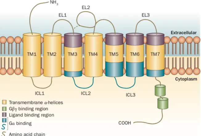
Figure 1: Schematic diagram of GPCR structure.
GPCR structure can be described as having 3 parts: (1) an extracellular domain comprising of the N terminus and 3 extracellular loops (EL1, EL2 and EL3); (2) the transmembrane (TM) domain consisting of 7 a-helices (TM1-TM7); and (3) the intracellular domain consisting of 3 intracellular loops (ICL1-ICL3), an intracellular amphipathic helix (H8), and the C terminus (Figure 1). A characteristic property of the extracellular domain of most GPCRs is the presence of disulfide bridges particularly at the top of TM3 that helps the receptor stability and limits the conformational freedom of this region during receptor activation (Venkatakrishnan et al., 2013).
4.0 5-HT regulated emotional and cognitive behaviours
4.1 Human association to depression phenotypes
Early studies on human subjects put forward a hypothesis of 5-HT deficiency in depression. This was proposed based on the findings of low 5-HIAA levels in the CSF of depressed patients (Sarna et al., 1983; Mignot et al., 1985) and suicidal victims (Asberg et al., 1986). These findings have been argued to reflect more the MAO activity than 5-HT release or utilization (Wolf et al., 1985). There have also been reports of low serotonergic activity inferred from allelic variation of genes involved in 5-HT synthesis, transmission and uptake in depressed individuals (Arango et al., 2003). In the late 90’s, Mayberg and colleagues proposed that depression involves a disruption of limbic-cortical pathways, based on evidence showing that regional glucose metabolic rate response (rCMRglu) is altered in depressed patients and that there are changes in brain metabolism after treatment with anti-depressants (Mayberg et al., 1997). This was further reinforced by the observation that depressed subjects exhibited significantly lower rCMRglu in the prefrontal and frontal cortices and in the inferior parietal lobe and the insula, compared to healthy subjects (Anderson et al., 2004).
Several 5-HT gene regulatory polymorphisms have been associated with negative emotionality and predisposition to depression and suicidality. In addition, stress hormones have been shown to be involved in regulating the expression of certain serotonin system genes. I will highlight a couple of the gene polymorphisms most linked to human emotional states.
The T allele is a functional polymorphism in the upstream regulatory region of TPH2 (SNP G-703T, rs4570625) which is responsible for low-expression of TPH2 (Gutknecht et al., 2007; Reuter et al., 2007). An impact of TPH2 variation on amygdala activation was reported by Milak and colleagues. Using positron emission tomography techniques on baboons, they found that transient reduction of cerebral 5-HT by acute tryptophan depletion increases amygdala’s responses to fearful faces. They argued that the increase amygdala response is likely due to a compensatory decrease of 5-HTT function and concomitant increase of synaptic 5-HTavailability (Milak et al., 2005).
Another genetic polymorphism of 5-HT regulatory gene linked to major depression is the C(-1019)G 5-HT1A promoter polymorphism. Negative feedback inhibition of serotonergic raphe neurons is facilitated by somatodendritic 5-HT1A autoreceptors (Mongeau et al., 1997) and several antidepressant compounds desensitize raphe 5-HT1A autoreceptors consequently enhancing 5-HT neurotransmission (Albert et al., 1996; Artigas et al., 1996). Conversely, postmortem brains from depressed suicidal victims have shown increased 5-HT1A receptor densities in the raphe nuclei but not at postsynaptic sites. This translates to decreased serotonergic activity in depressed patients (Stockmeier et al., 1998). Lemonde et al., 2003 observed a two-fold increase in expression of the homozygous G(-1019) allele in depressed patients and a four-fold increase in expression of the same allele in suicide victims compared to controls. They proposed a model where the C(-1019)G polymorphism prevents binding of the transcriptional repressor - nuclear deformed epidermal autoregulatory factor (NUDR) therefore resulting in enhanced 5-HT1A receptor expression in raphe neurons (Lemonde et al., 2003).
In an association study describing the possible association of a T941G single nucleotide polymorphism (SNP) in the monoamine oxidase A (MAOA) gene with generalized anxiety disorder (GAD), panic disorder (PD), and major disorder (MD), Tadic and colleagues reported no association of the MAOA-T941G polymorphism with MD and PD but patients with GAD of Caucasian descent had significantly higher frequency of the 941T allele as compared to healthy subjects (Tadic et al., 2001). This SNP is located in the third base of a codon and does not affect the amino acid sequence. However, association of the allele with lower MAOA enzyme activity has been reported in cell lines of known MAOA activity (Hotamisligil and Breakefield, 1991). Supporting the notion of MAOA polymorphism and emotional disorders, a study conducted by Brunner in 1993 is arguably one of the clearest genetic evidence implicating MAOA activity in human behaviour. The authors identified a C to T point mutation at position 936 in the MAOA gene. This mutation introduced a premature termination codon leading to a nonfunctional MAOA protein. The males manifested borderline mental retardation, impulsivity, aggression, arson, attempted rape and exhibitionism (Brunner et al., 1993).
By regulating the degree and extent of serotonergic responses, the 5-HT transporter (5-HTT) is central to the fine-tuning of brain serotonergic neurotransmission and of the peripheral actions of 5-HT. The human 5HTT is encoded by a single gene (SLC6A4) on chromosome 17q12. 5HTT is the target of selective serotonin reuptake inhibitors (SSRIs) that block 5-HT reuptake with the effect of increasing the half-life of 5-HT at the synapses. The SLC6A4 gene promoter contains a glucocorticoid response element that makes it responsive to stress-induced levels of corticosteroids (Glatz et al., 2003; Lopez and Higley, 2002). Polymorphism in the translational control region upstream of the 5HTT coding sequence (5HTTLPR) – has been reported to give a long and short variant (the polymorphism consists of 44-base pair insertion or deletion) with different translational efficiencies (Lesch et al., 1996).
In the literature, the involvement of this 5HTTLPR polymorphism in emotional behaviours have been inconsistent and somewhat contradictory. There has been documented studies showing significant association between the low-expressing 5HTTLPR short variant and neuroticism with traits related to anxiety, stress reactivity and depression (Caspi et al., 2003). Since this discovery in 2003, many studies have investigated whether interaction of these polymorphisms with adverse environmental factors could be a predisposing factor to major depressive disorders. Using genome-wide association studies (GWAS), particularly gene x environment association, quite a number of research papers have argued that subjects with either one or two copies of the 5HTTLPR short variant had higher scores of neuroticism than those who were homozygous for the 5HTTLPR long variant (Canli and Lesch, 2007; Lesch et al., 1996; Xie et al., 2009; Grabe et al., 2012). Additionally, postmortem studies have described decreased 5HTT expression in brains of suicidal victims. However, the causal role of 5HTT polymorphism on depression is still debated. Some investigators did not find any correlation between depression risk and 5HTTLPR, whereas the effect of environmental factors such as lifetime and recent stressful events, sexual abuse, childhood trauma; on the prevalence and course of major depressive disorders was confirmed (Peyrot et al., 2013; Fisher et al., 2012). It should be noted that the model postulated by researchers in favour of a predisposition to negative emotional states by subjects carrying the s-allele of the 5HTTLPR is quite counter-intuitive considering antidepressants further inhibit the actions of 5HTT. As discussed later in experimental models, this argued for a developmental effect of changed 5HTT expression.
In view of the ongoing debate on candidate gene x environment risk for depression, Border and colleagues performed a study where they empirically identified 18 genes, with polymorphisms and canonical risk alleles implicated in depression. This included 5HTT/SLc6a4. The selection of these candidates was done by literature search of depression-focused journal articles on PubMed (from 1991 to 2016). Then in a large case-control samples with GWAS (>500,000) from the population, they examined candidate gene and candidate gene-by-environment (GxE) interaction for evidence supporting a role in major depressive disorders (Border at al., 2019). Their results show no significant effect for any of the candidate gene polymorphisms selected for interaction or association to depression phenotypes. They conclude that previous studies reporting gene polymorphism association with depression phenotypes are false positives because the sample sizes employed in those studies are small and underpowered. They further argue that extracting significance for genetic effects from GWAS for depression would require approximately 34,000 individuals to be detectable with 80% power at a significance of 0.05 but the median sample sizes of previous studies is about 345, with a power of 65% (Border at al., 2019).
4.2 Animal model-based association to depression phenotypes
Studies looking at polymorphisms of 5-HT genes and association with depression phenotypes in nonhuman primates are few. A comparative study conducted by Lesch et al in 1997 described that variants of the 5HTTLPR are present only in simian primates (Lesch et al., 1997). They reported a long (419bp) and short (398bp) allelic variant of the 5HTTLPR in the rhesus monkey (rh-5HTTLPR). Because of their resemblance to humans in temperamental traits, Champoux and colleagues assessed the association between the rh-5HTTLPR and behavioural characteristics induced by 5-HT functioning in a sample of 115 infant rhesus monkeys with well characterized environmental histories. (Champoux et al., 2002) Their study showed 3 main observations – (1) a significant effect of genotype on emotionality – monkeys with the l/s genotype demonstrated more struggling, were less easily consoled, and manifested higher amounts of, and more frequent emotional distresses than the l/l homozygotic infants, (2) Nursery reared animals with the l/s genotype had substantially low orientation scores (an indirect measure of attention) compared to their maternally reared counterparts and (3) these observed behaviours became more obvious with increase in the age of the animals.
In a different experiment, the same authors assessed the effect of GxE in alcohol sensitivity. Here peer-reared (PR) and mother-reared (MR) monkeys with either the l/l or l/s genotype were infused with 16.8% ethanol through the saphenous veins and their intoxication scores were measured (Barr et al., 2003). They observed that animals homozygous for the l allele had significantly lower average intoxication scores compared to their l/s mates. Moreover, the PR animals with the l/l genotype had lower intoxication scores than did PR l/s animals. They concluded that, though their results cannot be directly translated to humans as quantifying alcohol intake is very complex, it is important to consider potential environmental influence on gene effects when studying the pathogenesis of alcohol dependence.
The rodent models support a significant role for serotonergic genotypes in determining psychiatric vulnerability. Investigating the effects of GxE as it relates to depressive phenotypes in rodents is only possible by the use of transgenic mouse or rat models. The down side of such models are the difficulty to really access the functional implication of the gene during developmental time point considering the life-long perturbation of the gene in question, leaving room for compensatory mechanisms. Therefore, using pharmacological tools (agonists and antagonists), the developmental function of a gene can be assessed during sensitive developmental windows.
In the rest of this chapter, while focusing on rats and mouse models, I will review concurrently, the effects of transgenic and pharmacological models (i.e. genetic inactivation of the gene and inactivation/activation during development respectively) of some 5-HT genes involved in depressive phenotypes within the scope of my thesis.
MAOA knockout mice were generated by the insertion of an interferon b (IFN-b) transgene between exons 1-4 of the MAOA gene. (Cases et al., 1995) the observed behaviours of pups were mostly locomotor, motor co-ordination defects. Similar to what had been observed in human males with a null MAOA mutation, adult mice showed increased aggressiveness, even among littermates. They had an anti-depressive phenotype when tested in the forced swim test, decreased anxiety as noted by longer time spent in the centre of the open field. At PND7, the authors observed disorganization of the barrel cortex, increased density of 5-HT staining by immunochemistry in the striatum, cortex, hilus of dentate gyrus and in neurons of the locus coeruleus and nigral complex, indicating an increase uptake of 5-HT (Cases et al., 1995, Vitalis et al., 1998). However, adult mice showed normal brain distribution and density. A reason for this could be the presence of the MAOB isoform present in non-neuronal cells in the adults (Cases et al., 1995; Vitalis et al., 1998). There is a gradual rise of MAOB specifically in postnatal life which could normalize adult monoamine metabolism (Tsang et al., 1986).
While MAOA action influences all monoamine levels, 5HTT function particularly regulates 5-HT signaling. Following the observation in humans that anti-depressants alleviates depressive symptoms, it was thought that generating a mouse model lacking 5HTT would be a good model to test the mechanisms underlying this. However, the contrary was observed because 5HTT knockout mice, had increased anxiety, learned helplessness, behavioural despair, and impaired social interaction (Ansorge et al., 2004; Kalueff et al., 2007; Lira et al., 2003; Muller et al., 2011). The 5-HTT knockout mice were generated by homologous recombination, by replacing the second exon of the SLC6A4 gene with a neomycin cassette (Bengal et al., 1998). This led to loss of the full length, functional 5HTT protein in the brain, and as expected, mice homozygous for the null mutation exhibit an absence of 5-HT reuptake, giving rise to a decreased rate of synaptic 5-HT clearance and a four- to six-fold increase in basal levels of forebrain extracellular 5-HT (Montanez et al., 2003).
Mutant mice lacking one SLC6A4 allele (5-HTT heterozygous) show a deficiency in 5-HT reuptake and clearance that is about 50% lower than controls (wildtype) (Bengal et al., 1998; Montanez et al., 2003). They reported only modest compensatory alterations in dopamine or norepinephrine function in 5HTT knockouts (Murphy et al., 2001; Montanez et al., 2003). There were, however, marked changes within the serotonergic system itself. 5HTT knockout mice showed a reduction in dorsal raphe neuronal firing and a desensitization and downregulation of somato-dendritic 5-HT1A autoreceptors (Gobbi et al., 2001; Li et al., 2000; Mannoury la Cour et al., 2001).
The 5HTT knockout mice showed reductions in the binding density of postsynaptic 5-HT1A
receptors (125p-MPPI – selective radioligand of 5-HT1A) in the frontal cortex, amygdala, septum, and
hypothalamus, but not the hippocampus (Li et al., 2000; Mannoury la Cour et al., 2001). Changes in the expression of other 5-HT receptor subtypes appeared to be less profound and more region-specific (Fabre et al., 2000).
In the hypothalamus and amygdala, the binding density of 5-HT2A and 5-HT2C receptors was increased respectively (Li et al., 2003). Not much is known about the status of the other 5-HT receptor subtypes.
Behaviorally, the 5HTT knockout mice bred on the C57BL/6J background spent less time in the aversive, open arms of the elevated plus-maze and showed less exploration of brightly lit areas in both the light/dark exploration and emergence tests. In the open field arena, knockouts showed general reduction in exploratory locomotion and greater thigmotaxic “wall-hugging” behaviours. Taken together, a robust increase in anxiety-like behavior was observed in 5HTT knockout mice. (Holmes et al., 2003; Gingrich et al., 2008) It is worth mentioning that these phenotypic abnormalities were not observed in some mice bred on other genetic backgrounds. The 5HTT knockouts that were bred onto a congenic 129S6 and 129 backgrounds had no observable anxiety-related phenotype (Holmes et al., 2003). Holmes et al argued that a possible reason for this could be that variable baseline behavioral phenotypes in these mice strains could mask the effects of slc6a4 null mutation.
In the test for depressive phenotypes - forced swim test (FST) and tail suspension (TST), the 5HTT knockout mice are reported to show normal levels of immobility on first exposure, but on second exposure, they had significantly higher immobility compared to WT (Zhao et al. 2006). Some studies however, reported an increased immobility following acute testing with the FST; in this case, the mutation was on a 129S6 genetic background whereas the increased depressive behaviour following repeated exposure were observed on the C57BL/6 background (Lira et al. 2003; Zhao et al. 2006). Increased immobility in the FST exemplifies a ‘despair-like’ response, in which the animal disengages from active forms of coping in a manner that may have presumed relevance to symptoms of hopelessness and entrapment (Porsolt 2000; Cryan and Holmes 2005). Taken together, it appears that the genetic background of the mice could determine sensitivity to the effects of stressful events in the 5HTT knockout mice and the C57BL/6J alleles might be conferring relative protection against the depressive phenotypes seen in the 5HTT null mutation (Caroll et al., 2007).
From the studies mentioned above, increases in extracellular 5-HT by genetic ablation of the 5HTT gene results in anxiety and depressive-like phenotypes in mice. Interestingly, constitutive mutants for
serotonin synthesis - TPH2 knock-in mice with drastic (about 80%) reduction in 5-HT levelsexhibit pro
depressive and anxiogenic behaviours (Beaulieu et al., 2008). The generation of these mouse mutants was based on the discovery of a rare human mutation – R441H TPH2 from a small cohort of elderly patients with major unipolar depression (Zhang et al., 2005). The R441H TPH2 mutation was engineered at a similar R439H amino acid residue of the mouse Tph2. This was done by homologous recombination resulting in the insertion of the mutation in exon 11 and of a residual loxP site in the ninth intron of the
gene (Beaulieu et al., 2008). A direct measure of Tph2 activity in vivo was done by monitoring accumulation of 5-HT precursor and Tph2 product – 5-HTP following treatment of mice with the L-aromatic amino acid decarboxylase inhibitor, m-hydroxybenzylhydrazine. (Zhang et al., 2004) Synthesis levels in the frontal cortex, striatum and hippocampus were reduced by approximately 40% and 80% in the heterozygous and homozygous R439H Tph2 knockin mice, respectively. Tissue contents of 5-HT were also substantially reduced in these same brain regions of R439H homozygous mice. The authors reported that both heterozygous and homozygous R439H Tph2 knockin mice showed prolonged immobility times in the TST, in the light/dark test for anxiety, they displayed longer latencies to cross to the lighted compartment, as well as reduced activity in this compartment. The homozygous mice exhibited more transitions between the two compartments. Their results indicated that expression of R439H Tph2 results in enhanced anxiogenesis in mice (Beaulieu et al., 2008).
Taken together, severe depletion and elevation of 5-HT establishes the set point for adult emotionality. Because of the trophic function of 5-HT during development, it was hypothesized that developmental 5-HT signalling could be establishing the set point for adult emotionality. To test this hypothesis requires temporal control over serotonergic signalling.
Therefore, pharmacological investigation of the 5-HT system during development, although it may lack 100% target specificity, has shed important light in advancing or confirming the developmental role of 5-HT in adult emotional behaviours. Ansorge and colleagues decided to test this hypothesis by temporarily inhibiting 5HTT function between postnatal days 4 and 21 (P4 and P21) by intraperitoneal administration of a known SSRI - fluoxetine (FLX) in mice (Ansorge et al., 2004). FLX is a common SSRI, with an extended half-life used in humans to alleviate adult depressive symptoms. They crossed
heterozygous mice with the 5-HTT mutation (5HTT+/-) to produce a Mendelian combination of 5HTT+/+,
5HTT+/-, and 5HTT-/- offspring. Mixed litters were randomly allocated to either saline or FLX (10 mg/kg, intraperitoneally) treatments beginning on P4 and lasting until P21. This strategy allowed to directly compare the behavioral effects of transient pharmacological 5HTT inhibition and constitutive disruption of the 5HTT gene (Ansorge et al., 2004). Adult mice we tested for anxiety related behaviours. Interestingly, they observed a decrease in exploratory and locomotor behaviours and time spent in the
open arm of the elevated plus maze in the postnatal FLX (PN-FLX) treated 5HTT+/+ and 5HTT+/- animals
in comparison to the saline-treated animals. PN-FLX treatment did not modify the already existing anxiety behaviours observed in the 5HTT-/-, demonstrating that the effects of FLX were particularly
mediated by 5-HTT blockade. Their study showed that PN-FLX treatment of 5HTT+/+ and 5HTT+/- mice
mimicked the anxiogenic behaviours of 5HTT-/- mice treated with either FLX or vehicle. They reported
that the decrease locomotor activity observed in the open field and the elevated plus-maze was probably due to the novelty of these environments, because no differences in locomotion were observed when mice were tested in their home cage.
Furthermore, they tested animals in the novelty suppressed feeding (NSF) paradigm. This test is believed to reflect anxiety- and depression related behaviors because chronic antidepressant administration and anxiolytics reduce the latency of animals to begin feeding (Santarelli et al., 2003) and because animal models of depression and anxiety behave abnormally in this test. They observed that
PN-FLX treated 5HTT+/- mice had longer latencies to begin feeding sessions, a behaviour seen also in
the 5HTT-/- mice but not in the 5HTT+/+ mice. Weight loss from food deprivation, and food consumption in the home cage were comparable across groups, suggesting that the observed differences in latency were not due to motivational factors. This study by Ansorge et al showed that 5HTT function modulates the development of brain systems involved in emotional and stress related responses and that these responses can be recapitulated by PN-FLX administration during a set developmental window.
In line with these findings, the same authors, went further on to understand and refine the 5-HT sensitive developmental period, and elucidate possible morpho-anatomical changes in emotional circuitry resulting from increase in extracellular 5-HT secondary to PN-FLX administration (Rebello et al, 2014). Their experiments showed that shortening the PN-FLX exposure window from P21 to P2-P11 was sufficient to impact adult emotional behaviours. P12-P21 and P21-P41 exposure had no impact on adult emotionality in mice. Interestingly, this developmental window coincides with the maturation period of both serotonergic afferents and cortical circuits (Lidov and Molliver., 1982; Kiyasova and Gaspar., 2011; Vitalis et al., 2013).
4.3 5-HT regulation of PFC circuits
Converging studies have shown the relevance of the PFC in the integration of behaviour (Duncan, 2001; Fuster, 2001; Miller and Cohen, 2001) and its implication in the pathophysiology of neuropsychiatric disorders (Weinberger et al., 2001; Harrison, 2002). In an effort to understand the cellular and molecular basis of PFC function, particularly its relevance to the induction of depressive-like phenotypes, some studies described the expression pattern of 5-HT receptors within regions of the PFC and corticolimbic circuits (Barnes and Sharp, 1999), physiology of PFC neurons, and their functions during behavioural tasks (Durstewitz et al., 2000; Miller and Cohen, 2001). A model of impaired 5-HT regulation, particularly the 5HTT knockout model, investigators have begun understanding how excess 5-HT in the synapse affect the cytoarchitecture of developing PFC neurons and corticolimbic circuits implicated in anxiety-like behaviours (Esaki et al., 2005; Hariri and Holmes, 2006).
The 5HTT knockout mice show deficiencies in anxiety related behaviors and have proved to be a good model of depressive phenotypes. (Bengel et al., 1998) A study found that 5HTT knockout mice
completion of fear conditioning. When checked for morphological abnormalities, 5HTT knockout mice had significantly greater spine density on the fourth-order basolateral amygdala dendritic branches of these neurons, relative to wildtype. (Nietzer et al., 2011; Wellman et al., 2007) This is in line with the increases in excitability of the amygdala observed in patients with anxiety disorders (Etkins and Wager, 2007). More so, Wellman and colleagues observed significantly elongated dendrites of the infralimbic pyramidal neurons of the mPFC compared with WT mice (Wellman et al., 2007). These findings along with others confirm the reciprocal connection (Canteras et al., 1992; Bacon et al., 1996; McDonald et al., 1996; Berretta et al., 2005) between the mPFC and amygdala and their dysfunctionality in the pathophysiology of depression and anxiety disorders (Siegle et al., 2002; Phillips et al., 2003; Phelps et al., 2004). Though these morphological changes observed in the 5HTT KO mice do not provide a direct evidence of functional alterations in the basolateral amygdala (BLA) and infralimbic (IL) mPFC, it provides reasonable correlates of neural substrates underlying the behavioral deficits observed in these mice. An explanation could be that increased spine density of the BLA resulting from the behavioural paradigm employed, could be interpreted as an increased excitatory drive (or hyperactivity) of the BLA neurons. With this in mind, the increased dendritic extent of the IL neurons of the mPFC (via its strong inhibitory inputs to the amygdala) may be an adaptive response to dampen this hyperactivity (Wellman et al., 2007).
There are reports of increased expression of the 5-HT1A mRNA in the layers II/III of the PFC of rats exposed to ELS (Gross et al., 2002). In line with these findings, Goodfellow and colleagues showed increased 5-HT1A mediated currents in layer II/III of PFC of these animals as compared to their wildtype counterparts at the third postnatal weeks. However, in adulthood, both groups of animals had comparable 5-HT1A responses (Goodfellow et al., 2009). Interestingly, they showed that pups exposed to ELS had attenuated 5-HT1A responses after exposure to social isolation in adulthood. They argued that since these upper layer neurons communicate with other cortical neurons like the amygdala, a disinhibition of these neurons in the PFC of ELS animals exposed to social isolation is responsible for the increase in excitability in the amygdala of those rats (Goodfellow et al., 2009).
Rebello and colleagues also showed that in the model of postnatal fluoxetine (PN-FLX) treatment, some of the aspects observed in the 5HTT KO mice could be recapitulated. They showed that chronic fluoxetine treatment precisely from PND 2 to PND 11, while sufficient to induce adult anxiety and depressive phenotypes, also produces similar dendritic morphological alterations in mPFC layers II/III pyramidal neurons as in the 5HTT-KO (Rebello et al., 2014). They showed that in the context of PN-FLX model, the IL layer II/III neurons had reduced apical dendritic arbours and that these neurons had decreased intrinsic excitability. In addition, they found that the prelimbic neurons (PL) in the PN-FLX mice responded differently. These neurons had increased intrinsic excitability. The frequency and amplitude of sEPSCs in the IL and PL regions were not different, implying preserved glutamatergic
inputs. Based on these findings, they investigated the probability that PN-FLX treatment would impair some aspects of behaviour. Since the targets of these neurons are the central nucleus of the amygdala (Milad and Quirk, 2002; Quirk et al., 2006; Quirk and Mueller, 2008), the authors hypothesized that the PN-FLX mice would have cued fear extinction (Rebello et al., 2014). They observed that the PN-FLX showed blunted responses to learning and memory components of fear extinction.
It is known that, local inhibition or lesions of the IL produce fear extinction deficits (Sierra-Mercado et al., 2006, 2011; Farrell et al., 2010; Santini and Porter, 2010); extinction recall is associated with increased burst firing in IL pyramidal neurons (Milad and Quirk, 2002; Rabinak et al., 2008; Santini et al., 2008); and enhanced PL excitability is associated with deficits in fear extinction learning and recall (Vidal-Gonzalez et al., 2006; Santini et al., 2008; Burgos-Robles et al., 2009; Sierra-Mercado et al., 2011)
Amat and colleagues in 2005, described a role for the descending inputs from the PFC unto the 5-HT neurons in the dorsal raphe nuclei (DRN). Their study was centered on the effects of inescapable stress on the 5-HT system (Amat et al., 2005). They based their findings on the fact that rat IL cortex sends descending projections to the DRN, where they synapse either directly on 5-HT neurons and exert inhibitory control indirectly via GABAergic interneurons (Celada et al., 2001; Soiza-Reilly et al., 2011; Weissbound et al., 2014). They observed that when adult rats are exposed to inescapable shock, there is heightened activation of the 5-HT DRN neurons as compared to escapable shock. The rats exposed to inescapable shock showed increased freezing in the fear conditioning test and impaired escape learning. They showed that temporarily inactivating the ventromedial prefrontal cortex (mPFCv), escapable (or controllable) shock heightened the activity of the DRN cells to a comparable degree as inescapable shock. Furthermore, without the influence of mPFCv, exposure to controllable shock heightened the behavioral expression of fear and impaired effective escape learning. They concluded the vulnerability of 5-HT neurons to stress can be moderated by influences relayed from the PFC (Amat et al., 2005; Robbins, 2005).
The DRN is particularly known for its implication in major depressive disorders (Maes and Meltzer, 1995), and one of the subcortical targets of the PFC (Vertes, 2004). The PFC also receives a dense serotonergic input mainly arising from the DRN (Abrams et al., 2004; Azmitia and Segal, 1978; Vertes, 1991; Wilson and Molliver, 1991). Using the cholera toxin subunit b (CTb) as a retrograde tracer, Goncalves and colleagues described that all the injections placed in DRN produced retrograde labelling in the medial, orbital, and lateral divisions of the PFC. The labelling was mostly located in layer V. Labelling in the medial PFC was denser in the IL and ventral PL cortices than in the dorsal PL, anterior cingulate and medial precentral cortices (Goncalves et al., 2008; Gabbott et al., 2005).
A behavioural implication of activating the mPFC to DRN projection was shown by Deisseroth’s group. In rats, they optogenetically activated axons of mPFC neurons in the DRN during the FST and observed that rats showed effortful swimming behaviours as compared to control rats expressing the SHAM virus. They showed that blocking incoming glutamatergic synaptic activity in the DRN during mPFC-DRN axon stimulation blocked the stimulation driven swimming behaviour. Furthermore, inhibition of the mPFC axons in the DRN resulted in longer immobility time in the FST (Warden et al., 2012).
5 The 5-HT7 Receptor
The 5-HT7 receptor (Htr7 gene) was initially known as the 5-HT1-like receptor because it had a relaxant effect on smooth muscles and responded to 5-HT and 5-CT but did not fit with the initially described 5-HT1 or 5-HT2 or with any of the subsequently cloned receptor subtypes (Feniuk et al., 1983, https://doi.org/10.1016/0014-2999(83)90530-7). Eventually, 10 years later, the receptor was cloned independently and simultaneously in humans (Bard et al., 1993), rat (Lovenberg et al., 1993; Meyerhof et al., 1993; Ruat et al., 1993), mouse (Plassat et al., 1993), and found to be conserved in guinea pig (Tsou et al., 1994), Xenopus laevis (Nelson et al., 1995), pig (Bhalla et al., 2002), Caenorhabditis
elegans (Hobson et al., 2003), and Honeybee (Schlenstedt et al., 2006). In humans, The Htr7 gene is
located on human chromosome 10q21-q24 (Gelernter et al., 1995).
5.1 Gene structure and alternative splicing
The 5-HT7 receptor is a protein consisting of between 404 (Shen et al., 1993) to 448 (Ruat et al., 1993) amino acids. The complete organization of the gene encoding this receptor has not been fully determined but based on earlier work, it is now known that the translated portion of the gene contains two intronic regions. The first intron corresponds to the highly conserved AspArgTyr motif at the beginning of the receptor second intracellular loop, and the second corresponds to a site within the C-terminal tail (Heidmann et al., 1997).
Alternative splicing of the Htr7 transcript revealed five (5-HT(7a)/(b)/(c)(d)(e)) interspecies variants. These isoforms show differences in the length of their intracellular carboxyl terminals but no differences have been observed in their pharmacological profile, their signaling and positive coupling to adenylyl cyclase (Krobert et al., 2001; Gellynck et al., 2013).
Splice variants (a) and (b) are homologous in rat, mouse, and man and results from alternative usage of 2 splice donor sites organized in tandem at the end of exon II (Figure 2). The splice variant (b) is short of 13 amino acids resulting from a premature termination of the open reading frame caused by an insertion of 5-bp within the coding sequence. The third splice variant in rat and mouse has 470 amino acids and is caused by the presence of a short exon C (97-bp) found between the junctions of exons II and III. There is the presence of a human exon C-like sequence (ψC) which shows strong similarity to rat exon C but this was found to be more or less redundant as no 5-HT7(c) receptor mRNA has been detected in humans. The third human splice variant, 5-HT7(d) results from the presence of a short exon D (98-bp) between the exon II and exon III. This short exon is human specific as sequence alignment does not show any homologue or part of this sequence in either rat or mouse. The most recently discovered splice variant identified in rat – 5-HT7(e) results from the presence of a short exon E (54-bp) which almost overlaps with exon C (Liu et al. 2001).