HAL Id: hal-02100374
https://hal-amu.archives-ouvertes.fr/hal-02100374
Submitted on 15 Apr 2019
HAL is a multi-disciplinary open access
archive for the deposit and dissemination of
sci-entific research documents, whether they are
pub-lished or not. The documents may come from
teaching and research institutions in France or
abroad, or from public or private research centers.
L’archive ouverte pluridisciplinaire HAL, est
destinée au dépôt et à la diffusion de documents
scientifiques de niveau recherche, publiés ou non,
émanant des établissements d’enseignement et de
recherche français ou étrangers, des laboratoires
publics ou privés.
faeces
S Ndongo, M Beye, Gregory Dubourg, Tt Nguyen, Carine Couderc, Fabrizio
Di Pinto, Pierre-Edouard Fournier, Didier Raoult, Emmanouil Angelakis
To cite this version:
S Ndongo, M Beye, Gregory Dubourg, Tt Nguyen, Carine Couderc, et al.. Genome analysis and
description of Xanthomonas massiliensis sp. nov., a new species isolated from human faeces. New
Microbes and New Infections, Wiley Online Library 2018, 26, pp.63-72. �10.1016/j.nmni.2018.06.005�.
�hal-02100374�
TAXONOGENOMICS: GENOME OF A KNOWN ORGANISM
Genome analysis and description of Xanthomonas massiliensis sp. nov., a
new species isolated from human faeces
S. Ndongo1, M. Beye1, G. Dubourg1, T. T. Nguyen1, C. Couderc1, D. P. Fabrizio1, P.-E. Fournier2, D. Raoult1,3and E. Angelakis2,4
1) Aix-Marseille Université, IRD, AP-HM, MEPHI, 2) Aix-Marseille Université, IRD, AP-HM, SSA, VITROME, IHU-Méditerranée Infection, Marseille,
France, 3) Special Infectious Agents Unit, King Fahd Medical Research Center, King Abdulaziz University, Jeddah, Saudi Arabia and 4) Laboratory of Medical Microbiology, Hellenic Pasteur Institute, Athens, Greece
Abstract
Xanthomonas massiliensis strain SN6Tis a Gram-negative bacterium which is aerobic, motile and nonsporulating. This new species isolated from human faeces exhibited the characteristic traits of members of this genus, such as yellow pigmentation and viscosity. Here we present the main phenotypic characteristics and the taxonogenomics description of this strain. The genome is 3 690 720 bp long with DNA G + C content of about 70.52%.
© 2018 Published by Elsevier Ltd.
Keywords: Culturomics, genome, human gut microbiota, taxonogenomics, Xanthomonas massiliensis Original Submission: 12 April 2018; Revised Submission: 8 June 2018; Accepted: 13 June 2018 Article published online: 21 June 2018
Corresponding author: S. Ndongo, MEPHI, Institut Hospitalo-Universitaire Méditerranée Infection, 19-21 Boulevard Jean Moulin 13005 Marseille, France.
E-mail:sokhnandongo@gmail.com
Introduction
Thefirst member of the genus Xanthomonas was described by Dowson [1], and the genus [2]contains plant-associated bac-teria that establish neutral, commensal or pathogenic relation-ships with plants. Taxonomically, the members of this genus were revised several times because the taxonomy had been previously based on host specificity. Each bacterium isolated from a new host was considered as a new species. Dye and Lelliott[2]reduced the number of species from about 120 to the followingfive: Xanthomonas campestris, Xanthomonas albili-neans, Xanthomonas axonopodis, Xanthomonas fragariae and Xanthomonas ampelina. The others were grouped together as nomenspecies in the X. campestris group (pathovar). In 1995, Vauterin et al. [3] partially clarified the classification and described 20 species among the three former species, X.
axonopodis, X. fragariae and X. albilineans, and 62 pathovars of X. campestris, on the basis of DNA ± DNA hybridization data and biochemical and physiological tests. However, members of the genus Xanthomonas can be differentiated from members of the phylogenetically closest genus Pseudoxanthomonas by the absence of reduction of nitrates to nitrites and the presence of C13:0 iso 3-OH fatty acid[4].
Members of the Xanthomonas genus were known exclusively as plant-associated organisms and did not durably colonize other niches[5]. However, during the study of the bacterial diversity of the human microbiota by culturomics[6], a strain of Xanthomo-nas was isolated from the stool sample of an obese French patient. It is thefirst Xanthomonas isolate identified in humans to date. Here we report the characterization of strain SN6 as a novel species of the genus Xanthomonas, Xanthomonas massiliensis strain SN6 (= CSUR P2129 = DSM 100900), with a description of the complete genomic sequence and its annotation.
Materials and methods
Organism information and strain isolation
Strain SN6 was discovered in the context of a study on the microaerophilic bacteria of the human digestive microbiota by
New Microbe and New Infect 2018; 26: 63–72 © 2018 Published by Elsevier Ltd This is an open access article under the CC BY-NC-ND license (http://creativecommons.org/licenses/by-nc-nd/4.0/)
culturomics in September 2015. The strain was isolated from a 41-year-old obese Frenchwoman hospitalized in September 2012 at the La Timone Hospital in Marseille, France. This study and the assent procedure were validated by the ethics com-mittee of the IFR48 Federative Research Institute Marseille under number 09-022, and we obtained the signed consent of the patient.
After collecting the stool, a portion of a sample was stored at−80°C until use. In June 2015, the stool sample was cultivated as part of an exploration of the human microbiome centred on microaerophilic bacteria the. Part of the frozen aliquot of the specimen (approximately 1 g) was taken out and diluted in 900μL of phosphate-buffered saline (Life Technologies, Carlsbad, CA, USA) following ten serial dilutions to obtain 1/10. Inoculum (50 μL) was seeded on Columbia agar supplemented with 5% sheep’s blood (bioMérieux, Marcy l’Etoile, France) and incubated under microaerophilic conditions (7% O2, 5% H2, 10% CO2, 85% N2)
using the generator CampyGen (Thermo Scientific, Villebon-sur-Yvette, France) at 37°C for 48 hours.
Strain identification
After 48 hours of incubation in microaerophilic conditions, pure colonies were isolated on Columbia agar and identified by proteomic analysis using matrix-assisted desorption ionization–time of flight mass spectrometry (MALDI-TOF MS) following the same protocol as previously described by Seng et al. [7] with a Microflex spectrometer (Bruker Daltonics,
Leipzig, Germany). All obtained spectra of strain SN6 were imported into the MALDI BioTyper software (version 2.0; Bruker) and analysed by standard pattern matching (with default parameter settings) and compared to those of the BioTyper database and our own collection. Thus, a score of >2 allowed identification at the species level, and a score of <1.7 did not allow any identification.
If the identification of the spectrum from colonies selected and purified several times by subculturing on Columbia agar failed, then the 16S rRNA gene was amplified and complete genome sequencing was carried out as previously described[8]. The nucleotide sequence obtained was corrected using Chromas Pro 1.34 software (Technelysium, Tewantin, Australia) and compared to the nucleotide database using the BLAST similarities web services in the online PubMed National Center for Biotechnology Information (NCBI) database (http://blast. ncbi.nlm.nih.gov.gate1.inist.fr/Blast.cgi). As suggested previ-ously, similarity level thresholds of 98.65% and 95% allowed the definition of a new species or a new genus, respectively[9,10].
Growth conditions and morphologic characterization Strain SN6 was isolated at first under microaerophilic condi-tions (CampyGen) at 37°C for 48 hours on Columbia medium
supplemented with 5% sheep’s blood (COS) agar (bioMérieux) and we also tested its growth under aerobic and anaerobic conditions generated by AnaeroGen (bioMérieux). The mini-mum and maximini-mum growth temperature ranges (28°C, 37°C, 45°C, 55°C) were determined as well as maximum salinity levels (0–5, 50–75, 100 g/L NaCl). The ability of the strain to grow on media with different pH was also tested. The colonies appeared on day 3 after culture on Columbia agar, and their diameter was measured. Cell morphology, Gram staining and motility were observed in fresh colonies using a DM1000 photonic microscope (Leica Microsystems, Nanterre, France) with a 40 × objective lens. Sporulation was tested by thermal shock, which consists of exposing the bacterium to a temper-ature of 60°C for 20 minutes and then watching its growth after 48 hours. Negative staining was carried out with detection Formvar-coated grids placed on a drop of 40μL of bacterial suspension (after an overnightfixation in glutaraldehyde 2.5%) and incubated at 37°C for 30 minutes, followed by a 10-second incubation in 1% ammonium molybdate. The grids were dried on blotting paper and finally observed using a Tecnai G20 transmission electron microscope (FEI Company, Limeil-Bré-vannes, France).
Biochemical characterization
The biochemical properties of strain SN6 were characterized using API ZYM, API 20NE and API 50CH strips, according to the manufacturer’s instructions (bioMérieux) for testing of carbon source utilization and enzyme activity. The presence of catalase and oxidase activities was tested by using a BBL Dry-Slide (Becton, Le Pont de Claix, France) according to the manufacturer’s instructions. The analysis of cellular fatty acid methyl ester composition was performed by gas chromatog-raphy/mass spectrometry (GC/MS). Two samples of approxi-mately 100 mg of bacterial biomass per tube collected fromfive fresh culture plates were used for the extraction of cellular fatty acid methyl esters with the protocol described by Sasser
[11]. GC/MS analyses were carried out as described by Dione et al.[12].
Antibiotic susceptibility
The sensitivity to classical antibiotics was tested to determine the antibiogram profile of strain SN6 using the disc diffusion method following the European Committee on Antimicrobial Susceptibility Testing 2016 recommendations (http://www. eucast.org). A suspension of 0.5 McFarland of the species was grown on Mueller-Hinton agar in a petri dish (bioMérieux), and the discs used were provided by i2a (Montpellier, France). The reading of inhibition diameters according to manual measure-ment by using a ruler was done after 48 hours of incubation at 37°C under aerobic conditions with the Sirscan system (i2a). © 2018 Published by Elsevier Ltd, NMNI, 26, 63–72
Genome sequencing and assembly
A EZ1 DNA tissue kit was used to extract the DNA of strain SN6 on the EZ1 biorobot (Qiagen, Courtaboeuf, France) after pretreatment by lysozyme incubation at 37°C as previously described[13].
Genomic DNA (gDNA) was quantified by a Qubit assay with the high sensitivity kit (Life Technologies) and sequenced on the MiSeq Technology (Illumina, San Diego, CA, USA) with the mate-pair strategy as previously described [14]. The Nextera Mate sample collection kit (Illumina) was used to mix DNA previously barcoded with 11 other projects. An assembly of six different software packages (Velvet[15], Spades[16]and Soap Denovo[17]), on trimmed (MiSeq and Trimmomatic [18]or untrimmed data (only MiSeq software) was created from a pipeline and allowed to perform genome assembly. GapCloser was used to reduce the gaps of each of the six assemblies performed [17]. The contamination with Phage Phix was identified by Blastn against Phage Phix174 DNA sequence and then eliminated. Finally, all scaffolds smaller than 800 bp or with a depth value lower than 25% of the mean depth were removed (identified as possible contaminants). On the basis of different criteria (number of scaffolds, N50, number of N), the best sembly was selected. For strain SN6, Spades gave the best as-sembly, with a depth coverage of 267.
Genome annotation and comparison
Prodigal allowed to predict open reading frames (ORFs) using default parameters [19] and those that were spanning a sequencing gap region (contained N) were excluded. BLASTP with an E value of 1e-03, coverage of 0.7 and 30% identity was used to search the predicted bacterial protein sequences against the Clusters of Orthologous Groups (COGs) database. If no hit was found, it searched against the NR database using BLASTP (E value of 1e-03, coverage of 0.7 and 30% identity) and an E value of 1e-05 was used if the sequence’s length was shorter than 80 aa. The tRNAScanSE[20]and RNAmmer[21]
tools were used to find transfer RNA genes and ribosomal RNA genes, respectively. The number of transmembrane heli-ces and the lipoprotein signal peptides were predicted using Phobius [22]. ORFans were identified if all the BLASTP per-formed did not yield positive results (E value smaller than 1e-05 for ORFs with sequence length inferior to 80 aa or E value smaller than 1e-03 for ORFs with sequence size larger than 80 aa). These different parameter thresholds had already been used in previous works to define ORFans.
The genomes of each species from the 16S RNA tree used in the comparison were automatically retrieved using Xegen software (PhyloPattern), and the NCBI FTP was used to recover the complete genome sequence, proteome sequence and Orfeome[23]. When the complete genome of one specific
strain was not available, we used the complete genome of the same species. All proteomes were analysed with proteinOrtho
[24]. For each couple of genomes, a similarity score defined by
the mean value of nucleotide similarity between all couples of orthologous genes was computed by average genomic identity of orthologous gene sequences (AGIOS) software. The AGIOS values were calculated from the genome of Xanthomonas and Stenotrophomonas genera. The genome of Xanthomonas massi-liensis strain SN6 (FCOY00000000) was compared to that of Xanthomonas vesicatoria ATCC_35937_LMG_911T (AEQV00000000), Xanthomonas gardneri DSM_19127 (AEQX00000000), Xanthomonas axonopodis LMG_538T (JPYE00000000), Xanthomonas sacchari LMG_471T(CP010409), Xanthomonas campestris ATCC_33913 (AE008922), trophomonas acidaminiphila AMX19 (CP012900) and Steno-trophomonas maltophilia IAM_12423(CP008838). Genome-to-Genome Distance Calculator (GGDC) analysis was also per-formed using the GGDC web server as previously reported by Meier-Kolthoff et al.[25].
Results
Strain identification and phylogenetic analyses
The first colonies of strain SN6 were isolated after direct inoculation of the stool sample on Columbia agar plates under microaerobic condition at 37°C for 48 hours. The bacterial spectrum obtained by MALDI-TOF MS did not match against the Bruker or our own database. Thus, it was incremented in our database (Fig. 1). The 16S rRNA sequenced showed that strain SN6 was phylogenetically clustered in the genus of Xanthomonas and presented a sequence identity of 98.08% with Xanthomonas campestris strain ATCC33913 (NR_074936), the phylogenetically closest species with standing in nomenclature
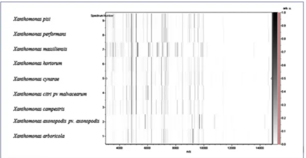
[3](Fig. 2), which putatively classifies strain SN6 as a member of a new species within the genus Xanthomonas in the phylum Proteobacteria. Thus, we propose the creation of the new spe-cies Xanthomonas massiliensis (Table 1). The 16S rRNA gene of Xanthomonas massiliensis strain SN6 is 1508 bp long and was deposited with the accession number AA00102 in the 16S IHU bank and LN881611 in GenBank. A comparison between the spectrum of the strain’s protein level and that of the closely related species on the 16S rRNA tree and present in our database was performed in a gel view (Fig. 3).
Phenotypic characteristics
Xanthomonas massiliensis strain SN6 grows between 28°C and 42°C; optimal growth was observed under aerobic conditions on COS at 37°C and pH7 after 48 hours of incubation. A smaller growth rate was observed under microaerobic
NMNI
Ndongo et al. Xanthomonas massiliensis 65© 2018 Published by Elsevier Ltd, NMNI, 26, 63–72 This is an open access article under the CC BY-NC-ND license (http://creativecommons.org/licenses/by-nc-nd/4.0/).
atmosphere after 48 hours of incubation, and no growth was observed under anaerobic conditions. Also, a smaller growth rate was observed at pH 7 and 8.5, and no growth was observed above 5% salinity. Colonies of the strain were yellowish, circular, viscous and smooth, nonhaemolytic and approximately 1 to 2 mm in diameter on Columbia agar under
aerobic conditions after 48 hours. The colonies became khaki green after 4 days of incubation. The yellow pigments, which are mono- or dibromo arylpolyenes called xanthomonadins
[26], are characteristic of this genus. Bacterial cells were Gram negative, rod shaped, motile and non–spore forming. Observed
FIG. 1.Reference mass spectrum from Xanthomonas massiliensis strain SN6T.
FIG. 2.Phylogenetic tree showing position of Xanthomonas massiliensis strain SN6Trelative to other close species. Sequences were aligned using
CLUSTALW and phylogenetic inferences were obtained with Kimura two-parameter models using maximum-likelihood method with 1000 bootstrap replicates, within MEGA software. Scale bar indicates 1% nucleotide sequence divergence.
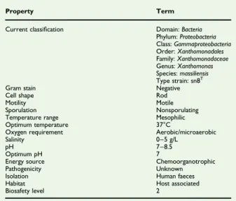
TABLE 1.Classification and general features of Xanthomonas massiliensis strain SN6T
Property Term
Current classification Domain: Bacteria Phylum: Proteobacteria Class: Gammaproteobacteria Order: Xanthomonadales Family: Xanthomonadaceae Genus: Xanthomonas Species: massiliensis Type strain: sn8T
Gram stain Negative
Cell shape Rod
Motility Motile
Sporulation Nonsporulating Temperature range Mesophilic Optimum temperature 37°C
Oxygen requirement Aerobic/microaerobic
Salinity 0–5 g/L
pH 7–8.5
Optimum pH 7
Energy source Chemoorganotrophic Pathogenicity Unknown Isolation Human faeces Habitat Host associated Biosafety level 2
© 2018 Published by Elsevier Ltd, NMNI, 26, 63–72
under electron microscopy, they occur singly or in chains and measure 0.6μm in diameter and 1.8 to 2.0 μm in length (Fig. 4). Biochemical analysis
The catalase activity test was positive, but the oxidase test was negative. Using API ZYM strip for the research of enzymatic activities of strain SN6, positive reactions were detected for alkaline phosphatase, esterase lipase (C8), leucine arylamidase, valine arylamidase, trypsin, acid phosphatase, naphthol-AS-BI-phosphohydrolase, β-galactosidase, β-glucosidase, N-acetyl-β-glucosaminidase and α-mannosidase. Esterase (C4), lipase (C14), cystine arylamidase, α-chymotrypsin, α-galactosidase, β-glucuronidase, α-glucosidase and α-fucosidase activities did
not show any sign of activity. The study of carbohydrate and its derivatives metabolism using API 50CH showed no fermen-tation of substrates, except for esculin. API 20NE strip revealed that there was neither nitrate reduction nor indole production, and urease was also negative. The reduction of nitrate to nitrite also makes it possible to differentiate the genus Xanthomonas from the genus Pseudoxanthomonas. On the same strip, positive reactions were observed for gelatin hydrolysis, malate and N-acetylglucosamine, and it also allowed to confirm the assimilation of esculin and β-galactosidase. A panel of 15 antibiotics was tested, and strain SN6 was sensitive to vancomycin, ceftriaxone, ciprofloxacin, clindamycin, doxy-cycline, erythromycin, gentamicin, penicillin, rifampicin, colistin, fosfomycin and trimethoprim/sulfamethoxazole but resistant to oxacillin, teicoplanin and metronidazole. Table 2
compares the phenotypic characteristics of strain SN6 with those of closely related species.
According to the cellular fatty acid methyl ester analysis, the most abundant fatty acid by far was branched 13-methyl-tet-radecanoic acid (58%). Many other branched structures were also described for this strain. Several specific 3-OH structures were detected. Minor amounts of unsaturated and saturated fatty acids were also identified. Regarding the differentiation between the species of the genera Pseudoxanthomonas and Xanthomonas, Xanthomonas massiliensis strain SN6 contains up to 4.5 ± 0.2% of 3-hydroxy-11-methyl-dodecanoic acid (C13:0 iso 3-OH) compared to the other species, which have none or only traces (Table 3).
FIG. 3.Gel view comparing Xanthomonas massiliensis strain SN6Tto other close species. Gel view displays raw spectra of strain SN6Tof loaded spectrumfiles arranged in a pseudo–gel like look. X-axis records m/z value. Left y-axis displays running spectrum number originating from subsequent spectra loading. Peak intensity is expressed by greyscale scheme code. Colour bar and right y-axis indicate relation between colour of peak and its intensity, in arbitrary units. Displayed species are indicated at left.
FIG. 4.Electron microscopy of Xanthomonas massiliensis strain SN6T.
NMNI
Ndongo et al. Xanthomonas massiliensis 67© 2018 Published by Elsevier Ltd, NMNI, 26, 63–72 This is an open access article under the CC BY-NC-ND license (http://creativecommons.org/licenses/by-nc-nd/4.0/).
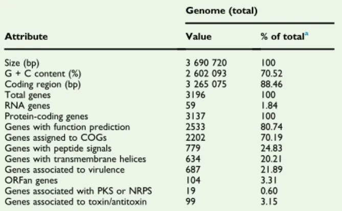
Genome properties
The genome of Xanthomonas massiliensis strain SN6 is 3 690 720 bp long with 70.52% GC content (Table 4, Fig. 5). It is composed of four scaffolds (composed of seven contigs). Of the 3196 predicted genes, 3137 were protein-coding genes and 59 were RNAs (two were 5S rRNA, two were 16S rRNA, two were 23S rRNA, 53 were tRNA genes). A total of 2533 genes (80.75%) were assigned as putative function (by COGs or by NR BLAST). A total of 104 genes were identified as ORFans (3.32%). The remaining genes were annotated as hypothetical proteins (350 genes, 11.16%). The distribution of genes into the different COGs functional categories is provided inTable 5. Genome comparison
The draft genome sequence of Xanthomonas massiliensis is smaller than that of Stenotrophomonas acidaminiphila,
Xanthomonas vesicatoria, Stenotrophomonas maltophilia, Steno-trophomonas rhizophila, Xanthomonas gardneri, Xanthomonas sacchari, Xanthomonas campestris and Xanthomonas axonopodis (3.69, 4.14, 5.53, 4.93, 4.65, 5.31, 4.93, 5.08 and 5.03 MB, respectively), but larger than the genome of Pseudoxanthomonas suwonensis (3.53 MB). The G + C content of Xanthomonas massiliensis is larger than that of Stenotrophomonas acid-aminiphila, Xanthomonas vesicatoria, Stenotrophomonas malto-philia, Stenotrophomonas rhizophila, Pseudoxanthomonas suwonensis, Xanthomonas gardneri, Xanthomonas sacchari, Xan-thomonas campestris and XanXan-thomonas axonopodis (70.523, 68.48, 64.07, 66.23, 67.30, 70.515, 63.53, 69.04, 65.07 and 64.89%, respectively). The gene content of Xanthomonas mas-siliensis is smaller than that of Stenotrophomonas acidaminiphila, Xanthomonas vesicatoria, Stenotrophomonas maltophilia,
TABLE 2.Differential characteristics of Xanthomonas massiliensis strain SN6, Xanthomonas campestris. pv. campestris ATCC33913, Xanthomonas sacchari LMG471, Xanthomonas vesicatoria ATCC35937_LMG911, Xanthomonas gardneri DSM 19127, Xanthomonas axonopodis LMG538 and Pseudoxanthomonas suwonensis 4M1
Property X. massiliensis X. campestris X. sacchari X. vesicatoria X. gardneri X. axonopodis P. suwonensis Cell diameter (μm) 0.5–0.6 0.4–0.6 0.4–0.6 0.4–0.6 0.4–0.6 0.4–0.6 0.3–0.5 Motility + + + + + + + Indole − − − − − − − Catalase + + + + + + + Oxidase − − − − − − + Nitrate reductase − − − − − − + Urease − − − − − − − N-Acetyl-glucosamine + − − + − ++/− + Acid from: L-Arabinose − − + − − − + D-Mannose − + + + + + − D-Mannitol − − +/− − − − D-Trehalose + + + + + + NA D-Glucose − + + + + + + D-Fructose − + + + + + NA D-Maltose − + + + + + + D-Lactose − − + − − − NA D-Raffinose − +/− − − − −/+ NA
Habitat Human gut Tomato/pepper Tomato/pepper Tomato/pepper Tomato/pepper Pasturage Cotton waste compost +, positive result;−, negative result; NA, data not available.
TABLE 3.Cellular fatty acid composition (%)
Fatty acid Name Mean relative %a
15:0 iso 13-Methyl-tetradecanoic acid 57.6 ± 0.4 11:0 iso 9-Methyl-decanoic acid 10.2 ± 0.5 17:1 iso 15-Methylhexadecenoic acid 4.9 ± 0.2 13:0 3-OH iso 3-hydroxy-11-methyl-Dodecanoic acid 4.5 ± 0.2 16:1n7 9-Hexadecenoic acid 3.7 ± 0.2 17:0 iso 15-methyl-Hexadecanoic acid 3.5 ± 0.1 12:0 3-OH 3-Hydroxydodecanoic acid 3.3 ± 0.1 16:0 9,10-methylene 2-hexyl-Cyclopropaneoctanoic acid 2.3 ± 0.2 15:1 iso 13-Methyltetradecenoic acid 2.2 ± 0.3 16:0 Hexadecanoic acid 1.7 ± 0.1 11:0 3-OH iso 3-hydroxy-9-Methyl-decanoic acid 1.4 ± 0.2 15:0 anteiso 12-methyl-Tetradecanoic acid 1.3 ± 0.1 16:1n9 7-Hexadecenoic acid TR 18:1 iso 16-Methylheptadecenoic acid TR 13:0 iso 11-methyl-Dodecanoic acid TR 18:1n9 9-Octadecenoic acid TR 10:0 Decanoic acid TR 17:0 anteiso 14-methyl-Hexadecanoic acid TR 14:0 Tetradecanoic acid TR
aMean peak area percentage; TR, trace amounts < 1 %.
TABLE 4. Nucleotide content and gene count levels of genome Attribute Genome (total) Value % of totala Size (bp) 3 690 720 100 G + C content (%) 2 602 093 70.52 Coding region (bp) 3 265 075 88.46 Total genes 3196 100 RNA genes 59 1.84 Protein-coding genes 3137 100 Genes with function prediction 2533 80.74 Genes assigned to COGs 2202 70.19 Genes with peptide signals 779 24.83 Genes with transmembrane helices 634 20.21 Genes associated to virulence 687 21.89 ORFan genes 104 3.31 Genes associated with PKS or NRPS 19 0.60 Genes associated to toxin/antitoxin 99 3.15 COGs, Clusters of Orthologous Groups database; NRPS, nonribosomal peptide synthase; PKS, polyketide synthase.
aTotal is based on either size of genome in base pairs or total number of
protein-coding genes in annotated genome. © 2018 Published by Elsevier Ltd, NMNI, 26, 63–72
Stenotrophomonas rhizophila, Xanthomonas gardneri, Xanthomo-nas sacchari, XanthomoXanthomo-nas campestris and XanthomoXanthomo-nas axono-podis (3137, 3617, 4927, 4565, 3938, 4228, 4168, 4181 and 3904, respectively), but larger than the genome of Pseudox-anthomonas suwonensis (3132). This comparison of genomes between X. massiliensis and the other genetically closest species is shown inTable 6. In all genomes compared, the distribution of genes into COGs categories is identical (Fig. 6).
Among Xanthomonas species with standing in nomenclature, AGIOS values ranged from 64.76% between Xanthomonas campestris pv. campestris and Stenotrophomonas acidaminiphila to 79.65% between Xanthomonas sacchari and Pseudoxanthomonas suwonensis. When comparing Xanthomonas massiliensis sp. nov. to other species, AGIOS values were in the same range, from 66.21% with Xanthomonas vesicatoria to 80.88% with Xantho-monas sacchari (Table 7). Among the species with standing in nomenclature, we found that by using the digital DNA-DNA hybridization (dDDH) with the GGDC software, values ranged from 20.8% between Xanthomonas vesicatoria and Pseu-doxanthomonas suwonensis to 32.1% between Xanthomonas gardneri and Xanthomonas axonopodis. When comparing Xan-thomonas massiliensis to other species, the dDDH value ranged
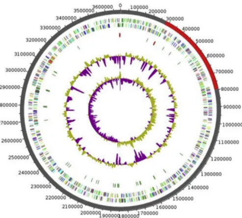
FIG. 5.Graphical circular map of genome of Xanthomonas massiliensis strain SN6T. From outside to center: Genes on forward strand coloured by COGs categories (only genes assigned to COGs), genes on reverse strand coloured by COGs categories (only gene assigned to COGs), RNA genes (tRNAs green, rRNAs red), GC content and GC skew. COGs, Clusters of Orthologous Groups database.
TABLE 5.Number of genes associated with 25 general COGs functional categories
Code Value % of total Description J 199 6.3436403 Translation
A 1 0.031877592 RNA processing and modification K 142 4.5266175 Transcription
L 93 2.9646158 Replication, recombination and repair B 1 0.031877592 Chromatin structure and dynamics D 32 1.020083 Cell cycle control, mitosis and meiosis Y 0 0 Nuclear structure
V 83 2.64584 Defense mechanisms T 104 3.3152692 Signal transduction mechanisms M 158 5.0366592 Cell wall/membrane biogenesis N 37 1.1794709 Cell motility
Z 1 0.031877592 Cytoskeleton W 34 1.0838381 Extracellular structures
U 72 2.2951865 Intracellular trafficking and secretion O 119 3.7934332 Posttranslational modification,
protein turnover, chaperones X 37 1.1794709 Mobilome: prophages, transposons C 164 5.227925 Energy production and conversion G 130 4.144087 Carbohydrate transport and metabolism E 193 6.152375 Amino acid transport and metabolism F 65 2.0720434 Nucleotide transport and metabolism H 103 3.283392 Coenzyme transport and metabolism I 146 4.654128 Lipid transport and metabolism P 157 5.0047817 Inorganic ion transport and metabolism Q 80 2.5502074 Secondary metabolites biosynthesis,
transport and catabolism R 227 7.236213 General function prediction only S 150 4.7816386 Function unknown
— 935 29.805548 Not in COGs COGs, Clusters of Orthologous Groups database.
NMNI
Ndongo et al. Xanthomonas massiliensis 69© 2018 Published by Elsevier Ltd, NMNI, 26, 63–72 This is an open access article under the CC BY-NC-ND license (http://creativecommons.org/licenses/by-nc-nd/4.0/).
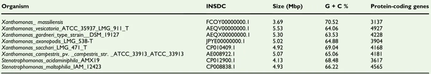
TABLE 6.Genome comparison of closely related species to Xanthomonas massiliensis strain SN6T.
Organism INSDC Size (Mbp) G + C % Protein-coding genes Xanthomonas_ massiliensis FCOY00000000.1 3.69 70.52 3137
Xanthomonas_vesicatoria_ATCC_35937_LMG_911_T AEQV00000000.1 5.53 64.06 4927 Xanthomonas_gardneri_type_strain__DSM_19127 AEQX00000000.1 5.30 63.53 4228 Xanthomonas_axonopodis_LMG_538-T JPYE00000000.1 5.02 64.88 3904 Xanthomonas_sacchari_LMG_471_T CP010409.1 4.92 69.04 4168 Xanthomonas_campestris_pv. _campestris_str. _ATCC_33913_ATCC_33913 AE008922.1 5.07 65.06 4181 Stenotrophomonas_acidaminiphila_AMX19 CP012900.1 4.13 68.48 3617 Stenotrophomonas_maltophilia_IAM_12423 CP008838.1 4.93 66.22 4565 INSDC, International Nucleotide Sequence Database Collaboration.
FIG. 6.Distribution of functional classes of predicted genes according to clusters of orthologous groups of proteins.
TABLE 7.Number of orthologous proteins shared between genomes (upper right) and AGIOS values obtained (lower left) P. suwonensis X. gardneri X. campestris S. maltophilia X. massiliensis X. sacchari X. vesicatoria S. acidaminiphila X. axonopodis S. rhizophila P. suwonensis 3132 1898 1925 1895 1678 1858 1914 1858 1902 1901 X. gardneri 66.11 4228 2945 2231 1859 2390 2962 2032 2880 2219 X. campestris 66.12 72.68 4181 2234 1848 2425 2970 2018 2909 2234 S. maltophilia 77.64 66.59 65.63 4565 1865 2137 2239 2174 2195 2466 X. massiliensis 79.82 67.19 66.64 78.60 3137 1788 1835 1778 1834 1854 X. sacchari 79.65 67.94 67.01 79.13 80.88 4168 2440 1973 2427 2127 X. vesicatoria 65.61 69.79 68.62 65.75 66.21 66.85 4927 2024 2934 2223 S. acidaminiphila 69.27 64.47 64.76 69.67 69.57 69.80 67.53 3617 1983 2125 X. axonopodis 66.01 70.07 63.74 66.33 66.92 67.70 71.02 67.35 3904 2197 S. rhizophila 66.12 65.42 61.96 68.84 66.81 67.04 66.54 67.77 66.98 3938 Numbers of proteins per genome are indicated in bold. AGIOS, average genomic identity of orthologous gene sequences.
Pseudoxanthomonas suwonensis 4M1, Xanthomonas gardneri DSM 19127, Xanthomonas campestris pv. campestris ATCC33913, Stenotrophomonas maltophilia IAM12423, Xanthomonas massiliensis SN6T, Xanthomonas sacchari LMG471T, Xanthomonas vesicatoria ATCC35937_LMG911T, Stenotrophomonas acidaminiphila AMX19,
Xanthomonas axonopodis LMG538T, Stenotrophomonas rhizophila ep10.
© 2018 Published by Elsevier Ltd, NMNI, 26, 63–72
from 21.3% with Stenotrophomonas maltophilia to 23.5% with Xanthomonas sacchari (Table 8).
Conclusion
Phenotypic characteristics as well as phylogenetic and genomic analyses of strain SN6 suggest that it represents a novel species within the Xanthomonas genus, for which the name Xanthomo-nas massiliensis sp. nov. is proposed. This bacterial strain was isolated from the faecal flora of an obese Frenchwoman, and the description was based on a single isolate.
Description of Xanthomonas massiliensis sp. nov. Xanthomonas massiliensis (mas.si.li.en’sis, L. fem. adj. massiliensis, ‘of Massilia,’ the Latin name of Marseille where strain SN6T
was first cultivated).
X. massiliensis is a rod-shaped (0.6 × 1.8–2.0 μm), aerobic and Gram-negative bacterium occurring singly or in chains. Growth was also observed under microaerophilic conditions. Cells are motile with a flagellum and nonsporulating. Fresh colonies were yellow, circular, smooth and viscous with a diameter of 1 to 2 mm on COS. Optimal growth of strain SN6 occurred at 37°C under aerobic atmosphere with a pH of 7 but did not grow at 5% of salinity or under anaerobic conditions. The strain was catalase positive. Tests for nitrate reduction, indole production and urease were negative. API 50CH showed that the only substrate used as a carbon source was esculin. Positive reactions were detected for alkaline phosphatase, esterase lipase (C8), leucine arylamidase, valine arylamidase, trypsin, acid phosphatase, naphthol-AS-BI-phosphohydrolase, β-galactosidase, β-glucosidase, N-acetyl-β-glucosaminidase, α-mannosidase, gelatin hydrolysis, malate and N-acetylglucos-amine. The strain was sensitive to ceftriaxone, ciprofloxacin, clindamycin, doxycycline, erythromycin, gentamicin, penicillin, rifampicin, colistin, vancomycin, fosfomycin and trimethoprim/ sulfamethoxazole but resistant to metronidazole, oxacillin and teicoplanin. Predominant fatty acids were 13-methyl-tetrade-canoic acid followed by 9-methyl-de13-methyl-tetrade-canoic acid.
The DNA G + C content is about 70.52%. The 16S rRNA gene and genome sequences were deposited in GenBank under accession number LN881611 and FCOY00000000, respectively. The type strain is Xanthomonas massiliensis strain SN6T(= CSUR P2129 = DSM 100900) and was isolated from human faeces.
Acknowledgements
The authors thank the Xegen Company (http://www.xegen.fr/) for automating the genomic annotation process, and
TABLE 8. Pairwise comparison of Xan thom onas mass iliensis wit h other species us ing GGDC , formula 2 (DD H estimat es based on identities/HSP lengt h), a upper right XM XV XS XG XCC XA SR SM SA PS XM 100 22 (19.7 – 24. 4%) 23.5 (21.2 – 26%) 21.9 (19. 6 – 24.3%) 21.9 (19.7 – 24.4%) 22 (19. 8 – 24.5%) 2 1.9 (19.7 – 24.4%) 21.3 (19 .1 – 23.7%) 22.7 (20.4 – 25.2%) 22.2 (20 – 24.7%) XV 100 23.1 (20.8 – 25.6%) 32 (29.6 – 34. 6%) 29.8 (27.4 – 32.3%) 31.4 (29 – 33.9%) 2 2 (19.8 – 24.5%) 21.6 (19 .4 – 24.1%) 22.1 (19.8 – 24.5%) 20.8 (18.6 – 23.3%) XS 100 23.3 (21 – 25. 8%) 23.3 (21 – 25.8%) 23.6 (21 .3 – 26%) 2 3.6 (21.3 – 26%) 22.7 (20 .5 – 25.2%) 23.7 (21.4 – 26.2%) 22.3 (20 – 24.7%) XG 100 31.1 (28.7 – 33.6%) 32.1 (29 .7 – 34.6%) 2 2.2 (19.9 – 24.6%) 21.6 (19 .3 – 24%) 22 (19.8 – 24.5%) 21 (18 .7 – 23.4%) XCC 100 29.6 (27 .2 – 32.1%) 2 2.2 (19.9 – 24.6%) 21.6 (19 .3 – 24%) 22.1 (19.8 – 24.5%) 21.1 (18.8 – 23.5%) XA 100 2 2.5 (20.2 – 24.9%) 22.1 (19 .8 – 24.5%) 22.3 (20.1 – 24.8%) 21 (18 .8 – 23.5%) SR 100 24 (21 .7 – 26.5%) 23.9 (21.5 – 26.3%) 21.6 (19.3 – 24%) SM 100 23.1 (20.9 – 25.6%) 21.1 (18.8 – 23.5%) SA 100 22.2 (20 – 24.7%) PS 100 Bold indicates comparison between strain and itself. DDH, DNA-DNA hybridization ; GGDC, Genome-to-Genome Distan ce Calculator ; HSP, high-sco ring segm ent pairs; PS, Pseudox anthomonas suw onensis; SA, Ste notrophomonas acidamin iphila; SM, Stenotrophomonas maltophilia; SR, Stenotrophomonas rhizophila; XA, Xan thomonas axonopodis; XCC, Xant homonas campes tris pv. Campes tris; XG, Xant homonas gardneri; XM, Xan thomonas massilien sis; XS, Xan thomonas sacch ari; XV, Xanthom onas vesicatoria. aCon fidence intervals indicate inherent uncerta inty in estimating DDH values from intergenomic distanc es based on models derived from empirica l test dat a sets (wh ich are always lim ited in size).
NMNI
Ndongo et al. Xanthomonas massiliensis 71© 2018 Published by Elsevier Ltd, NMNI, 26, 63–72 This is an open access article under the CC BY-NC-ND license (http://creativecommons.org/licenses/by-nc-nd/4.0/).
M. Lardiere (MEPHI) for English-language editorial review. This study was supported by the Fondation Méditerranée Infection and the French government under the ‘Investissements d’ave-nir’ with the reference Mediterranée Infection 10-IAHU-3.
Con
flict of Interest
None declared.
References
[1] Dowson D. On the systematic position and generic names of the Gram negative bacterial plant pathogens. Zbl Bakteriol Parasitenkd Infekt Hyg Abt 2 100 1939;100:177–93.
[2] Dye DW, Lelliott RA. Genus 11. Xanthomonas Dowson 1939. In: Buchanan RE, Gibbons NE, editors. Bergey’s manual of determinative bacteriology. 8th ed. Baltimore: Williams & Wilkins; 1974. p. 243–9. [3] Vauterin L, Hoste B, Kersters K, Swings J. Reclassification of
Xantho-monas. Int J Syst Bacteriol 1995;45:472–89.
[4] Yang P, Vauterin L, Vancanneyt M, Swings J, Kersters K. Application of fatty acid methyl esters for the taxonomic analysis of the genus Xan-thomonas. Syst Appl Microbiol 1993;16:47–71.
[5] Darrasse A, Carrère S, Barbe V, Boureau T, Arrieta-Ortiz ML, Bonneau S, et al. Genome sequence of Xanthomonas fuscans subsp. fuscans strain 4834-R reveals thatflagellar motility is not a general feature of xanthomonads. BMC Genomics 2013;14:761.
[6] Lagier JC, Khelaifia S, Alou MT, Ndongo S, Dione N, Hugon P, et al. Culture of previously uncultured members of the human gut micro-biota by culturomics. Nat Microbiol 2016;1:16203.
[7] Seng P, Drancourt M, Gouriet F, La Scola B, Fournier PE, Rolain JM, et al. Ongoing revolution in bacteriology: routine identification of bacteria by matrix-assisted laser desorption ionization time-of-flight mass spectrometry. Clin Infect Dis 2009;49:543–51.
[8] Morel AS, Dubourg G, Prudent E, Edouard S, Gouriet F, Casalta JP, et al. Complementarity between targeted real-time specific PCR and conventional broad-range 16S rDNA PCR in the syndrome-driven diagnosis of infectious diseases. Eur J Clin Microbiol Infect Dis 2015;34:561–70.
[9] Stackebrandt E, Ebers J. Taxonomic parameters revisited: tarnished gold standards. Microbiol Today 2006:152–5.
[10] Kim M, Oh HS, Park SC, Chun J. Towards a taxonomic coherence between average nucleotide identity and 16S rRNA gene sequence
similarity for species demarcation of prokaryotes. Int J Syst Evol Microbiol 2014;64:346–51.
[11] Sasser M. Bacterial identification by gas chromatographic analysis of fatty acids methyl esters (GC-FAME). Newark, NY: Microbial ID; 2006. [12] Dione N, Sankar SA, Lagier JC, Khelaifia S, Michele C, Armstrong N, et al. Genome sequence and description of Anaerosalibacter massiliensis sp. nov. New Microbes New Infect 2016;10:66–76.
[13] Lagier JC, Ramasamy D, Rivet R, Raoult D, Fournier PE. Non contiguous-finished genome sequence and description of Cellulomonas massiliensis sp. nov. Stand Genomic Sci 2012;7:258–70.
[14] Lagier JC, Bibi F, Ramasamy D, Azhar EI, Robert C, Yasir M, et al. Non contiguous-finished genome sequence and description of Clostridium jeddahense sp. nov. Stand Genomic Sci 2014;9:1003–19.
[15] Zerbino DR, Birney E. Velvet: algorithms for de novo short read as-sembly using de Bruijn graphs. Genome Res 2008;18:821–9. [16] Bankevich A, Nurk S, Antipov D, Gurevich AA, Dvorkin M,
Kulikov AS, et al. SPAdes: a new genome assembly algorithm and its applications to single-cell sequencing. J Comput Biol 2012;19:455–77. [17] Luo R, Liu B, Xie Y, Li Z, Huang W, Yuan J, et al. SOAPdenovo2: an empirically improved memory-efficient short-read de novo assembler. GigaScience 2012 27;1:18.
[18] Bolger AM, Lohse M, Usadel B. Trimmomatic: aflexible trimmer for Illumina sequence data. Bioinformatics 2014;30:2114–20.
[19] Hyatt D, Chen GL, Locascio PF, Land ML, Larimer FW, Hauser LJ. Prodigal: prokaryotic gene recognition and translation initiation site identification. BMC Bioinform 2010;11:119.
[20] Lowe TM, Eddy SR. tRNAscan-SE: a program for improved detection of transfer RNA genes in genomic sequence. Nucleic Acids Res 1997;25:955–64.
[21] Lagesen K, Hallin P, Rødland EA, Stærfeldt HH, Rognes T, Ussery DW. RNAmmer: consistent and rapid annotation of ribosomal RNA genes. Nucleic Acids Res 2007;35:3100–8.
[22] Käll L, Krogh A, Sonnhammer EL. A combined transmembrane to-pology and signal peptide prediction method. J Mol Biol 2004;338: 1027–36.
[23] Gouret P, Thompson JD, Pontarotti P. PhyloPattern: regular expres-sions to identify complex patterns in phylogenetic trees. BMC Bio-inform 2009;10:298.
[24] Lechner M, Findeiß S, Steiner L, Marz M, Stadler PF, Prohaska SJ. Proteinortho: detection of (co-)orthologs in large-scale analysis. BMC Bioinform 2011;12:124.
[25] Meier-Kolthoff JP, Auch AF, Klenk HP, Göker M. Genome sequence–based species delimitation with confidence intervals and improved distance functions. BMC Bioinform 2013;14:60.
[26] Andrewes AG, Jenkins CL, Starr MP, Shepherd J, Hope H. Structure of xanthomonadin I, a novel dibrominated arylpolyene pigment produced by the bacterium Xanthomonas juglandis. Tetrahedron Lett 1976;45: 4023–4.
© 2018 Published by Elsevier Ltd, NMNI, 26, 63–72