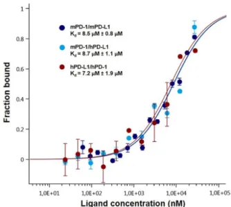
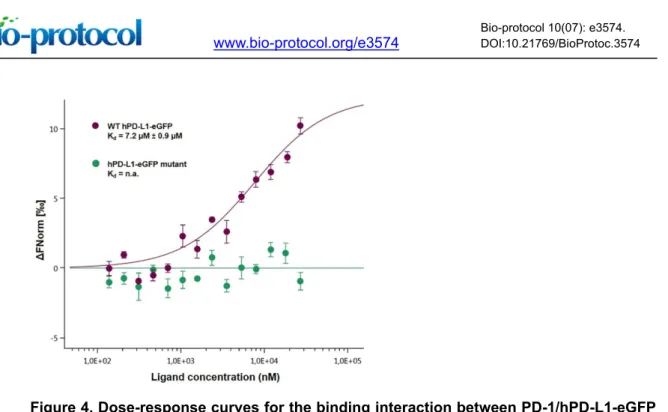
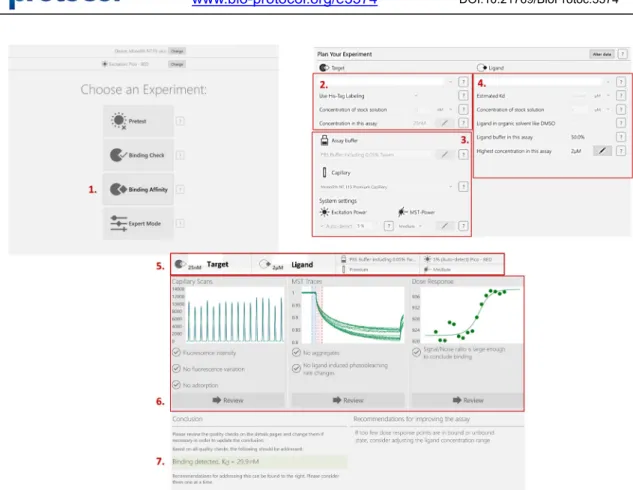
Measurement of Protein-Protein Interactions through Microscale Thermophoresis (MST) Magnez Romain, Bryan Thiroux, Morgane Tardy, Bruno Quesnel and Xavier Thuru*
10
0
0
Texte intégral
(2)
(3)
(4)
(5)
(6)
(7)
(8)
(9)
(10)
Figure


+2
Documents relatifs
Four residues of the extracellular N-terminal domain of the NR2A subunit control high-affinity Zn2+ binding to NMDA receptors... &
However, we believe that the main contribution of this paper is not PairClass itself, but the extension of supervised word pair classification beyond the classification of
We finally, provide some new sharp bounds for the first Dirichlet eigenvalue of planar convex sets and a new sharp upper bound for triangles which is better than the conjecture
The Bacterial and Fungal Diversity of an Aged PAH- and Heavy Metal-Contaminated Soil is Affected by Plant Cover and Edaphic Parameters... Although the communities may be
labelled 15 N 2 O were flowing through each soil sample, containing a background of 0.82 mg to 1.91 mg of 15 N in organic matter (Table 1).. Figure 2 presents the fraction of
One-shot kinetic assay in nanofluidic sensors with integrated concentration gradient generator With an optimized flow velocity of the injected solutions at the inlet reservoirs,
Whereas, under the current regime of private international law, it is the insurer who hesitates to provide cross border services, the policyholder would be reluctant to
Pour les salariés ayant conservé le même emploi et la même profession entre 2013 et 2016, les chances d’améliorer ses conditions de travail sont plus faibles, surtout en
