B.S. M.S.
DNA Target Selection by Calicheamicin
byAaron A. Salzberg
Aerospace Engineering, University of Maryland, 1983 Aerospace Engineering, University of Maryland, 1986
SUBMITTED TO THE DIVISION OF TOXICOLOGY IN PARTIAL FULFILLMENT OF THE REQUIREMENTS FOR THE DEGREE OF
Ph.D. IN TOXICOLOGY AT THE
MASSACHUSETTS INSTITUTE OF TECHNOLOGY
May, 1998
© 1998 Massachusetts Institute of Technology. All rights reserved.
Signature of Author... ...
Division of Toxicology May 15, 1998
C ertifie d b y ... ... ...
Peter C. Dedon Associate Professor of Toxicology Thesis Supervisor
A ccep ted by ... .4...
St
Peter C. DedonMASSACHUSTTS O ,irman, Committee on Graduate Students
OF TECHNOLOGY Division of Toxicology
JJUNO
0
8
7/filwi~fj
This doctoral thesis has been examined by a Committee of the Division of Toxicology as follows:
Professor William Thilly... Chairman Division of Toxicology Massachusetts Institute of Technology
P rofessor P eter C . D edon ...:... ... Supervisor
Division of Toxicology Massachusetts Institute of Technology
Professor John Essigm ann ... ... Divisio fTo1xic and Department of Chemistry
Massachusetts Institute of Technology
P rofessor A lexan der R ich ... ... ... ... Department of Biology Massachusetts Institute of Technology
Professor Jam es W illiam son... ... . ...
Department of Molecular Biology and the Skaggs Institute of Chemical Biology The Scripps Research Institute
DNA Target Selection by Calicheamicin
byAaron A. Salzberg
Submitted to the Division of Toxicology on May 15, 1998 in Partial Fulfillment of the Requirements for the Degree of Ph. D. in Toxicology
Abstract
The research presented here has evolved from the general hypothesis within our laboratory that DNA in vivo has characteristics which modulate its sensitivity to DNA damaging compounds. As one component of this overall paradigm, this work sets out to explore the material and structural properties of DNA that mediate the targeting of drugs to specific sites within the DNA.
To examine these issues, we have studied the clinically relevant antitumor antibiotic calicheamicin. Calicheamicin binds DNA non-covalently in the minor groove and abstracts hydrogen atoms from both sugar phosphate backbones causing site-specific double-stranded DNA damage. The reactivity of the drug is affected by both the topology and sequence context of the binding site, suggesting that
calicheamicin recognizes some form of structural perturbation rather than sequence per se. However, the true mechanism for calicheamicin target selection is
unknown.
To investigate the role of both calicheamicin structure and DNA structure and dynamics in target recognition, two approaches were taken: First, gel mobility studies were performed to evaluate whether calicheamicin binding sites possessed a common element of curvature. Second, DNA cyclization experiments were
conducted to assess and measure the structural changes that occur in DNA when calicheamicin or its oligosaccharide binds. (Novel modifications were introduced to facilitate the rapid detection and analysis of DNA bending.) Our results show that calicheamicin binding sites are not curved, but that both the drug and
oligosaccharide bend DNA upon binding. These data support a model for
calicheamicin target selection where the drug prefers to bind those sequences that can be easily bent (i.e., flexible) into a conformation that maximizes its electrostatic and hydrophobic interactions with DNA.
In addition to these experiments, we exploited calicheamicins' ability to bend DNA to study the affect of topology on the kinetics of DNA bimolecular association. We show, that when the ends of the DNA molecule are in close proximity,
intermolecular reactions are inhibited; a result that calls into question indirect methods for measuring bimolecular association reaction rates.
These results contribute to our understanding of small molecule-DNA interactions and lay the groundwork for understanding how molecules select genomic targets.
Thesis Supervisor: Associate Professor Peter C. Dedon Title: Associate Professor of Toxicology
Dedication
Bruce Becker David Monius
David Price Samuel Rosenthal
Acknowledgments
I am extremely grateful to the Division of Toxicology at MIT, the National Institutes of Health and the Hugh Hampton Young Fellowship Committee for their support and commitment to my work.
Table of Contents
Title Page ... . . . 1
Com m ittee Page ... 2
A bstract ... . . . 3
D edication ... . . . 5
A cknow ledgm ents ... ... 6
Table of Contents ... 7
List of A bbreviations and Sym bols ... 9
List of Figures ... 11 List of Tables ... 14 Biographical N ote ... 15 Publications ... 16 A w ards ... 18 Prologue ... ... 19 Chapter I: Introduction ... 20 Calicheam icin ... 21
Calicheam icin and D N A ... 22
Measuring the Cyclization Properties of DNA ... 26
Chapter II: The Intrinsic Properties of Calicheamicin Binding Sites ... 30
Experim ental D esign ... ... 30
M aterials and M ethods ... 33
Results and D iscussion ... 34
Figures ... 37
Chapter III: The Effects of Calicheamicin Binding on DNA Topology ... 41
Experim ental D esign ... 41
M aterials and M ethods ... 45
Results and D iscussion ... ... 47
Sum m ary ... ... 57
Tables ... 59
Figures ... ... ... ... 61
Chapter IV: Calicheamicin Binding and DNA Cyclization Kinetics ... 76
Theoretical Background ... 76
Experimental Design ... ... 86
M aterials and M ethods ... 90
M ethods of A nalysis ... 91
Results and D iscussion ... 95
Tables Figures
... ... ... 99 ... 1 0 1 Chapter V: The Influence of DNA Length and Structure on the Kinetics of
Bimolecular Association ... E xp erim en tal M eth od ... M ethod s an d M aterials ... R esu lts an d D iscu ssion ... F ig u re s ... Chapter VI: Determinants of Calicheamicin-Induced DNA Damage ...
Sequence-Specific Electrostatic Interactions in Calicheamicin Binding ... Shape Recognition and Hydrophobic Forces in Calicheamicin Binding ... DNA Structure and Dynamics in Calicheamicin Binding ... Calicheamicin Binding to DNA ... F ig u re s ... 112 112 113 114 117 123 123 125 127 129 131 R e feren ce s ... 133 Appendix A: A Compendium of Calicheamicin Binding Sites ... 141
Abbreviations and Symbols
(in alphabetical order)
a, b Mathematical constants as defined in the text
bp Base-pairs
AL Apparent length (base pairs)
[B] Concentration of binding sites (molar)
[B-CAL] Concentration of occupied binding sites (molar)
Ci Integration constants
CAL Calicheamicin 711
[CAL] Concentration of calicheamicin 711 (molar)
CAL-E Calicheamicin E
[CAL-e] Concentration of calicheamicin F (molar)
C(t)r Radioactivity measured in the circularized DNA band (cpm) [C(t)] Concentration of circles (molar)
d Change in mean circle size (mul - mll)/100
D(t)r Radioactivity measured in the dimerized DNA band (molar) [D] Concentration of uncyclized substrate (molar)
[D'] Concentration of undimerized substrate (molar) [Di(t)] Concentration of dimers (molar)
[Dm(t)] Concentration of measured undimerized dimerized substrate (molar)
[Dmol Total measured concentration of undimerized substrate (molar) [E] Free enzyme concentration (molar)
[Eo] Total enzyme concentration (molar)
[ES] Concentration of the enzyme-substrate complex (molar) fd Fraction of unligatable substrate
fs Ratio of ligatable substrate to total DNA (circularization)
fs' Ratio of ligatable substrate to total DNA (bimolecular association) [H] Concentration of half molecules (molar)
J
Ring closure probability (molar) kl Rate constant for cyclization (s-1)kxy Rate constant going from step "x" to step "y" Ka Bimolecular association equilibrium constant Kc Cyclization equilibrium constant
Km Rate constant for DNA ligase
[L] Concentration of DNA substrate (molar) [Mo] Concentration of monomers (molar)
m Mean circle size
m 11 Lower limit of the mean circle size mul Upper limit of the mean circle size M W Molecular weight (grams/mole)
[P] Concentration of covalently closed circles (molar) [P'] Concentration of covalently linked monomers (molar)
pUC19-Sap The pUC19 cloning vector with the Sap I restriction site removed pAS1 Cloning vector created from pUC19 as described in Figure 1
pAS1/nmx The pAS1 cloning vector incorporating the "n"th polymer of the "x" monomer
RL Ratio of apparent length to actual length
[S] Concentration of ligatable substrate (cyclization)
[Sal Concentration of ligatable substrate (bimolecular association)
X Migration distance (cm)
a Bend angle of a regular polygon with respect to a local tangent (degrees)
acal Bend angle caused by calicheamicin binding (degrees per site)
af Average bending due to the intrinsic flexibility of DNA (degrees per base-pair)
SEnclosed angle of a regular polygon (degrees) 8- Partial charge (negative)
List of Figures
Chapter II: Figure 2.1. Figure 2.2. Figure 2.3. Figure 2.4. Figure 2.5. Chapter III: Figure 3.1. Figure 3.2. Figure 3.3. Figure 3.4. Figure 3.5. Figure 3.6. Figure 3.7. Figure 3.8. Figure 3.9. Figure 3.10. Figure 3.11. Figure 3.12. Figure 3.13. Figure 3.14. Figure 3.15. Figure 3.16.Synthesis of the pAS1 cloning vector ... ... 37
Damage produced by calicheamicin Y11 in the binding site constructs ... ... 38
Gel migration of polymerized DNA constructs ... 39
Curve fit of the BamnH I linker data ... 40
RL values for the in-phase, out-of-phase, no-site and A-tract ... 40
HPLC purification of calicheamicin e ... ... 61
Calicheamicin
e
induced DNA damage in the out-of-phase construct ... 62Resolving circular from linear DNA by two-dimensional gel electrophoresis ... 63
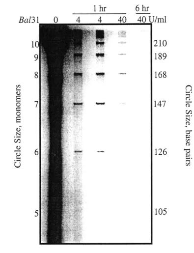
Effect of BAL-31 on the products of T4 ligase reaction with the calicheam icin binding site construct ... ... 64
Effect of BAL-31 concentration on the distribution of DNA circles from ligation of the in-phase calicheamicin binding site construct ... ... 65
Effect of BAL-31 reaction conditions on the distribution of circles resulting from the ligation of the A-tract construct ... 66
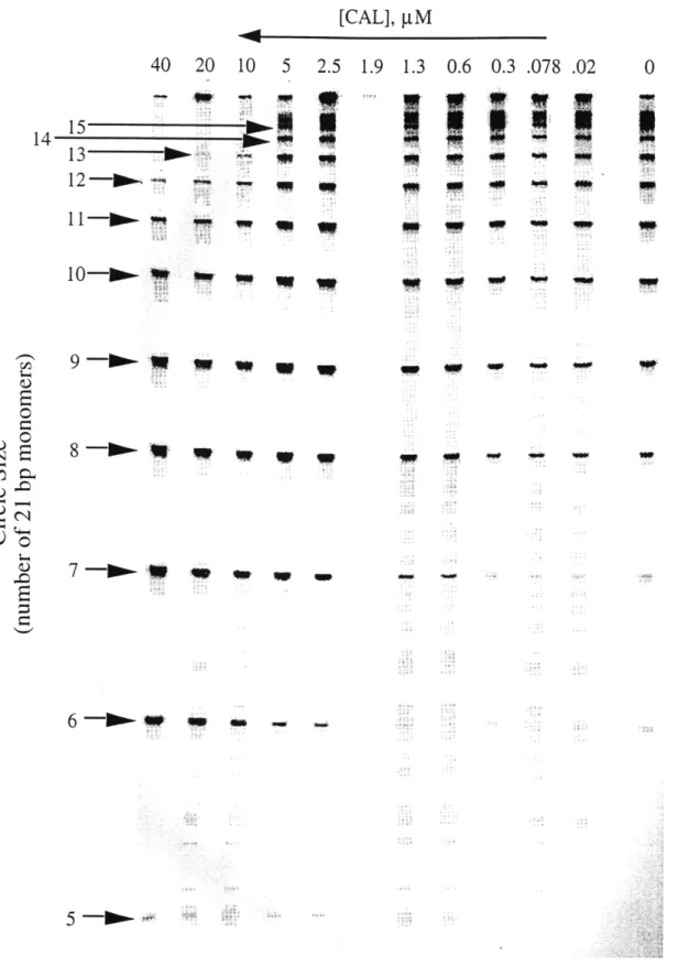
Effect of calicheamicin e on the formation of DNA circles by the in-phase D N A construct ... 67
Identification of circle size by denaturing gel electrophoresis ... 68
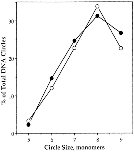
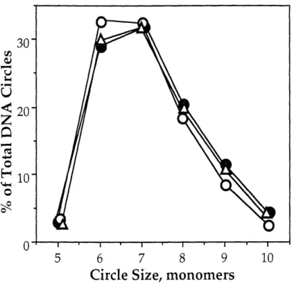
Distribution of DNA circles as a function of calicheamicin a concentration for the in-phase DNA construct ... 69
Plot of the mean circle size as a function of calicheamicin e concentration for the in-phase DNA construct ... 69
Effect of calicheamicin E on the formation of DNA circles by the out-of-phase construct ... ... 70
Distribution of DNA circles as a function of calicheamicin F concentration for the out-of-phase DNA construct ... 71
Plot of the mean circle size as a function of calicheamicin F concentration for the in-phase construct ... 71
Mean circle size as a function of calicheamicin a concentration for both the in-phase and out-of-phase DNA constructs ... 72
Mean circle size versus oligosaccharide concentration ... 73
Figure 3.17. Chapter IV: Figure 4.1. Figure 4.2. Figure 4.3. Figure 4.4. Figure 4.5. Figure 4.6. Figure 4.7. Figure 4.8. Figure 4.9. Figure 4.10. Figure 4.11. Chapter V: Figure 5.1. Figure 5.2. Figure 5.3. Figure 5.4.
Effect of BAL-31 on the products of T4 ligase reaction with the
in-phase calicheamicin binding site construct ... 75
The cyclization and bimolecular association behavior of a 273 bp polymer of the in-phase binding site construct ... 101
The effect of 0.1 tM calicheamicin E on the cyclization and bimolecular association behavior of a 273 bp polymer of the in-phase binding site construct ... 102 The effect of 0.32 tM calicheamicin c on the cyclization and
bimolecular association behavior of a 273 bp polymer of the in-phase binding site construct ... 103 The effect of 1 gM calicheamicin c on the cyclization and
bimolecular association behavior of a 273 bp polymer of the in-phase binding site construct ... 104 The effect of 3.2 jiM calicheamicin E on the cyclization and
bimolecular association behavior of a 273 bp polymer of the in-phase binding site construct ... 105 The effect of 10 jgM calicheamicin E on the cyclization and
bimolecular association behavior of a 273 bp polymer of the in-phase binding site construct ... 106 The effect of 32 gM calicheamicin e on the cyclization and
bimolecular association behavior of a 273 bp polymer of the in-phase binding site construct ... 107 The effect of 100 giM calicheamicin E on the cyclization and
bimolecular association behavior of a 273 bp polymer of the in-phase binding site construct ... 108 Reaction substrate and products versus time for the cyclization of the 273 bp polymer of the in-phase construct ... 109 The rate constants for cyclization and bimolecular association
of the 273 bp in-phase polym er ... The effect of calicheamicin e and its oligosaccharide on the cyclization kinetics of the 273 bp in-phase polymer ...
HPLC purification of pAS1 restriction fragments ... The bimolecular association kinetics of the 63 bp polymer of
110
... 111
... 117
the in-phase binding site construct ... 118 The effect of 4 4M calicheamicin E on the bimolecular
association kinetics of a 63 bp polymer of the in-phase binding
site construct ... 119 The effect of 40 tM calicheamicin E on the bimolecular
association kinetics of a 63 bp polymer of the in-phase binding
Figure 5.5.
Figure 5.6.
Chapter VI: Figure 6.1. Figure 6.2.
The effect of 400 tM calicheamicin e on the bimolecular
association kinetics of a 63 bp polymer of the in-phase binding
site construct ... 121 Rate of bimolecular association for the 63 and 273 bp polymers of the in-phase DNA construct ... 122
Hydrogen bonding of calicheamicin to DNA as determined by
N M R ... 131 A slide/roll map illustrating the positions of the calculated
List of Tables
Chapter III:
Table 3.1. Reaction conditions for the DNA polymerization
exp erim en t ... ... 59 Table 3.2 Calculating the circle distribution ... 60 Chapter IV:
Table 4.1. Table 4.2.
General reaction conditions and calculated calicheamicin binding data for the DNA kinetic experiments ... 99 Estimating rate constants kl and k2 for pAS1/13m9 with 0.32 gM calich eam icin ... 100
Publications and Conference Presentations
Biochemistry
Salzberg, A.A., and P.C. Dedon (1997) Calicheamicin target recognition: DNA structure and dynamics in small molecule-DNA interactions. Proc. Natl. Acad. Sci.
USA, submitted.
Salzberg, A.A., and P.C. Dedon (1997) An improved method for the rapid
assessment of DNA bending by small molecules. J. Biomol. Struc. Dyn. 15: 277-284.
Salzberg, A.A., and P.C. Dedon (1997) DNA bending by small molecules: Studies with calicheamicin, in Structure, Motion, Interaction and Expression of Biological Macromolecules (ed. R.H. Sarma and M.H. Sarma) Adenine Press (in press).
Salzberg, A.A., P. Mathur, and P.C. Dedon (1996) The intrinsic flexibility and drug-induced bending of calicheamicin DNA targets, in DNA Cleavers and
Chemotherapy of Cancer or Viral Diseases (ed. B. Meunier). Kluwer Academic Publishers: The Netherlands, pp. 23-36.
Yu, L., A.A. Salzberg, and P.C. Dedon (1995) New insights into calicheamicin-DNA interactions derived from a model nucleosome system. Bioorg. Med. Chem. 3: 729-741.
Dedon, P.C., A.A. Salzberg, and J. Xu (1993) Exclusive production of bistranded DNA damage by calicheamicin. Biochemistry 32: 3617-3622.
Salzberg, A.A. (1997) DNA bending by small molecules: Studies with calicheamicin, 10th Conversation in the discipline of Biomolecular Stereodynamics.
Salzberg, A.A., and P.C. Dedon (1997) Calicheamicin bends DNA, Proc. of the Amer. Assoc. Cancer Res. 38: 228.
Salzberg, A.A., and P.C. Dedon (1997) Determinants of calicheamicin binding, New England Pharmacologists.
Salzberg, A.A. (1995) Does DNA flexibility play a role in target selection by genotoxins? (Observations with calicheamicin), Boston Mutagenisis Society.
Salzberg, A.A., and P.C. Dedon (1995) The structure and dynamics of calicheamicin binding sites, Proc. of the Amer. Assoc. Cancer Res. 36: 353.
Engineering
Haughton, J., and A.A. Salzberg (1990) Combined high level acoustic and
mechanical vibration testing and analysis. Proceedings of the 8th International Modal Analysis Conference.
Salzberg, A.A. (1989) Validation of LACE spacecraft vibroacoustic prediction model,
J.
Env. Sciences, 32: 53-59.Salzberg, A.A. (1989) Deriving component pyroshock test specifications from system level test data, Proceedings of the 60th Shock and Vibration Symposium 1989. Salzberg, A.A., and J. Haughton (1989) Combined high level acoustic testing and mechanical vibration testing for vibration prediction, 35th Institute of
Environmental Sciences technical meeting.
Salzberg, A.A. (1988) Spacecraft vibroacoustic response prediction, NRL Review, 93-94.
Salzberg, A.A. (1988) Validation of LACE spacecraft vibroacoustic prediction model, Proceedings of the 59th Shock and Vibration Symposium 1988.
Salzberg, A.A., and I. Chopra (1986) Tailoring the structural stiffness coupling in composite blade spars, RPI Workshop on Composite Materials and Structures for Rotorcraft.
Ethics and Sociology
Salzberg, A.A. (1997) Informed Discussion / Formal Authority, in Research Ethics: Fifteen Cases and Commentaries 1 (ed. B. Schrag ). Association for Practical and Professional Ethics, Bloomington.
Salzberg, A.A. (1997) Commentary on "The Social Responsibilities of Biological Scientists." J. Science Eng. Ethics, 3:149-152.
Awards
AAAS Science and Engineering Diplomacy Fellowship (1998) Hugh Hampton Young Fellowship (1997)
AACR Young Investigator's Award (1995) Alan Berman Research Publication Award (1990)
Prologue
Seminal to the research presented in this dissertation, is the view that DNA is a structure whose material and conformational properties play a role in regulating the intracellular environment. Within this paradigm, two hypotheses have emerged:
First, the degeneracy in the genetic code suggests that structurally different sequences can be used -- interchangeably -- to code for the same protein. In other words, while coding for the same gene product, the sequence -- by virtue of its
intrinsic material and topological properties -- can use structure and dynamics to interact uniquely with its environment.
Second, as an anisotropic structure, DNA contains discontinuities in its material properties that lead to localized regions of high stress when the molecule undergoes dynamic motion. On an atomic level, this stress results in an increase in the reactivity of specific bonds within the structure. We call these hyper-reactive regions "hot spots".
The importance of these observations is the suggestion that there is more to DNA than first meets the eye and that to properly understand its behavior, we must discover and accept what it means to be a structure built from nucleotide bricks. Indeed, it is from this exploration of DNA structure that this thesis emerges.
CHAPTER I
Introduction
Deoxyribonucleic acid (DNA) is a molecule composed of two complementary polynucleotide chains twisted about each other in the form of a regular
right-handed helix. The type and order of nucleotides within the chain directs the construction and function of cellular components necessary to sustain "life." If damage should occur to the DNA, the instructions may become modified or lost. In a healthy individuals, such alterations may result in physiological changes that can cause sickness or death. However, in unhealthy people, we may actually want to damage cellular DNA intentionally to selectively kill and destroy cells that are part of, or promote, a diseased state.
In addition to the "genetic code," the sequence of nucleotides in DNA creates series of uniquely charged chemical surfaces within the grooves of the double-helix that can act as recognition sites for proteins and other molecules. These regions play an important role in the proper functioning of the cell by regulating critical
processes such as gene expression and DNA packaging. They also contribute to the targeting of DNA damaging compounds to specific regions within the molecule. However, DNA is more than a collection of chemical charges. As a polynucleotide chain, DNA possesses both material and structural properties that can also provide unique features that contribute to the recognition of specific sequences by DNA-reactive chemicals.
The research presented here has evolved from the general hypothesis within our laboratory that DNA in vivo has characteristics that modulate its sensitivity to DNA damaging compounds. These include phenomena that affect DNA generally -- such as chromatin packaging, DNA transcription and supercoiling -- and locally --as in sequence-defined bending or flexibility. As one component of this overall paradigm, this work sets out to explore the material and structural properties of DNA that mediate the targeting of drugs to specific sites within the molecule.
To examine these issues, I have chosen to study the antitumor antibiotic calicheamicin and its interactions with DNA. Calicheamicin is a clinically relevant [1, 2], extremely toxic [3, 4] chemical that binds non-covalently to the minor groove of DNA causing site-specific strand cleavage. My goals were to 1) determine
whether the structural or dynamic properties intrinsic to specific DNA sequences were responsible for the sequence selectivity of calicheamicin-induced DNA
damage, and 2) discover what, if any, components of calicheamicin were responsible for these interactions.
This work is significant for several reasons. First, by defining -- at the local level -- the structural properties that govern DNA binding by calicheamicin, we can
begin to interpret differences in drug-induced DNA damage that are observed under physiologically relevant conditions in whole cells. In addition, since calicheamicin targets a wide variety of sequences, we may uncover a common structural motif unique to specific DNA sequences that increases their susceptibility to damage by certain compounds. Next, by developing an assay for quickly measuring and quantifying the effects of calicheamicin binding on DNA structure, we further the techniques available for the study of similar DNA distorting phenomena. This is particularly significant given the extreme hydrophobicity and weak binding affinity
of calicheamicin for DNA. And last, by rigorously examining the empirically
observed results from carefully designed cyclization experiments, it may ultimately be possible to refine theoretical models of DNA structure and to extract
--biochemically -- the topological changes that occur in DNA as a result of drug
binding. In all cases, the research adds to the growing body of data that may one day allow us to design drugs that target sequence-defined structural or dynamic
properties rather than sequence per se. It will, perhaps, allow us to better
understand why specific drugs target certain areas within the genome and, at the same time, begin to decipher this secondary "language" of DNA structure and dynamics.
Calicheamicin
Calicheamicin, a member of the enediyne family of antitumor antibiotics, has received significant attention both for its potency as a cytotoxic agent [5, 6] and for the chemistry of its DNA damage [7, 8]. Isolated from Micromonospora
greater activity against someiumors than other similar compounds such as adriamycin [6]. Shown below, the drug consists of a reactive enediyne core (common to enediyne antitumor antibiotics) attached to a five membered oligosaccharide chain (aryltetrasaccharide).
0 0 Aglycone NH OCH3 CH SSS H CH3 O 3 CH 0 O NH-CH 2CH3 CH 3 C CH3 NH OH OCH 3 HO 0" 7-C o -OCH 30 _r , Ethylamino
OH Thiosugar amino sugar
Thiosugar Thiobenzoate
Rhamnose
Calicheamicin 71Y
Aside from its practical relevance as a tumor fighting agent, the benefit in using calicheamicin as a model compound for investigating the interactions of DNA and drug structure, is that it represents one member of a group of functionally
similar, yet structurally diverse, enediyne compounds. This allows us to investigate DNA-drug interactions by comparing and contrasting the effects of drug structure on the affinity and reactivity of these compounds with DNA under a variety of
functional and topological settings [9-12].
Calicheamicin and DNA
Like other members of the enediyne family, calicheamicin produces DNA damage via a diradical intermediate that, when positioned in the minor groove, abstracts deoxyribose hydrogen atoms from the DNA backbone (see figure below). The drug is activated by reduction of the trisulfide moiety which initiates formation of a relatively stable dihydrothiophene intermediate [13]. The activated drug,
positioned in the minor groove of DNA, then begins a Bergman-type
cycloaromatization to the highly reactive diradical species which then abstracts hydrogen atoms from the deoxyribose sugars [7, 8]. As a result, the diradical
intermediate is reduced to its inactive form, calicheamicin e, and the oxidized sugar radicals undergo oxygen-dependent reactions to form abasic sites or strand breaks
[14].
0
SRA R 0
R
R
R
HO, R HO DNA HOW
H OR HOR H *"OR HOR H
RS- SSCH3
thiol activation dihydrothiophene diradical intermediate CAL-E
of CAL intermediate
Single molecules of calicheamicin produce predominantly double-stranded DNA lesions with damage in each strand staggered by two base pairs (bp) [6, 7, 15, 16]. The oxidative damage to the deoxyribose results in products that are specific to the position of the abstracted hydrogen atoms and are detectable in gel-shift mobility assays [17-21]. These experiments, combined with deuterium transfer studies, have shown that hydrogen abstraction occurs in the deoxyribose sugars at the 4' and 5' positions [13, 22]; the latter, as shown below, positioned three base pairs in a
3'-direction on the complementary strand.
aglycone 5'-hydrogen abstracted
5 ' XAyi 3'
minor groove
3'X X X Pu Pu Pu Pu X X 5'
4'-hydrogen abstracted
These results are consistent with hydroxyl radical footprinting and NMR solution structures of the calicheamicin-DNA complex which suggest that the drug binds DNA such that the aglycone warhead spans the minor groove with the ethylamino sugar reaching over the phosphate backbone and the carbohydrate tail adopting a right-handed screw conformation that extends down the minor groove in a 3'-direction [22-27].
Calicheamicin damages DNA site-specifically; showing a general preference for a wide variety of tetrapurine sequences such as AGGA, AGAG, AGGT, TGGA, AGGC, GAGA and AAAA [6, 7, 28, 29]. This targeting may reflect differences in binding affinity, if every drug molecule bound leads to a measured damage event; or in reactivity, if the binding site sequence plays a role in the activation of the drug to the diradical intermediate or in the rate of the formation of the final damage
products. Most likely, DNA targeting involves some combination of both. In the case of calicheamicin, it is now generally accepted that the apparent tetrapurine sequence selectivity reflects a recognition of sequence-dependent local DNA
conformation and dynamics rather than a direct reading of specific base sequences or issues of reactivity [6, 24-27, 30-37].
The oligosaccharide tail of calicheamicin appears to be the structure responsible for the sequence selectivity. Damage studies with derivatives of calicheamicin have shown that the molecule retains site specificity without the ethylamino or rhamnose sugars, but requires the remaining elements of the oligosaccharide tail for sequence recognition [38]. The aglycone alone, or with the hydroxyl amino and ethylamino sugars, is not sequence specific; it binds and
damages DNA indiscriminately with significantly lower affinity [35]. Interestingly, hydroxyl radical footprinting experiments demonstrate that the oligosaccharide does not bind in the exact same location as the parent calicheamicin but is instead shifted slightly [34, 38]. Taken together, these results suggest that the affinity of
calicheamicin for specific DNA sequences is primarily a function of the
oligosaccharide tail with some "small," as yet unknown, contribution from the aglycone.
How the aryltetrasaccharide recognizes DNA sequence is unclear. In target sequences with a 5' penultimate cytidine, it has been shown that the presence of the exocyclic amine from the complementary guanosine (located in the minor groove) is important to, but not necessary for, recognition [34, 39]. Indeed, molecular
dynamics studies suggest that the thiobenzoate ring changes its orientation
completely when binding to DNA without such an amine present [40]. However, despite these observations, no pattern of bonding interactions appears to account for the ability of calicheamicin to uniquely recognize the wide variety of purine
sequences it does.
Several observations suggest that calicheamicin targeting may reflect the ability of specific sequences to undergo structural changes in response to drug binding. NMR studies indicate that the relatively rigid carbohydrate side chain
contacts the pyrimidine strand and that the minor groove widens to accommodate the aglycone [26, 27, 39]. Additionally, observations have been made that
calicheamicin binding causes DNA overwinding and increases the negative
superhelical content of DNA [41, 42]. These data, combined with experiments that show base changes outside the target recognition sequence affects calicheamicin-induced damage [38], suggest that DNA topology and dynamics is influenced by calicheamicin binding. As a result, it may be energetically favorable for
calicheamicin to bind those sequences that can undergo these changes most easily. As yet, however, there is no unified model for the calicheamicin target recognition process.
Recent work within the Dedon laboratory has contributed significantly to resolving many of these issues. In a comparison of calicheamicin-induced damage in naked DNA to DNA reconstituted into nucleosomes we found that: 1) bending of nucleosome DNA increased drug-induced cleavage at one site; 2) there is little
correlation between minor groove width per se and calicheamicin target selection; and 3) calicheamicin appears to recognize the structural discontinuity at the 3'-ends of purine tracts [43].
The observation that bending could influence DNA damage suggested that calicheamicin may have a preference for curved DNA; DNA that could
accommodate drug structure with a smaller expenditure of energy. This is consistent with previous observations that calicheamicin binding caused some, albeit small, modifications to DNA structure and that some form of an "induced fit" was responsible for drug binding.
Together, the last two observations suggest the existence of a structural feature, common to the 3' ends of purine tracts, that increases the susceptibility of DNA to damage by calicheamicin. This finding is further supported by a review of the literature which shows that many of the most reactive sites occur at the 3' ends of purine tracts, as shown below.
CACAAGGGGAGGAAAAGTCAGCCTT Yu et al, GCAGGAGMGCATAAGGGAGAGC1 Nicolaou et al., GTGTTCCCCTCCTTTTCATCGGAA Bioorg. Med. Chlem. CGTCCTCAGCGTATTCCCTCTCGC J. Am. Che. Soc.
A A CG 3: 729, 1995 ,A 114: 7555, 1992
' 4 Mah et al., Myers et al.
ACCGCAAAAAAAAAAAAAAACCCGC Tetrahedron CGTTGCAGCAAGAGGATCAGCGTGC . Cer.
Soc.
TGGCGTTTTTTTTTTTTTTTTGGGCG 50: 4 GCAACGTCGTTCTCCTAGTCGCACG
Mah et al. came to a similar conclusion in their work [24]. They propose that the 3' end of the A-tract is curved, creating a structure which deviates from canonical B-DNA making it a better target for calicheamicin.
These observations suggest a new model for calicheamicin's interaction with its DNA targets [43]. Within this paradigm, the drug does not recognize sequence per se. Instead, a sequence-dependent structural perturbation (i.e., a bend or hinge), lying 3' to the purine tract at or beyond the purine-pyrimidine junction, facilitates the reaction of the drug with DNA. It is further suggested that the aglycone plays some role in the recognition of these DNA features. This is somewhat analogous to observations of anthramycin and tomaymycin binding to, and bending, target DNA sequences [44].
To investigate the appropriateness of this model, experiments were performed to:
1) Identify, if any, the intrinsic characteristics of calicheamicin binding sites (Chapter II).
2) Qualitatively determine the effects of calicheamicin and oligosaccharide binding on DNA structure (Chapter III).
3) Quantitatively measure the effect of calicheamicin and oligosaccharide binding on the kinetics of DNA cyclization (Chapter IV).
To accomplish these objectives, it was necessary to develop techniques to measure the changes in DNA structure under conditions which allowed the drug to bind the DNA.
Measuring the Cyclization Properties of DNA
Three methods are most often used to detect DNA curvature or bending:1
electron microscopy, gel electrophoresis and circularization of DNA molecules [45]. The first, electron microscopy, is useful for observing macroscopic structural
phenomena that can be captured and visualized within the operating conditions of the microscope. Typical results have been estimates of large bend angles or
population profiles, reflecting the proportion of molecules captured in a curved or 1 Curvature is a change in the direction of the helical axis that is sequence defined. In other words, a property intrinsic to the DNA. Bending is a change in the direction of the helical axis due to external forces, such as protein or drug binding.
bent conformation [46]. Small changes, however, are often undetectable. Gel
electrophoresis methods take advantage of the abnormally slow migration rate that macroscopically curved or bent DNA molecules exhibit when compared with
"straight" DNA [47]. The technique is versatile and reliable, in some cases allowing -- with the proper experimental design -- the estimate of bend angles, the direction of curvature/bending and even, under some circumstances, changes in twist and flexibility [48-50]. Unfortunately, the electrophoretic properties of a DNA molecule are relatively insensitive to small changes in curvature and non-planar superhelical conformations [51]. Furthermore, it is unclear how flexibility and twist affect gel migration. As a result, it is often difficult to ascribe specific changes in
electrophoretic mobility to a unique structural or dynamic change in the DNA molecule.
The T4 DNA ligase-catalyzed cyclization assay, originally described by Shore et al. [52], has become the most sensitive method for measuring the static and dynamic properties of a DNA molecule [53]. Under the proper conditions, both bend and twist changes can be identified and quantitatively defined. The extreme sensitivity of this technique derives from the conditions required for a DNA molecule to circularize. To successfully ligate into a circle, a DNA molecule must:
* have ends that are in close proximity to one another, * have helical axes that are nearly aligned at the ends, and
* be torsionally aligned such that the helical backbone is continuous at the site of the newly formed bond.
Changes in the first two, due to planar curvature, bending or anisotropic flexibility;2
or the third, as a result of over- or under-winding and torsional stiffness, dramatically affect the overall rate of cyclization.
Cyclization experiments are often conducted in one of two ways. The first allows a DNA molecule, with self-complementary ends, to self-ligate and form either circles or polymers [54, 55]. The process is allowed to continue for long a period of time after which the circular and linear products are separated by two-dimensional gel electrophoresis. For each polymer size, the ratio of circles to linear 2 Isotropic flexibility implies that the stiffness of the molecule is the same in all directions. An example would be a round broom handle. Anisotropic flexibility means that the stiffness properties are dependent upon the direction that the molecule is being bent. A flat plastic ruler, easily bent across the thickness but almost impossible to bend across the width, is an example of an anisotropically flexible structure.
products gives a qualitative measure of the probability of cyclization. The change in this ratio over the entire range of polymer sizes gives a relative measure of the cyclization properties of the DNA molecule with respect to other similarly constructed and tested DNA molecules. The method has several disadvantages. Since the measured linear products do not faithfully represent the amount of a specific size polymer that was, or was not, a substrate for further polymerization; no kinetic information, concerning the rates of cyclization or dimerization, can be obtained. In addition, it is not clear how, or if, these observations can be analyzed in a quantitative fashion to reveal the underlying characteristics of the DNA or the response of DNA to external forces. In Chapter III, an approach is developed to overcome these difficulties and this assay is used to qualitatively examine the effects of calicheamicin binding on DNA structure.
The second form of the circularization assay consists of a time course experiment in which a single DNA molecule is ligated under conditions which allow either circularization, dimerization or both to occur but limits further polymerization [56, 57]. The reaction is performed over a short period of time so that all the reaction products, and their relative proportions, can be identified and measured. As described in detail in Chapter IV, this allows the individual reaction kinetics to be determined and the probability of cyclization to be calculated for a specific DNA molecule under defined conditions.
The relationship between the probability of cyclization and changes in DNA trajectory or twist for a given molecule have been worked out using statistical
mechanics and polymer chain theory [58]. Monte Carlo simulations have been used to extract twist and bend data from empirical determinations of the probability of cyclization over a range of conditions [56, 59, 60]. Therefore, with the proper experimental approach, it may be possible to precisely determine the structural changes occurring within a DNA molecule biochemically.
Fundamental to many of these calculations, is the assumption that the rate of bimolecular association is independent of the rate of cyclization [52]. In other words, if a DNA molecule is highly curved and cyclizes easily, the rate of bimolecular
association is assumed to be the same as if it were a "straight" DNA that had little, or no, propensity to circularize. In performing these experiments, we observed that this may not be the case. In Chapter V, additional experiments are performed to determine whether DNA length and topology can affect intramolecular reactions. Regardless, by conducting experiments in which we incrementally change -- by the addition of drug -- the structural or dynamic characteristic of the same DNA
molecule, we will establish a benchmark for the validation of theoretical models predicting the dynamic behavior of DNA.
CHAPTER II
The Intrinsic Properties of Calicheamicin Binding Sites
As discussed Chapter I, there is evidence to suggest that calicheamicin targets DNA sequences that.possess some form of a structural perturbation. In other words, is it possible that the drug has an affinity for a topological characteristic unique to specific DNA sequences. Since the nucleosome studies described in Chapter I indicate that calicheamicin-induced DNA damage is sensitive to DNA bending, it may be that the drug targets regions of DNA that are intrinsically curved. To test this hypothesis, an experiment was designed to qualitatively assess whether
different calicheamicin binding sites possessed similar curvatures. While it may be important in some cases, the results shown here demonstrate that DNA curvature is not a necessary perquisite for calicheamicin binding.
Experimental Design
To determine whether calicheamicin binding sites are -- as a group
--intrinsically curved, we examined the migration of DNA polymers containing two different, but phased, calicheamicin binding sites on non-denaturing polyacrylamide gels. The studies are premised upon the anomalously slow migration of curved DNA sequences when compared to "straight" DNA of the same size [45, 47-50, 61, 62]. This behavior is thought to arise from the fact that a rigid, curved molecule has a larger enclosed volume or surface area which excludes gel fibers. This interferes with the ability of the curved DNA to reptate smoothly. In general, the greater the enclosed area the slower the DNA migrates. Therefore, long DNA molecules will migrate much more slowly than short molecules with an identical radius of
curvature. As a result, small DNA bends can often be detected by repeating the bent sequences in-phase with the helical repeat of canonical DNA (10.5 bp) such that all
the bends occur in the same direction to form a longer DNA molecule with an observable degree of planar curvature.
To establish that any electrophoretic anomaly was the result of curvature at the calicheamicin binding sites, three duplex oligonucleotides were tested. The "in-phase" DNA construct, shown below, contained two calicheamicin binding sites, AGGA*TCCT [7] and ACAA*TTGT [35], spaced 10 base-pairs (bp) apart in a 21 bp DNA duplex.
GTGCGA CCT ATC TT
GCTAQGG ATAGCAACACAC
I<- 10bp -- 1 in-phase construct
These sequences have been shown to be high affinity binding sites for both calicheamicins 711 and E [33, 35]. At each site, the purine-rich sequence was positioned on the same strand to ensure that the aglycone portion of the drug
molecule was at the 3' end with the oligosaccharide tail extending in the 5' direction across the recognition site [22, 23, 33]. When self-ligated, the 21 bp DNA monomer would form polymers with binding sites aligned -- on average -- with the 10-5 bp helical repeat of B-DNA. Since all the binding sites in the polymer were in-phase with each other, any small localized sequence-dependent curvature would add constructively, effectively magnifying a small localized bend into a planar macroscopic curve observable by polyacrylamide gel electrophoresis.
The three base pair CAC*GAG complementary ends of the DNA oligomers were designed to allow the cloning and preparation of large quantities of purified polymers. No other combination of two or three base pairs would allow integration of the polymer into a standard cloning vector without introducing an additional. calicheamicin binding site at the junction between two monomers formed during self-ligation.
To verify the dependence of any observed DNA curvature on the
calicheamicin binding sites, two additional constructs were tested. The
"out-of-GTGATCGATCCT TCPTT GA
GCTCTGG-AGLAAL TCAC
-1 8 bp
phase " 21 bp duplex oligonucleotide has the same binding site sequences as the
in-phase monomer except that they are now spaced 8 bp apart. If the calicheamicin binding sites were curved, then polymers formed from the self-ligation of this construct would form a 3-dimensional zig-zag or superhelical structure which
would migrate more like "straight" DNA than the planar curved generated from the in-phase polymers.
GTGCGATPT CTATC Cf ACTA GCTALC GATAGCTGATCAC
1--0 bp --> no-site construct
The "no-site" construct contains the same sequence as the in-phase DNA duplex except that the calicheamicin binding sites have been abolished by exchanging the central two base pairs on opposite strands. If polymers created by the self-ligation of this construct migrate normally, then any observed anomaly with the in-phase monomer must be the result of the intact calicheamicin binding sites and not
curvature arising from adjacent sequences. (This is necessary since all the sequences are repeated every 21 bp and are therefore in-phase with each other.)
Two additional duplex oligomers served as positive and negative controls. When self-ligated, the A-tract monomer forms curved polymers that exhibit
anomalous migratory behavior on polyacrylamide gels [54]. Conversely, polymers of the 10 bp BamH I linker DNA have been used historically as a benchmark for
"straight" DNA in gel mobility assays [49].
TCTC TA AAA TAT I CGGGATCCCG
TTTTTTATATTTTTTAGAGA GCCCTAGGGC
A-tract BamH I linker
To verify that each of the above constructs contained the desired
calicheamicin binding sites, damage studies were performed using calicheamicin Y1I
on restriction fragments from cloned vectors containing polymers of the DNA monomer constructs.
Materials and Methods
Synthesis and purification of DNA oligonucleotides. DNA oligomers were either synthesized (391 DNA synthesizer from Applied Biosystems, all reagents from Biogenex) or purchased from Oligo's Etc. (Portland, OR). Synthesized oligomers (2 pmoles) were deprotected by an overnight incubation at 55°C in 1 ml of ammonium hydroxide, lyophilized and resuspended in 500 ml of water. All oligomers were purified by gel elution from 20% polyacrylamide sequencing gels [63] and desalted using C18 Sep Pack cartridges (Waters).
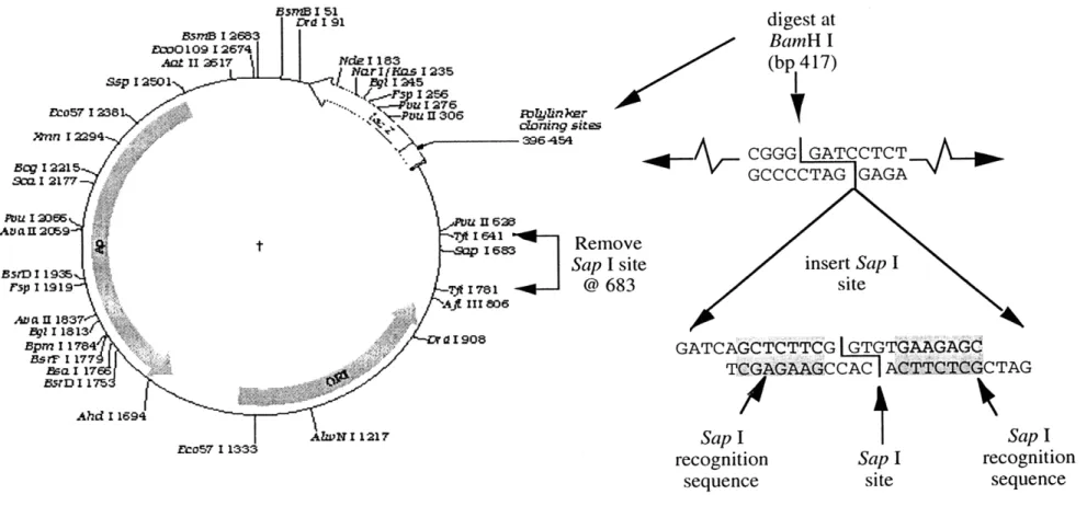
Construction of the pAS1 cloning vector. Plasmids containing DNA polymers of multiple monomer units were created by inserting polymers into a modified pUC19 cloning vector and transfecting the vector into a non-methylating E. coli host for culture. To modify the pUC19 vector for this purpose, the native Sap I site at 683 was removed by digesting pUC19 with Tfi I, gel purifying the larger DNA fragment and religating the vector (pUC19-Sap). The pUC19-Sap was prepared to accept the polymer constructs by inserting a new Sap I restriction site at the BamH I site of the polylinker region as shown in Figure 2.1. The 24 bp insert contained a Sap I
recognition element that would leave, after digestion, a GTG overhang at the 3' end and a CAC overhang on the 5' end. Since the insert was small and reading frame maintained, the ability of the new vector, pAS1, to u-complement was preserved. Construction of pAS1/nmx plasmids. A family of clones containing the same monomer element "x" repeated "n" times was created by digesting pAS1 with Sap I,
dephosphorylating the vector, and ligating in either gel purified polymers of a specific size or a mixture of polymers resulting from the self-ligation of monomers. The ligation was performed at micromolar duplex monomer concentrations for 4 minutes at 0°C to prevent further polymerization or cyclization of the polymers. The mixture of polymers was then ligated into Sap I-digested pAS1 at a molar ratio of 50:1, vector to polymer. GM2163 E. coli cells were transformed with the resulting vectors and the individual clones characterized by plasmid minipreps, DNA
sequencing and gel electrophoresis [63, 64]. Particularly useful in this process was the modified mini-prep protocol developed by Lee and Suraiya [65]. The GM2163 strain was chosen since it is dam methylase deficient and can undergo (-complementation (NEB). Each clone was characterized by the type and size of the polymer insert. For example, a pAS1 vector containing a polymer of 12 repeated monomer elements of
the in-phase DNA duplex was named pAS1/12m9 (9 for in-phase, 10 for out-of-phase and 11 for no-site constructs). For larger quantities, plasmids containing the desired insert were grown under appropriate conditions for the host GM2163 cells (NEB fact sheet #316) with chloramphenicol to increase copy number and reduce contamination by genomic DNA [63]. Plasmid DNA was then extracted and purified using Qiagen Giga Preps.
Identification of calicheamicin binding sites. To verify the location of calicheamicin binding sites, specific cloned DNA constructs (i.e., pAS1/5m9, pAS1/6m10 and pAS1/3m11) were digested with EcoR I, 5'-[3 2p] end-labeled with T4 polynucleotide
kinase and
7-[
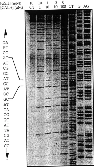
3 2P]-ATP, purified on G25 Quick-spin Columns (Boehringer), anddigested with Hind III [63]. The labeled fragment containing the insert was purified by elution from a 10% polyacrylamide gel [63]. Damage reactions consisted of labeled DNA (-50,000 cpm), 30 jgg/ml of calf thymus DNA, calicheamicin Y11 (0-30 nM), 1% methanol, 10 mM glutathione, 50 mM HEPES, 1 mM EDTA, pH 7. The reaction was initiated by adding calicheamicin. After 1 hr at O'C, the DNA was purified and resolved on an 8% sequencing gel.
Gel mobility assay. The electrophoretic migration studies on the DNA constructs were conducted as follows. Monomers were 5'-[32p] end-labeled and ligated with T4
DNA ligase in a reaction containing 7-5 mM labeled monomer, 50 mM Tris-HCl (pH 7.8), 10 mM MgC12, 10 mM dithiothreitol, 1 mM ATP and 50 gg/ml bovine serum albumin. Ligase was added to a final concentration of 8 units/ml and the mixture was incubated overnight at 160C. The DNA was extracted once with
phenol/chloroform, precipitated in ethanol, and resolved by electrophoresis on an 8% non-denaturing polyacrylamide gel. Gels were then dried and subject to
autoradiography or phosphorimager analysis (Molecular Dynamics).
Results and Discussion
Synthesis, cloning and purification of DNA constructs. In general, the cloning strategy worked well and a variety of plasmids containing different sized polymers of the three monomer inserts were created. Transformed cells showed a propensity for removing whole monomers from the polymer chain, converting from a
the vector would loose the entire insert. E. coli strains deficient in a variety of recombinant proteins fared worse that the GM2163 strain. In any case, the presence of multiple polymer sizes within a recovered plasmid sample had no effect on the damage assays.
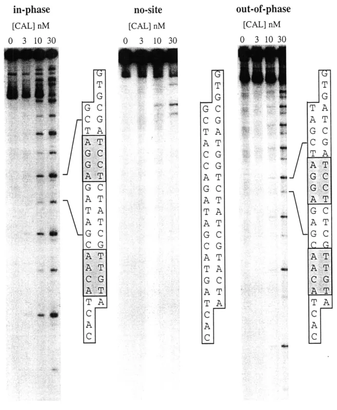
Calicheamicin damage in DNA constructs. To ensure that the DNA constructs possessed the predicted binding sites, multimers of each monomer were damaged by calicheamicin 711 and the cleavage products analyzed on sequencing gels. As shown in Figure 2.2, the mixed-site polymers were damaged only at the expected sites. Consistent with previous studies, damage at the AGGA site occurred more
frequently than the ACAA site [24, 35]. The absence of a similar damage pattern in the no-site construct, under the same conditions, indicates that the primary binding sites have been abolished by the change in sequence. However, a closer look at Figure 2.2 does show the presence of one, perhaps two, low affinity binding sites at different locations.
If we assume that the frequency of drug-induced damage reflects the binding affinity of calicheamicin for these sequences then the drug binds the DNA oligomers as designed.
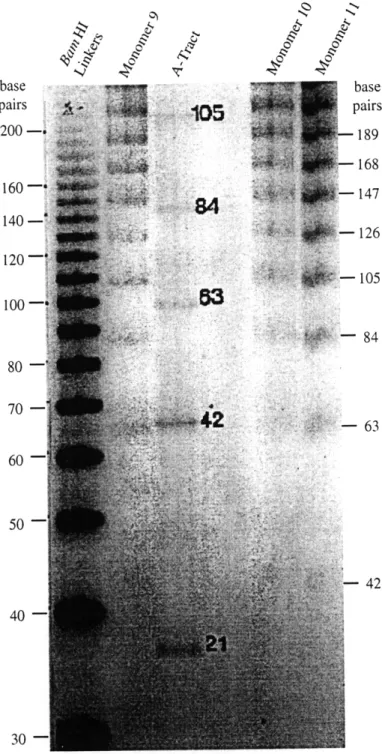
Calicheamicin binding sites are not intrinsically curved. To determine whether calicheamicin binding sites possessed any intrinsic curvature, each monomer was 5'-end labeled, self-ligated and resolved on an 8% non-denaturing polyacrylamide gel. From Figure 2.3, it appears that the binding site constructs migrated normally with respect to the BamH I linker. As expected, the curved A-tract migrated much slower than similarly sized DNA in the adjacent lanes.
The mobility of these polymers relative to the BamH I linker DNA can be quantitatively expressed as the ratio of the polymers apparent length to its true length (RL). The apparent length is determined by measuring the migration
distance of a specific DNA polymer and determining the length of "straight" DNA that would migrate the same distance. The following equation, describing the migration behavior of "straight" DNA, was derived empirically by finding the best fit to the BamH I Linker data (R2 > 0-995, see Figure 2.4; least squares analysis
performed in Cricket Graph by Cricket Software).
where X is the migration distance in centimeters and MW is the molecular weight of the polymer. Rearranging and assuming the molecular weight of each base pair is 650g/mole, gives the following equation for the apparent length (in base pairs) of a DNA polymer migrating X (cm) on the gel:
ALbp = (6.13 x 10⁶) X-3-45 (2.2)
Dividing equation 2.2 by the true length of the polymer, gives the RL value for that specific DNA molecule. The RL values for the polymers shown in Figure 2.3 are plotted in Figure 2.5.
None of the binding site constructs exhibited significant migration anomalies relative to a "straight" DNA standard [66]. These results stand in striking contrast to the abnormally slow migration of the A-tract. These data suggest that neither in-phase, out-of-phase or no-site constructs possess a significant degree of intrinsic curvature. This does not, however, rule out the possibility that calicheamicin targets curved or bent DNA. Therefore, it can only be concluded that two of the DNA sequences recognized by calicheamicin are not curved and that DNA curvature is not necessary for calicheamicin binding. It is important to note that these results do not preclude the possibility that other sequence dependent
properties such as flexibility or twist are responsible for calicheamicin target recognition.
digest at BamH I (bp 417) BlsmB I 51 Drd 1 91 EM0109 12674 / Nar IfKas 1235 S5p I 2501 I 245 .AzPvu 1127306
fto57 I238 u II306 It. r-b.ylinker
doning sites 8cg I 2215 Sco= 12177 PUUU 13065 II 628 AD 4 II 2059 T I641 Ren t I6Re B5sD 1193s5 Sap rsp 11919 1 r1781 @ 1Af 11806 Ava II 1637 B9 11813 Isa 117 ;D I 175 Ahd I 1694 AIwN 11217 bco57 1 1333 nove I site 683 insert Sap I site GATCAGCTCTTCGIGTGTGAAGAGC TCGAGAAGCCAC ACTTCTCGCTAG
/4
4
Sap I recognition sequence Sap I site Sap I recognition sequenceFigure 2.1. Synthesis of the pAS1 cloning vector. pUC19 was digested with Tfi I and religated to remove the existing
Sap I site at 683. The 24 bp sequence, shown on the right, was then inserted into the polylinker cloning region at the BamH I site; creating two new Sap I sites with recognition sequences on both sides of the shared restriction site.
in-phase
[CAL] nM 0 3 10 30no-site
[CAL] nM 0 3 10 30out-of-phase
[CAL] nM 0 3 10 30 T G G C C G TA AT G C GC AT G CA ATT A A T G C C G AT AT CG AT TA C A C'HIP"'
T G G C C G TA AT CG CG AT G C AT TA AT G C C G AT TA G C AT TA C A CI T G TA AT GC CG TA AT GC A T G C ATGC AT G C ..C....A AT A TC G .A T TA C A CFigure 2.2. Damage produced by calicheamicin Y11 in the binding site constructs. Cloned polymers of each monomer were end-labeled, treated with calicheamicin Y11 (0-30 nM) and resolved on sequencing gels. Sequence diagrams, indicating the location of the damage sites in each of the constructs are shown to the right.
:*":
base base pairs
105
pairs 200 -i 18918 160 - 147 ow 147 140-126 30 - - 105100
-- 84 80 - ::: 60 50 -42 40-30-Figure 2.3. Gel migration of polymerized DNA constructs. Monomers were
end-labeled, self-ligated and resolved on an 8% non-denaturing gel. BamH I linker represents a "straight" control whereas the A-tract is known to polymerize into a molecule exhibiting planar curvature. The actual lengths for the polymers are shown, in base pairs, in the margins.
9 10 11
In(molecular weight of polymer) Figure 2.4. Curve fit of BamH I linker data.
3 5 7 9
Polymer Size
(number of 21 bp monomers) Figure 2.5. RL values for
value reflects the relative
the in-phase, out-of-phase, no-site and A-tract. The RL migration of the DNA compared to a "straight" standard.
A-tract' .
in-phase, out-of-phase and no-site
constructs
/
Ir
CHAPTER III
The Effects of Calicheamicin Binding on DNA Topology
DNA is a chain-like structure whose mechanical properties are governed by the hydrophobic and electrostatic forces acting between its individual elements. The balance of these forces determines the overall trajectory, or shape, of the molecule while their interaction defines how the structure will respond to its external environment. In other words, like a solid bar, DNA has properties of form (i.e., curvature and twist) and function (i.e., lateral and torsional stiffness).
As discussed in Chapter I, there is evidence to suggest that calicheamicin selects its targets based -- at least in part -- on the topological characteristics of the binding site. In Chapter II, however, data were presented to show that DNA sequences bound by calicheamicin are not necessarily curved. To 1) explore this apparent inconsistency and 2) further understand the role of DNA flexibility in the target selection process, experiments were performed to investigate the changes that occur in DNA structure when calicheamicin binds. By finding what, if any,
topological changes occur when the drug binds, we may be able to infer the sequence dependent properties that facilitate drug binding.
Experimental Design
As described in Chapter I, the biochemical methods for looking at DNA structure are rather limited. Measuring the changes in electrophoretic mobility of drug bound DNA can reveal drug-induced changes in DNA structure. However, the extreme hydrophobicity and weak binding affinity of calicheamicin for DNA make this approach difficult. The T4 ligase DNA cyclization assay, described in Chapter I, overcomes these problems and provides greater detection of drug-induced structural changes. The assay can be run under a wide variety of conditions and, because it relies on circle closure, is much more sensitive to bend and twist changes.
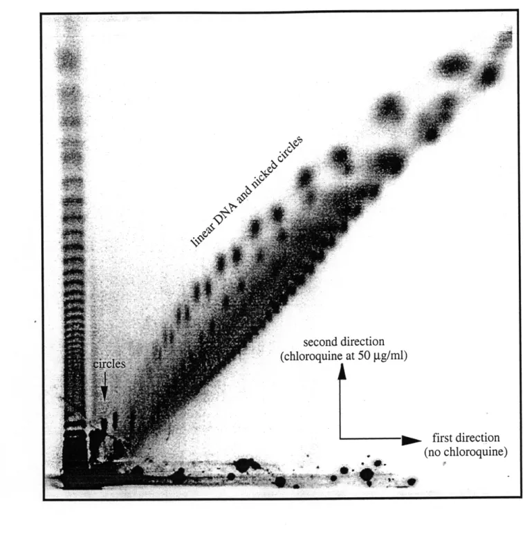
To determine whether a particular sequence is curved, bent or anisotropically flexible by DNA circularization, the sequence of interest is repeated in-phase with the helical repeat of B-DNA and the probability of the molecule forming a circle measured with respect to a random sequence DNA molecule of the same length. The propensity of these DNA molecules to form circles is then measured by "trapping" transiently cyclized products with T4 DNA ligase to form covalently closed circles which can then be separated from uncircularized reaction products [49, 52, 54, 57]. (The ends are "transiently" joined by non-covalently hydrogen bonding between complementary bases.) The proper phasing of the binding sites ensures that the structural changes at each site add constructively to magnify their
individual effects. If the in-phase binding sites are bent, then the DNA molecule will curve in a plane and the ends will move closer together, thus increasing the probability of non-covalent binding of the ends and therefore, the number of covalently closed circles.
As discussed in Chapter I, factors other than bending can affect the rate of circle formation. They are:
1) Molecule length - Longer molecules possess greater intrinsic flexibility
making it easier to bring the ends close together.
2) End sequence - Both the length and sequence of the complementary ends
affect the association and disassociation rates of non-covalently closed DNA circles. Longer, or G*C rich, sequences promote non-covalent binding over shorter, or A*T rich, sequences.
3) End Alignment - When the phosphate backbones are aligned, hydrogen bonding occurs easily and facilitates covalent closure. Ends out-of-phase by ninety degrees hydrogen bond with much lower frequency and
therefore, form fewer circles. This is not the case for long pieces of DNA (approximately 250 bp and larger) where an accumulation of torsional flexibility throughout the chain effectively decouples the rotational position of one end from the other. As a result, the sensitivity of DNA circularization to twist changes within the molecule decreases with increasing molecule length.
Each of these factors must be considered in both the design and interpretation of DNA cyclization experiments.
To assess the bend and twist changes that occur in DNA when calicheamicin binds, circularization studies were performed on the in-phase and out-of-phase DNA duplexes described in Chapter II. The oligomers were annealed with their
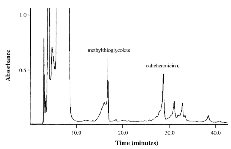
complementary strands, radiolabeled and polymerized with T4 DNA ligase under conditions that allowed circles to form once the DNA molecules had reached a reasonable length. Since components of the ligation reaction buffer activated calicheamicin into the DNA-damaging diradical species, the experiments were performed with calicheamicin r, an inactive form of the drug. While the two compounds bind DNA at the same sequences [33], calicheamicin e appears to bind with 10- to 15-fold lower affinity [67].
NH OCH3 HO \ NH-CH2CH 3 13 CH3. HO CH 30 calicheamicin E
To assess qualitatively the effects of calicheamicin binding on DNA structure, circularization experiments were performed with both the in-phase and out-of-phase DNA constructs over a broad range of drug concentrations. The reactions were repeated with the oligosaccharide portion of calicheamicin to determine which component of the parent compound was responsible for any observed effects.
CH3
CH 30
OCH 3
oligosaccharide (aryltetrasaccharide)
The circular reaction products were resolved by gel electrophoresis using a novel approach that obviated the need for two-dimensional electrophoresis. Identification