HAL Id: hal-02961598
https://hal.inrae.fr/hal-02961598
Submitted on 8 Oct 2020
HAL is a multi-disciplinary open access
archive for the deposit and dissemination of sci-entific research documents, whether they are pub-lished or not. The documents may come from teaching and research institutions in France or abroad, or from public or private research centers.
L’archive ouverte pluridisciplinaire HAL, est destinée au dépôt et à la diffusion de documents scientifiques de niveau recherche, publiés ou non, émanant des établissements d’enseignement et de recherche français ou étrangers, des laboratoires publics ou privés.
endosymbiotic signaling pathway in actinorhizal plant
species
Mireille Chabaud, Joëlle Fournier, Lukas Brichet, Iltaf Abdou-Pavy, Leandro
Imanishi, Laurent Brottier, Elodie Pirolles, Valérie Hocher, Claudine Franche,
Didier Bogusz, et al.
To cite this version:
Mireille Chabaud, Joëlle Fournier, Lukas Brichet, Iltaf Abdou-Pavy, Leandro Imanishi, et al.. Chitote-traose activates the fungal-dependent endosymbiotic signaling pathway in actinorhizal plant species. PLoS ONE, Public Library of Science, 2019, 14 (10), pp.e0223149. �10.1371/journal.pone.0223149�. �hal-02961598�
Chitotetraose activates the fungal-dependent
endosymbiotic signaling pathway in
actinorhizal plant species
Mireille Chabaud1, Joe¨lle Fournier1, Lukas Brichet1, Iltaf Abdou-Pavy1, Leandro Imanishi2, Laurent BrottierID3¤a, Elodie Pirolles3¤b, Vale´rie Hocher3, Claudine Franche4,
Didier Bogusz4, Luis G. WallID2, Sergio Svistoonoff3, Hassen Gherbi3, David G. BarkerID1*
1 Laboratory of Plant-Microbe Interactions (INRA/CNRS/University of Toulouse), Castanet-Tolosan, France, 2 Laboratory of Biochemistry, Microbiology and Soil Biological Interactions, Department of Science and
Technology, National University of Quilmes, CONICET, Bernal, Argentina, 3 Laboratory of Tropical and Mediterranean Symbioses (IRD/INRA/CIRAD/University of Montpellier/Supagro), Montpellier, France,
4 Plant Diversity, Adaptation and Development (IRD/University of Montpellier), Montpellier, France ¤a Current address: Laboratory of Biochemistry & Plant Molecular Physiology (INRA/SupAgro/CNRS/ University of Montpellier), Montpellier, France.
¤b Current address: Laboratory of Biology and Genetics of Plant-Pathogen Interactions (INRA/CIRAD/ SupAgro), Montpellier, France.
*David.Barker@inra.fr
Abstract
Mutualistic plant-microbe associations are widespread in natural ecosystems and have made major contributions throughout the evolutionary history of terrestrial plants. Amongst the most remarkable of these are the so-called root endosymbioses, resulting from the intra-cellular colonization of host tissues by either arbuscular mycorrhizal (AM) fungi or nitrogen-fixing bacteria that both provide key nutrients to the host in exchange for energy-rich photo-synthates. Actinorhizal host plants, members of the Eurosid 1 clade, are able to associate with both AM fungi and nitrogen-fixing actinomycetes known as Frankia. Currently, little is known about the molecular signaling that allows these plants to recognize their fungal and bacterial partners. In this article, we describe the use of an in vivo Ca2+reporter to identify symbiotic signaling responses to AM fungi in roots of both Casuarina glauca and Discaria tri-nervis, actinorhizal species with contrasting modes of Frankia colonization. This approach has revealed that, for both actinorhizal hosts, the short-chain chitin oligomer chitotetraose is able to mimic AM fungal exudates in activating the conserved symbiosis signaling pathway (CSSP) in epidermal root cells targeted by AM fungi. These results mirror findings in other AM host plants including legumes and the monocot rice. In addition, we show that chitote-traose is a more efficient elicitor of CSSP activation compared to AM fungal lipo-chitooligo-saccharides. These findings reinforce the likely role of short-chain chitin oligomers during the initial stages of the AM association, and are discussed in relation to both our current knowledge about molecular signaling during Frankia recognition as well as the different microsymbiont root colonization mechanisms employed by actinorhizal hosts.
a1111111111 a1111111111 a1111111111 a1111111111 a1111111111 OPEN ACCESS
Citation: Chabaud M, Fournier J, Brichet L,
Abdou-Pavy I, Imanishi L, Brottier L, et al. (2019) Chitotetraose activates the fungal-dependent endosymbiotic signaling pathway in actinorhizal plant species. PLoS ONE 14(10): e0223149.
https://doi.org/10.1371/journal.pone.0223149 Editor: Richard A. Wilson, University of
Nebraska-Lincoln, UNITED STATES
Received: May 29, 2019
Accepted: September 13, 2019 Published: October 10, 2019
Copyright:© 2019 Chabaud et al. This is an open access article distributed under the terms of the
Creative Commons Attribution License, which permits unrestricted use, distribution, and reproduction in any medium, provided the original author and source are credited.
Data Availability Statement: All relevant data are
within the manuscript and its Supporting Information files.
Funding: Funding was provided by grants from the
French National Research Agency (ANR-12-BSV7-0007-02), which also covered the salaries of IA-P, EP, L Brottier and L Brichet, from the National University of Quilmes, Argentina (0395/07, 1411/ 15), the Argentinian National Council for Scientific & Technical Studies (CONICET; PIP 2271 and Bernardo Houssay, 2011), the French National
Introduction
A limited number of soil microorganisms have acquired the remarkable capacity to colonize plant roots and form mutually beneficial endosymbiotic associations. The most widespread of these are the obligate biotrophic fungi belonging to the Glomeromycota phylum, able to asso-ciate with the majority of terrestrial plants resulting in the formation of highly ramified intra-cellular root cortical structures known as arbuscules [1,2]. Arbuscular mycorrhizal (AM) fungi scavenge vital soil nutrients such as phosphorus, which are then delivered to the host plantvia
the elaborate symbiotic interface created within arbuscule-containing cortical cells, in exchange for energy-rich metabolites. The AM association is thought to have contributed to the successful establishment of the earliest terrestrial plant species over 400 million years ago [3].
In addition to this ubiquitous fungal symbiosis, certain dicot species belonging to the Euro-sid 1 clade subsequently evolved additional beneficial associations with nitrogen-fixing soil bacteria. Gram-negative rhizobia primarily associate with legume Fabales hosts, whilst gram-positive filamentous actinobacteria known asFrankia are able to colonize actinorhizal host
species belonging to the Fagales, Cucurbitales and Rosales orders. In the majority of cases the nitrogen-fixing bacteria multiply and differentiate intracellularly withinde novo specialized
plant organs known as root nodules, thereby providing a valuable source of nitrogen to the host in return for photosynthate-derived resources. Interestingly, initial root colonization by nitrogen-fixing bacteria can be either intra- or intercellular depending upon the host plant [4]. Whilst intracellular root entryvia infection thread compartments constructed within root
hairs has been well-documented in model legume hosts such asMedicago truncatula [5,6], intercellular modes of colonization in legumes are only poorly understood at the molecular/ cellular levels. Both types of root colonization byFrankia have also been observed for
actinor-hizal hosts [4,7]. For example, intracellular entryvia root hairs appears to be common amongst
Fagales species such asAlnus glutinosa or Casuarina glauca, whereas intercellular modes of
root colonization appear to be predominant for Rosales species typified byCeanothus spp, Elaeagnus angustifolia or Discaria trinervis [8–10]. In the case of initial root colonization by AM fungi, experiments performed on both legume and non-legume hosts have demonstrated that AM fungal hyphae traverse atrichoblast (non-root hair) cells of the root epidermis within host-constructed transcellular compartments by a mechanism reminiscent of root hair infec-tion thread-mediated colonizainfec-tion by nitrogen-fixing bacterial symbionts [2,3].
An important parameter for the establishment of both AM and nitrogen-fixing endosymbi-oses is the capacity for mutual recognition of the respective partners. In particular, it is now well-established that host plant perception of appropriate secreted microbial molecules leads to the activation of a Conserved Symbiotic Signaling Pathway (CSSP) required for subsequent microbe root entry [11,12]. A characteristic feature of the CSSP is the triggering of sustained nuclear-associated calcium oscillations which can be conveniently monitoredin vivo using
fluorescent Ca2+reporters [13,14]. For the large majority of rhizobial-legume associations, decorated lipo-chitooligosaccharides (LCOs) known as nodulation (Nod) factors play this key signaling function, and these LCOs are specifically recognized by host Lysin Motif (LysM) receptor like kinase (RLK) receptor complexes [15,16]. Based on research on legumes such as
M. truncatula, there is evidence that chitin-based molecules are also involved in pre-infection
AM fungal–host signaling. These include both AM fungal LCOs known as Myc LCOs [17] and AM fungal short-chain chitin oligomers (referred to collectively as Myc COs) and typified by chitotetraose (CO4) [18]. Although the precise roles of these chitinaceous factors remain to be determined, recent studies focused on the monocot rice have confirmed the importance of
Research Institute for Sustainable Development (IRD-DPF) and ECOS-SUD (A07B02 and A13B03). LI received a PhD fellowship from CONICET (Argentina). This study is part of the ‘TULIP’ Laboratory of Excellence (ANR-10-LABX-41). The funders had no role in study design, data collection and analysis, decision to publish, or preparation of the manuscript.
Competing interests: The authors have declared
CO4 in AM fungal-host signaling [19], and identified the LysM RLK OsCERK1 as a putative component of the host perception machinery for these fungal-secreted molecules [20].
In contrast withRhizobium-legume associations, relatively little is known about
pre-infec-tion microsymbiont-host signaling during the establishment of actinorhizal symbioses. Although strong evidence suggests that intracellular-colonizingFrankia which nodulate A. glu-tinosa and C. glauca do secrete symbiotic signaling factors, the precise chemical nature of these
molecules has not yet been determined. The use of root hair deformation assays, nuclear-local-ized Ca2+reporters to monitor CSSP activation and the expression of symbiotic reporter genes have together led to the conclusion that theseFrankia make use of alternative
non-chitinac-eous signals [21–23]. On the other hand, it is currently unknown whether chitin-based mole-cules are involved in AM fungal signaling to actinorhizal hosts. In order to investigate this important question, we have made use of thein vivo Ca2+reporter approach to evaluate CSSP activation in epidermal root tissues in response to treatment with crude AM fungal exudates as well as potential Myc CO and Myc LCO signaling molecules. The two evolutionarily distant actinorhizal hosts,C. glauca and D. trinervis, were selected as typical examples of either
intra-cellular or interintra-cellular modes ofFrankia root colonization. Finally, it is important to
under-line that, in the absence of either morphological root responses or available AM-related reporter genes for the two actinorhizal hosts, this assay is the only technique currently available for assaying CSSP activation in response to potential AM fungal signaling.
Results presented in this article show that, for both actinorhizal hosts, AM fungal exudates are efficient activators of sustained nuclear Ca2+spiking in root atrichoblasts, the cellular tar-gets for AM root entry. Furthermore, sub-micromolar concentrations of CO4 were able to mimic CSSP activation for the two host plants and were significantly more efficient elicitors of Ca2+spiking compared to the tested Myc LCOs. Interestingly, in contrast to the observed CSSP activation in AM-targeted atrichoblasts, CO4 treatment did not elicit Ca2+spiking in root hairs, the epidermal cells ofC. glauca which are not colonized by AM fungi. Finally,
although responding robustly to AM fungal signaling with CSSP activation, we show thatD. trinervis atrichoblasts are unresponsive to crude Frankia supernatants. Together, these
find-ings underline the cellular specificity of these responses and indicate that actinorhizal host plants represent excellent models for studying endosymbiont-host signaling mechanisms for both intracellular and intercellular colonization.
Materials and methods
Plant materials and growth conditions
TransgenicC. glauca plants expressing the nuclear-localised cameleon reporter gene p35S: NUP-YC2.1 [24] were generatedvia Agrobacterium tumefaciens transformation and
propa-gated by taking cuttings as previously described [22]. Transgenic composite plants ofD. triner-vis (also now known as Ochetophila trinertriner-vis) expressing the p35S:NUP-YC2.1 reporter in
roots were obtained using ARquaIA. rhizogenes-mediated transformation according to the
protocol described in [25]. Transgenic plants were grown in liquid Broughton and Dillworth (BD) medium [26].
Preparation of germinated spore exudates of
Rhizophagus irregularis and
Frankia supernatants
R. irregularis (Agronutrition, Labège, France) germinated spore exudates (GSEs) were kindly
provided by Soizic Rochange and prepared as described in [18] by incubating 125,000 sterile spores in 40 ml of H2O for 7 d in the dark. These GSEs were then concentrated 40-fold and
applied to freshly excised root segments. Cell-free supernatants were prepared from “induced” cultures ofFrankia strains which nodulate either C. glauca (Frankia CcI3, referred to in this
article asF. casuarinae [27]) orD. trinervis (Frankia BCU110501, referred to in this article as F. discariae [28]). For bothF. casuarinae (Fci) and F. discariae (Fdi) supernatants the post-fix
“i” refers to the induction of theFrankia cultures by addition of the respective host plant
exu-dates 5 days before harvesting as described in [22,29]. These inducedFrankia supernatants
were applied to root segments at a 100-fold dilution.
Chitin-based elicitors
Chitotetraose (CO4) was purchased from Megazym (LIBIOS, Pontcharra, France). An aque-ous stock solution of 10−3M CO4 was diluted to either 10−6or 10−8M. Sulphated and non-sulphated Myc LCOs were kindly provided by Fabienne Maillet and prepared as described in [18]. Since Myc LCO solutions were prepared in diluted acetonitrile, comparative experiments with chitotetraose were performed using CO4 diluted with identical acetonitrile concentra-tions (0.5% acetonitrile for 10−6M CO4 and 0.005% acetonitrile for 10−8M CO4).
Nuclear Ca
2+spiking responses in
C. glauca and D. trinervis roots
expressing the
p35S:NUP-YC2.1 reporter
Assays for nuclear Ca2+spiking were performed using young lateral root segments (0.5 to 0.8 cm long) freshly excised from hydroponically-grown transgenic plants expressing thep35S: NUP-YC2.1 cameleon reporter. Root explants were placed in a microchamber and treated with
150μl of the solution to be tested as described in [18]. Confocal FRET-based ratio imaging for detecting and plotting relative changes of nuclear Ca2+levels corresponding to YFP-to-CFP fluorescence intensity changes over time was performed according to [24] using a Leica TCS SP2 AOBS confocal laser-scanning microscope. Imaging was performed on both atrichoblasts and trichoblasts (root hairs) in the case ofC. glauca. In contrast, Ca2+spiking responses were only studied inD. trinervis atrichoblasts since few root hairs developed on D. trinervis roots
under our growth conditions. It should be underlined that despite limited root hair formation on hydroponically-grownD. trinervis roots, efficient intercellular Frankia colonization and
nodulation can be observed on these roots [e.g. 25]. Experiments were repeated several times for each treatment using independent roots (detailed inS1 Table).
Results
Initial outer root colonization by AM fungi is intracellular for both
C.
glauca and D. trinervis
Late stages of AM fungal colonization have been described for bothC. glauca [29–32] andD. trinervis [33]. In both cases, the presence of intracellular symbiotic arbuscules located within inner root cortical cells, accompanied by axially growing intercellular fungal mycelia is consis-tent with classical Arum-type root colonization [34]. Since the filamentous nitrogen-fixing
Frankia microsymbiont penetrates the root outer tissues of these two actinorhizal hosts via
contrasting mechanisms, it was essential to examine the initial stages of AM fungal root entry for both species. The images shown inFig 1and the accompanying Gif animations (S1andS2 Figs) are consistent with AM hyphopodium formation on the root surface, followed by
intra-cellular hyphal root penetration of the outer root tissues for the two host plants. This suggests that the AM fungus crosses the epidermal and outer cortical cell layersvia a transcellular
apo-plastic mechanism analogous to what has already been observed for both legume and non-legume hosts [35–37]. Furthermore, initial AM fungal entry for the two actinorhizal hosts
occurs exclusivelyvia epidermal atrichoblasts, again similar to earlier findings for other AM
hosts [38]. Thus, despite striking differences in the modes ofFrankia root colonization for C. glauca and D. trinervis, it is likely that comparable cellular mechanisms operate during initial
root entry of the AM fungal partner.
AM fungal spore exudates trigger sustained Ca
2+spiking in the root
epidermis of actinorhizal host plants
Our next objective was to investigate the capacity of AM fungal signals to activate the CSSP in actinorhizal host epidermal tissues, focusing primarily on AM-targeted atrichoblast cells. For this we exploited transgenic plant tissues expressing the cameleon sensor Nup-YC2.1 in order to detect nuclear-associated Ca2+oscillations characteristic of CSSP activation [24]. In the case ofC. glauca, stable Nup-YC2.1-expressing lines were already available as described in [22]. For
D. trinervis, composite plants expressing Nup-YC2.1 in root tissues were obtained via A. rhizo-genes-mediated transformation (seeMaterials & Methods; [25,39]).
Initially, we examined whether crude exudates of germinated AM spores were able to acti-vate the characteristic symbiotic nuclear Ca2+spiking response in atrichoblasts of both actinor-hizal host plants. Young lateral root segments were harvested from plants growing in liquid medium and treated with a 40x concentrated solution of aR. irregularis germinated spore
exu-date (GSE) as described in [18]. Changes in nuclear Ca2+levels within root atrichoblasts were recorded over a 20 min period. Compared with H2O controls (Fig 2A and 2B), GSE treatment
activated sustained nuclear Ca2+oscillations (spiking) in 80–90% of root atrichoblasts, whether fromC. glauca or D. trinervis roots (Fig 2C and 2D). Having established that the CSSP can be activated by AM fungal exudates in theD. trinervis epidermis, we then evaluated responses to
a diluted supernatant from theF. discariae strain that nodulates D. trinervis (supernatants
from host root exudate-induced cultures were prepared as described in [22]). Strikingly, theF. discariae (Fdi) supernatant was unable to elicit Ca2+spiking in theD. trinervis root (Fig 2F), despite the fact that intercellularFrankia root colonization takes place between atrichoblast
Fig 1.Rhizophagus irregularis hyphae enter actinorhizal host roots intracellularly via atrichoblasts. Roots of C.
glauca (A, B) and D. trinervis (C, D) plants were observed 15d after inoculation with spores of R. irregularis. Asterisks mark AM fungal hyphopodia formed on the root surface at sites of hyphal root entry. Single and double arrowheads indicate transcellular hyphae that cross respectively epidermal and outer cortical cells. The two focal planes were selected to highlight either the surface hyphopodia (A, C) or the transcellular hyphae (B, D). To facilitate appreciation of these data, superimposed images (C and D) are presented as an animated Gif inS1 Fig, as well as an animated Gif of a second site of AM fungal colonization of aD. trinervis root (S2 Fig). Bars: 10μm.
cells [10,25]. This contrasts with the situation forC. glauca, where, as previously shown,
super-natants from the inducedF. casuarinae strain (Fci) trigger sustained spiking in C. glauca root
hairs, the target cells for intracellular colonization [22] (Fig 2E).
CO4 mimics the activation of symbiotic Ca
2+spiking by fungal GSEs in the
two actinorhizal hosts
The short-chain chitin oligomer chitotetraose (CO4) is able to activate the AM-dependent CSSP of both legume and non-legume hosts at sub-micromolar concentrations [18,19]. We therefore tested the capacity of CO4 to trigger nuclear Ca2+spiking in root atrichoblasts ofC. glauca and D. trinervis at both 10−6M and 10−8M. Data presented in Fig (3A–3D) show that
both concentrations of CO4 elicit pronounced and sustained Ca2+spiking responses similar to those observed with AM fungal GSEs (Fig 2). Percentages of responding cells ranged from 75– 90% and were marginally higher when CO4 was added atμM levels. In contrast to AM fungi,
F. casuarinae initially colonizes C. glauca via root hairs, and as shown above, Frankia
superna-tants elicit Ca2+spiking in these epidermal cells (Fig 2E). For this reason, we also evaluated the reactivity ofC. glauca root hairs to exogenous 10−8M CO4. Out of the 20 root hairs examined (from three independent root segments), not a single cell responded positively to the Myc CO elicitor (Fig 3E). These findings therefore demonstrate that chitotetraose can mimic the activa-tion of the CSSP by AM fungal GSEs in root atrichoblasts of both actinorhizal host plants, but is not able to trigger nuclear Ca2+spiking inC. glauca root hairs.
Fig 2. Nuclear Ca2+spiking elicited in root epidermal cells of bothC. glauca and D. trinervis in response to either
AM fungal exudates orFrankia supernatants. Freshly excised root segments of C. glauca and D. trinervis were
treated with either an H2O control (a, b), 40x concentrated AM fungal germinated spore exudates (GSE) (c, d) or 100x diluted supernatants of the appropriate inducedFrankia supernatants (SN-Fci and SN-Fdi: seeMaterials & Methods) (e, f). Ca2+spiking responses were monitored in either atrichoblasts (Atr) or root hairs (RH) over 20 min periods following root treatment. These experiments show that fungal GSEs elicit sustained spiking responses in bothC. glauca andD. trinervis atrichoblasts (c, d), the cellular targets for AM colonization. A typical Ca2+spiking response elicited in C. glauca root hairs by an induced F. casuarinae supernatant is shown in (e). By comparison, the negative response to inducedF. discariae supernatant treatment of D. trinervis atrichoblasts is illustrated in (f). Percentages of positively responding cells are indicated for each treatment with the total number of cells examined in brackets.
Myc LCOs are less efficient elicitors of nuclear Ca
2+spiking in actinorhizal
hosts as compared to CO4
In addition to Myc COs such as CO4, other chitin-based molecules have been identified as potential AM fungal symbiotic signals. LCOs, which structurally resemble rhizobial Nod fac-tors, are also present in AM fungal exudates, and have been shown to activate the CSSP in legume hosts [17–19]. Myc LCOs are present in both sulphated (S-Myc LCOs) and non-sulphated forms (NS-Myc LCOs) in exudates ofR. irregularis [17]. For comparative purposes, we therefore evaluated nuclear Ca2+spiking initiation in bothC. glauca and D. trinervis root
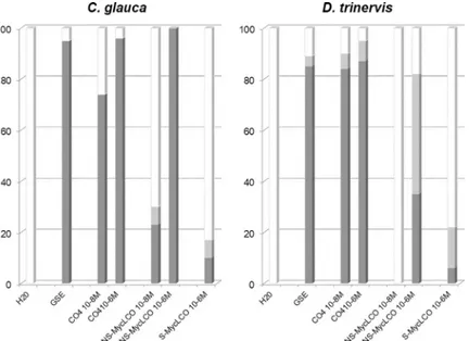
atrichoblasts in response to either S-Myc LCOs or NS-Myc LCOs. These experiments, summa-rized inS1 Tableand in histogram form inFig 4, reveal that both sulphated and
non-sulphated Myc LCOs are capable of triggering Ca2+oscillations in bothC. glauca and D. triner-vis atrichoblasts (illustrated for NS-Myc LCOs inS3 Fig). However, these data also show that Myc LCOs are significantly less active than CO4 in triggering the CSSP in both actinorhizal hosts, and that S-Myc LCOs are even less active than NS-Myc LCOs. In the case ofC. glauca,
the difference between CO4 and NS-Myc LCO activity can be best appreciated at the lower 10−8M concentration. Note also that, since spiking was only observed in 20% of atrichoblasts at 10−6M S-Myc LCO, experiments were not performed at the lower concentration of 10−8M. Although a similar overall trend was found for both actinorhizal hosts, the ability to discrimi-nate between CO4 and NS-Myc LCOs appears greater forD. trinervis by comparison with C. glauca (Fig 4). This difference can also be appreciated in the representative Ca2+spiking pro-files shown inS3 Fig, where 10−6M NS-Myc LCO elicits a response with lower spiking period-icity in theD. trinervis epidermis as compared to C. glauca. Taken together, we conclude that
Fig 3. Chitotetraose (CO4) elicits similar nuclear Ca2+spiking to AM fungal GSEs in root atrichoblasts of both
actinorhizal hosts. Root segments of bothC. glauca and D. trinervis were treated with either 10−6M (a, b) or 10−8M (c-e) CO4, and Ca2+spiking responses monitored in epidermal tissues over 20 min periods. (a-d) Both concentrations of chitotetraose elicited sustained spiking responses in atrichoblasts (Atr) of the two hosts resembling those observed with AM fungal exudates (Fig 2). (e) In contrast, 10−8M CO4 failed to trigger Ca2+spiking in
C. glauca root hairs (RH). Percentages of positively responding cells are indicated for each treatment with the total number of cells monitored in brackets. Note that these figures combine all CO4 treatments (seeS1 Table).
chitotetraose is a more efficient elicitor of CSSP activation in the actinorhizal host root epider-mis compared to the two types of Myc LCOs evaluated in this study.
Discussion
In this article we have investigated early symbiotic signaling between AM fungi and the two distantly-related actinorhizal host plants,C. glauca and D. trinervis, examples of either
intracel-lular or intercelintracel-lularFrankia root colonization respectively. Host perception of AM fungal
sig-nals was studied for both plants using a nuclear-localized Ca2+reporter to monitor the activation of the conserved symbiotic signal transduction pathway (CSSP) in epidermal root tissues. This approach has revealed that AM fungal spore exudates (GSEs) elicit sustained nuclear Ca2+spiking in root atrichoblasts of bothC. glauca and D. trinervis (Fig 2) when applied at doses equivalent to those used previously for the legumeM. truncatula [18]. This finding is coherent with the observation that, for all three host plants, AM fungal root entry is intracellular, with atrichoblasts as the principal epidermal target (Fig 1).
Earlier studies had shown that short-chain chitin oligomers such as CO4/CO5 are present in AM fungal GSEs and furthermore that 10−8M CO4 is sufficient to mimic GSE-elicited Ca2+ spiking inM. truncatula roots [18]. We show here that the same concentration of CO4 is able to trigger spiking responses in root atrichoblasts of bothC. glauca and D. trinervis which
closely resemble those observed following GSE treatment (Fig 3). Our data also reveal that, as forM. truncatula, CO4 is a significantly more active elicitor of the CSSP in roots of both
acti-norhizal hosts compared to either sulphated or non-sulphated Myc LCOs (Fig 4). These find-ings now add two actinorhizal hosts to the growing list of AM fungal host plants for which 10−8M CO4 is able to elicit nuclear Ca2+spiking [18,19]. Not only are Myc COs active on all the AM hosts examined to date, but in all cases CSSP activation is observed in non-root hair
Fig 4. Chitotetraose is a more efficient elicitor of Ca2+spiking in root atrichoblasts of bothC. glauca and D. trinervis by comparison with Myc LCOs. Root segments of both actinorhizal host plants were treated with either
GSEs (40x concentrated), CO4 (10−8& 10−6M), non-sulphated (NS)-Myc LCOs (10−8& 10−6M), or sulphated (S)-Myc LCOs (10−6M). For each treatment the dark grey bars indicate the percentage of atrichoblast cells with more than 2 spikes within the 20 min imaging period, the light grey bars 1–2 spikes, and the white bars represent non-spiking cells. SeeS1 Tablefor details of the number of cells examined for each condition and note that the data presented in this figure for all the CO4 and Myc-LCO treatments were obtained in the presence of acetonitrile (0.005% for 10−8M dilutions and 0.5% for 10−6M dilutions).
atrichoblasts, the cellular targets for AM colonization. Furthermore, for certain hosts, such as rice [19] andC. glauca (Fig 3) this response is cell-specific by comparison with root hairs (see below). Additional direct evidence favoring the role of Myc COs as signaling molecules in the initial establishment of the AM association has come from the recent finding that AM-defec-tiveOscerk1 rice mutants are also defective in responding to CO4 [20], thus suggesting that Myc COs are perceived by a novel rice receptor complex comprising OsCERK1 associated with a second CO-binding LysM-containing membrane protein. Future research now needs to be directed towards identifying the corresponding actinorhizal receptor components capable of recognizing these AM fungal signals and thereby triggering the downstream CSSP. Finally, since LCOs do not appear to be involved in earlyFrankia-host signaling in the case of C. glauca
(see below), the observed low-level responses to Myc LCOs are unlikely to be due to crosstalk with aFrankia-related pathway. In consequence, we interpret this Myc LCO activity as
result-ing from less efficient perception by the AM-associated receptor compared to Myc COs such as CO4.
As stated earlier, initial root colonization ofC. glauca by F. casuarinae occurs intracellularly via root hairs [7]. A variety of host bio-assays, including the expression of transgenic Ca2+ reporters, have together revealed that the symbiotic factors present within theFrankia exudate
are unlikely to be either LCOs or COs [22,23], and hence this raises the question of the nature of the receptors forFrankia signals in C. glauca. As expected, C. glauca root hairs are the
prin-cipal cellular targets forFrankia symbiotic signals [29]. The fact that short-chain COs present in AM fungal exudates can only activate the CSSP in root atrichoblasts (Fig 3) leads us to pro-pose that, in the case ofC. glauca, receptors responding to either Frankia or AM fungal signals
are distinct and specifically localized to the appropriate epidermal target cell. In contrast, the situation differs forD. trinervis, since initial Frankia root colonization occurs intercellularly
between adjacent atrichoblasts [10,25]. We show that, although both AM fungal exudates and CO4 trigger nuclear Ca2+spiking inD. trinervis atrichoblasts, spiking is not observed in these
cells in response toF. discariae supernatants (Figs2and3). One possible interpretation of these findings is that pre-infection symbiotic signaling leading to host CSSP activation is absent during the initial stages ofFrankia colonization of D. trinervis. This question clearly
merits further investigation since there is evidence from legumes that CSSP-related signaling may not be required during rhizobial intercellular “crack entry” invasion of certain host spe-cies [40,41]. In conclusion, the results presented in this article indicate that actinorhizal host plants are particularly valuable model systems for comparative studies of endosymbiont-host signaling mechanisms associated with either intracellular or intercellular root colonization.
Supporting information
S1 Fig. Animated Gif of superimposed images from a site of AM fungal colonization of a D. trinervis root corresponding to (Fig 1C and 1D).
(GIF)
S2 Fig. Animated Gif of superimposed images from a second site of AM fungal coloniza-tion of aD. trinervis root.
(GIF)
S3 Fig. Nuclear Ca2+spiking profiles in root atrichoblasts of the two actinorhizal host plants in response to 10−6M NS-Myc LCOs. These representative profiles reflect the lower
reactivity ofD. trinervis atrichoblasts to NS-Myc LCOs as illustrated in histogram form in
Fig 4. (PDF)
S1 Table. Summary of the Ca2+spiking responses for each treatment including the number of independent roots and the number of cells observed. For each treatment, cells were
assigned to one of the three categories presented in histogram format inFig 4(non-responding cells, cells with 1–2 spikes/20 min and cells with more than 2 spikes/20 min). Note that, in the case of CO4 treatment, the numbers in brackets refer to the roots/cells treated with CO4 in the presence of acetonitrile (0.005% for 10−8M CO4 and 0.5% for 10−6M CO4; seeMaterials & Methods) and correspond to the cells assigned to the three spiking categories.
Atr = atrichoblast; RH = root hair. (DOCX)
Acknowledgments
We would like to thank Soizic Rochange (LRSV, University Paul Sabatier-CNRS, Castanet-Tolosan, France) for kindly providing us withR. irregularis GSEs and Fabienne Maillet (LIPM,
CNRS-INRA, Castanet Tolosan, France) for providing both sulphated and non-sulphated Myc LCOs. Confocal imaging was performed using the Imagery Platform of the Federated Research Institute (FRAIB) at Castanet-Tolosan.
Author Contributions
Conceptualization: Mireille Chabaud, Joe¨lle Fournier, Claudine Franche, Didier Bogusz,
Ser-gio Svistoonoff, Hassen Gherbi, David G. Barker.
Funding acquisition: Didier Bogusz, Sergio Svistoonoff, Hassen Gherbi, David G. Barker. Investigation: Mireille Chabaud, Joe¨lle Fournier, Lukas Brichet, Iltaf Abdou-Pavy.
Methodology: Lukas Brichet, Iltaf Abdou-Pavy, Leandro Imanishi, Laurent Brottier, Elodie
Pirolles, Luis G. Wall, Sergio Svistoonoff.
Project administration: Hassen Gherbi, David G. Barker.
Resources: Leandro Imanishi, Laurent Brottier, Elodie Pirolles, Vale´rie Hocher, Luis G. Wall,
Sergio Svistoonoff, Hassen Gherbi.
Supervision: Mireille Chabaud, Joe¨lle Fournier, Vale´rie Hocher, Luis G. Wall, Sergio
Svistoon-off, Hassen Gherbi, David G. Barker.
Visualization: Mireille Chabaud, Joe¨lle Fournier. Writing – original draft: David G. Barker.
Writing – review & editing: Mireille Chabaud, Joe¨lle Fournier, Claudine Franche, Luis G.
Wall, Sergio Svistoonoff, Hassen Gherbi, David G. Barker.
References
1. Bonfante P, Genre A. Plants and arbuscular mycorrhizal fungi: an evolutionary-developmental perspec-tive. Trends in Plant Science. 2008; 13:492–8.https://doi.org/10.1016/j.tplants.2008.07.001PMID:
18701339
2. Parniske M. Arbuscular mycorrhiza: the mother of plant root endosymbioses. Nature Reviews Microbiol-ogy. 2008; 6:763–75.https://doi.org/10.1038/nrmicro1987PMID:18794914
3. Martin FM, Uroz S, Barker DG. Ancestral alliances: Plant mutualistic symbioses with fungi and bacteria. Science. 2017; 356:aad4501.
4. Ibañez F, Wall L, Fabra A. Starting points in plant-bacteria nitrogen-fixing symbioses: intercellular inva-sion of the roots. Journal of Experimental Botany. 2017; 68:1905–18.https://doi.org/10.1093/jxb/ erw387PMID:27756807
5. Fournier J, Timmers ACJ, Sieberer BJ, Jauneau A, Chabaud M, Barker DG. Mechanism of infection thread elongation in root hairs of Medicago truncatula and dynamic interplay with associated rhizobial colonization. Plant Physiology. 2008; 148:1985–95.https://doi.org/10.1104/pp.108.125674PMID:
18931145
6. Gage DJ. Infection and invasion of roots by symbiotic, nitrogen-fixing rhizobia during nodulation of tem-perate legumes. Microbiology and Molecular Biology Reviews. 2004; 68:280–300.https://doi.org/10. 1128/MMBR.68.2.280-300.2004PMID:15187185
7. Svistoonoff S, Hocher V, Gherbi H. Actinorhizal root nodule symbioses: what is signalling telling about the origins of nodulation? Current Opinion in Plant Biology. 2014; 20:11–18.https://doi.org/10.1016/j. pbi.2014.03.001PMID:24691197
8. Liu Q, Berry AM. The infection process and nodule initiation in the Frankia-Ceanothus root nodule sym-biosis–a structural and histochemical study. Protoplasma. 1991; 163:82–92.
9. Miller IM, Baker DD. The initiation, development and structure of root nodules in Elaeagnus angustifolia L. (Elaeagnaceae). Protoplasma. 1985; 128:107–119.
10. Valverde C, Wall LG. Time course of nodule development in the Discaria trinervis (Rhamnaceae) Fran-kia symbiosis. New Phytologist. 1999; 141:345–54.
11. Gutjahr C, Parniske M. Cell and developmental biology of arbuscular mycorrhiza symbiosis. Annual Review of Cell and Developmental Biology. 2013; 29:593–617. https://doi.org/10.1146/annurev-cellbio-101512-122413PMID:24099088
12. Oldroyd GED. Speak, friend, and enter: signalling systems that promote beneficial symbiotic associa-tions in plants. Nature Reviews Microbiology. 2013; 11:252–63.https://doi.org/10.1038/nrmicro2990
PMID:23493145
13. Barker DG, Chabaud M, Russo G, Genre A. Nuclear Ca2+signalling in arbuscular mycorrhizal and acti-norhizal endosymbioses: on the trail of novel underground signals. New Phytologist. 2017; 214:533–8.
https://doi.org/10.1111/nph.14350PMID:27918078
14. Miwa H, Sun J, Oldroyd GED, Downie JA. Analysis of calcium spiking using a cameleon calcium sensor reveals that nodulation gene expression is regulated by calcium spike number and the developmental status of the cell. Plant Journal. 2006; 48:883–94.https://doi.org/10.1111/j.1365-313X.2006.02926.x
PMID:17227545
15. Antolin-Llovera M, Petutsching EK, Ried MK, Lipka V, Nurnberger T, Robatzek S, et al. Knowing your friends and foes—plant receptor-like kinases as initiators of symbiosis or defence. New Phytologist. 2014; 204:791–802.https://doi.org/10.1111/nph.13117PMID:25367611
16. Zipfel C, Oldroyd GED. Plant signalling in symbiosis and immunity. Nature. 2017; 543:328–36.https:// doi.org/10.1038/nature22009PMID:28300100
17. Maillet F, Poinsot V, Andre O, Puech-Pages V, Haouy A, Gueunier M, et al. Fungal lipochitooligosac-charide symbiotic signals in arbuscular mycorrhiza. Nature. 2011; 469:58–63.https://doi.org/10.1038/ nature09622PMID:21209659
18. Genre A, Chabaud M, Balzergue C, Puech-Pages V, Novero M, Rey T, et al. Short-chain chitin oligo-mers from arbuscular mycorrhizal fungi trigger nuclear Ca2+ spiking in Medicago truncatula roots and their production is enhanced by strigolactone. New Phytologist. 2013; 198:179–89.https://doi.org/10. 1111/nph.12120
19. Sun J, Miller JB, Granqvist E, Wiley-Kalil A, Gobbato E, Maillet F, et al. Activation of symbiosis signaling by arbuscular mycorrhizal fungi in legumes and rice. Plant Cell. 2015; 27:823–38.https://doi.org/10. 1105/tpc.114.131326PMID:25724637
20. Carotenuto G, Chabaud M, Miyata K, Capozzi M, Takeda N, Kaku H, et al. The rice LysM receptor-like kinase OsCERK1 is required for the perception of short-chain chitin oligomers in arbuscular mycorrhizal signaling. New Phytologist. 2017; 214:1440–6.https://doi.org/10.1111/nph.14539PMID:28369864 21. Ce´re´monie H, Debelle´ F, Fernandez MP. Structural and functional comparison of Frankia root hair
deforming factor and rhizobia Nod factor. Canadian Journal of Botany. 1999; 77:1293–301.
22. Chabaud M, Gherbi H, Pirolles E, Vaissayre V, Fournier J, Moukouanga D, et al. Chitinase-resistant hydrophilic symbiotic factors secreted by Frankia activate both Ca2+spiking and NIN gene expression in the actinorhizal plant Casuarina glauca. New Phytologist. 2016; 209:86–93.https://doi.org/10.1111/ nph.13732PMID:26484850
23. Cissoko M, Hocher V, Gherbi H, Gully D, Carre-Mlouka A, Sane S, et al. Actinorhizal signaling mole-cules: Frankia root hair deforming factor shares properties with NIN-inducing factor. Frontiers in Plant Science. 2018; 9:1494.https://doi.org/10.3389/fpls.2018.01494PMID:30405656
24. Sieberer BJ, Chabaud M, Timmers AC, Monin A, Fournier J, Barker DG. A nuclear-targeted cameleon demonstrates intranuclear Ca2+spiking in Medicago truncatula root hairs in response to rhizobial
nodulation factors. Plant Physiology. 2009; 151:1197–206.https://doi.org/10.1104/pp.109.142851
PMID:19700563
25. Fournier J, Imanishi L, Chabaud M, Abdou-Pavy I, Genre A, Brichet L, et al. Cell remodeling and subti-lase gene expression in the actinorhizal plant Discaria trinervis highlight host orchestration of intercellu-lar Frankia colonization. New Phytologist. 2018; 219:1018–30.https://doi.org/10.1111/nph.15216
PMID:29790172
26. Svistoonoff S, Sy MO, Diagne N, Barker DG, Bogusz D, Franche C. Infection-specific activation of the Medicago truncatula Enod11 early nodulin gene promoter during actinorhizal root nodulation. Molecular Plant-Microbe Interactions. 2010; 23:740–7.https://doi.org/10.1094/MPMI-23-6-0740PMID:20459313 27. Nouioui I, Ghodhbane-Gtari F, Montero-Calasanz MD, Goker M, Meier-Kolthoff JP, Schumann P, et al.
Proposal of a type strain for Frankia alni (Woronin 1866) Von Tubeuf 1895, emended description of Frankia alni, and recognition of Frankia casuarinae sp nov and Frankia elaeagni sp nov. International Journal of Systematic and Evolutionary Microbiology. 2016; 66:5201–10.https://doi.org/10.1099/ijsem. 0.001496PMID:27624710
28. Nouioui I, Montero-Calasanz MD, Ghodhbane-Gtari F, Rohde M, Tisa LS, Klenk HP, et al. Frankia dis-cariae sp nov.: an infective and effective microsymbiont isolated from the root nodule of Discaria triner-vis. Archives of Microbiology. 2017; 199(5):641–7.https://doi.org/10.1007/s00203-017-1337-6PMID:
28105505
29. Clavijo F, Diedhiou I, Vaissayre V, Brottier L, Acolatse J, Moukouanga D, et al. The Casuarina NIN gene is transcriptionally activated throughout Frankia root infection as well as in response to bacterial diffusible signals. New Phytologist. 2015; 208:887–903.https://doi.org/10.1111/nph.13506PMID:
26096779
30. Gherbi H, Markmann K, Svistoonoff S, Estevan J, Autran D, Giczey G, et al. SymRK defines a common genetic basis for plant root endosymbioses with arbuscular mycorrhiza fungi, rhizobia, and Frankia bac-teria. Proceedings of the National Academy of Sciences of the United States of America. 2008; 105:4928–32.https://doi.org/10.1073/pnas.0710618105PMID:18316735
31. Diagne N, Escoute J, Lartaud M, Verdeil JL, Franche C, Kane A, et al. Uvitex2B: a rapid and efficient stain for detection of arbuscular mycorrhizal fungi within plant roots. Mycorrhiza. 2011; 21:315–21.
https://doi.org/10.1007/s00572-010-0357-8PMID:21225294
32. Svistoonoff S, Benabdoun FM, Nambiar-Veetil M, Imanishi L, Vaissayre V, Cesari S, et al. The indepen-dent acquisition of plant root nitrogen-fixing symbiosis in Fabids recruited the same genetic pathway for nodule organogenesis. PloS One. 2013; 8:e64515.https://doi.org/10.1371/journal.pone.0064515
PMID:23741336
33. Obertello M, Wall LG. Interactions between Frankia BCU110501 (actinorhiza) and Gigaspora rosea (arbuscular mycorrhiza) with Discaria trinervis studied by spot inoculation. Symbiosis. 2015; 66:13–20.
34. Smith SE, Reed DJ. Mycorrhizal Symbiosis. London, UK: Elsevier. 2008.
35. Bonfante P, Genre A, Faccio A, Martini I, Schauser L, Stougaard J, et al. The Lotus japonicus LjSym4 gene is required for the successful symbiotic infection of root epidermal cells. Molecular Plant-Microbe Interactions. 2000; 13:1109–20.https://doi.org/10.1094/MPMI.2000.13.10.1109PMID:11043472 36. Genre A, Chabaud M, Timmers T, Bonfante P, Barker DG. Arbuscular mycorrhizal fungi elicit a novel
intracellular apparatus in Medicago truncatula root epidermal cells before infection. Plant Cell. 2005; 17:3489–99.https://doi.org/10.1105/tpc.105.035410PMID:16284314
37. Genre A, Chabaud M, Faccio A, Barker DG, Bonfante P. Prepenetration apparatus assembly precedes and predicts the colonization patterns of arbuscular mycorrhizal fungi within the root cortex of both Med-icago truncatula and Daucus carota. Plant Cell. 2008; 20:1407–20.https://doi.org/10.1105/tpc.108. 059014PMID:18515499
38. Chabaud M, Genre A, Sieberer BJ, Faccio A, Fournier J, Novero M, et al. Arbuscular mycorrhizal hyphopodia and germinated spore exudates trigger Ca2+spiking in the legume and non-legume root epidermis. New Phytologist. 2011; 189:347–55.https://doi.org/10.1111/j.1469-8137.2010.03464.x
PMID:20880223
39. Imanishi L, Vayssieres A, Franche C, Bogusz D, Wall L, Svistoonoff S. Transformed hairy roots of Dis-caria trinervis: A valuable tool for studying actinorhizal symbiosis in the context of intercellular infection. Molecular Plant-Microbe Interactions. 2011; 24:1317–24.https://doi.org/10.1094/MPMI-03-11-0078
PMID:21585269
40. Goormachtig S, Capoen W, James EK, Holsters M. Switch from intracellular to intercellular invasion during water stress-tolerant legume nodulation. Proceedings of the National Academy of Sciences of the United States of America. 2004; 101:6303–8.https://doi.org/10.1073/pnas.0401540101PMID:
41. Madsen LH, Tirichine L, Jurkiewicz A, Sullivan JT, Heckmann AB, Bek AS, et al. The molecular network governing nodule organogenesis and infection in the model legume Lotus japonicus. Nature Communi-cations. 2010; 1:10.https://doi.org/10.1038/ncomms1009PMID:20975672