I
NVESTIGATIONS OF CANDIDATE
PLASTOGLOBULE PROTEINS
Characterization of chloroplast NON-INTRINSIC ABC PROTEINS 13 and -14
in Arabidopsis thaliana
&
Constitutive expression of CAROTENOID CLEAVAGE DIOXYGENASE 4
leads to a reduced level of photosynthesis-related carotenoids during senescence
A dissertation submitted to the UNIVERSITY OF NEUCHATEL
For the degree of
Doctor of Philosophy in Biological Sciences
Presented by
Sarah ROTTET
Laboratory of Plant Physiology, Institute of Biology
Accepted on the recommendation of Prof. Felix Kessler, thesis director
Dr. Didier Schaefer PD Dr. Markus Geisler
Faculté des sciences Secrétariat-décanat de Faculté Rue Emile-Argand 11 2000 Neuchâtel - Suisse Tél: + 41 (0)32 718 2100 E-mail: secretariat.sciences@unine.ch
Imprimatur pour thèse de doctorat www.unine.ch/sciences
IMPRIMATUR POUR THESE DE DOCTORAT
La Faculté des sciences de l'Université de Neuchâtel
autorise l'impression de la présente thèse soutenue par
Madame Sarah ROTTET
Titre:
“Investigations of candidate
plastoglobule proteins”
sur le rapport des membres du jury composé comme suit:
- Prof. Felix Kessler, directeur de thèse, Université de Neuchâtel, Suisse - Dr Didier Schaefer, Université de Neuchâtel, Suisse
- PD Dr Markus Geisler, Université de Fribourg, Suisse
Neuchâtel, le 3 mai 2016 Le Doyen, Prof. B. Colbois
5
Keywords: Chloroplasts, plastoglobules, lipids, albino phenotype, ABC transporter,
carotenoid cleavage
Mot-clés: Chloroplastes, plastoglobules, lipides, phénotype albinos, transporteur ABC,
7
Table of contents
Abstract ... 9
Résumé ... 11
List of abbreviations ... 13
Chapter I General introduction ... 17
1 Abstract ... 17
2 Plastids are malleable plant organelles ... 17
3 Thylakoid membranes possess microdomains termed plastoglobules ... 18
4 Role of plastoglobules in plastid differentiation... 21
4.1 Photomorphogenesis: From the etioplast to the chloroplast ... 21
4.2 Reproduction: From the chloroplast to the chromoplast ... 24
4.3 Senescence: From the chloroplast to the gerontoplast ... 27
5 Role of plastoglobules in chloroplast during stress conditions ... 28
5.1 Implication of PG in thylakoid maintenance during high light stress ... 29
5.2 PG may be involved in the tolerance to the heavy metal cadmium ... 31
6 Aim of this work ... 32
Chapter II Characterization of chloroplast NON-INTRINSIC ABC PROTEINS 13 and -14 in Arabidopsis thaliana ... 33
1 Abstract ... 33
2 Background ... 33
3 Results ... 37
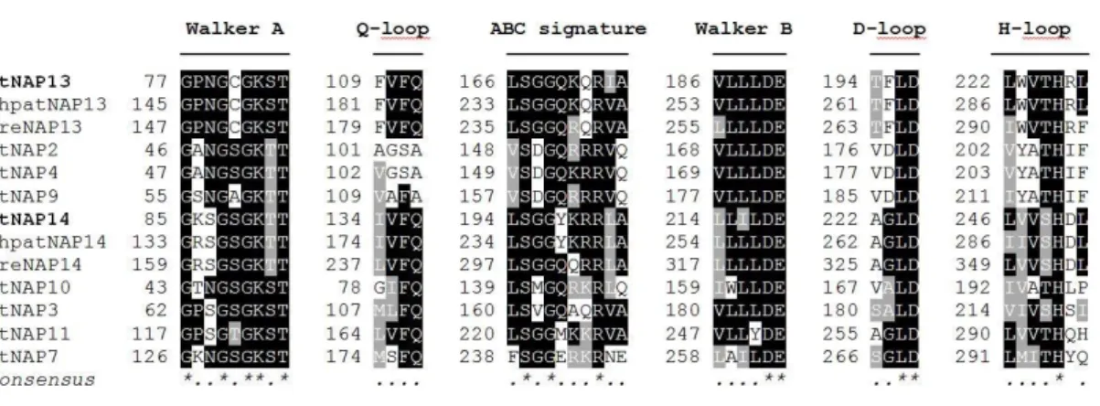
3.1 Structure of AtNAP13 and AtNAP14 proteins ... 37
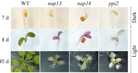
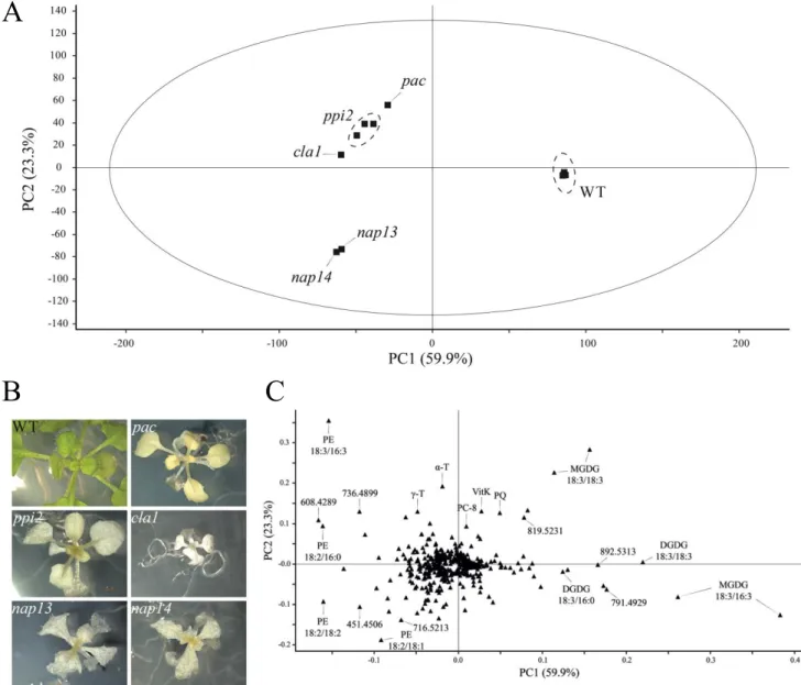
3.2 nap13 and nap14 are albino mutants already affected in etioplast development ... 39
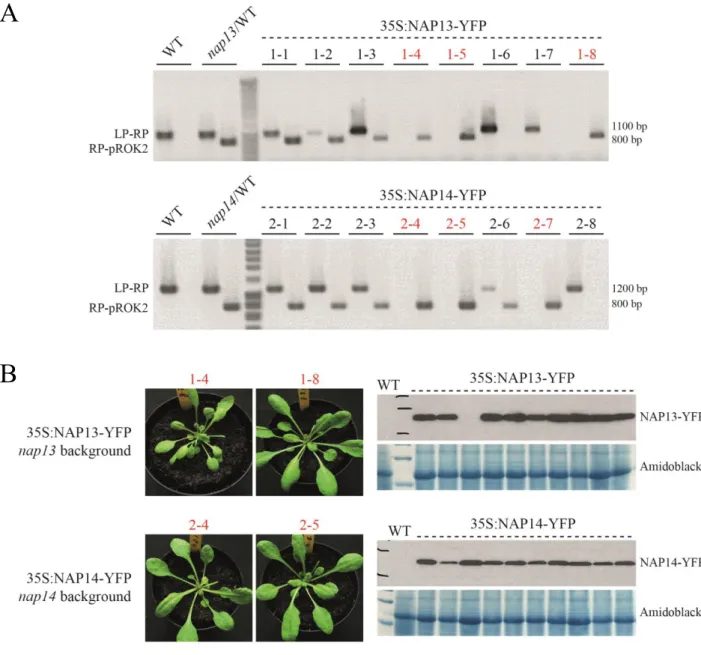
3.3 nap13 and nap14 are complemented by NAP13- and NAP14- derived cDNA constructs, respectively ... 41
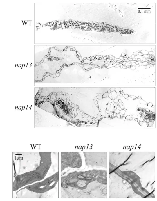
3.4 Leaf organization and plastid ultrastructure are affected in nap13 and nap14 ... 43
3.5 Lipid profiling of nap13 and nap14 reveals perturbation in phosphatidylethanolamine species ... 45
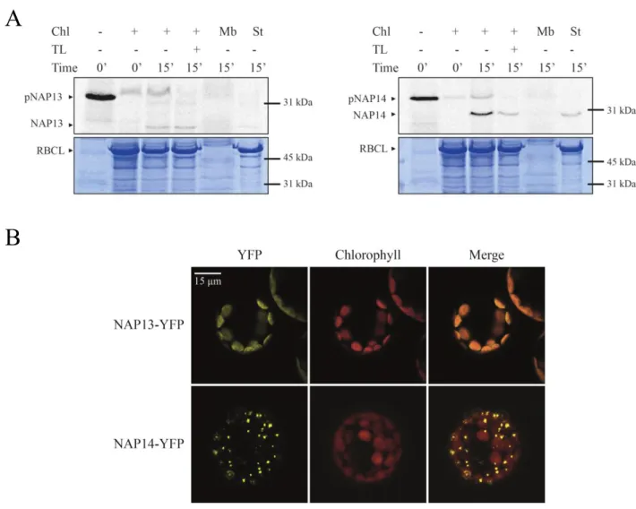
3.6 NAP13 is an extrinsic membrane protein localized at the chloroplast envelope ... 47
3.7 NAP14 might be an extrinsic membrane protein at the thylakoid membrane ... 49
3.8 NAP13 and NAP14 may physically interact with ABCI12 ... 53
3.9 Implication in fatty acid biosynthesis : A shared metabolite with ACC and PDHC? ... 56
4 Discussion ... 57
4.1 Severe phenotypes that emphasize the essential roles played by NAP13 and NAP14 ... 57
4.2 The distinct lipid phenotypes of nap13 and -14 points to a defect in lipid metabolism ... 57
4.3 Is NAP14 really associated with the thylakoid membrane? ... 58
4.4 NAP13 is conditionally associated with the chloroplast inner envelope membrane ... 59
4.5 Discovery of a novel prokaryotic-type ABC transporter in A. thaliana ... 60
5 Conclusion ... 61
Chapter III Constitutive expression of CAROTENOID CLEAVAGE DIOXYGENASE 4 leads to a reduced level of photosynthesis-related carotenoids during senescence ... 63
1 Abstract ... 63
2 Background ... 63
3 Results ... 68
3.1 Mutant characterization through loss-of-function and complementation ... 68
3.2 Subcellular localization of AtCCD4 in plastoglobules ... 71
3.3 Carotenoid accumulation in ccd4 occurs mainly in senescence ... 73
3.4 Plastoglobules of senescent ccd4 contained more β-carotene and lutein ... 76
3.5 Enhanced carotenoid degradation in 35S:CCD4 ... 80
3.6 Deciphering protein-protein interaction to understand regulatory pathway ... 83
Table of contents
8
4.1 Plastoglobule proteomes revealed CCD4, a member of the carotenoid cleavage dioxygenase family ... 85
4.2 Plastoglobule localization of CCD4 and its significance ... 86
4.3 Two approaches to evaluate the implication of CCD4 in carotenoid breakdown during senescence... 87
4.4 Potential interactions with NCED5 and the chloroplast NDH complex ... 88
5 Conclusion ... 89
Chapter IV Materials and methods ... 91
1 Materials ... 91 1.1 Biological material ... 91 1.2 Oligonucleotides ... 91 1.3 cDNA clones ... 93 1.4 Antibodies... 93 1.5 Plasmids ... 93 1.6 Chemicals ... 93 2 Methods ... 94 2.1 Bioinformatics resources ... 94 2.2 Physiological methods ... 94
2.3 Methods for Gateway cloning ... 95
2.4 Agrobacterium-mediated transformation of A.thaliana ... 95
2.5 Diagnostic PCR on plants ... 96
2.6 RNA extraction and qRT-PCR analysis ... 96
2.7 Protein extraction and Western blot analysis ... 96
2.8 Transmission electron microscopy ... 97
2.9 Estimation of carotenoid contents by spectrophotometry ... 98
2.10 Lipid extraction and HPLC analysis ... 98
2.11 Subcellular localization ... 99
2.12 Solubilization assays ... 100
2.13 Co-immunoprecipitation and mass spectrometry ... 101
Acknowledgements ... 103
9
Abstract
Photosynthesis is the key bioenergetic process taking place in the chloroplast. The components of the photosynthetic machinery are embedded in a highly dynamic matrix, the thylakoid membrane. The galactolipids are the major polar lipid components of the thylakoid membrane conferring bilayer properties, while neutral thylakoid lipids such as the prenyllipids and carotenoids contribute to essential functions such as electron transport and photoprotection. Despite a large number of studies, the intriguing processes of thylakoid membrane biogenesis and dynamics remain unsolved. Plastoglobules, thylakoid-associated lipid droplets, appear to actively participate in thylakoid function from biogenesis to senescence. Recruitment of specific proteins enables the plastoglobules to act in metabolite synthesis, repair and disposal under changing environmental conditions and developmental stages. In the first chapter of this thesis, we review plastoglobules as thylakoid membrane microdomains and discuss their involvement in lipid remodeling during stress and in the conversion from one plastid type to another.
The second chapter is dedicated to the characterization of NAP13 (ABCI10) and -14 (ABCI11). These two proteins belong to the large ABC family. Each member of the ABCI subfamily consists of only one of the typical domains of a full ABC transporter. NAP13 and -14 encode two predicted nucleotide-binding domains. Based on earlier results (Shimoni-Shor et al. 2010), our first hypothesis was that NAP14, possibly together with NAP13, are involved in lipid translocation between PG and thylakoids. However, this thesis demonstrates that NAP13 is extrinsically associated with the chloroplast inner envelope while NAP14 cofractionated with the thylakoid membrane. Both the nap13 and -14 mutants had albino phenotypes, emphasizing essential functions in the chloroplast. Interestingly, nap13 and -14 showed similar lipid profiles but distinct from three unrelated albino mutants. The most relevant observation was their reduced level of phosphatidylethanolamine 16:0/18:3. The striking similarities shared by nap13 and -14 suggest that they contribute to the same functional pathway. We hypothesize that this may be lipid transport at the envelope rather than between PG and thylakoids.
In the third chapter, we describe the characterization of CCD4, a carotenoid cleavage dioxygenase, and its physical and functional association with PG. Fluorescence-tagged CCD4 resulted in a punctate pattern inside chloroplasts typical for PG localization in vivo. To discover the function of CCD4, a comparative lipidomics study of Arabidopsis ccd4 mutants and wild type was carried out under various conditions (light stress, senescence, Pseudomonas syringae infection). The results indicated that CCD4 is implicated in β-carotene and lutein degradation during leaf senescence. To further investigate the function of the enzyme, we engineered 35S:CCD4-YFP overexpressing lines. In conclusion, our findings indicate that β-carotene, lutein and violaxanthin are the principle substrates of CCD4 in vivo during chloroplast to gerontoplast differentiation.
11
Résumé
La photosynthèse est le processus clé se déroulant au sein du chloroplaste. La machinerie photosynthétique est intégrée à une matrice très dynamique, la membrane des thylakoïdes. Les galactolipides, les principaux lipides polaires de cette membrane, lui confèrent la propriété de bicouche alors que les lipides neutres tels que les prényllipides et les caroténoïdes contribuent à d’essentielles fonctions telles que le transport d’électrons et la photoprotection. Malgré de nombreuses études, la biogénèse et la dynamique des thylakoïdes restent irrésolus. Les plastoglobules (PG), des gouttelettes lipidiques associées aux thylakoïdes, participent activement aux fonctions des thylakoïdes. Sous un environnement changeant et lors des différents stades de développement, le recrutement d’enzymes spécifiques permet aux PG de participer à la synthèse, la réparation et l’élimination des métabolites. Dans le premier chapitre de cette thèse, nous passons en revue les PG en tant que microdomaines des thylakoïdes et discutons leur implication dans le remodelage lipidique lors de stress et lors de la conversion d’un type de plaste à l’autre.
Le chapitre II est dédié à la caractérisation de NAP13 (ABCI10) et -14 (ABCI11). Ces deux protéines appartiennent à la grande famille des ABC. Chaque membre de la sous-famille ABCI présente un seul des domaines typiques des transporteurs ABC. NAP13 et -14 codent deux domaines prédits pour la liaison des nucléotides. En se basant sur de précédents résultats (Shimoni-Shor et al. 2010), notre première hypothèse impliquait NAP14, éventuellement avec NAP13, dans un transport de lipides entre les PG et les thylakoïdes. Cependant, cette thèse démontre que NAP13 est extrinsèquement associée à l’enveloppe interne du chloroplaste, alors que NAP14 cofractionne avec les thylakoïdes. Les mutants nap13 et -14 ont un phénotype albinos, ce qui souligne une fonction essentielle au sein du chloroplaste. Notons que nap13 et -14 ont un profile lipidique similaire, bien que distinct de trois autres mutants albinos. L’observation la plus pertinente étant leur taux réduit de phosphatidyléthanolamine 16:0/18:3. Les similarités partagées par nap13 et -14 suggèrent que les protéines correspondantes contribuent à la même voie métabolique. D’après nos résultats, il s’agirait d’un transport de lipides au niveau de l’enveloppe.
Dans le chapitre III, nous décrivons la caractérisation de CCD4, une enzyme qui clive les caroténoïdes, et son association physique et fonctionnelle avec les PG. La fusion d’une protéine fluorescente avec CCD4 a donné lieu à un signal ponctué dans les chloroplastes typique d’une localisation aux PG in vivo. Pour élucider la fonction de CCD4, une étude de lipidomique comparative de mutants ccd4 et de plantes sauvages a été effectuée sous diverses conditions. Les résultats indiquent que CCD4 est impliquée dans la dégradation du β-carotène et de la lutéine lors de la sénescence des feuilles. Pour approfondir, nous avons conçu des lignées de surexpresseurs 35S:CCD4-YFP. En conclusion, nos résultats indiquent que le β-carotène, la lutéine et la violaxanthine sont les principaux substrats de CCD4 in vivo durant la différenciation du chloroplaste en gérontoplaste.
13
List of abbreviations
% (v/v) mL / 100mL % (w/v) g / 100 mL 35S CaMV 35S promoter 6xHIS hexahistidinyl-tagA. thaliana Arabidopsis thaliana
ABA abscisic acid
ABC ATP-binding cassette
ABC1K activity of bc1 complex-like kinase
ABCISSE ABC: information on sequence, structure and evolution
ACC acetyl-coenzyme A carboxylase
ANOVA analysis of variance
At Arabidopsis thaliana
ATP adenosine triphosphate
bp base pairs
BP substrate binding protein
CaMV cauliflower mosaic virus
CBP calmodulin binding peptide
CCD carotenoid cleavage dioxygenase
Cd cadmium
cDNA complementary DNA
CFP cyan fluorescent protein
CFU colony-forming unit
Chl chloroplast
Cit Citrus unshiu
CLA1 CLOROPLASTOS ALTERADOS 1
clb5 chloroplast biogenesis 5
Cm Chrysanthemum morifolium Ramat.
CoA coenzyme A
CrtR-β β-carotene β-hydroxylase
Cs Crocus sativus
CTAB cetyltrimethylammonium bromide
Ctrl control
DAG diacylglycerol
DAPI 4',6-diamidino-2-phenylindole
DGAT diacylglycerol acyltransferase
DGDG digalactosyldiacylglycerol
DMF dimethylformamide
DMPBQ 2,3-dimethyl-6-phytyl-1,4-benzoquinone
DNA deoxyribonucleic acid
dNTP deoxy nucleotide triphosphate
List of abbreviations
14
DTT 1,4-dithio-DL-threitol
DXP 1-deoxyxylulose 5-phosphate
EDTA ethylenediamine-N,N,N',N'-tetraacetic acid
Env. envelopes
EtOH ethanol
FA fatty acid
FAPE fatty acid phytyl ester
FAS fatty acid synthase
FBN fibrillin
Fd ferredoxin
fmol femtomole
For forward
FW fresh weight
gDNA genomic DNA
GFP green fluorescent protein
HA human influenza hemagglutinin
HPLC high performance liquid chromatography
HRP horseradish peroxidase
IVT in vitro translated
JA jasmonic acid (jasmonate)
kb kilo base pairs
kDa kilo Dalton
LB T-DNA left border or lysogeny broth
LD lipid droplet
LHC light harvesting complex
LHCBII PSII light-harvesting chlorophyll a/b-binding protein 2
LP left genomic primer
LSD lignostilbene dioxygenases
LYC-β Lycopene β-cyclase
MeOH methanol
MEP methylerythritol 4-phosphate
MES 2-morpholinoethanesulfonic acid
MGDG monogalactosyldiacylglycerol
mRNA messenger RNA
MS Murashige and Skoog or mass spectrometry
NADPH nicotinamide adenine dinucleotide phosphate
NAP non-intrinsic ABC protein
NBD nucleotide binding domain
NCED 9-cis-epoxycarotenoid dioxygenase
NDC1 NAD(P)H dehydrogenase C1
NDH NADH dehydrogenase-like complex
NPQ non-photochemical quenching
P.syringae Pseudomonas syringae
List of abbreviations
15
PC-8 plastochromanol
PCA principal component analysis
PCR polymerase chain reaction
PDHC pyruvate dehydrogenase complex
PE phosphatidylethanolamine
PES phytyl ester synthase
PG plastoglobules
PGL plastoglobulin
PLB (paracrystalline) prolamellar body
PLS-DA partial least squares-discriminant analysis
PMSF phenylmethylsulfonyl fluoride
pNAP13 precursor of NAP13
pNAP14 precursor of NAP14
PPH pheophytin pheophorbide hydrolase
ppi2 plastid protein import 2
PQ plastoquinone
PQH2 plastoquinol
PSI photosystem I
PSII photosystem II
PSM peptide spectral match
PSY phytoene synthase
PYP pale yellow petal
qRT-PCR quantitative real-time PCR
RB T-DNA right border
RBCL RuBisCo large subunit
Rev reverse
RNA ribonucleic acid
ROS reactive oxygen species
RP right genomic primer
RT room temperature or retention time
RT-PCR reverse transcription-PCR
RuBisCo ribulose-1,5-bisphosphate carboxylase/oxygenase
SD standard deviation
SDS sodium dodecyl sulfate
SDS-PAGE SDS-polyacrylamide gel electrophoresis
SE standard error
SSU RuBisCo small subunit
TAG triacylglycerol
TAIL-PCR thermal asymmetric interlaced-PCR
TAP tandem affinity purification
T-DNA transfer DNA
TEM transmission electron microscopy
TEV tobacco etch virus nuclear inclusion A endopeptidase
List of abbreviations
16
THF tetrahydrofuran
Thyl. thylakoids
TIC translocon at the inner chloroplast membrane
TL thermolysin
TM transmembrane domain
TOC translocon at the outer chloroplast membrane
Tricine N-tris(hydroxymethyl)methylglycine
Tris tris(hydroxymethyl)aminomethane
TX triton X-100
UTR untranslated region
VAR3 VARIEGATED 3
VitK phylloquinone
VTE vitamin E-deficient
Vv Vitis vinifera
WT wild type
YFP yellow fluorescent protein
ZDS ζ-carotene desaturase
α-T α-tocopherol
α-TQH2 α-tocopherol quinol
γ-T γ-tocopherol
Upper case, italic gene, WT allele (e.g. NAP13)
Lower case, italic mutant allele (e.g. nap13)
17
Chapter I
General introduction
1
1 Abstract
Photosynthesis is the key bioenergetic process taking place in the chloroplast. The components of the photosynthetic machinery are embedded in a highly dynamic matrix, the thylakoid membrane. This membrane has the capacity to adapt during developmental transitions and under stress conditions. The galactolipids are the major polar lipid components of the thylakoid membrane conferring bilayer properties, while neutral thylakoid lipids such as the prenyllipids and carotenoids contribute to essential functions such as electron transport and photoprotection. Despite a large number of studies, the intriguing processes of thylakoid membrane biogenesis and dynamics remain unsolved. Plastoglobules, thylakoid-associated lipid droplets, appear to actively participate in thylakoid function from biogenesis to senescence. Recruitment of specific proteins enables the plastoglobules to act in metabolite synthesis, repair and disposal under changing environmental conditions and developmental stages. In this introduction, we describe plastoglobules as thylakoid membrane microdomains and discuss their involvement in lipid remodeling during stress and in the conversion from one plastid type to another.
2 Plastids are malleable plant organelles
During the last 1.5 billion years, plastids evolved from cyanobacteria to become the most versatile organelle in plant cells (Timmis et al. 2004; Reyes-Prieto et al. 2007). In angiosperms, totipotent proplastids are maternally inherited (but exceptions exist) (Reboud and Zeyl 1994; Daniell et al. 2005; Hagemann 2010). They are able to differentiate into a large diversity of functional entities, from light harvesting photosynthetic factories to starch storage sites (Thomson and Whatley 1980). Depending on tissue, developmental stage and environmental conditions, proplastids morph into etioplasts, chloroplasts or leucoplasts, while chromoplasts and gerontoplasts are normally derived from chloroplasts (Vothknecht and Westhoff 2001). Even conversions of differentiated plastids into another plastid type are possible and require extensive ultrastructural reorganization (Li and Yuan 2013). Here, we will focus on chloroplasts and three developmentally related forms, namely etioplasts, chromoplasts and gerontoplasts.
Etioplasts occur in photosynthetic tissues in absence of light and represent the chlorophyll-less chloroplast precursor. Chromoplasts are found in reproductive tissues and lend attractiveness to pollinators and disseminators by storing colored carotenoids. Gerontoplasts represent senescent
I – General introduction
18
chloroplasts and appear during leaf senescence. Highly regulated catabolic activities take place in gerontoplasts, while resources are relocalized to seeds or perennial tissues.
Chloroplasts emerge upon illumination (photomorphogenesis) (Waters and Langdale 2009). Characterized by the presence of chlorophyll and a complex network of internal membranes, the thylakoids, chloroplasts allow plants to convert sunlight into chemical energy by photosynthesis. The light reactions of photosynthesis occur at the thylakoid membrane, a highly dynamic lipid matrix that must continuously adapt to changing environmental conditions and developmental cues.
3 Thylakoid membranes possess microdomains termed plastoglobules
The thylakoid bilayer is composed of four glycerolipids (Boudière et al. 2014). These include two galactolipids, monogalactosyldiacylglycerol (MGDG) and digalactosyldiacylglycerol (DGDG); one sulfolipid, sulfoquinovosyldiacylglycerol and one phospholipid, phosphatidylglycerol. Beside these polar lipids, the thylakoid membrane also contains neutral lipids. These play essential roles in electron transport between photosystem II (PSII) and cytochrome b6f (plastoquinone, (Millner and
Barber 1984)) and within PSI (phylloquinone), others act as photoprotective agents at the membrane level (tocopherol) or protect the photosynthetic complex by non-photochemical quenching (NPQ) (xanthophylls).
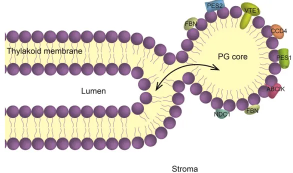
The thylakoids consist of a lipid bilayer enclosing a lumen, resulting in disc-shaped “sacs”. The majority of thylakoids are stacked (granal thylakoids) and interconnected by non-stacked lamellae (stromal thylakoids) (Daum and Kühlbrandt 2011). At the curved regions of the thylakoid membrane, globular lipid droplets, named plastoglobules (PG), are present (Austin II et al. 2006). PG were discovered by electron microscopy owing to their highly osmiophilic properties that are due to the presence of unsaturated lipids (Greenwood et al. 1963). They consist of a membrane lipid monolayer surface surrounding a core of neutral lipids. This membrane lipid surface is studded with proteins (Kessler et al. 1999), many of which are implicated in lipid metabolism (Grennan 2008; Eugeni Piller et al. 2012) (Figure 1.1).
Since the characterization of PG proteomes from Arabidopsis chloroplasts and red pepper chromoplasts, the concept has emerged that PG are not only implicated in the storage of neutral lipid molecules but actively contribute to their synthesis (Vidi et al. 2006; Ytterberg et al. 2006; Lundquist et al. 2012b). Around 30 proteins are associated with the PG polar monolayer (Table 1.1, see (Lundquist et al. 2012b) for a complete list).
Their expression may be regulated in response to stress or developmental conditions (Bréhélin and Kessler 2008). For example, an effect of abiotic conditions, such as prolonged darkness or high light, was observed on the level of PG protein accumulation (Ytterberg et al. 2006; Lundquist et al. 2013). However, the mechanisms of their regulation at the protein level and their targeting to PG remain largely unsolved. Although none of the known PG proteins have predicted transmembrane domains, (Austin II et al. 2006) showed that part of the PG-localized tocopherol cyclase VTE1 (vitamin E-deficient 1) is exposed at the PG surface while another extends across the polar lipid monolayer, allowing direct contact with the neutral lipid content of the PG (Austin II et al. 2006; Kobayashi and
I – General introduction
19
DellaPenna 2008; Mène-Saffrané and DellaPenna 2010). PG proteins belong to three different functional groups: (1) presumed structural proteins called fibrillins (FBN) or plastoglobulins; (2) enzymes implicated in lipid metabolism and (3) uncharacterized proteins. Proteins are recruited to the PG in response to stress or developmental stage. Changes of the PG protein composition may directly and indirectly affect PG lipid composition, because several prenyllipid biosynthetic enzymes and regulatory kinases are located in PG.
It has been suggested that the membrane curvature facilitates PG formation whereby the PG stay physically connected to the thylakoid membrane through the outer membrane lipid leaflet of the thylakoid membrane (Austin II et al. 2006). Moreover, a specific bilayer composition might influence the formation of PG as it has been proposed for lipid droplets (LD). LD are structurally related to the PG and arise at the endoplasmic reticulum membrane. LD serve as lipid reservoir and site of lipid metabolism and are essential in membrane formation and maintenance (Murphy 2012). The molecular mechanisms of LD formation are still under debate (Walther and Farese 2012). One of the models proposes that the products of specifically recruited neutral lipid biosynthetic enzymes accumulate between the lipid leaflets of the membrane bilayer leading to LD formation. This model is equally applicable to PG has they also host enzymes that contribute to neutral lipid biosynthesis and accumulation.
Figure 1.1 Representation of the physical connection between the thylakoid membrane and the plastoglobule
(PG).
Within the chloroplast, the thylakoids are immersed in the stroma. The thylakoid membrane, composed of a polar lipid bilayer (mainly MGDG and DGDG), surrounds the lumen. At the curved regions of the thylakoid membrane, a PG buds from the outer thylakoid leaflet. Surrounded by a polar lipid monolayer, the PG core contains neutral lipids such as prenylquinones, carotenoids, fatty acid phytyl ester and triacyglycerol. The monolayer surface of PG is studded with specific proteins many of which are involved in prenyllipid metabolism. The contact site between PG and thylakoid membrane may provide a conduit for lipid trafficking (arrow). ABC1K, activity of bc1 complex-like kinases; CCD4, carotenoid cleavage dioxygenase 4; FBN, fibrillins; NDC1, NAD(P)H dehydrogenase C1; PES1, phytyl ester synthase 1; PES2, phytyl ester synthase 2; VTE1, tocopherol cyclase. Figure from (Rottet et al. 2015).
I – General introduction
20
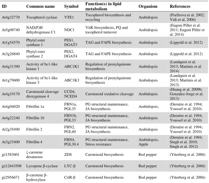
Table 1.1 Characterized plastoglobule (PG) proteins. This table provides a link between PG proteins and
their roles in lipid remodeling. Despite quite numerous studies, the functions of many PG proteins remain unknown (c.f. (Lundquist et al. 2012b) for a complete list). Table from (Rottet et al. 2015). ID Common name Symbol Function(s) in lipid
metabolism Organism References
At4g32770 Tocopherol cyclase VTE1 Tocopherol biosynthesis and
recycling Arabidopsis
(Porfirova et al. 2002; Vidi et al. 2006) At5g08740 NAD(P)H
dehydrogenase C1 NDC1
VitK biosynthesis, PQ and
tocopherol turnover Arabidopsis
(Eugeni Piller et al. 2011; Eugeni Piller et al. 2014)
At1g54570 Phytyl ester synthase 1
PES1,
DGAT3 TAG and FAPE biosynthesis Arabidopsis (Lippold et al. 2012) At3g26840 Phytyl ester
synthase 2
PES2,
DGAT4 TAG and FAPE biosynthesis Arabidopsis (Lippold et al. 2012) At4g31390 Activity of bc1-like
kinase 1 ABC1K1 Regulation of prenylquinone biosynthesis Arabidopsis (Lundquist et al. 2013; Martinis et al. 2014)
At1g79600 Activity of bc1-like
kinase 3 ABC1K3 Regulation of prenylquinone biosynthesis Arabidopsis (Lundquist et al. 2013; Martinis et al. 2013)
At4g19170 Carotenoid cleavage dioxygenase 4
CCD4,
NCED4 Carotenoid oxidative cleavage Arabidopsis
(Huang et al. 2009b; Gonzalez-Jorge et al. 2013)
At4g04020 Fibrillin 1a FBN1a,
PGL35 PG structural maintenance, JA biosynthesis Arabidopsis, (Deruère et al. 1994; Youssef et al. 2010) At4g22240 Fibrillin 1b FBN1b, PGL33 PG structural maintenance, JA biosynthesis Arabidopsis (Deruère et al. 1994; Youssef et al. 2010) At2g35490 Fibrillin 2 FBN2, PGL40 PG structural maintenance, JA biosynthesis Arabidopsis (Deruère et al. 1994; Youssef et al. 2010) At3g23400 Fibrillin 4 FBN4, PGL30.4 PG structural maintenance, Stress resistance Arabidopsis, Apple (Deruère et al. 1994; Singh et al. 2010; Singh et al. 2012) gi|1583601 ζ-carotene
desaturase ZDS Carotenoid biosynthesis Red pepper (Ytterberg et al. 2006) gi|12643508 Lycopene β-cyclase LYC-β Carotenoid biosynthesis Red pepper (Ytterberg et al. 2006) gi|2956671 carotene
β-hydroxylase CrtR-β Carotenoid biosynthesis Red pepper (Ytterberg et al. 2006) VTE, vitamin E-deficient; VitK, phylloquinone; PQ, plastoquinone; DGAT, diacylglycerol acyltransferase; TAG,
I – General introduction
21
Thin layer chromatography, and recently mass spectrometry have allowed the identification of lipids in PG, but the complete lipidomic of PG is not available (Lichtenthaler and Tevini 1970; Ghosh et al. 1994; Zbierzak et al. 2010; Besagni et al. 2011; Martinis et al. 2011; Lippold et al. 2012; Singh et al. 2012; Martinis et al. 2013; Eugeni Piller et al. 2014; Kessler and Glauser 2014; Martinis et al. 2014). The neutral lipid core contains prenylquinones, carotenoids, triacylglycerol (TAG) and phytyl esters. The fact that PG and thylakoid membrane share the same kinds of neutral lipids must be emphasized even though the proportions may vary between the compartments. Concerning the polar lipid monolayer, the situation is ambiguous. In the past, galactolipids were either considered components of PG (Greenwood et al. 1963; Leggett Bailey and Whyborn 1963; Lichtenthaler 1968) or thylakoid contaminants because they were undetected in some studies but not in others (Steinmüller and Tevini 1985; Tevini and Steinmüller 1985). Topologically, PG are contiguous with the outer leaflet of the thylakoid membrane bilayer and therefore logically should share the same polar lipids.
It has been proposed that the thylakoid membrane adapts to changing environmental conditions and developmental cues by lipid remodeling. Lipid turnover is essential to ensure longevity and stress resistance of membranes. Such dynamics are clearly evident in extraplastidic membranes under phosphate deficiency where a large proportion of phospholipids are replaced by DGDG (Härtel et al. 2000; Andersson et al. 2003; Dörmann 2007; Gaude et al. 2007).
Interestingly, Lichtenthaler in 1968 already suggested that PG have a dynamic composition according to the stage of development (Lichtenthaler 1968). This hypothesis has been expanded during the last few years as it became clear that PG are sites of lipid metabolism. It can now be proposed that the PG serve to supply and maintain (neutral) lipids required for remodeling in response to environmental or developmental changes (Figure 1.2).
It is therefore of high interest to elucidate the functions of PG. To achieve this goal, a wide range of methods are being used. Application of stresses and description of the resulting macro- and microphenotypes of plants were among the early approaches, followed by reverse genetics in conjunction with metabolomics analyses. In the following, the implication of PG in thylakoid lipid remodeling under various developmental and stress conditions will be reviewed.
4 Role of plastoglobules in plastid differentiation
4.1 Photomorphogenesis: From the etioplast to the chloroplast
In the dark or under low light intensity, proplastids develop into etioplasts, the chloroplast precursor organelles (Solymosi and Schoefs 2010). Etioplasts are characterized by the presence of the paracrystalline prolamellar body (PLB) that contains molecular building blocks for the chloroplast thylakoid membrane. Light triggers the conversion of the etioplast to the chloroplast. During this conversion, the PLB disappears while the thylakoid membrane emerges accompanied by the production of large quantities of chlorophyll. Dark-grown etiolated tissues have a pale yellow color due to the presence of carotenoids. In addition, etioplasts accumulate the chlorophyll precursor
I – General introduction
22
protochlorophyllide, the conversion of which to chlorophyllide and then chlorophyll occurs almost instantly in response to light (Philippar et al. 2007).
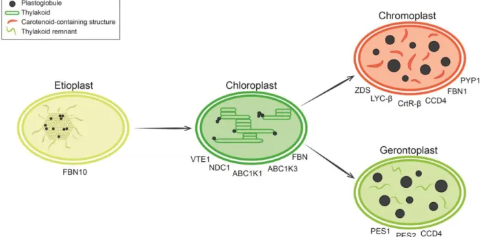
Figure 1.2 Roles of plastoglobules (PG) in plastid differentiation.
Etioplasts, chloroplast precursors, are present in the dark. They are characterized by the paracrystalline prolamellar body (PLB) that is interspersed with numerous small PG. As a known lipid reservoir, PG may assist in the rapid formation of thylakoid membranes upon illumination. Ytterberg et al. (2006) identified the homolog of the thylakoid-associated FBN10 in low-density lipid particles that correspond to the PLB of rice etioplasts (Ytterberg et al. 2006). FBN10 may serve to channel lipids to the nascent thylakoids. Chloroplasts contain the internal thylakoid membrane network. PG are coupled to the thylakoid membrane and involved in lipid remodeling. Tocopherol metabolism is upregulated under high light, involving the PG-located proteins tocopherol cyclase (VTE1), NAD(P)H dehydrogenase C1 (NDC1), activity of bc1 complex-like kinases (ABC1K) and fibrillins (FBN). Chromoplasts in reproductive tissues (fruits and flowers) differentiate from chloroplasts to attract pollinators and seed disseminators. These plastids are characterized by thylakoid disassembly and an appearance of carotenoids stored and produced in PG or carotenoid fibrils with the help of ζ-carotene desaturase (ZDS), lycopene β-cyclase (LYC-β) and two β-carotene β-hydroxylases (CrtR-β). Carotenoid cleavage dioxygenase 4 (CCD4) cleaves carotenoids to release apocarotenoids that contribute to scent and flavor. The tomato pale yellow petal 1 (PYP1) is required for xanthophyll esterification and hence normal yellow petal pigmentation. Gerontoplasts differentiate from chloroplasts during senescence. Chlorophyll and thylakoid membranes are dismantled, releasing large amounts of fatty acids and phytol. These catabolic products are sequestered in triacyglycerol (TAG) and fatty acid phytyl ester (FAPE) by phytyl ester synthase 1 (PES1) and 2 (PES2), two PG-located enzymes. TAG and FAPE contribute to the formation of giant PG. Carotenoids remaining in gerontoplasts are degraded by CCD4. Figure from (Rottet et al. 2015).
I – General introduction
23
Thylakoid membrane formation requires the coordinated assembly of the lipid bilayer with membrane proteins. These are mostly nuclear-encoded, imported proteins, and many are assembled to photosynthetic complexes together with cofactors such as pigments or metal ions. Galactolipids constitute 80% of the thylakoid membrane lipids. Synthesized at the inner envelope membrane, they contain fatty acids (FA) originating from the endoplasmic reticulum. FA are then incorporated into phosphatidic acid, imported into the chloroplast by the trigalactosyldiacylglycerol complex at the inner envelope and converted into diacylglycerol (DAG) (Benning 2009; Wang et al. 2012; Mehrshahi et al. 2013; Mehrshahi et al. 2014). Monogalactosyldiacylgycerol synthase 1 and digalactosyldiacylglycerol synthase 1 are required for galactolipid synthesis. Both enzymes are embedded in the inner envelope of the chloroplast and catalyze the conversion of DAG into MGDG and DGDG respectively, the two main galactolipids present in thylakoid membranes (Aronsson et al. 2008).
Several mechanisms of lipid transfer from the site of synthesis at envelope membrane to the nascent thylakoid membrane have been proposed. These include transport vesicles as well as membrane tubes emerging from the inner envelope or the PLB (Andersson et al. 2001; Kobayashi et al. 2007; Chigri et al. 2012; Kobayashi et al. 2012; Vothknecht et al. 2012; Rütgers and Schroda 2013).
During chloroplast biogenesis, lamellar prothylakoids have been observed to emerge from the PLB (Gunning 2001; Grzyb et al. 2013). The PLB itself consists of tubular structures interspersed with numerous PG (Lichtenthaler 1968). Both PLB tubules and prothylakoids are composed mainly of MGDG and DGDG, the PLB being enriched in MGDG (Aronsson et al. 2008). Apart from membrane lipids, the PLB also contains protochlorophyllide and proteins that are required for rapid chloroplast biogenesis. Proteomics analysis of purified PLB revealed two major functional groups of proteins: (1) Proteins implicated in pigment synthesis, such as the abundant NADPH protochlorophyllide oxidoreductases A/B and chlorophyll synthase. (2) Known thylakoid proteins implicated in photosynthetic reactions (e.g. ATP synthases, cytochrome b6f, ferredoxin-NADPH
oxidoreductases, PSII subunits). The protein and metabolite compositions provide the evidence that the PLB plays a role in chloroplast biogenesis and functions as a precursor of the chloroplast thylakoid membranes (Blomqvist et al. 2008; Solymosi and Schoefs 2010).
In etioplasts, the PLB cages a high number of PG. After exposure to light, PG number decreases in de-etiolated tissue simultaneously with the rise of thylakoid membranes from the PLB (Lichtenthaler and Peveling 1966; Sprey and Lichtenthaler 1966; Lichtenthaler 1968; Nacir and Bréhélin 2013). PG as a lipid reservoir may assist in the rapid formation of thylakoid membranes in greening tissues by releasing structural thylakoid lipids (MGDG) and their precursors (TAG, DAG). Supplying lipid building blocks for membrane expansion may explain why PG become small and rare during thylakoid formation and larger and more abundant in the mature chloroplast (Lichtenthaler 1968). PG also contain tocopherol and other prenylquinones, therefore PG have also been proposed to protect the nascent membrane by supplying lipid antioxidants during chloroplast biogenesis (Eugeni Piller et al. 2014).
However, two questions remain unsolved: how small hydrophobic molecules can be transferred from PG to thylakoid membrane and the extent to which PG and associated proteins are involved in thylakoid membrane formation.
I – General introduction
24
While most likely the contact sites between PG and thylakoids provide the conduit for small molecules, FBN may also be involved in lipid transfer. Indeed, FBN possess a predicted lipocalin motif that has the ability to bind and transport small hydrophobic molecules (Flower 1994; Flower 1996; Flower et al. 2000; Kim et al. 2001; Singh and McNellis 2011). The members of the FBN family constitute the most abundant proteins in PG. Together seven FBN (out of 13 identified in Arabidopsis) represent 53% of the PG proteome mass (FBN1a-At4g04020, FBN1b-At4g22240, FBN2-At2g35490, FBN4-At3g23400, FBN7a-At3g58010, FBN7b-At2g42130, FBN8-At2g46910) (Lundquist et al. 2012b). Initially considered structural proteins, recent studies indicate that FBN function may extend to chromoplast pigment accumulation, resistance to biotic and abiotic stresses as well as protection of the photosystem from photodamage (Giacomelli et al. 2006; Leitner-Dagan et al. 2006; Singh et al. 2010; Youssef et al. 2010). These roles are in agreement with FBN expression patterns, which vary with plant developmental stage, hormonal treatment and various stresses (Pozueta-Romero et al. 1997; Gillet et al. 1998; Singh and McNellis 2011; Lundquist et al. 2013; Gámez-Arjona et al. 2014). Unfortunately, a PG proteome of etioplasts is still missing. Ytterberg et al. (2006) attempted to establish the PG proteome of rice etioplasts (Ytterberg et al. 2006). However, the isolated low-density particles are dominated by protochlorophyllide reductase and showed no overlap with other PG proteomes. For this reason, it was concluded that these low-density particles most likely represented PLB. Interestingly, a member of the FBN family was identified in these low-density particles of rice etioplasts. In Arabidopsis, the probable ortholog is FBN10 (At1g51110). FBN10 is mainly present in thylakoids but not particularly enriched in PG. Nevertheless, it is tempting to speculate on the implication of FBN10 in lipid trafficking during thylakoid formation.
In conclusion, the main ultrastructural modification induced by light during the transition from etioplast to chloroplast is the formation of thylakoid membrane that emerges from the PLB and is presumably assisted by the PG lipid reservoir.
4.2 Reproduction: From the chloroplast to the chromoplast
Higher plants have evolved flowers and fruits, which are specialized tissues for reproduction. Often the color of fruits and flowers are conferred by chromoplasts. Chromoplasts differentiate from chloroplasts during floral development and fruit maturation. During the process the well-developed thylakoid membrane network that characterizes chloroplasts is dismantled and replaced by carotenoid-containing structures and enlarged PG in chromoplasts (Egea et al. 2010). During this transition, the degradation of chlorophyll is concomitant with carotenoid biosynthesis and sequestration (Li and Yuan 2013; Othman et al. 2014).
Carotenoids are widespread pigments synthesized and localized in plastids. They are subdivided in two classes, carotene hydrocarbons that may be cyclized at one or both ends and xanthophylls that contain extra oxygen atoms in the form of hydroxyl or epoxide groups. Carotenoids are highly esterified in fruits and flowers (Hornero-Méndez and Mínguez-Mosquera 2000; Breithaupt and Bamedi 2001). The esterification seems to enhance carotenoid stability (Schweiggert et al. 2006) and to increase their lipophilic properties, which may facilitate their sequestration into carotenoid-containing structures (Deruère et al. 1994).
I – General introduction
25
During chloroplast to chromoplast conversion in red pepper, thylakoids and chlorophyll disappear whereas PG strongly enlarge. In chromoplasts, PG may transform into rod-like structures (fibrils) filled with carotenoids. The major protein associated with the fibrils was termed FBN and turned out to be the first identified member of the FBN family (Deruère et al. 1994; Vishnevetsky et al. 1999). Interestingly, chromoplast fibril self-assembly occured when FBN was added to a mixture of carotenoids and polar lipids (MGDG, DGDG and phospholipids). Fibril reconstitution only occurred when the lipid-protein ratio was close to the in vivo ratio. Furthermore, linear carotenoids such as lycopene inhibited fibril assembly. Cyclic carotenoids (β-carotene, zeaxanthin and capsanthin) gave intermediate results, while esterified carotenoids (zeaxanthin diester and capsanthin diester) worked best (Deruère et al. 1994). This important experiment suggested that FBN play a role in the formation and stabilization of fibrils and PG.
High expression of pepper FBN1 in tomato fruits leads to enlarged PG and protects the thylakoids from degradation during chloroplast-to-chromoplast transition (Simkin et al. 2007). Indeed, an increase in carotenoid content and a delay in thylakoid disintegration were observed under FBN overexpression. These data were interpreted in two different ways. FBN may act directly on the thylakoid membrane. Alternatively, FBN may act indirectly by enhancing PG formation and/or stabilizing their structure, including their thylakoid connections. FBN are often upregulated under abiotic and biotic stresses (for review see (Singh and McNellis 2011)), supporting the idea of their protective role for thylakoid membranes, be it direct or indirect.
In chloroplasts as well as chromoplasts, PG participate in carotenoid metabolism. Ytterberg et al. (2006) identified four enzymes in the PG proteome of red pepper chromoplasts that are implicated in carotenoid biosynthesis, including ζ-carotene desaturase (ZDS), lycopene β-cyclase (LYC-β) and two β-carotene β-hydroxylases (Ytterberg et al. 2006). ZDS introduces conjugated double bonds which are required for the biosynthesis of lycopene, the red pigment of tomato chromoplasts (Ben-Shaul and Naftali 1969), from phytoene. LYC-β catalyzes the cyclization of one or both ends of lycopene to yield α- or β-carotene. The next step in carotenoid biosynthesis is to generate xanthophyll products by the incorporation of oxygen molecules into carotene cycles. By the action of carotenoid hydroxylases, α- and β-carotene are converted into lutein and zeaxanthin, respectively (Ruiz-Sola and Rodríguez-Concepción 2012).
Carotenoids can also be metabolized further by oxidative cleavage to produce apocarotenoids (Auldridge et al. 2006b). Apocarotenoids have important metabolic functions since they act as phytohormones (abscisic acid, strigolactone), pigments (citraurin), aromatic volatiles (ionone, β-cyclocitral, geranial) or signaling molecules (β-cyclocitral). Phytohormones are essential for growth regulation, stress responses and much more (Hirayama and Shinozaki 2007; Gomez-Roldan et al. 2008; Jia et al. 2013). As an aromatic volatile, β-ionone is a pollinator attractant in flowers but also contributes to fruit flavor (Simkin et al. 2004a). β-cyclocitral functions as a stress signal produced under high light. It increases the tolerance to photooxidative stress by modulating the expression of genes involved in the singlet oxygen response (Ramel et al. 2012). The oxidative cleavage of carotenoids may occur spontaneously under oxidative stress or require enzymatic activity (Havaux 2014). The enzymes involved in carotenoid cleavage are grouped into the family of carotenoid cleavage dioxygenases (CCD). Members of this enzyme family have divergent roles in different tissues and species (Auldridge et al. 2006a; Ohmiya 2009). A subgroup termed
9-cis-I – General introduction
26
epoxycarotenoid dioxygenases is essential for abscisic acid biosynthesis (Schwartz et al. 2003; Tan et al. 2003). Notably, PG proteomes contain CCD4 which cleaved 8’-apo-β-caroten-8’-al to release β-ionone in Arabidopsis thaliana (Huang et al. 2009b). Interestingly, simultaneous absence of PG-localized ABC1K1 and ABC1K3 (activity of bc1 complex-like kinase) resulted in a threefold downregulation of CCD4, whereas protein abundance levels of several other PG-localized metabolic enzymes were not affected (Lundquist et al. 2013). Thus, CCD4 stability may depend upon phosphorylation by these two PG kinases. CCD4 is a good example for the divergent roles of the CCD family even though the substrates are not fully known. The following examples strongly suggest a range of CCD4 substrates in different species.
In chrysanthemum (Chrysanthemum morifolium Ramat.), CmCCD4a is responsible for white color formation in petals. The expression level of CmCCD4a in yellow-flowered cultivars is extremely low, while it occurs at high levels in petals of white-flowered cultivars. Since carotenoid biosynthesis is unaffected in both cultivars, and lutein is the predominant carotenoid in yellow petals (Zhu et al. 2010), it is likely that CmCCD4a cleaves lutein into colorless apocarotenoids thus disrupting the accumulation of carotenoids. Despite the fact that CmCCD4a shares 61% of homology with AtCCD4, its association with PG remains experimentally unexplored (Ohmiya et al. 2006; Yoshioka et al. 2012).
During Satsuma mandarin (Citrus unshiu) fruit ripening, the upregulation of CitCCD4 correlates with the accumulation of β-citraurin, a red pigment typical for the Yamashitabeni-wase variety. Actually, CitCCD4 was shown to cleave β-cryptoxanthin and zeaxanthin in vitro at the 7,8 and 7’,8’ position to yield β-citraurin. It was concluded that CitCCD4 is substrate specific since no activity was detected with lycopene, α-carotene, β-carotene and violaxanthin (Ma et al. 2013).
Heterologous expression of the saffron (Crocus sativus) protein shows that CsCCD4 preferentially cleaves β-carotene (but also lutein, neoxanthin and violaxanthin) into β-ionone and β-cyclocitral during stigma development. Furthermore, the expression of CsCCD4-GFP resulted in a spot-like pattern inside chloroplasts reminiscent of PG localization (Rubio et al. 2008; Rubio-Moraga et al. 2014).
Recent insight into carotenoid sequestration stems from a study on tomato flowers (Ariizumi et al. 2014). The pyp1 (pale yellow petal 1) mutant shows reduced yellow pigmentation in flowers and altered chromoplast development due to a lack of xanthophyll esters (mainly neoxanthin and violaxanthin esterified with 14:0 and 16:0 FA). Esterification enhances carotenoid stability (Schweiggert et al. 2006) and may therefore be favored in the chloroplast-to-chromoplast transition. Interestingly, tomato PYP1 shared 58% identity (73% similarity) with phytyl ester synthase 1 (PES1) (Ariizumi et al. 2014) which is localized in PG of A. thaliana. Previously named diacylglycerol acyltransferase 3 (DGAT3), PES1 contributes to TAG and fatty acid phytyl ester (FAPE) synthesis in chloroplasts (Lippold et al. 2012). Besides, its implication in carotenoid esterification is probable as PES1 was found in PG of chromoplasts of red pepper (Ytterberg et al. 2006).
In conclusion, the chromoplast PG proteome differs strongly from that of the chloroplast (Ytterberg et al. 2006). It is customized to assist the chloroplast-to-chromoplast transition. This is particularly
I – General introduction
27
apparent by the presence of the carotenoid metabolic enzymes. These are specifically recruited for the purpose of depositing carotenoids in the emerging colored PG and fibrils.
4.3 Senescence: From the chloroplast to the gerontoplast
During senescence, nutrients are relocated from senescing tissue to the developing seeds and perennial tissues which must survive the challenges of winter (Quirino et al. 2000; Hörtensteiner and Feller 2002). Leaf senescence has aspects of a recycling process and is important for reproduction and plant fitness. Thus, plant senescence is a highly regulated process composed of a cascade of events, ultimately leading to cell death (Besagni and Kessler 2013). It is induced by external factors (e.g low/high temperature, shading or lack of nutrient) or internal factors such as plant age and phytohormones. Developmental factors and stresses lead to the synthesis of jasmonate (JA), abscisic acid, salicylic acid or auxin, which initiate the transcription of genes implicated in senescence. Highly expressed during senescence, the large set of senescence associated genes plays a central role throughout senescence. A number of senescence associated genes function to catalyze catabolic events leading to the transition from chloroplast to gerontoplast. This transition is accompanied by dramatic ultrastructural reorganization that is characterized by the dismantling of the thylakoids and the appearance of giant PG (Ghosh et al. 2001).
Chlorophyll breakdown is the most striking activity taking place in chloroplast to gerontoplast transition (Matile et al. 1999). However, mutants have been identified in which chlorophyll degradation is perturbed. In the most dramatic cases this results in a stay green phenotype that may be either functional (photosynthetically active) or just cosmetic (green but photosynthetically inactive) (Thomas and Howarth 2000).
Free phytol is a catabolic product released from chlorophyll by pheophytin pheophorbide hydrolase (PPH) (Schelbert et al. 2009; Zhang et al. 2014). This hydrolytic product has to be metabolized to minimize its toxicity (Ischebeck et al. 2006). A first pathway implicates the conversion of free phytol to phytol monophosphate by phytol kinase VTE5 followed by a second phosphorylation step and its incorporation into tocopherol (Ischebeck et al. 2006; Valentin et al. 2006). In a second pathway, phytol is incorporated into FAPE by the esterification with free FA. These FA may either be synthesized de novo or result from hydrolysis of galactolipids during thylakoid dismantling. The hydrolysis of galactolipids by lipases releases a pool composed mainly of C18:3 and C16:3 free FA. FA may also be deposited in TAG.
It appears likely that the huge size of PG in gerontoplasts is at least partly due to the accumulation of these senescence-associated catabolic products. Moreover, TAG and FAPE are excellent examples to illustrate the participation of PG in chloroplast lipid remodeling during senescence both at the biochemical and ultrastructural levels. Biochemically, remodeling here refers to the synthesis of FAPE and TAG while galactolipids as well as chlorophyll are broken down. Ultrastructurally, remodeling is observed by electron microscopy: thylakoids disappear and are replaced by massively enlarged PG. Moreover, PG do not only serve to deposit FAPE and TAG but actively participate in their synthesis by the presence of three PG enzymes.
I – General introduction
28
PPH (At5g13800), which dephytylates pheophytin and thereby releases free phytol, was recently identified in the PG of the abc1k1abc1k3 double mutant lacking two PG-associated kinases (Lundquist et al. 2013). The transient association of PPH with PG may be viewed as a response to the increased chlorophyll turnover observed in abc1k1abc1k3 light-stressed leaves (see chapter I, 5.1).
A reverse genetic study revealed that PES1 and PES2, previously named DGAT3 and DGAT4, are overexpressed during senescence and required for FAPE and TAG production in gerontoplasts (Lippold et al. 2012). Indeed, heterologous expression in yeast and in vitro assay show that both proteins, members of the esterase/lipase/thioesterase family, have diacylglycerol acyltransferase activity allowing the production of TAG and FAPE from FA esterified respectively to DAG or phytol (Lippold et al. 2012; Besagni and Kessler 2013). In addition, pes1pes2 mutant plants under nitrogen starvation accumulate far less TAG and FAPE.
PES1 and PES2, however, are not predicted to function exclusively as acyltransferases. It is tempting to speculate that they function as galactolipid lipases at the thylakoid membrane. This idea is supported by the presence of a predicted lipase domain in both proteins, their ability to release FA from MGDG in vitro (Lippold et al. 2012) and because the pes1pes2 double mutant exhibits a delay in senescence characterized by a prolonged persistence of thylakoid membranes and of pale green color whereas wild type (WT) plants are yellow.
In autumn, the progressive disappearance of chlorophyll in tree leaves reveals the carotenoids that are degraded more slowly (Keskitalo et al. 2005). CCD4 identified in the PG proteome has recently been shown to cleave β-carotene during seed maturation and senescence (Gonzalez-Jorge et al. 2013). Moreover, Tevini and Steinmüller (1985) observed in beech leaves (Fagus sylvatica) that carotenoids liberated from the thylakoid membrane were esterified and deposited in PG during senescence (Tevini and Steinmüller 1985). This suggested an implication of carotenoids in FA sequestration as carotenoid esters. This is reminiscent of the PES1 homolog PYP1 in tomato flowers that is required for normal carotenoid ester production in petal chromoplasts (Ariizumi et al. 2014). It therefore appears likely that PES1 and PES2 may catalyze the equivalent reaction in senescent chloroplasts.
In conclusion, the transition from chloroplast to gerontoplast occurs in close relationship with PG which serve not only to store catabolic products but also participate in their release and transformation into storage compounds.
5 Role of plastoglobules in chloroplast during stress conditions
Plants are confronted with various stress conditions, including nitrogen starvation, drought, heavy metal toxicity, temperature variations, pathogen infection and high light. In many cases, these conditions induce oxidative stress within the chloroplast.
I – General introduction
29
5.1 Implication of PG in thylakoid maintenance during high light stress
Noxious reactive oxygen species (ROS) are formed during high light stress, especially singlet oxygen that is responsible for most chloroplast photooxidative damage (Triantaphylidès et al. 2008). Under high light, the major photoprotective mechanism at the thylakoid membrane is NPQ of excess light energy (Jahns and Holzwarth 2012) and the production of membrane antioxidants. NPQ refers to the xanthophyll cycle that dissipates the excessive light energy in the form of heat, thereby reducing ROS formation (Demmig-Adams and Adams 1996; Jahns and Holzwarth 2012), while carotenoids and most of the prenylquinones accumulated in PG are antioxidant molecules playing an important role in photoprotection (Avalos and Limón 2014).
Tocopherol is a well-studied membrane antioxidant and its production is strongly increased under high light (Havaux et al. 2005). Belonging to the tocochromanol family, tocopherol, commonly called vitamin E, refers to four compounds. They differ by the methylation degree of their chromanol ring (α, β, γ and δ). They are lipid-soluble antioxidant molecules (Mène-Saffrané and DellaPenna 2010). α-tocopherol (α-T) is the predominant form in photosynthetic tissues, while γ-tocopherol (γ-T) accumulates mainly in seeds. Tocopherols protect membrane lipids against peroxidation and preserve PSII from photoinactivation (Havaux et al. 2005). Vitamin E-deficient mutants led to the identification of the enzymes involved in tocopherol biosynthesis (Porfirova et al. 2002; DellaPenna 2005). VTE1 is tocopherol cyclase and catalyzes the cyclisation of 2,3-dimethyl-6-phytyl-1,4-benzoquinone to γ-T (Vidi et al. 2006). Surprisingly, VTE1 appears to be localized mainly in PG whereas the remainder of the enzymes of the pathway (VTE2, VTE3 and VTE4) were localized at the chloroplast inner envelope membrane (Soll et al. 1985; Cheng et al. 2003; Vidi et al. 2006; Kobayashi and DellaPenna 2008; Zbierzak et al. 2010). The differential localization of VTE1 in PG and the other enzymes at the inner envelope suggest that tocopherol metabolites need to traffick between the compartments (Bréhélin et al. 2007).
VTE1 is not only implicated in the de novo synthesis of tocopherol but also in the recycling of the tocopherol oxidation product α-tocopherol quinol (α-TQH2) that is produced upon reaction with lipid
peroxides. Cyclization by VTE1 completes the conversion of α-TQH2 into α-T (Kobayashi and
DellaPenna 2008; Eugeni Piller et al. 2014). It is interesting to note that reduced quinones are the preferred substrate of VTE1 (Grütter et al. 2006; Kobayashi and DellaPenna 2008).
Interestingly, this regeneration pathway implicates another PG protein, NDC1 (NAD(P)H dehydrogenase 1), which reduces α-tocopherol quinone to α-TQH2. In the ndc1 mutant α-tocopherol
quinone accumulates to a higher degree than in the WT. NDC1 has a wide substrate specificity and also catalyzes the reduction of plastoquinone (PQ) to plastoquinol contained within PG. In the ndc1 mutant more PQ is present than in the WT under high light conditions. In higher plants, however, this reaction is inconsequential for bulk photosynthetic electron flow unlike Chlamydomonas where NDC1 rather than the NAD(P)H-plastoquinone oxidoreductase complex is responsible for cyclic electron flow around PSI (Jans et al. 2008). NDC1 is also required for the efficient production of plastochromanol (PC-8) from PQ that also depends on VTE1. PC-8 is also known to accumulate in PG. PC-8 inhibits lipid peroxidation and is an efficient oxygen scavenger (Olejnik et al. 1997; Gruszka et al. 2008) primarily produced during senescence rather than under high light (Szymańska and Kruk 2010; Eugeni Piller et al. 2014). Finally and surprisingly NDC1 is also required for the
I – General introduction
30
conversion of 2-phytyl-1,4-naphthoquinone, the immediate precursor of phylloquinone, to phylloquinone in A. thaliana (Eugeni Piller et al. 2011; Eugeni Piller et al. 2014). This step requires methylation of the naphthoquinone intermediate by AtMenG (Lohmann et al. 2006). The role of NDC1 in the methylation step is not clear (Eugeni Piller et al. 2011). It is interesting to note, however, that neither atmenG nor the ndc1 mutant have an apparent phenotype and the naphthoquinone intermediate can functionally replace phylloquinone except under high light conditions where oxidative damage of PSI was observed in atmenG.
The Arabidopsis PG proteomes revealed a surprisingly high number of ABC1-like kinases
(ABC1K9-At5g05200, ABC1K1-At4g31390, ABC1K3-At1g79600, ABC1K5-At1g71810,
ABC1K6-At3g24190, ABC1K7-At3g07700) (Lundquist et al. 2012a; Lundquist et al. 2012b). In bacteria and mitochondria, ABC1 homologs regulate ubiquinone synthesis (Poon et al. 2000; Xie et al. 2011). Recently, it has been demonstrated by two independent studies that two PG ABC1-like kinases, ABC1K3 and ABC1K1, regulate prenylquinone metabolism probably through the phosphorylation of VTE1 (Martinis et al. 2013; Martinis et al. 2014). Martinis et al. characterized the single mutants abc1k1 and abc1k3, while Lundquist et al. concluded that ABC1K1and ABC1K3 are likely to form a complex and consequently focus their work on the characterization of the double mutant abc1k1abc1k3 (Lundquist et al. 2013). abc1k1 and abc1k1abc1k3 showed sensitivity to light stress, expressed by a rapid leaf bleaching and an inability to accumulate anthocyanin. In the abc1k1 and abc1k3 single mutants, VTE1 failed to localize in PG leading to reduced vitamin E (α-T, γ-T and δ-tocopherol) content under high light stress. Surprisingly, however the transcript level of VTE1 was increased. Comparable findings were also made for FBN1a and FBN2 in both single mutants (Martinis et al. 2013; Martinis et al. 2014). This hints at an underlying post-translational regulation of protein stability. ABC1-like kinase-dependent phosphorylation may stabilize the proteins and/or affect their recruitment to PG. But these results are partially in conflict with those for abc1k1abc1k3. A very robust mass spectrometry-based quantification (Lundquist et al. 2013) showed that the levels of VTE1, FBN1a and FBN2 are unaffected in isolated PG of abc1k1abc1k3 compared to WT. In addition, CCD4, ABC1K5 and ABC1K6 levels are strongly downregulated, suggesting that they are stabilized by ABC1K1 and ABC1K3 is required. Moreover, an enrichment of lipoxygenase 3 and 4 (respectively At1g17420 and At1g72520) was observed in isolated PG of abc1k1abc1k3 under light stress. These two newly identified PG proteins are involved in the synthesis of the plant hormone JA using FA. Recruitment of the JA biosynthetic pathway to PG suggests an increased lipid remobilization for JA production in abc1k1abc1k3. An extensive quantification of prenylquinones and carotenoids was also provided, with a specific focus on their distribution and redistribution between PG and thylakoid membrane. Compared to the WT, abc1k1abc1k3 showed an increased amount of PQ, phylloquinone and β-carotene that are relocated to the PG rather than to the thylakoid membranes. In accordance with a reduced activity of VTE1, they also observed a lower level of PC-8 and α-T that are redistributed toward the thylakoid membranes. Interestingly, a novel quinone (molecular mass of 746.6) was identified in isolated PG of abc1k1abc1k3. This putative quinone could be an unknown intermediate of the quinone biosynthesis pathway or an oxidation product of prenyllipids.
The degreening phenotype of abc1k1abc1k3 is not a strict light stress effect as the same phenotype was obtained under drought stress or nitrogen starvation (Lundquist et al. 2013). The inability of
I – General introduction
31
abc1k1abc1k3 to adapt to elevated light intensity, water or nitrogen deficiency probably leads to excessive singlet oxygen production. The implication of singlet oxygen retrograde signaling in the degreening phenotype is supported by the elevated level of β-cyclocitral found in abc1k1abc1k3 under light stress (Ramel et al. 2012).
In conclusion, regulation of VTE1 activity by the phosphorylation of ABC1K3 and ABC1K1 is a common finding, which is strongly supported by predictions and experimentation. The PhosPhAt database (The Arabidopsis Protein Phosphorylation Site Database, http://phosphat.uni-hohenheim.de/) shows that VTE1 has a phosphorylation hotspot near its N-terminus. Conceivably, phosphorylation at this site affects the activity of the protein. Moreover, VTE1 was shown to be phosphorylated in a phosphoproteome analysis upon nitrogen deficiency (Nakagami et al. 2010).
5.2 PG may be involved in the tolerance to the heavy metal cadmium
Heavy metals are either essential (Cu, Zn, Mn) or toxic (Cd, Pb) for plants. Small amounts of essential metals are sufficient to ensure their metabolic role while larger amounts of heavy metals are toxic to the plant (Jasinski et al. 2008). To overcome metal toxicity, phytochelatins, glutathione and ABC transporters are produced in order to sequestrate and/or extrude heavy metals (Clemens et al. 2002; Kim et al. 2007). Thus, heavy metal resistance requires the activation of a specific set of genes.
Cadmium (Cd) has a destructive effect on the chloroplast. Upon Cd treatment, the disorganization of the photosynthetic apparatus was observed. More specifically, PSI and associated (light-harvesting) antenna proteins are dismantled while PSII, cytochrome b6f and ATP-synthase seem less affected
(Fagioni et al. 2009; Villiers et al. 2011). Cd can replace other metal ions (Zn, Fe, Ca, Mg) in proteins and may affect the redox state by generating, directly or indirectly, ROS. Regarding lipids, spinach basal leaves showed an increase in xanthophyll content (neoxanthin, violaxanthin and lutein) concomitant with a chlorophyll decrease (chlorosis) in the presence of Cd. The lutein overproduction may represent a defensive mechanism against ROS (Fagioni et al. 2009). Surprisingly, upon Cd treatment, plants seem unable to produce zeaxanthin, a well-described ROS scavenging defense (Havaux et al. 2007; Dall’Osto et al. 2012). In the chloroplast, Cd toxicity mimics the symptoms observed during senescence. A notable change is a considerable increase in the number and size of PG (Jin et al. 2008). Indeed, Cd-treated Pisum sativum plants showed disorganized thylakoid membranes in conjunction with an increase in size and number of PG as well as in the size of starch grains. It was suggested that Cd toxicity induces oxidative stress as in leaf senescence (Sandalio et al. 2001).
Interestingly, the expression of the envelope-located ABC1K8 (or OSA1, oxidative-stress related ABC1-like protein) is strongly affected by Cd treatment (Jasinski et al. 2008). To investigate the implication of PG in Cd resistance, effect of Cd on root growth was evaluated for WT, abc1k1, abc1k3 and abc1k1abc1k3 (Lundquist et al. 2013). However all genotypes showed the same reduced root growth, indicating that ABC1K1 and ABC1K3 are not involved in Cd tolerance. Nevertheless, the PG proteome contains 4 other ABC1-like kinase candidates that have not yet been characterized.