thalamus
Common physiopathology for sensory, motor and limbic
positive symptoms
D. Jeanmonod, M. Magnin and A. Morel
Laboratory for Functional Neurosurgery, Neurosurgical Clinic, University Hospital, Zurich, Switzerland
Correspondence to: D. Jeanmonod, Laboratory for Functional Neurosurgery, Neurosurgical Clinic, University Hospital, Rdmistrasse 100, 8091 Zurich, Switzerland
Summary
Positive symptoms arise after lesions of the nervous system. They include neurogenic pain, tinnitus, abnormal movements, epilepsy and certain neuropsychiatric disorders. Stereotactic medial thalamotomies were performed on 104 patients with chronic therapy-resistant positive symptoms. Peroperative recordings of 2012 single units revealed an overwhelming unresponsiveness (99%) to sensory stimuli or motor activa-tion. Among these unresponsive cells, 45.1% presented a rhythmic or random bursting activity. Rhythmic bursting activities had an average interburst interval of 263±46 ms corresponding to a frequency of 3.8±0.7 Hz. Frequency variations among the different symptoms were not statistically
different. Intraburst characteristics such as the highest frequency encountered in the burst (480±80 Hz) or the mean frequency of the burst (206±44 Hz) were also similar in all patients. All bursts, rhythmic or random, fulfilled the extracellular criteria of low-threshold calcium spike (LTS) bursts. After medial thalamotomy and depending on the symptom, 43-67% of the patients reached a 50-100% relief with sparing of all neurological functions. On the basis of these electrophysiological and clinical results, we propose a unified concept for all positive symptoms centred on a self-perpetuating thalamic cell membrane hyperpolarization, similar to the one seen in slow-wave sleep.
Keywords: low-threshold calcium spike bursts; neurogenic pain; abnormal movements; epilepsy; tinnitus; neuropsychiatric disorders
Abbreviations: LTS = low-threshold calcium spike
Introduction
British medicine of the second half of last century (Reynolds, 1858; Monro, 1862; Jackson, 1889; for review, see Berrios, 1985) laid the clinical foundations for the distinction between negative and positive symptoms arising after a lesion of the nervous system. As for positive symptoms, the expressions 'loss of equilibrium' and 'state of overfunction' were proposed and thought to be mainly related to the 'release' phenomenon, i.e. a neuronal hyperactivity due to the loss of afferent inhibition. Since then, data have accumulated showing that injuries involving excitatory projections can be at the origin of positive symptoms, e.g. spinothalamic tract lesions resulting in neurogenic pain. What are then the mechanisms allowing a loss of excitatory afferents to trigger the appearance of such a 'state of overfunction'? Recent © Oxford University Press 1996
experiments have demonstrated that thalamic neurons show a specific activity in strict relationship to a certain state of membrane hyperpolarization (Llinas and Jahnsen, 1982; Jahnsen and Llinas, 1984a, b\ Steriade et al., 1990a). This activity is composed of spike bursts initiated by the deinactivation of calcium channels, the so-called low-threshold calcium spike bursts. A lesion producing a membrane hyperpolarization by either a loss of excitation or an increase of inhibition, can thus lead to the appearance of a bursting activity at the origin of a positive symptom. Along this line, LTS bursts were hypothesized to be at the origin of parkinsonian tremor, action tremor, neurogenic pain or absence epilepsy (Buzsaki et al., 1990; Par6 et al., 1990; Roberts et al., 1992; Llinas and Par6, 1995). Bursting
activities were recorded in the thalamus of patients with neurogenic pain (Modesti and Waszak, 1975; Rinaldi et al., 1991) and abnormal movements (Raeva, 1986), but were first recognized as LTS bursts by Lenz et al. (1989, 1994) in the lateral thalamus (ventral posterior nucleus) and by Jeanmonod et al. (1993) in the medial thalamus.
A causal role of a medial 'essential thalamic centre' in the physiopathology of neurogenic pain was proposed initially by Head and Holmes (1911). This hypothesis was later supported by the beneficial effect of stereotactic medial thalamic lesions (medial thalamotomy) against positive symptoms in man (Adams and Rutkin, 1965; Ojemann and Ward, 1975; Sano, 1977; Hitchcock and Teixeira, 1981; Spiegel, 1982), obtained together with a remarkable sparing of neurological functions. The present work confirms and extends the therapeutic possibilities of the medial thalamo-tomy against positive symptoms and, on the basis of the presence of medial thalamic LTS bursts, gives firm support for a common physiopathology for all positive symptoms considered here.
Methods
Patient population
The stereotactic medial thalamotomy was approved by the University Hospital ethics committee and proposed to 104 patients suffering from chronic severe therapy-resistant positive symptoms. The patients gave informed consent to the procedure. Ages ranged between 13 and 82 years, and symptom duration between 3 months (cancer neurogenic pain) and 29 years (dystonia). Seventy-four patients had peripheral or central neurogenic pain, 22 had abnormal movements (parkinsonian tremor, rigidity and hypo-bradykinesia, athetodystonia, spasticity, cerebellar tremor, thalamic dyskinesia, hemifacial spasm), four had frontal or parietal epilepsy, three tinnitus and two neuropsychiatric symptoms (obsessive-compulsive disease, endogenous depression and psychosis). This list describes only the dominant, i.e. clinically most relevant, symptomatology, as 31% of patients had an additional, more or less disturbing positive symptom. One patient had even the association of hemifacial spasm, neurogenic pain and tinnitus. The most frequent association was neurogenic pain and spasticity.
Technique
Surgery took place under local anaesthesia in fully awake patients. A stereotactic frame (Radionics, Burlington, Mass., USA) compatible with MRI allowed MRJ localization of medial thalamic targets using an adaptation of a CT technique (Jeanmonod and Thomas, 1989). The electrode reached the computer-calculated target through a prefrontal approach, under impedance monitoring. All targets were confined to the medial thalamus, including the central lateral nucleus, the centre mddian, the parafascicular nucleus, the ventromedial
nucleus and the posterior thalamic complex comprising the suprageniculate-limitans complex and the posterior nucleus. Single unit recordings were obtained using tungsten microelectrodes (impedance 0.5-1 Mil). Unit responses were searched with tests of motor voluntary function, and tactile, nociceptive, proprioceptive and auditory stimuli. Unit recordings and stimulation data guided the location of radiofrequency thermolesions (volume 40-120 mm3).
Quantitative data analysis was carried out off-line using the software Discovery (Data Wave Technologies) to produce interspike interval and autocorrelation histograms. Four categories of unit activities were established according to the following criteria. Bursting units were considered as rhythmic if the autocorrelation histogram (centred form: time analysis, 4 s; bin size, 10 ms) of their activities exibited at least four successive peaks and their interspike interval histogram (time analysis, 2.2 s; bin size, 1 ms) showed a marked bimodal distribution. Random bursting units were characterized by autocorrelation histograms displaying less than four peaks and by interspike interval histograms with weak bimodal distributions. Activities which did not fulfil the above criteria were classified as sporadic and further subdivided into responsive or unresponsive.
Quantitative analysis of the intraburst characteristics was based on two parameters given by an interspike interval histogram centred on the burst (time analysis, 30 ms; bin size, 0.1 ms): (i) the shortest interval which corresponds to the highest frequency found within the bursts; and (ii) the mean interval representative of the mean frequency of the bursts. Student's t test was used for data comparisons, considering P values < 0.05 as an indicator of a statistically significant difference.
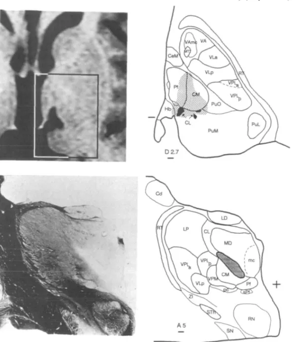
Patients underwent a post-operative three-dimensional MRI examination to control lesion location. Target position, lesion reconstruction and unit localization were projected on sections of our stereotactic atlas of the human thalamus. Two examples of medial thalamic lesions are illustrated in Fig. 1. In the first case (Fig. 1, upper row), three targets (the central lateral nucleus, the parafascicular nucleus and the posterior thalamic complex) were aimed at during two different interventions in a patient suffering from central neurogenic pain. At the level shown in the figure, the three lesions merge with each other, the lateral one being the dorsal extension of the posterior thalamic complex target. In the second case (Fig. 1, lower row), the histological reconstruction of a medial thalamotomy in a patient who suffered from neurogenic pain and died from cancer, shows the localization of the lesion confined to the targeted central lateral nucleus.
Assessment of clinical results
Clinical results were assessed using visual analogue scales for pain and tinnitus (0-100), activities of daily living, drug intake, audiometric masking for tinnitus, fit calendar for epilepsy, 24-h assessment of motor activity with an accelerometer, video recordings for abnormal movements
Fig. 1 Upper row: 3 months post-operative gadolinium enhanced MRI (left) and atlas projection (right) of medial thalamic therapeutic lesions in a patient suffering from central neurogenic pain. The insert on the axial MRI and the corresponding atlas section are 2.7 mm dorsal to the intercommissural plane (the posterior commissure level is indicated on the left-hand side of the atlas section). Black areas
correspond to densocellular islands. Lower row: frontal section stained for myelin (left) and lesion reconstruction (right) of a medial thalamotomy in a patient who suffered from peripheral neurogenic pain and died from cancer. The level of the section is 5 mm anterior to the posterior commissure. A plus sign indicates the intersection between intercommissural and midsagittal planes. Cd = caudate nucleus; CeM = central medial nucleus; CL = central lateral nucleus; CM = centre median nucleus; Hb = habenular nucleus; LD = lateral dorsal nucleus; LP = lateral posterior nucleus; MD = mediodorsal nucleus; me = magnocellular division of MD; MTT = mamiliothalamic tract; Pf = parafascicular nucleus; PuL, PuM, and PuO = lateral, medial and oral pulvinar nuclei; VLa = ventral lateral anterior nucleus; VLp = ventral lateral posterior nucleus; RN = red nucleus; RT = reticular nucleus; SN = substantia nigra; sPf = subparafascicular nucleus; STh = subthalamic nucleus; VA = ventral anterior nucleus; VAmc = ventral anterior nucleus, magnocellular division; VPLpj = ventral posterior lateral nucleus, posterior and anterior divisions, respectively; VPM = ventral posterior medial nucleus; pc = parvocellular division of VPM; ZI = zona incerta. Scale bars = 1 mm.
and the unified Parkinson's disease rating scale. In motor disorders, the symptom and relief intensities were rated between 0 (absent) and 4 (very marked). For all symptoms,
the patient's estimation in percent of the global post-operative improvement was asked. In neurogenic pain and tinnitus, this estimation was accepted as such, even if indirect assessments
(activities of daily living, drug intake, emotional improve-ments) would have spoken for a larger relief. These indirect assessments never indicated a more limited relief than the patient's estimation did.
Results
Unit recordings
Two thousand and twelve units were recorded along 189 penetrations directed at medial thalamic targets. Unit activities were classified into four types: (i) rhythmic bursting; (ii) random bursting; (iii) sporadic; (iv) sporadic with evoked response. A very large majority of cells (>99%) were unresponsive to sensory stimuli or motor activation. Among the unresponsive cells, 45.1% presented random or rhythmic bursting activities and tended to concentrate (localization error ± 1 mm) in the central lateral nucleus. Figure 2 shows typical examples of rhythmic bursting activities found in patients presenting different positive symptoms. These rhythmic bursting units were characterized by interburst intervals ranging from 176 ms (2.5 Hz) to 397 ms (5.6 Hz) (average: 263 ms±46 ms, 3.8±0.7 Hz). When each symptom was considered independently, interburst interval average was 260±44 ms for pain, 258±42 ms for abnormal movements, 276±22 ms for tinnitus, 256±41 ms for neuropsychiatric symptoms and 306 ±74 ms for epilepsy. Two by two statistical comparisons (Student's t test; P < 0.05) of these averages did not show any significant differences between the five symptoms. Sufficient data for inter-patient comparisons were obtained in 14 pain patients and eight patients with abnormal movements. Only three of the pain patients showed an interburst interval average statistically different from the others (P ranging from 0.0002 to 0.029 according to the patient compared). One of them presented an average interval lower (195±17 ms) than the average interval (260±44 ms) of the whole group while the other two exhibited higher values (290±l ms; 323±21 ms). In only one of the eight patients with abnormal movements, interburst interval average (326±26 ms) was statistically different from the average of this group (258±42 ms; P = 0.016).
The random bursting units showed a more or less marked rhythmic tendency characterized by the presence of two peaks in their autocorrelation histograms and a weak bimodal distribution of their interspike intervals. The second mode of this distribution was due to the presence of a significant number of intervals corresponding to frequencies between 3 and 5 Hz.
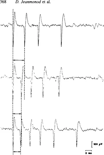
In each burst, be it random or rhythmic, the first spike was often of higher amplitude and interspike intervals within a burst increased with each successive interval (Fig. 3). The shorter the first interspike interval in a burst, the larger the number of spikes (two to eight) within this burst. Quantification of these features demonstrates that they are common to all five symptoms (Fig. 4). All these characteristics are the hallmarks of LTS bursts, found in
the context of thalamic relay cell hyperpolarization (Steriade et al., 1990£>). The highest intraburst frequency averaged throughout all patients was 480±80 Hz and the mean intraburst frequency 206±44 Hz. When values obtained for each symptom were compared with the above values, no statistically significant differences could be found.
Some unresponsive sporadic units of our sample exhibited rare LTS bursts, suggesting that they are under a more limited inhibitory influence than cells showing random or rhythmic bursts. Such an interpretation is supported by experimental data showing the occurrence of isolated LTS after a moderate, and of LTS bursts after a stronger hyperpolarization of thalamic relay cells (Steriade et al., 1990a). This suggests a continuum of increasingly inhibited thalamic neurons.
As to the sporadic cells showing evoked responses elicited either by somatosensory stimuli or by voluntary movements, they were rare (14 out of 2012 cells) and only found in neurogenic pain (0.6%) and abnormal movements (1.2%). In pain patients, five out of eight units, located in the rostrodorsal part of the central lateral nucleus, were activated in relation to voluntary saccadic eye movements. The other three units exhibited somatosensory receptive fields covering the entire contralateral hemibody in two cases and restricted to the third dermatome of the trigeminal nerve in the third one. Considering the relatively small size of this last receptive field, this cell may well have to be located outside of the central lateral nucleus. In patients with dyskinesias, voluntary movements elicited responses in six cells. Three were related to tongue movements, two to leg movements and one to hand movements. When spontaneous activity of all these sporadic cells with evoked responses was considered, none of them showed any sign of bursting activity.
Figure 5 displays the proportions of the four types of activities associated with the different positive symptoms. The percentage of bursting activities is smaller in abnormal movements than in the other categories.
Clinical results
The mean post-operative follow-up for the neurogenic pain patient group is 2 years and 1 month, for abnormal movements
1 year and 6 months, for epilepsy 1 year and 10 months, for tinnitus 7 months, and for neuropsychiatry 2 years and 2 months.
Neurogenic pain
A complete relief was achieved in 20% of patients with neurogenic pain, a good improvement (50-99%) in 47% (Jeanmonod et al., 1994). A 50-100% improvement was found in 71% of patients with peripheral neurogenic pain and in 60% of patients with central pain. Steady pain resisted more than intermittent pain and allodynia, deep pain qualities more than superficial ones. The mean of the highest post-operative pain intensity values on the visual analogue scale
OBSCMVE COMPULSIVE DtSEAM AND MAJOR DEPRESSION
1
1
I I IDV8T0MA
MflKMSON Fig. 2 Examples of unit activities recorded in medial thalamus (CL and SG/Li, see below) in
seven positive symptoms. This activity, analysed by interspike interval {left) and autocorrelation histograms (right), is characterized by rhythmic bursts with frequencies between 3 and 5 Hz. They all correspond to the rhythmic bursting category defined in the text. The localization of each unit is shown on a sagittal section of our stereotaxic atlas of the human thalamus, 7 mm from the medial border of the thalamus. The two plus signs correspond to the position of the anterior (right) and posterior (left) commissures. Horizontal and vertical scale bars for unit activities are 200 ms and 100 mV, respectively. Po = posterior nucleus; SG/Li = suprageniculate-limitans complex; VLpd v = ventral lateral posterior nucleus, dorsal and ventral divisions; VM = ventromedial nucleus.
For other abbreviations, see Fig. 1.
was 49, compared with 85 pre-operatively. Significant the lateral spinothalamic tract always explained the observed decrease of the drug intake was observed in 54% of patients.
Significant improvement of the activities of daily living was seen in 45 % of the patients. The majority of the patients (91 %) showed no post-operative somatosensory deficits (including pain). In six patients, accidental lateral expansion of the
somatosensory deficits, completely reversible in half of them.
Abnormal movements
Forty-three percent of patients with motor disorders had a lesion (bleeding, oedema) into the ventral posterior nucleus or 50-100% relief. The strongest resistance to medial
i
Fig. 3 Intrinsic organization of three bursts from the rhythmic
bursting unit shown in Fig. 2 for epilepsy. They fulfil all
extracellular characteristics of LTS bursts (see text). Arrows mark the first interspike interval of each burst, the duration of which is inversely correlated with the number of spikes in the burst.
thalamotomy was shown by cerebellar tremor. In the parkinsonian patients, the mean pre-operative rating for tremor was 3.5, for rigidity 2.3 and for hypo-bradykinesia 2.7. Post-operatively, these ratings amounted to 2.5, 1.2 and 1.7, respectively. The mean unified Parkinson's disease rating scale was 47 pre-operatively and 37 post-operatively.
either only tinnitus (two) or tinnitus plus neurogenic pain (one).
Epilepsy
Three of the six patients with focal frontal and/or parietal epilepsy had a relief between 50 and 100%. The first patient with a fronto-parietal epilepsy who had somatosensory or sensorimotor fits three times per day on average had a post-operative reduction of the fits to two per month, with only pure motor residual symptomatology. The second patient had the association of neurogenic pain with sensorimotor epilepsy after a parietal lesion, with a fit frequency of one per month. The thalamotomy brought her a long-term 60% pain relief and a reduction of the fits to only one over 5 years. The third patient with a sensorimotor epilepsy of parietal origin and two fits per day on average had a post-operative reduction of the fit frequency to two per month, with a full restoration of his working capacity.
Neuropsychiatry
The psychotic patient experienced an excellent relief after a single left-sided thalamotomy, but relapsed to only 30% improvement. The other patient with an obsessive-compulsive and depressive symptomatology gained, after a bilateral thalamotomy, a complete relief of the obsessive-compulsive symptoms and only a very partial effect on depression.
These clinical results are not to be considered as definitive, because possible improvements or relapses may be observed over time, and because a significant number of patients have not yet received the complete, often bilateral surgical treatment they need. None of our patients showed a iatrogenic increase of their positive symptom(s), and a sparing of all neurological functions was observed even after a bilateral operation. Moreover, 47% of patients with neurogenic pain experienced an improvement of their somatosensory functions and preliminary data indicate neuropsychological improve-ments in about one-third of the examined patients.
Tinnitus
A 50-100% relief was obtained in three of the six patients with tinnitus, with a complete relief in only one. These three patients suffered from a combination of tinnitus and neurogenic pain, one of them with the addition of hemifacial spasm. The three other patients with insufficient relief had
Discussion
A unified concept for all positive symptoms
Unresponsiveness of medial thalamic neurons and their tendency to generate LTS bursts in patients with seemingly unrelated positive symptoms leads us to propose a common basis for their physiopathology and the therapeutic effect of
Fig. 4 Quantification of the intrinsic characteristics of bursts recorded in the five different symptoms. A burst is defined as a group of
contiguous action potentials separated by interspike intervals not longer than 20 ms. Duration of interspike intervals is plotted as a function of their position within the burst (graphs on the left). In each case, the progressive lengthening of successive intervals within the burst is obvious whatever the number of spikes in this burst. Graphs on the right are obtained by plotting, for the same cells, the duration of the first interval in a burst as a function of the number of spikes within this burst. These two latter parameters show an inverse relationship best characterized by a logarithmic function. ISI = interspike interval.
1 2 1 2 3 1 2 3 4 1 2 3 4 5 1 2 3 4 5 6 1 2 3 4 5 6 7 1 2 3 4 5 6 7 1 = -1.8846Ln(x)+5.4197 r2 = 0.9048 0 1 2 3 4 5 6 7 8 12 - 108 6 -4 • 2 0 -ABNORMAL MOVEMENTS
j J
nl - 3 2 bunt n2 - 2S2 mttrvtfa 1 1 2 1 2 3 1 2 3 4 1 2 3 4 5 1 2 3 4 5 6 1 2 3 4 5 6 7 1 2 3 4 5 6 7 8 0 1 2 3 4 5 6 7 8 TINNITUS n l - 3 6 bint D2 - 178 tntcrvlls 1 1 2 1 2 3 1 2 3 4 1 2 3 4 5 1 2 3 4 5 6 1 2 3 4 5 6 7 1 2 3 4 5 6 7 8 2 3 4 5 6 7 8 EPILEPSY 2 2 • 20: 18 • 16-14 • 1 2 • 10 : 8-6 : 4 -2; 0: n l - 4 8 tern n2 - 204 mtervilj -2.0172LuM +5.9872 r2 = 0.9344 1 2 1 2 3 1 2 3 4 1 2 3 4 5 1 2 3 4 5 6 1 2 3 4 5 6 7 1 2 3 4 5 6 7 8 0 1 2 3 4 5 6 7 8 NEUROPSYCHIATRIC SYMPTOMS 10 8 • 6 • 4 • 2 • 0-J
n l - 3 9 bunt n i - l 4 0 i m e r v i I s 1 2 1 2 3 1 2 3 4 1 2 3 4 5 1 2 3 4 5 6 1 2 3 4 5 6 7 1 2 3 4 5 6 7 8 ISI Ordinal 2 3 4 5 6 7 8 Number of ISI / B u n tn=1362 n = 484 100%- 80%- 60%-(A) 40%-
20%-Fig. 5 Relative distribution of the four defined types of unit activities in the five positive symptoms studied: rhythmic bursting (black), random bursting (obliquely hatched), sporadic (white) and sporadic with evoked response (vertically hatched), n = number of recorded units in each category.
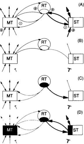
the medial thalamotomy. The following anatomophysiological elements (Desiraju et al., 1969; Jones, 1975, 1985; Steriade et al., 1984; Velayos et al., 1989) have to be considered (Fig. 6A): (i) the medial thalamus, the lateral or specific thalamus and the thalamic reticular nucleus are interconnected by inhibitory (GABAergic) reticulothalamic and excitatory thalamoreticular pathways; (ii) the medial thalamus has more multimodal convergent and divergent connections with centres other than the specific thalamus, whereas the specific thalamus has dense projections to and from restricted, unimodal and topologically organized areas.
The indispensable causal event for the appearance of a positive symptom is an alteration of the normal inputs to the specific thalamus. Any decrease of its excitatory or increase of its inhibitory inputs affect the specific thalamus more efficiently than the medial thalamus (Fig. 6B). In both cases, a portion of the specific thalamus sends less excitatory inputs to the corresponding part of the reticular nucleus (Fig. 6B). The deactivation of the medial thalamus is in comparison much less pronounced, and so is the subsequent influence of this deactivation on the reticular nucleus. The affected part of the reticular nucleus (shaded in Fig. 6C) is placed in a situation where its cell membrane potentials are hyper-polarized because of the lack of excitatory inputs from the specific thalamus, and where excitatory postsynaptic potentials, coming from the medial thalamus or from the cortex, can trigger the appearance of LTS bursts, as already shown experimentally (Pare" et al., 1990; Bal and McCormick, 1993; Llinas and Pare\ 1995). These bursts are sent back to the medial thalamus and the specific thalamus (Fig. 6C), inducing a potent GABAergic inhibition. Cells in the medial thalamus and the specific thalamus are in turn hyperpolarized
Fig. 6 Display of the central thalamic network and its sequential
alterations leading to the appearance of a positive symptom in a case of thalamic deafferentation. In A, thicker arrows represent denser projections. In B and C, the decrease of a projection is represented by a thinner arrow. Shading (arrows and areas) represents LTS bursts. The causal change in the network is represented by the interrupted arrows, whether an anatomical or functional deactivation or an increase of inhibition of ST is relevant. See text for the description of steps A to D. ST = specific thalamus; MT = medial thalamus; RT = thalamic reticular nucleus.
(shaded in Fig. 6D) and produce LTS bursts. These are projected back to the reticular nucleus, reinforcing its bursting tendency (Steriade et al., 1993a) and thus closing a circle (Fig. 6D). This circle can be maintained and amplified by the widespread projections of the medial thalamus on the reticular nucleus, as supported by the privileged role of the medial thalamus in recruiting responses (Morison and Dempsey, 1942). Moreover, the medial thalamus was shown to be preponderant in the genesis of rhythmic thalamic oscillations (Steriade et al., 1971). Although this scenario focuses on thalamic mechanisms, the potential physiopatho-logical relevance of thalamo-cortico-thalamic reverberating and/or synchronizing loops (Steriade et al., 1976; Canavero, 1994; Steriade and Contreras, 1995) should not be under-estimated.
The above concept is reminiscent of earlier hypotheses (Roberts et al., 1992; Llinas and Par6, 1995) to explain the
posterior nucleus or to the ventral lateral posterior nucleus of the thalamus. However, they only involve the reciprocal connections of these latter structures with the reticular nucleus and do not explain the beneficial effect of the medial
thalamotomy. t
The thalamic LTS bursts, reaching the cortex, are thought to cause the appearance of the positive symptoms and/or of functional deficits. The latter can be centred on the system involved by a given positive symptom (e.g. somatosensory deficits in pain patients) or unrelated to it (neuropsychological deficits), and have been found to be reversible after medial thalamotomy. The widespread cortical projections of the intralaminar nuclei, which have been shown to influence the activity of a large number of cortical areas when functioning in bursting mode (Fox and Armstrong-James, 1986), may explain such phenomena. Further, considering functional deficits, it is worth noting that hypo-bradykinesia of the parkinson syndrome and endogenous depression are also improved by the medial thalamotomy, indicating thus that they are due to interference of thalamic LTS bursts with cortical functions.
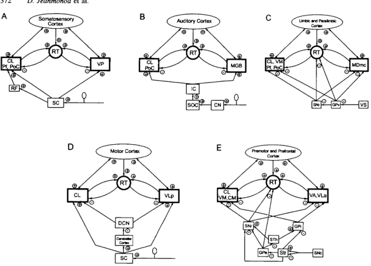
Figure 7 shows the networks relevant to the positive symptoms considered in this study. Figure 7A relates to the somatosensory system and neurogenic pain: lesions causing such pain can be localized at all levels of the somatosensory pathways, from primary afferent fibres to spinal cord, brainstem, the ventral posterior nucleus and cortex. They always produce a loss of excitatory input to the ventral posterior nucleus, anatomical (spinothalamic or cortico-thalamic lesion) or functional (peripheral lesion). Figure 7B is for the auditory system and tinnitus: peripheral lesions decrease the activation of the medial geniculate body trans-synaptically, i.e. functionally, and typically cause the appearance of tinnitus. So do probably other lesions along the pathways from the cochlear nuclei to cortex. Figure 7C illustrates the limbic system and the related neuropsychiatric symptoms: evidence is accumulating for hypofunction and/ or anatomical abnormalities in limbic and paralimbic cortices (for review, see Harrison, 1995). A loss of corticothalamic activation and/or an increase of pallidothalamic inhibition can explain the appearance of obsessive-compulsive, depressive and psychotic symptoms. Figure 7D describes the first part of the motor system centred on the ventral lateral posterior nucleus: lesions of the deep cerebellar nuclei and their excitatory output to the ventral lateral posterior nucleus produce action tremor. Lesions of spinothalamic or corticothalamic inputs produce spasticity or motor epilepsy. Peripheral nerve lesions, causing a functional spinothalamic deactivation of the ventral lateral posterior nucleus, can also induce abnormal movements, e.g. hemifacial spasm, 'epilepsie du moignon' and other tonico-clonic contractions associated with neurogenic pain. Figure 7E illustrates the striatopallidal part of the motor system: in Parkinson's disease,
part of the pallidum (Wichmann and DeLong, 1993; Hutchison et al, 1994), resulting in thalamic cell membrane hyperpolarization. As for epilepsy (parietal, limbic and frontal types), one can refer to parts A, C, D or E of Fig. 7 depending on the ictal symptoms considered.
Surgical targets against positive symptoms
As discussed above, positive symptoms are considered to be the result of a thalamus-centred network imbalance attributed to extrathalamic influences (Fig. 7). Indeed, in addition to the medial thalamus targets which were explored here, interventions at the level of the other thalamic partner, the specific thalamus, the cortex and the pallidum have also been shown to have therapeutic value. Lesions of the specific thalamus have been performed (Cooper, 1969; Ojemann and Ward, 1975; Spiegel, 1982; Gybels and Sweet, 1989) with a certain success, but at the cost of more or less marked deficits and with the risk of increasing the physiopathology. Cortical resections, widely used in epilepsy surgery, and subcortical limbic lesions against neuropsychiatric symptoms have indisputable value but also present a risk, potentially serious, of post-operative deficits. The therapeutic action of the specific thalamus or of cortical and subcortical lesions could well be related to the descent of thalamic and reticular cell membrane potentials to values lower than -75 mV, where LTS bursts are no longer produced and where neurons are silenced. Therapeutic effect can also be obtained with chronic stimulation of the specific thalamus (Gybels and Sweet, 1989; Caparros-Lefebvre et al, 1993), which specifically counteracts its hyperpolarization, i.e. raises the cell membrane potential above LTS threshold (-65 mV), but is limited by topographic, temporal and technical factors. A medial pallidal lesion is also able to decrease thalamic cell membrane hyperpolarization, but here through a suppression of afferent inhibition. This is indeed the case of ventroposterior lesions in the internal part of the pallidum, which relieve tremor, rigidity and hypo-bradykinesia in parkinsonian patients (Laitinen et al, 1992).Our clinical results demonstrate certain limitations of the effect of the medial thalamotomy. This operation alleviates the positive symptoms most probably by reducing the recruiting, reverberating and/or synchronizing properties of the medial thalamus neurons, but simultaneously suppresses some excitatory inputs to the reticular nucleus. By lowering membrane potentials toward the LTS producing zone, the latter effect may play a negative role, the importance of which remains unclear.
Clinical and experimental data indicate a role of the medial thalamus, particularly the central lateral nucleus and the centre median, in attentional mechanisms (Heilman et al, 1985). The fact that lesions in these nuclei in patients with positive symptoms do not produce attentional or other deficits
A
B
Fig. 7A-E This series of diagrams displays the relevant connectional data for each system affected by the positive symptoms studied and collected by modem tract tracing methods. Each arrow represents a direct projection, the plus or minus signs indicate the known excitatory or inhibitory quality of this projection. The heavily outlined boxes relate to Fig. 6. CN = cochlear nuclei; DCN = deep cerebellar nuclei; GPe = external part of globus pallidus; GPi = internal part of globus pallidus; GPv = ventral part of globus pallidus; IC = inferior colliculus; MGB = medial geniculate body; PoC = posterior thalamic complex; RF = reticular formation; SC = spinal cord dorsal horn; SNc = substantia nigra pars compacta; SNr = substantia nigra pars reticulata; SOC = superior olivary complex; Str =
striatum; VP = ventral posterior nucleus; VS =ventral striatum. For other abbreviations, see Figs 1 and 2.
suggests that they are no longer functional. This is supported by the unresponsiveness of the recorded units, and implies that their function is overtaken by other areas through plastic reorganizations.
The positive symptoms revisited
The new concept of positive symptoms presented here suggests reconsiderations of large domains of contemporary neurology and psychiatry. First to be stressed is the existence of central suprasegmental causal mechanisms for peripheral neurogenic pain, tinnitus, spasticity and hemifacial spasm, whereas most investigations and hypotheses have concen-trated on nerves and spinal cord segments. Similarly, a thalamic site of the dysregulation responsible for motor and limbic positive symptoms challenges current interpretations centred on the basal ganglia and cortex. The systemic approach proposed here integrates periphery, spinal cord,
basal ganglia, cerebellum and cortex into the affected networks, but lesions in these areas are only the triggers of the thalamic physiopathology. Spasticity illustrates such a systemic approach. If, as is generally accepted, lesion of the descending fibres to the spinal cord was responsible for spasticity, its intensity should be directly related to the extent of the lesion. This is contradictory to the observation that total spinal cord lesions do not give rise to marked spasticity, and that'. . . spasticity is the clinical expression of preserved brain influences on the segmental stretch reflex' (Dimitrijevic, 1988). Our concept supports the relevance of the lesion of spinothalamic fibres in triggering a thalamic physiopathology between the ventral lateral posterior nucleus and the central lateral nucleus, which then invades cord segments caudal to the lesion through spared thalamo-cortico-spinal pathways.
As for epilepsy, the separation into focal (cortical) and generalized (subcortical or cortico-subcortical) epilepsies would have to be left in favour of a unique causal
cortico-seizures, a well-known form of generalized epilepsy (Gloor and Fariello, 1988; Steriade et al., 1993a).
The genetic factor
Positive symptoms do not appear systematically after very similar lesions in the PNS or CNS. This points to an idiosyncratic, probably genetic factor. The development of positive symptoms in several members of the same family (as an extreme example, in the family of one of our patients, up to nine persons over two generations affected by three positive symptoms, i.e. Parkinson's disease, major depression and epilepsy) supports such a genetic predisposition. Experimental evidence (Seltzer et al., 1993) showed that the mechanisms underlying neurogenic pain and epilepsy share the same genetic determinant(s), encoding for GABA-related inhibition in the CNS. According to our data, this genetically coded dysfunction would be expressed at the level of the reticulothalamic projection.
Positive symptoms and slow-wave sleep
In contrast to what was reported in the medial thalamus of animals (Schlag-Rey and Schlag, 1984; Willis, 1985) and humans (Ishijima et al., 1975) without positive symptoms, the rarity of evoked responses shown in this study suggests per se thalamic cell inhibition and is specifically confirmed by the presence of LTS bursts. Decreased responsiveness to peripheral stimuli and thalamic rhythmic LTS bursting activities are the hallmarks of slow-wave sleep, with spindles at 7-14 Hz and delta waves at 1-4 Hz (Steriade et al., 1993a). As our patients were fully awake during the opera-tion, parts of their thalamic network, displaying rhythmic LTS bursts with delta to theta frequencies (2.5-5.6 Hz), can be conceived to function on a modulation mode close or identical to the one seen in slow-wave sleep. Indeed, we have observed theta to delta EEG activity in positive symptoms other than epilepsy (D. Jeanmonod, M. Magnin and A. Morel, unpublished data). In addition, recent studies using PET showed, in patients suffering from neurogenic pain, a thalamic hypometabolism (Di Piero et al., 1991; Iadarola et al., 1995) which was also demonstrated in slow-wave sleep (Madsen and Vorstrup, 1991).
Could sleep then be conceived as based on a tripartite thalamic network similar to those proposed above for pathological conditions? Recent developments in the under-standing of sleep generation demonstrate a decrease of thalamic activation by the hypothalamus (Koyama and Hayaishi, 1994), either directly (Palkovits and Zaborszky, 1979) or through cholinergic and monoaminergic projections (Steriade et al., 1971; Steriade, 1993; Steriade et al., 1993a, b). Experimental and clinical data on sleep induction and suppression suggest the central lateral nucleus, midline nuclei
symptoms and sleep is reinforced by the existence of the narcolepsy syndrome, which may be considered as a true positive symptom, i.e. a pathological transformation of the normal sleep function. Although no causal lesions were found in the most common idiopathic form, lesions in the posterior hypothalamus were correlated with the appearance of symptomatic narcolepsies (Castaigne and Escourolle, 1967).
Acknowledgements
We wish to thank Professor K. Akert for his helpful critical review of the manuscript, Professor M. G. Yasargil for his support, J. Dodd, S. Richter, P. Roth, R. Stillhard and V. Streit for technical assistance, and V. Cucci for secretarial help. The study was supported by grants from the University of Zurich and from the Swiss National Science Foundation (31-36330.92).
References
Adams JE, Rutkin BB. Lesions of the centrum medianum in the treatment of movement disorders. Confin Neurol 1965; 26: 231-45. Akert K. The anatomical substrate of sleep. Prog Brain Res 1965;
18:9-19.
Bal T, McCormick DA. Mechanisms of oscillatory activity in guinea-pig nucleus reticularis thalami in vitro: a mammalian pacemaker. J Physiol (Lond) 1993; 468: 669-91.
Bernos GE. Positive and negative symptoms and Jackson: a conceptual history. Arch Gen Psychiatry 1985; 42: 95-7.
Buzsaki G, Smith A, Berger S, Fisher LJ, Gage FH. Petit mal epilepsy and parkinsonian tremor: hypothesis of a common pacemaker. [Review]. Neuroscience 1990; 36: 1-14.
Canavero S. Dynamic reverberation. A unified mechanism for central and phantom pain. [Review]. Med Hypotheses 1994; 42: 203-7.
Caparros-Lefebvre D, Blond S, Vermersch P, Pecheux N, Guieu J-D, Petit H. Chronic thalamic stimulation improves tremor and levodopa induced dyskinesias in Parkinson's disease. J Neurol Neurosurg Psychiatry 1993; 56: 268-73.
Castaigne P, Escourolle R. Etude topographique des lesions anatomiques dans les hypersomnies. Rev Neurol (Pans) 1967; 116: 547-84.
Cooper IS. Involuntary movement disorders. New York: Hoeber, 1969.
Desiraju T, Broggi G, Prelevic S, Santini M, Purpura DP. Inhibitory synaptic pathways linking specific and nonspecific thalamic nuclei. Brain Res 1969; 15: 542-3.
Dimitrijevic MR. Spasticity and rigidity. In: Jankovic J, Tolosa E, editors. Parkinson's disease and movement disorders. Baltimore: Urban and Schwarzenberg, 1988: 385-94.
Di Piero V, Jones AKP, Iannotti F, Powell M, Perani D, Lenzi GL, et al. Chronic pain: a PET study of the central effects of percutaneous high cervical cordotomy. Pain 1991; 46: 9-12.
Fox K, Armstrong-James M. The role of the anterior intralaminar nuclei and N-methyl D-aspartate receptors in the generation of spontaneous bursts in rat neocortical neurones. Exp Brain Res 1986; 63: 505-18.
Gloor P, Fariello RG. Generalized epilepsy: some of its cellular mechanisms differ from those of focal epilepsy. [Review]. Trends Neurosci 1988; 11: 63-8.
Gybels JM, Sweet WH. Neurosurgical treatment of persistent pain. Basel: Karger, 1989.
Harrison PJ. On the neuropathology of schizophrenia and its dementia: Neurodevelopmental, neurodegenerative, or both? [Review]. Neurodegeneration 1995; 4: 1-12.
Head H, Holmes G. Sensory disturbances from cerebral lesions. Brain 1911; 34: 102-254.
Heilman KM, Watson RT, Valenstein E. Neglect and related disorders. In: Heilman KM, Valenstein E, editors. Clinical neuropsychology. 2nd ed. New York: Oxford University Press, 1985: 243-93.
Hitchcock ER, Teixeira MJ. A comparison of results from center-median and basal thalamotomies for pain. Surg Neurol 1981; 15: 341-51.
Hutchison WD, Lozano AM, Davis KD, Saint-Cyr JA, Lang AE, Dostrovsky JO. Differential neuronal activity in segments of globus pallidus in Parkinson's disease patients. Neuroreport 1994; 5: 1533-7.
Iadarola MJ, Max MB, Berman KF, Byas-Smith MG, Coghill RC, Gracely RH, et al. Unilateral decrease in thalamic activity observed with positron emission tomography in patients with chronic neuropathic pain. Pain 1995; 63: 55-64.
Ishijima B, Yoshimasu N, Fukushima T, Hori T, Sekino H, Sano K. Nociceptive neurons in the human thalamus. Confin Neurol 1975; 37: 99-106.
Jackson JH. On post-epileptic states: a contribution to the comparative study of insanities. J Ment Sc Lond 1889; 34: 490-500. Jahnsen H, Llinas R. Electrophysiological properties of guinea-pig thalamic neurones: an in vitro study. J Physiol (Lond) 1984a; 349: 205-26.
Jahnsen H, Llinas R. Ionic basis for the electro-responsiveness and oscillatory properties of guinea-pig thalamic neurones in vitro. J Physiol (Lond) 1984b; 349: 227^17.
Jeanmonod D, Thomas DGT. Application of CT-directed stereotaxy in the determination of functional neurosurgical targets in the diencephalon and cerebral hemisphere. Br J Neurosurg 1989; 3: 337-42.
Jeanmonod D, Magnin M, Morel A. Thalamus and neurogenic pain: physiological, anatomical and clinical data [published erratum appears in Neuroreport 1993; 4: 1066]. Neuroreport 1993; 4: 475-8. Jeanmonod D, Magnin M, Morel A. A thalamic concept of neurogenic pain. In: Gcbhart GF, Hammond DL, Jensen TS, editors.
Progress in pain research and management. Seattle: IASP Press, 1994: 767-87.
Jones EG. Some aspects of the organization of the thalamic reticular complex. J Comp Neurol 1975; 162: 285-308.
Jones EG. The thalamus. New York: Plenum Press, 1985. Koyama Y, Hayaishi O. Firing of neurons in the preoptic/anterior hypothalamic areas in rat: its possible involvement in slow wave sleep and paradoxical sleep. Neurosci Res 1994; 19: 31-8. Laitinen LV, Bergenheim AT, Hariz MI. Leksell's posteroventral pallidotomy in the treatment of Parkinson's disease [see comments], J Neurosurg 1992; 76: 53-61. Comment in: J Neurosurg 1992; 77: 487-8.
Lenz FA, Kwan HC, Dostrovsky JO, Tasker R. Characteristics of the bursting pattern of action potentials that occurs in the thalamus of patients with central pain. Brain Res 1989; 4%: 357-60. Lenz FA, Kwan HC, Martin R, Tasker R, Richardson RT, Dostrovsky JO. Characteristics of somatotopic organization and spontaneous neuronal activity in the region of the thalamic principal sensory nucleus in patients with spinal cord transection. J Neurophysiol 1994; 72: 1570-87.
Llinas R, Jahnsen H. Electrophysiology of mammalian thalamic neurones in vitro. Nature 1982; 297: 406-8.
Llinas R, Par6 D. Role of intrinsic neuronal oscillations and network ensembles in the genesis of normal and pathological tremors. In: Findley LJ, Koller WC, editors. Handbook of tremor disorders. New York: Marcel Dekker, 1995: 7-36.
Lugaresi E, Montagna P, Gambetti P. The thalamus regulates the sleep-wake cycle and autonomic and endocrine functions. In: Minciacchi D, Molinari M, Macchi G, Jones EG, editors. Thalamic networks for relay and modulation. Oxford: Pergamon Press, 1993: 395-400.
Madsen PL, Vorstrup S. Cerebral blood flow and metabolism during sleep. [Review]. Cerebrovasc Brain Metab Rev 1991; 3: 281-96. Modesti LM, Waszak M. Firing pattern of cells in human thalamus during dorsal column stimulation. Appl Neurophysiol 1975; 38: 251-8.
Monro H. Remarks on insanity: its nature and treatment. London: Churchill, 1851.
Monson RS, Dempsey EW. A study of thalamocortical relations. Am J Physiol 1942; 135: 281-92.
Ojemann GA. Ward AA Jr. Stereotactic and other procedures for epilepsy. [Review], Adv Neurol 1975; 8: 241-63.
Palkovits M, Zaborszky L. Neural connections of the hypothalamus. In: Morgane PJ, Panksepp J, editors. Handbook of the hypothalamus. Vol. 1. New York: Marcel Dekker, 1979; 379-509.
Pare1 D, Curro Dossi R, Steriade M. Neuronal basis of the
parkinsonian resting tremor: a hypothesis and its implications for treatment. [Review]. Neuroscience 1990; 35: 217-26.
Raeva S. Localization in human thalamus of units triggered during 'verbal commands', voluntary movements and tremor. Electro-encephalogr Clin Neurophysiol 1986; 63: 160-73.
Spontaneous neuronal hyperactivity in the medial and intralaminar thalamic nuclei of patients with deafferentation pain. J Neurosurg
1991; 74: 415-21.
Roberts WA, Eaton SA, Salt TE. Widely distributed GABA-mediated afferent inhibition processes within the ventrobasal thalamus of rat and their possible relevance to pathological pain states and somatotopic plasticity. Exp Brain Res 1992; 89: 363-72.
Sano K. Intralaminar thalamotomy (thaJamolaminotomy) and postero-medial hypothalamotomy in the treatment of intractable pain. Prog Neurol Surg 1977; 8: 50-103.
Schlag-Rey M, Schlag J. Visuomotor functions of central thalamus in monkey. I. Unit activity related to spontaneous eye movements. J Neurophysiol 1984; 51: 1149-74.
Seltzer Z, Attal U, Devor M, Levi Z, Neuman S, Shavit Y. Neuropathic pain-related behaviour and epileptogenesis are co-inherited in rats. In: Proceedings of the 7th World Congress on Pain. Seattle: IASP Publications, 1993; 513.
Spiegel EA. Guided brain operations. Basel: Karger, 1982. Steriade M. Central core modulation of spontaneous oscillations and sensory transmission in thalamocortical systems. [Review]. Curr Opin Neurobiol 1993; 3: 619-25.
Steriade M, Contreras D. Relation between cortical and thalamic cellular events during transition from sleep patterns to paroxysmal activity. J Neurosci 1995; 15: 623-^12.
Steriade M, Apostol V, Oakson G. Control of unitary activities in
after discharges in thalamic neurons. Electroencephalogr Clin Neurophysiol 1976; 41: 641^4.
Steriade M, Parent A, Hada J. Thalamic projections of nucleus reticularis thalami of cat: a study using retrograde transport of horseradish peroxidase and fluorescent tracers. J Comp Neurol 1984; 229: 5 3 1 ^ 7 .
Steriade M, Jones EG, Llinas RR. Thalamic oscillations and signalling. New York: Wiley, 1990a: 115-38.
Steriade M, Jones EG, Llinas RR. Thalamic oscillations and signalling. New York: Wiley, 1990b: 167-237.
Steriade M, McCormick DA, Sejnowski TJ. Thalamocortical oscillations in the sleeping and aroused brain. [Review]. Science 1993a; 262: 679-85.
Steriade M, Contreras D, Curro Dossi R, Nunez A. The slow (<1 Hz) oscillation in reticular thalamic and thalamocortical neurons: scenario of sleep rhythm generation in interacting thalamic and neocortical networks. J Neurosci 1993b; 13: 3284-99. Velayos JL, Jimenez-Castellanos J Jr, Reinoso-Suarez F. Topo-graphical organization of the projections from the reticular thalamic nucleus to the intralaminar and medial thalamic nuclei in the cat. J Comp Neurol 1989; 279: 457-69.
Wichmann T, DeLong MR. Pathophysiology of parkinsonian motor abnormalities. [Review]. Adv Neurol 1993; 60: 53-61.
Willis WD. The pain system. Basel: Karger, 1985.
Received June 13, 1995. Revised November 28, 1995. Accepted December 14, 1995