Recovery of Evoked Potentials, Metabolic
Activity and Behavior in a Mouse Model of
Somatosensory Cortex Lesion: Role of the
Neural Cell Adhesion Molecule (NCAM)
E. Troncoso1, D. Muller2, K. Korodi1, T. Steimer3, E. Welker4 and J.Z. Kiss1
1Department of Morphology, University of Geneva Medical School, 1 rue Michel Servet, CH-1211 Geneva 4, Switzerland, 2Department of Neuropharmacology, University of Geneva Medical School, 1 rue Michel Servet, CH-1211 Geneva 4, Switzerland, 3Department of Psychopharmacology, University of Geneva Medical School, 1 rue Michel Servet, CH-1211 Geneva 4, Switzerland and 4IBCM, University of Lausanne, Switzerland
Understanding the processes that underlie functional recovery after cortical injury is a major challenge for neurobiology and clinical neurology. The aim of the present study was to establish a mouse model of functional recovery that would facilitate the investigation of the molecular and cellular events involved in cortical dynamics. We show that a focal injury of ∼0.5 mm of diameter and 1 mm depth made in the barrel cortex of adult mice induced a transitory deficit that could be characterized using somatosensory evoked potential (SEP), metabolic mapping and a behavioral test. SEP recordings of short latency responses using an epicranial multi-array system showed a decreased cortical activity in the peri-lesion regions 2 weeks after the injury and a partial recovery to normal pattern 6 weeks after the lesion. Delayed SEP signals over the motor cortex were not altered by the injury. Metabolic mapping with [14 C]deoxy-glucose uptake in the surround of the injury reproduced the time course of deficit and recovery. Finally, a deficit in vibrissae related performance in a gap-crossing test 1 week after injury was followed by a functional recovery in the following 2 weeks. We show in addi-tion that the recovery process is deficient and significantly delayed in NCAM knockout mice lacking all isoforms of NCAM (neural cell adhesion molecule)and PSA-NCAM. These results support the hypothesis that impairment and recovery of functions after focal cortical lesion involves remodeling of intact circuits surrounding the lesion and that the NCAM molecule participate in this process. The model opens new possibilities for investigating the role of candidate molecules in functional recovery using genetically modified mice. Keywords: behavior, evoked potentials, metabolic activity, mouse model, neural cell adhesion molecule, somatosensory cortex
Introduction
The transitory or permanent functional deficit produced by a traumatic brain injury is very often a consequence of focal cortical damage. A large body of experimental and clinical studies suggests that functional recovery after focal lesions is possible, though often limited (Geschwind, 1985; Jenkins and Merzenich, 1987). The degree and the time course of restora-tion is highly variable and appears to depend on a large number of factors, including the lesion size, patient age, sex, previous experience, stimulation, emotional status and motivation (Geschwind, 1985). An emerging view is that functional recovery after focal injury mainly relies on a dynamic reorgani-zation of preserved networks (Buonomano and Merzenich, 1998; Witte, 1998). The bulk of evidence for this plasticity stems from experiments in the somatomotor cortex (Jenkins and Merzenich, 1987). Subtotal lesions in the primary somato-sensory cortex of owl monkeys were shown to be followed by behavioral recovery due to remodeling of cortical representa-tional maps (Jenkins and Merzenich, 1987). In this model,
intact zones in the neighborhood of a focal lesion took over the function of the damaged areas. Similar restorative mechanisms have been reported after unilateral focal lesions of the sensori-motor cortex in a number of species and the same observations were made in human cases (Geschwind, 1985). While the capacity for functional recovery after cortical damage is well documented, little is known about the physiology of recov-ering cortical networks and the molecular mechanisms contributing to the repair process. Molecules such as growth factors, proteases or cell–cell adhesion molecules, that shape the formation of neuronal networks during development, are particularly interesting candidates. Recent data suggest that these molecules play a role in several aspects of cellular and synaptic plasticity, mechanisms that are proposed to underlie the re-organization of cortical maps associated with functional recovery (Buonomano and Merzenich, 1998). The role of PSA-NCAM has been established in axon growth, lesion induced sprouting, cell migration and activity-dependent synaptic plas-ticity (Kiss et al., 2001). Importantly, removal of alpha 2,8-linked sialic acid (polysialic acid; PSA) from NCAM prevents induction of long-term potentiation (LTP) and long-term depression (LTD) in the CA1 (Muller et al., 1996) and CA3 regions of hippocampal slice cultures and NCAM knockout mice (Cremer et al., 1998) and results in deficit in spatial learning (Cremer et al., 1994; Becker et al., 1996). Based on these data, PSA-NCAM has emerged as an attractive candidate for performing a role in functional recovery after lesion.
In an attempt to develop a clinically relevant analysis of cortical recovery in animals, we recently introduced an epi-cranial approach for multi-electrode recording of vibrissal sensory evoked potentials (SEP) in the mouse (Troncoso et al., 2000). The vibrissae–barrel-field circuit is of particular interest in mice. Mystacial whiskers are sensitive tactile organs that selectively activate a predictable area, the barrel in the somato-sensory cortex (Woolsey and Van der Loos, 1970). The topo-graphic disposition of barrel-like structures in this cortical region obeys the same arrangement as the vibrissae on the mystacial pad. Due to this modular somatotopic characteristic, this sensory system has become one of the major models for assessing the cortical reorganization. Using an epicranial multi-array recording system, we showed that SEP could be repeat-edly assessed over different cortical areas in the same animal (Troncoso et al., 2000). By applying this approach to mice, we report here that the time course of deficit and recovery pro-duced by a focal cortical injury can be monitored over time and that this recovery process is correlated with corresponding changes in cortical metabolic activity and behavior. Applica-tion of this model to NCAM knockout mice reveals major deficits in the recovery process. This mouse model of func-tional recovery opens therefore new possibilities to investigate
the cellular, molecular and genetic determinants of cortical reorganization triggered by a focal lesion.
Materials and Methods
Animals
A total number of 38 male C57BL/6 mice aged 14–20 weeks and weighing 27–30 g were individually housed with water and food ad libitum at 12 h light/12 h dark cycle and removed from the housing environment only on experimental days. NCAM-deficient mice have been described previously (Cremer et al., 1994). All analyses were performed on a C57BL/6J background (five backcrosses) in mice between 3 and 6 months of age. All experimental procedures were in accordance with Swiss laws, previously approved by the Office Vétér-inaire Cantonal of Geneva.
Lesions
Cortical injuries were performed under general anesthesia (pento-barbital 60 mg/kg, with an additional dose of 20 mg/kg if it was neces-sary) and sterile conditions. The skull was exposed and a small hole of 0.5 mm was made with a stereotaxic micromotor drill over the left hemisphere 1.5 mm posterior to the bregma and 2.3 mm lateral to the midline. These coordinates targeted the δ barrel. Through the hole on the skull, a solid needle with 0.5 mm diameter was introduced in the cortex (1 mm deep from the surface) with a rotation rate of 20 000 r.p.m.; finally the skin was closed with surgical suture (Safil® 5/0). There was no evident morbidity associated with this injury. In the control (sham operated) group, injury was made outside the barrel cortex 2 mm posterior to the bregma and 2.0 mm lateral to the midline.
Histological Control of Cortical Injury
The localization and extension of the lesion were histologically confirmed on cytochrome oxidase or Nissl-stained sections. Mice were killed 2 or 6 weeks after the injury with an overdose of pento-barbital and their brains were removed and fixed by chilled (4°C) 4% paraformaldehyde. Brains were kept in the same fixative for 24 h and then dipped in a 30% sucrose for another 24 h. For cytochrome oxidase reaction, cerebral hemispheres were frozen and serially sectioned at 40 µm tangential to the pial surface. Sections were treated for reaction according to a protocol described by Wong-Riley (1979). Alternatively, 20 µm thick coronal sections were cut with a cryostat and stained with cresyl violet. Injury of all mice was histo-logically verified
SEP Recordings
These experiments were conducted on two control groups of eight animals each (sham operated, and barrel cortex injury) and two groups of four NCAM mutant animals each (sham operated and
injured). SEP were recorded in both groups three times (before and 2 and 6 weeks after cortical injury). Epicranial SEP recordings were per-formed as described previously (Troncoso et al., 2000). Briefly, the mouse head was placed in a stereotaxic frame under pentobarbital anesthesia (60 mg/kg i.p.) and loss of eye blink and withdrawal reflexes were observed for the assessment of anesthesia depth. Body temperature was maintained at ∼37°C. An array of five stainless steel electrodes (0.45 mm in external diameter and 2 cm length), were pos-itioned over the skull, in a row with the following coordinates related to bregma: AP +1/L 1.5, AP 0/L 2.0, AP –1/L 2.5, AP –2/L 3.0 and AP –3/L 3.5 (distances in millimetres). Series of 10 electromechanical stimuli driven by a computer-controlled signal were applied unilater-ally at 10 min intervals to all whiskers at a distance of 10 mm from the face, with a vertical excursion of 300 µm in the dorso-ventral direction and an inter-stimulus-interval of 3 s. Signals were amplified (×10 000) and filtered (high pass 4 Hz, low pass 300 Hz), then hooked up and digitally converted (16 bits, 2 kHz with triggered scan) and stored for post-hoc analysis. At the end of the recordings, the skull was carefully cleaned and the skin closed with surgical suture.
Data were then processed by statistical elimination of responses that fulfilled the rejection criteria (to be beyond 2 standard deviation from the original mean). The mean of three series obtained after signal processing was calculated for each experimental condition. The values obtained by the three series of stimulation were very constant. The peak positive and negative values between 10 and 30 ms post-stimulus were measured. Results are expressed as mean ± SEM of voltage amplitude (µV) and latency (ms).
Assessment of Metabolic Activity by 2 Deoxy-D-glucose (DG) Uptake
Three control groups of four animals each (before and 2 and 6 weeks survival after lesion) and three groups of NCAM knockout animals of four animals each (before and 2 and 6 weeks survival after lesion) were used for deoxy-D-glucose (DG) experiments. These animals were recorded before and 2 or 6 weeks after lesion. At the end of the last trial of SEP recording and before the recovery from anesthesia, all whiskers excepting δ, E1 and E2 were bilaterally trimmed. Then animals were food-deprived overnight. On the next day, animals received a single injection of DG (167 nCi/kg i.p. of [1-14
C]deoxy-D-glucose MC355S; Moravek Biochem, Brea, CA), as described previ-ously (Welker et al., 1992) and bilateral δ, E1 and E2 barrels were spontaneously activated during 45 min, by stimulation of the corres-ponding untrimmed whiskers. At the end of the experiment, animals received a lethal dose of pentobarbital (i.p.) and were perfused tran-scardially (cold formalin 3.3%, 0.1 M phosphate buffer, for 10 min). Then brains were removed and immediately frozen on dry ice. Serial 20 µm thick sections were cut in a cryostat in a plane tangential to the pial surface overlying the barrel cortex. Design of experimental groups for SEP and DG uptake analysis is shown in Table 1.
Table 1
Design of experimental groups for SEP and DG uptake analysis
Group name Experience Before injury 2 weeks later 6 weeks later
Injured C57/Bl6 SEP recordings Eight animals Eight animals Eight animals
Sham C57/Bl6 SEP recordings Eight animals Eight animals Eight animals
Control C57/Bl6 DG uptake Four animals
2 weeks C57/Bl6 DG uptake Four animals
6 weeks C57/Bl6 DG uptake Four animals
Injured NCAM knockout SEP recordings Four animals Four animals Four animals
Sham NCAM knockout SEP recordings Four animals Four animals Four animals
Control NCAM knockout DG uptake Four animals
2 weeks NCAM knockout DG uptake Four animals
Data Analysis
To localize the site of lesion, the outlines of barrels in layer IV were drawn from Nissl-stained sections using a camera lucida. The corres-ponding autoradiograms were digitized with a digital video camera (Sony TRV-900) mounted on a macroscope. The images obtained were aligned with the images of the drawings. Then, three consecutive Nissl-stained sections where the D or E row barrels were clearly iden-tifiable, were considered as representative for layer IV, with two for layer II/III and two for layer V/VI (10 sections above and 10 sections under those from layer IV, respectively). The corresponding optical density of stimulated and inactive areas was measured with image analysis software (NIH Image). Digitized autoradiograms were cali-brated using the co-exposed C14-microscales (RPA504; Amersham,
UK). At 200 µm from the center of the lesion, an area situated over the stimulated barrels in the intact tissue was measured (400 × 400 µm, black square in Fig. 4A). Another similar area over inactive barrels, 600 µm away from the injury (gray square in Fig. 4A), was considered as a reference for background activity. The same procedure was applied to the contralateral non-injured cortex, where equivalent areas from stimulated (over E1 barrel) and inactive barrels (over C1) were measured. The ratio between stimulated/inactive areas was compared between injured and normal barrel cortex. The results are expressed as a percentage of activity related to uninjured barrel cortex.
Statistical Analysis
Post-hoc analysis with unpaired t-test was used to compare the rela-tive DG uptake between groups 2-w and 6-w of experiment III. One-way analysis of variance (ANOVA) followed by paired t-test corrected for multiple comparisons (Dunnett) was performed where the time course after cortical injury was assessed (GCt performance and SEP monitoring).
Behavioral Assessment by the Gap-crossing Test (GCt)
The gap-crossing test was carried out as adapted to mice by Barnéoud et al. (1991) with the following modifications: mice were placed on a small platform (6 × 6 cm) and trained to cross spontaneously to a transparent tube (54 mm in inner diameter, 12 cm length, built in transparent acrylic), without food reward; during the training period (2 weeks before the test), the distance from the platform to the tube was incremented by steps of 0.5 cm, from 3 to 6 cm. Whiskers were always trimmed to the skin surface on one side and mice were not blinded; vibrissa sensory function on the other side was sufficient to limit the performance. Trials were recorded with a video camera (Sony CCD-TRV69E) and the maximum distance crossed by each animal was measured. Video recordings allowed for precisely meas-uring the time spent to cross the gap and to observe the different strategies adopted.
Two groups of 10 mice each were tested. In the first group, five mice with all left whiskers trimmed, the innervation of right mystacial
pad was blocked with local anesthesia (bupivacaine 0.5%, 100 µl, s.c.), while the five control mice received 100 µl,.NaCl (9%) s.c. In the second group of 10 mice, animals were trained for a period of 2 weeks and the maximum distance that mice could cross was assessed before and after injury. Trials with video recordings were carried out before and 1, 2 and 4 weeks after a cortical lesion similar to that described above. All animals were injured and in all mice all whiskers but three (δ, D1 and E1), corresponding to those represented at the injured site, were trimmed. In the test group (n = 5), the spared whiskers were contralateral to the lesion, while in the control group (n = 5) the spared whiskers were ipsilateral to the injured cortex.
Results
Histological Verification of the Lesion
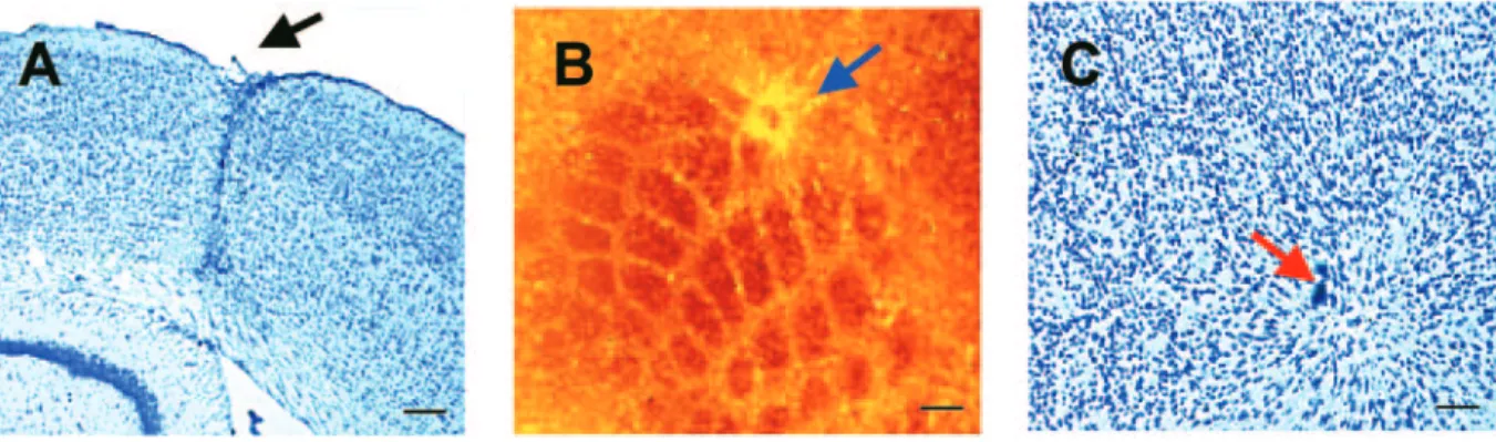
In order to produce a small localized lesion in the barrel cortex a focal lesion was performed with coordinates that targeted the δ barrel in the left hemisphere. The injured area extended through the entire cortical thickness down to the subcortical white (Fig. 1A). Most of the lesions produced a superficial injury to the subcortical white matter. On tangential cyto-chrome oxidase stained sections, the lesion was centered on the δ barrel and often extended into neighboring barrels (D1, E1, E2; Fig. 1B). The injured area shrank over the first 2 weeks after the lesion and by 6 weeks post-lesion, it corresponded to a discreet glial scar of ∼200 µm (Fig. 1C). Although there was some variability in the extent and position of the lesion, no major difference in the measured physiological and behavioral data could be traced to apparent differences in lesion place-ment and size. The lesion did not result in any apparent changes in food intake and body weight and caused no mortality.
Somatosensory Evoked Potential
To investigate whether and to what extent focal lesions affected the global activity of the barrel cortex, we applied multielectrode epicranial recordings. Sensory evoked poten-tials were elicited by stimulating all contralateral whiskers and recorded using an array of five electrodes placed in line at 1 mm distance from each other (Fig. 2B). The typical responses recorded over the barrel cortex before and 2 and 6 weeks after injury are shown in Figure 2A. Before lesion, the characteristic biphasic response was composed of a rapid positive peak (P1; mean delay of 13–14 ms) followed by a negative wave (N1;
Figure 1. Photomicrographs illustrating the lesion site in the barrel cortex 6 weeks after injury. (A) A cresyl violet stained coronal section showing a discreet glial scar (arrow) at
the site of the injury. (B) A tangential 40 µm section stained for cytochrome oxidase shows barrels and the lesion. (C) A cresyl violet stained tangential section showing the cytoarchitecture of injured barrels and the surround. Scale bars = 200 µm (A, B); 100 µm (C).
mean delay of 25–27 ms). At ∼8 ms there is another smaller positive peak that was more clearly distinguishable from P1 after the lesion. These short latency responses concur with those recorded earlier in the rat somatosensory cortex (Di and Barth, 1991; Seo, 1992). An almost complete absence of P1 as well as a shorter N1 latency was typically observed 2 weeks after injury. However, a clear recovery was observed 6 weeks post lesion. A detailed quantitative analysis of the responses was performed using a group of 12 injured mice. To quantify data, we used as an index, the peak-to-peak amplitude (P1/N1), calculated as the difference between P1 and N1 values. SEP recorded in absence of injury showed that contralateral
stimu-lation evoked similar responses in the left (LH) and right (RH) hemispheres. Mean P1/N1 values were 84.5 ± 7.9 and 89.9 ± 8.0 µV for LH and RH, respectively, while P1 and N1 latencies were 13.6 ± 0.4 and 26.7 ± 0.9 ms at LH and 13.1 ± 0.5 and 25.9 ± 1.2 ms at RH (Fig. 2C). Since there was no evident asymmetry in amplitude and latency, the right uninjured hemisphere was considered as control and all values measured in the left, injured cortex normalized to right values. Two weeks after cortical injury P1/N1 amplitude decreased significantly from 104.7 ± 15.6 to 29.7 ± 4.9% (n = 8, P < 0.001). Also, N1 latency decreased from 98.5 ± 1.8 to 79.1 ± 5.4% (P < 0.01). However, P1 latency did not change significantly (99.7 ± 1.3 versus 96.7
Figure 2. Epicranial SEP recording. (A) Representative barrel cortex responses contralateral to the stimulated whiskers in the same mouse before and 2 and 6 weeks after the
injury. Each trace is the average of three trials of 10 consecutive responses recorded with an electrode placed at 1 mm posterior to bregma (see B). The responses recorded over the barrel cortex are characterized by biphasic primary responses composed of a rapid positive peak followed by a negative wave (N1). Note the changes in peak-to-peak amplitude (P1–N1) depicted by circles. (B) Schematic drawing displaying the relative position of recording sites (black circles) over the skull. The red open circle indicates the site of barrel cortex injury and the blue open circle shows the site of sham injury outside of the barrel field (stippled outline). (C) Quantitative analysis of PN1–N1 amplitude. The changes observed at 2 and 6 weeks after the injury of the barrel cortex (inside, n = 8) is compared to an injury produced outside the cortical representation of whiskers (outside, n = 4). While the responses decreased significantly 2 weeks after inside-injury and recover 4 weeks later, the injury outside the barrel cortex did not affect these responses significantly. Results represent the amplitude (mean ± SEM) of the peak-to-peak (PN1–N1) responses, normalized to the SEP recorded over the contralateral uninjured cortex (100% = 89.9 ± 8µV). *2w, Significantly different from responses recorded before the lesion. *6w, Significantly different from values recorded 2 weeks after the lesion. Pre, before lesion; 2w, 2 weeks after injury; 6w, 6 weeks after injury. (D) Typical spatio-temporal representation of SEP multielectrode recordings, obtained in the same mouse before and 2 and 6 weeks after the injury in the barrel cortex. Horizontal axis represents time in milliseconds, vertical axis represents the distance in millimetres related to bregma and pseudo-color scale represents the amplitude in µV (red, positive; green, negative values). Arrows show the region (1 mm behind bregma, AP: –1 mm) where the most significant changes appear over the barrel cortex. Note that in the intact cortex (before lesion) the earlier positive responses are typically recorded above barrel cortex. Similar but delayed responses were consistently recorded with the electrode placed over the facial motor cortex (AP: +1 mm). The deficit observed 2 weeks after injury is characterized by a dramatic decrease in positive potentials in the barrel cortex. The positive response recorded over motor cortex seems to be unaffected. Six weeks after injury, there is the recovery of positive potentials at AP: –1 mm.
± 6.5%). Records performed 6 weeks after the injury showed a partial reversion of these changes: P1/N1 amplitude recovered to 88.4 ± 12.4% and N1 latency to 94.4 ± 5.8% (n = 8, 1 < 0.01, for comparison between recordings performed 2 and 6 weeks after injury; t-test corrected for multiple comparisons). In another group of eight mice, a similar lesion was made outside the barrel cortex. In this group, P1/N1 amplitude and N1 latency values in the recordings performed before and 2 and 6 weeks after the cortical injury, showed no significant changes (Fig. 2C).
Figure 2D illustrates spatio-temporal maps obtained by analyzing responses before and after cortical injury. In these graphs, the horizontal scale represents time (ms), vertical scale the spatial topography (mm from bregma) and the color range is the amplitude (µV) normalized to the maximum positive or negative peak responses (P1 and N1, respectively). As a rule, the main, earlier positive peak is recorded with the electrodes placed at –1 and –2 mm behind bregma (0.5 mm from the site of injury), in proximity to barrel field. However, responses recorded over the motor cortex area did not show significant changes (data not shown). A clear recovery of P1, recorded by the electrode placed at –1 mm behind bregma, was noticed 6 weeks after cortical injury. These results indicate that a focal lesion in the somatosensory barrel cortex induce deficit in global cortical activity that is followed by a partial recovery. These changes are specific for the barrel cortex.
[14C]2-DG Uptake
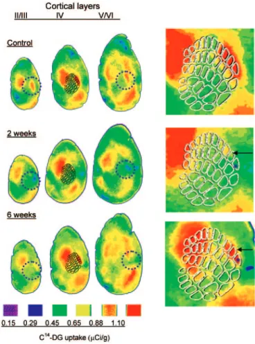
We investigated whether the recovery process demonstrated by SEP recording could be confirmed using quantitative [14 C]2-deoxyglucose (DG) autoradiography. To do this, we measured DG uptake in response to whisker activation in injured mice 2 and 6 weeks after lesion. In these experiments, all whiskers except three (δ, E1 and E2) were clipped on both sides, so as to make sure that activity was evoked only in the area of the lesion. Following DG injection, animals were allowed to explore a cage during 45 min to obtain spontaneous activation of intact whiskers (see Material and Methods). In tangential sections from control mice, the metabolic uptake occurred in areas that corresponded to the three stimulated barrels (Fig. 3). For the quantitative analyses, the metabolic activity of stimu-lated barrels was normalized to that of an inactive reference region (C1; Fig. 4A). In the right, uninjured hemisphere, this value, expressed as a percentage of a reference barrel, was very constant before and 2 weeks (33.8 ± 3.4%, n = 3) and 6 weeks (30.4 ± 3.5%, n = 3) after injury (Fig. 4B). Therefore we consid-ered these values as control for comparisons with changes in relative DG uptake in the injured barrel cortex. The effect of injury on metabolic uptake was assessed in the intact barrels adjacent to the lesion. The quantitative analysis indicated that DG uptake was significantly (P < 0.05) lower in the injured than in intact barrel cortex in all layers studied 2 weeks after lesion. This deficit in metabolic uptake partially recovered 6 weeks after lesion; the difference between the 2 and 6 weeks groups being significant (P < 0.05).
Behavioral Performances
An important question with regard to the recovery of electrical and metabolic activity in the barrel cortex concerned the possi-bility that this process could have functional consequences. To address this issue, we used a gap-crossing test to assess the
functional importance of whisker-mediated information. In these experiments, whiskers were clipped on the left side and right vibrissae-dependent performance was analyzed by meas-uring the size of the gap that the mouse was capable of crossing using its whiskers. Infiltration of mystacial pad with local anesthetics (on one side and saline on the other; see Fig. 5) was used to test the reliability of gap-crossing task. Figure 5 shows that local anesthesia in the right mystacial pad produced a significant (P < 0.01) decrease in the performance of animals compared to the control saline injected group. The control group with local anesthesia on the trimmed side did not show any decrease of the performance in the task. Injury in the left barrel cortex, however, resulted in a significant deficit in right vibrissae-dependent gap-crossing task 1 week after the injury (6.8 ± 0.14 versus 6.0 ± 0.25 cm, P < 0.01). Nevertheless, animals were always able to make the movements required to cross the gap. Mice made use of their nose tips or paws to gauge the distance to cross the gap in the dark. The perform-ance then recovered and no significant deficit was detected when mice were tested 4 weeks after the injury. Clipping
Figure 3. DG- uptake labeling. Representative maps are shown on tangential sections
of the cortex taken from a control animal and from animals that survived 2 or 6 weeks following the injury. II/III, supragranular layers; IV, granular layer with schematic of the barrel field; V/VI, infragranular layers. In the control brain strong activation is seen over the three activated barrels (δ, E1 and E2) and the corresponding supragranular and infragranular layers. Note the decrease in uptake on stimulated barrels after 2 weeks and the partial recovery 6 weeks after lesion. Circles represent 1 mm. At the right, magnified maps of the barrel cortex are shown illustrating the injured area centered on the δ barrel (asterisk) and the nearest activated activated barrel (E1 depicted by the arrows) where the uptake measurements were performed.
whiskers on the right side abolished the functional recovery (not shown), indicating that this phenomenon depended on the intact vibrissae–barrel cortex pathway. The control group, with spared whiskers ipsilateral to the lesion (unaffected cortical representation), did not show any deficit 1 week after the injury. In all these experiments, the sensory deficit, whether produced at the peripheral or cortical level, corres-ponded to a distance (0.7–0.8 cm) that is detected by whiskers. Recovery in NCA M Knockout Mice
Since previous research had identified a key role for NCAM in different processes that could participate to functional recovery (see review in Kiss et al., 2001), we decided to take advantage of NCAM knockout mice lacking all isoforms of NCAM to evaluate the recovery process using this model. As previously reported (Cremer et al., 1994), NCAM knockout mice exhibit a reduced size of the olfactory bulb, but no other identified abnormalities in the size and organization of the central nervous system. On Nissl-stained sections, the rest of the brain displayed a normal cytoarchitectonics. The cerebral cortex had a typical six-layer organization and on tangential cytochrome oxidase-stained sections we observed a normal barrel pattern (not shown). Finally, the whisker pad in wild type and mutant animals showed no difference. The histolog-ical appearance of the lesion and the subsequent repair had the same characteristics in wild type and mutant animals. In a group of four animals, SEP responses were recorded before and 2 and 6 weeks after the lesion. As illustrated in Figure 6, SEP responses showed a similar deficit in control and mutant animals 2 weeks after the lesion. However, after 6 weeks, almost no recovery was perceptible in transgenic mice: the positive and negative peaks remained essentially unchanged. Overall, the peak-to-peak amplitude obtained by subtraction of the voltages measured at the times of the positive and negative peaks showed only ∼10% recovery, which is in marked contrast with what was observed in wild type animals (Fig. 6C). Simi-larly, measurement of DG uptake confirmed a reduced recovery of metabolic activity 6 weeks after the lesion. Figure 7 illustrates the level of uptake observed in an NCAM knockout
mouse before and 2 and 6 weeks after a lesion made in the region of the δ barrel. While some activity is taken over by the adjacent barrels, the recovery at 6 weeks is significantly less than in control animals (compare with Fig. 4). A quantitative analysis (Fig. 7B) indicates that the level of metabolic activity recovered in the adjacent barrels is significantly lower in NCAM knockout mice than in control animals both 2 and 6 weeks after the lesion. These results thus clearly indicate that functional recovery is deficient in the NCAM mutant animal. Whether this defect also resulted in behavioral deficits could unfortunately not be tested, because, surprisingly, NCAM knockout mice used a different strategy than wild type animals in the gap-crossing test: they jumped over the gap rather than extending one leg. The reasons for this difference are unknown, but this prevented comparison between control and transgenic mice.
Discussion
The results of the present study provide evidence that the production of a small lesion in the somatosensory cortex of the mouse leads to a functional deficit that recovers over a period of 4–6 weeks. Both the deficit and the functional recovery could be monitored using repeated, multielectrode SEP record-ings, analysis of metabolic activity and a behavior test, the gap-crossing test. Application of this model to NCAM knockout mice allowed us to detect a deficit in functional recovery that suggests an involvement of the NCAM molecule in the repara-tion process. Together, these data indicate that this mouse model of lesion-induced recovery may represent an interesting tool for the analysis of the cellular and molecular mechanisms underlying brain injury and repair.
In these studies, activity in the barrel cortex was analyzed by recording potentials evoked by whisker deflection using an epicranial multielectrode recording system (Troncoso et al., 2000). These surface potentials are thought to reflect the acti-vation of apical dendrites of thousands of pyramidal cells sharing architectural and temporal coherence and generating post-synaptic activity. Intra-cortical laminar recordings of evoked field potentials in the rat barrel cortex have established
Figure 4. Quantitative analysis of DG uptake. (A) Schema of barrel field indicating the position with respect to the lesion site of the activated and reference (whisker trimmed)
barrels where DG uptake measurement was carried out. A rectangular area with a constant size of ∼400 × 400 µm was used to determine DG uptake in the different layers. The measured values over the activated barrels (E1 barrel) were standardized to DG uptake in reference areas (C1 barrel) in both intact and injured hemispheres. DG uptake in the injured barrel field is expressed as percentages of values calculated in the contralateral homotop area of the barrel cortex. (B) Photomicrograph of a cresyl violet stained tangential section illustrating the injured barrel (δ) and the surround. (C) Results of the quantitative analysis showing a significant difference between the DG uptake measured 2 weeks and 6 weeks after lesion (n = 4, P < 0.05).
that the primary responses of the surface SEP complex are produced by synchronized activation of both supra- and infra-granular pyramidal cells (Di et al., 1990). In the current study, SEP recordings were performed after deflection of all contra-lateral whiskers in order to evaluate the impact of a small focal lesion on the global activity of the barrel cortex. These record-ings, made 2 weeks after injury, disclosed a measurable deficit that involved mainly the (first) main positive peak of the SEP complex, occurring ∼13–14 ms after the vibrissae were mechanically stimulated. This positive peak of the SEP complex most likely reflects intra-cortical integrative processes (Di and Barth, 1991). The present results indicate that a focal lesion of a few barrels markedly affected this integration, making it an interesting tool to monitor recovery. It is of interest that this peak is preceded by an earlier and lower amplitude positive response (occurring at a latency of ∼8 ms). This earliest positive signal is most likely due to direct thalamo-cortical activation and, as illustrated in Figure 3, was not significantly modified after the lesion upon stimulation of all contra-lateral whiskers. Importantly also, spatiotemporal
anal-yses indicated that the deficit and recovery observed 4 weeks later were restricted to the barrel cortex. No changes of activity were observed in the somatomotor cortex. Together, these results indicate therefore that a focal injury to the barrel cortex is associated with a loss of the cortical activity evoked by stimulation of all contra-lateral whiskers and a recovery process that take place within a few weeks.
In agreement with the results of previous studies (Glassman, 1971), the present data are consistent with and support the interpretation that functional recovery may take place due to a reorganization of cortical activity in the regions around the lesion. The lesion-induced SEP deficit was paralleled by a hypo-metabolism (28% decrease) detected by stimulus-induced DG uptake in the region surrounding the lesion. Similarly also, the recovery specifically occurred in the barrels adjacent to the lesion within the next 4 weeks. These observations are also consistent with previous reports showing dynamic changes in the functional representation of whiskers after focal lesions. Schiene et al. (1999) demonstrated in a rat model that photo-chemically induced ischemic lesions resulted in an enlarge-ment of cortical vibrissa representation in the surrounding cortical area. They attributed the expansion of the activated area to a decreased GABA-ergic inhibition that facilitates a remapping of the cortical representation in neighboring brain areas. Similar changes in representation area of whiskers after kainate lesions were detected using repeated optical imaging (Nguyen et al., 2000). These results indicate that dynamic changes in juxtalesional areas could play an important role in remapping of cortical representation. In this regard, lesion studies are in agreement with earlier publications showing expansion or retraction of whisker cortical maps after activa-tion (Polley et al., 1999) or inactivaactiva-tion by peripheral deaffer-entation (Silva et al., 1996; Bronchti et al., 1999; Kossut and Juliano, 1999). In contrast to small focal lesions, however, larger injuries appear to produce persistently depressed meta-bolic activity in adjacent regions (Dunn-Meynell and Levin, 1995; Passineau et al., 2000).
Interesting information added by this study is that the small deficit and recovery in cortical activity had functional conse-quences at the behavioral level. Using the gap-crossing test we could find a correlated deficit and recovery of performance. This test is sensitive and clearly depended upon a functionally intact vibrissae–somatosensory cortex pathway, as both anesthesia of the vibrissae and lesion of barrels resulted in behavioral deficit. Recovery of behavioral performance took place within 4 weeks, a time course very consistent with those found by previous studies using the gap-crossing test and diverse lesion models (Hutson and Masterton, 1986; Barnéoud et al., 1991; Pazos et al., 1995). Also, the time course of behav-ioral recovery closely corresponded to that of functional cortical activity (present study). A simple explanation for this observation could be a reorganization of whisker representa-tions in the barrels adjacent to the lesion, although others factors, such as learning of new strategies to cross the gap, cannot be excluded. Similar conclusions were reached by other authors (Hutson and Masterton, 1986) who suggested that destruction of the barrel may impair a ‘higher-order integ-ration’ necessary for a complex task such as sensory-guided movement and that the motor system involved in gap crossing might require sensory input from the somatosensory cortex. Studies in animal models and human data suggest that small lesions in the motorcortex or in the somatosensory cortex
Figure 5. Gap-crossing test. (A) Video images showing a mouse crossing the gap
between the platform and the tunnel made of transparent acrylic. Mice need to contact the opposite border with their whiskers before crossing. (B) Schematic drawing of the gap illustrating the distance measured in millimetres. (C) The effect of blocking the mystacial pad innervation with local anesthesia on the gap-crossing scores. In these experiments whiskers were clipped on the left side and the right vibrissae-dependent performance was tested with or without local anesthesia (see Materials and Methods). Values are mean ± SEM, n = 5. *Significantly different (P < 0.01) from controls. (D) The effect of barrel field injury on the gap-crossing score. Injury was made in the left barrel field and all whiskers but three (δ, D1 and E1) were trimmed. In the test group, the spared whiskers were contralateral to the lesion, while in the control (sham) group the spared whiskers were ipsilateral to the injured cortex. Values are mean ± SEM, n = 5. *Significantly different (P < 0.01) from controls.
recover from the ipsilateral cortex adjacent to the lesion, whereas large lesions recover from the contralateral cortex. (Jenkins and Merzenich, 1987; Witte et al., 2000). Human data also indicate that a new lesion in the surround of the lesioned cortex abolishes previous recovery (Jenkins and Merzenich, 1987; Witte et al., 2000). It would be of particular interest to test in our model if lesions of the adjacent cortex 6 weeks after the first lesion would affect functional recovery.
The present model also opens the way to analyze the cellular and molecular mechanisms underlying the recovery. Several possibilities have been considered. One involves the resolution of acute pathological mechanisms such as edema, vascular perturbation and excitotoxicity. These events, however, are thought to be at least partially resolved by 2 weeks after the lesion. Another mechanism contributing to recovery could be a gradual reversal of diaschisis, a temporal suppression of surrounding and remote cortical tissues after focal lesions
(Witte, 1998). The effect of diaschisis has been substantiated by recent metabolic studies showing a persistent hypoactivity in perilesional areas (Schiene et al., 1999; Passineau et al., 2000). Finally, growth mechanisms and functional reorganiz-ation of cortical networks could also account for the take over of function (vicariation of function) of the damaged area by adjacent or remote areas (Witte, 1998). This process is likely to be complex and involve several cellular and molecular events, including a modulation of the expression of receptors (Schiene et al., 1996), axonal sprouting and new synapse formation (Stroemer et al., 1993). The use of transgenic models could reveal of interest to distinguish between all these possibilities. The experiments with the NCAM knockout mice demon-strate the interest of this recovery model to identify, through the use of transgenic animals, the role of candidate molecules in functional recovery. While the functional organization of the whisker-barrel system in WT and NCAM mutant animals
Figure 6. SEP recording in NCAM knockout mice. (A) Representative SEP responses recorded over the barrel cortex of an NCAM knockout animal contralateral to the stimulated
whiskers in the same mouse before and 2 and 6 weeks after the injury. Notice that the absence of positive response observed 2 weeks after the cortical injury remains pronounced 4 weeks later (6 weeks after injury). (B) Voltage values measured at a latency of 13 ms after stimulation. This latency corresponds to the mean positive peak values observed before cortical injury. Compared to control mice, 6 weeks after injury voltage at this latency was negative in al NCAM knockout mice (n = 5 in both groups). (C) Typical spatio-temporal representation of SEP multielectrode recordings, obtained in the same NCAM knockout mouse before and 2 and 6 weeks after the injury in the barrel cortex. All three parameters (time, distance and voltage) are the same as described for Figure 3. Arrows indicates the loss of positivity and absence of recovery at AP: –1 mm. Responses over the motor cortex were not affected by the injury.
displayed no apparent differences, the functional recovery 6 weeks after the lesion was significantly reduced. This conclu-sion was reached through the use of epicranial SEP recordings and analyses of metabolic activity. The precise mechanism of the deficient recovery in the mutant mice remains unknown. It is of particular interest that NCAM/PSA-NCAM has been local-ized in synapses and that long-term synaptic potentiation (LTP) is impaired in CA1 synapses of the hippocampus in NCAM knockout animals (Muller et al., 1996). It is thus possible that an altered synaptic plasticity in cortical circuits underlies the observed deficit in functional recovery. Alternatively, the absence of NCAM during development could results in a modi-fied organization of cortical circuits that could lead to reduced plasticity and capacity to reorganize after a lesion. Clearly, further studies involving intra-cortical recordings and analyses of the synaptic organization of the barrel cortex will be required to verify these hypotheses.
Conclusions
In this study we have assessed the electrical and metabolic activity of recovering circuits in the somatosensory barrel
cortex of the mouse. A time window has been identified between 2 and 6 weeks after injury, during which behaviorally relevant impairment and recovery was reliably measured in the surround of injury. This mouse model therefore opens new ways to investigate, at the molecular level, the mechanisms contributing to functional recovery.
Notes
We are grateful to Cynthia Saadi and Nathalie Moulin for technical assistance, especially with the histological processing. This work was supported by the Swiss National Science foundation grants 31-64030.00 to J.Z.K. and PNR-38: 4038-0044151 to J.Z.K. and D.M.
Address correspondence to J.Z. Kiss, Department of Morphology, University of Geneva Medical School, 1 rue Michel Servet, CH-1211 Geneva 4, Switzerland. Email: [email protected].
References
Barnéoud P, Gyger M, Andres F, van der Loos H (1991) Vibrissa-related behavior in mice: transient effect of ablation of the barrel cortex. Behav Brain Res 44:87–99.
Becker CG, Artola A, Gerardy-Schahn R, Becker T, Welzl H, Schachner M (1996) The polysialic acid modification of the neural cell
Figure 7. DG- uptake labeling in NCAM knockout mice. (A) Representative DG maps of tangential barrel cortex sections taken from a NCAM knockout mice. Notice that the
activated barrel (E1 depicted by the arrow) which recovers its activity after 6 weeks in wild type animals (see Fig. 3), displays low uptake compared to the reference barrel. (B) Quantitative analysis of C14-DG uptake obtained in wild type animals (C57/Bl6) and NCAM knockouts. Values are mean ± SEM (n = 4 animals in each group) normalized to the contralateral homotop area of the barrel cortex (see legend for Fig. 4) and expressed as percentage of the value measured before the lesion (pre) (100%). *Significantly different from the wild type group, P < 0.05).) (C) Representative cresyl violet stained tangential section showing the cortical injury in a NCAM knockout animal killed 2 weeks after the lesion. Red dashed line shows the position of E2 barrel. Scale bar = 200 µm.
adhesion molecule is involved in spatial learning and hippocampal long-term potentiation. J Neurosci Res 45:143–152.
Bronchti G, Corthesy ME, Welker E (1999) Partial denervation of the whiskerpad in adult mice: altered patterns of metabolic activity in barrel cortex. Eur J Neurosci 11:2847–2855.
Buonomano DV, Merzenich MM (1998) Cortical plasticity: from synapses to maps. Annu Rev Neurosci 21:149–186.
Cremer H, Lange R, Christoph A, Plomann M, Vopper G, Roes J, Brown R, Baldwin S, Kraemer P, Scheff S (1994) Inactivation of the N-CAM gene in mice results in size reduction of the olfactory bulb and deficits in spatial learning. Nature 367:455–459.
Cremer H, Chazal G, Carleton A, Goridis C, Vincent JD, Lledo PM (1998) Long-term but not short-term plasticity at mossy fiber synapses is impaired in neural cell adhesion molecule-deficient mice. Proc Natl Acad Sci USA 95:13242–13247.
Di S, Barth DS (1991) Topographic analysis of field potentials in rat vibrissa/barrel cortex. Brain Res 546:106–112.
Di S, Baumgartner C, Barth DS (1990) Laminar analysis of extracellular field potentials in rat vibrissa/barrel cortex. J Neurophysiol 63:832–840.
Dunn-Meynell AA, Levin BE (1995) Lateralized effect of unilateral somatosensory cortex contusion on behavior and cortical reorgan-ization. Brain Res 675:143–156.
Geschwind N (1985) Mechanisms of change after brain lesions. Ann N Y Acad Sci 457:1–11.
Glassman RB (1971) Recovery following sensorimotor cortical damage: evoked potentials, brain stimulation and motor control. Exp Neurol 33:16–29.
Hutson KA, Masterton RB (1986) The sensory contribution of a single vibrissa’s cortical barrel. J Neurophysiol 56:1196–1223.
Jenkins WM, Merzenich MM (1987) Reorganization of neocortical representations after brain injury: a neurophysiological model of the bases of recovery from stroke. Prog Brain Res 71:249–266. Kiss JZ, Troncoso E, Djebbara Z, Vutskits L, Muller D (2001) The role
of neural cell adhesion molecules in plasticity and repair. Brain Res Rev 36:175–184.
Kossut M, Juliano SL (1999) Anatomical correlates of representational map reorganization induced by partial vibrissectomy in the barrel cortex of adult mice. Neuroscience 92:807–817.
Muller D, Wang C, Skibo G, Toni N, Cremer H, Calaora V, Rougon G, Kiss JZ (1996) PSA-NCAM is required for activity-induced synaptic plasticity. Neuron 17:413–422.
Nguyen TT, Yamamoto T, Stevens RT, Hodge CJ Jr (2000) Reorganiza-tion of adult rat barrel cortex intrinsic signals following kainic acid induced central lesion. Neurosci Lett 288:5–8.
Passineau MJ, Zhao W, Busto R, Dietrich WD, Alonso O, Loor JY, Bramlett HM, Ginsberg MD (2000) Chronic metabolic sequelae of traumatic brain injury: prolonged suppression of somatosensory activation. Am J Physiol Heart Circ Physiol 279:H924–H931. Pazos AJ, Orezzoli SL, McCabe PM, Dietrich WD, Green EJ (1995)
Recovery of vibrissae-dependent behavioral responses following barrelfield damage is not dependent upon the remaining somato-sensory cortical tissue. Brain Res 689:224–232.
Polley DB, Chen-Bee CH, Frostig RD (1999) Varying the degree of single-whisker stimulation differentially affects phases of intrinsic signals in rat barrel cortex. J Neurophysiol 81:692–701.
Schiene K, Bruehl C, Zilles K, Qu M, Hagemann G, Kraemer M, Witte OW (1996) Neuronal hyperexcitability and reduction of GABAA-receptor expression in the surround of cerebral photothrombosis. J Cereb Blood Flow Metab 16:906–914.
Schiene K, Staiger JF, Bruehl C, Witte OW (1999) Enlargement of cortical vibrissa representation in the surround of an ischemic cortical lesion. J Neurol Sci 162:6–13.
Seo ML (1992) Effect of environmental complexity on the latency of cortical vibrissa potentials. Dev Psychobiol 25:67–76.
Silva AC, Rasey SK, Wu X, Wall JT (1996) Initial cortical reactions to injury of the median and radial nerves to the hands of adult primates. J Comp Neurol 366:700–716.
Stroemer RP, Kent TA, Hulsebosch CE (1993) Acute increase in expression of growth associated protein GAP-43 following cortical ischemia in rat. Neurosci Lett 162:51–54.
Troncoso E, Muller D, Czellar S, Kiss JZ (2000) Epicranial sensory evoked potential recordings for repeated assessment of cortical functions in mice. J Neurosci Meth 97:51–58.
Welker E, Rao SB, Dorfl J, Melzer P, van der Loos H (1992) Plasticity in the barrel cortex of the adult mouse: effects of chronic stimulation upon deoxyglucose uptake in the behaving animal. J Neurosci 12:153–170.
Witte OW (1998) Lesion-induced plasticity as a potential mechanism for recovery and rehabilitative training. Curr Opin Neurol 11:655–662.
Witte OW, Bidmon HJ, Schine K, Redecker C, Hagemann G (2000) Functional differentiation of multiple perilesional zones after focal cerebral ischemia. J Cereb Blood Flow Metab 20:1149–1165. Wong-Riley M (1979) Changes in the visual system of monocularly
sutured or enucleated cats demonstrable with cytochrome oxidase histochemistry. Brain Res 171:11–28.
Woolsey TA, Van der Loos H (1970) The structural organization of layer IV in the somatosensory region (SI) of mouse cerebral cortex. The description of a cortical field composed of discrete cytoarchi-tectonic units. Brain Res 17:205–242.