Calystegine degradation capacities of microbial rhizosphere
communities of Zea mays (calystegine-negative)
and Calystegia sepium (calystegine-positive)
Daniel Guntli, Steèphane Burgos, Yvan Moeënne-Loccoz
1, Genevieéve Deèfago *
Phytopathology Group, Institute of Plant Sciences, Swiss Federal Institute of Technology (ETH), Universitaëtstr. 2, CH-8092 Zuërich, Switzerland Received 18 June 1998; received in revised form 23 September 1998; accepted 28 September 1998
Abstract
Calystegines are tropane alkaloids produced by the roots of a few plant species. A bioassay was developed to identify roots with a microbial rhizosphere community capable of calystegine degradation (i.e. MCD roots). In a field survey, the proportion of MCD roots of Zea mays (calystegine-negative) varied from 20 to 80%. In field experiments, the proportions of MCD roots of Z. mays and Calystegia sepium (calystegine-positive) grown in a particular plot were similar to each other but varied with time and, overall, were higher than those of Z. mays roots from adjacent plots free of C. sepium. In autoclaved soil, no root of C. sepium or Z. mays plants propagated as seeds was MCD, indicating that calystegine-degrading microorganisms were not seed-borne. However, MCD roots were found as early as 1 day after planting of rhizomes of C. sepium in autoclaved soil or planting of axenic seedlings of either plant in natural soil microcosms. In total, microorganisms capable of degrading calystegines were harboured not only in the rhizosphere of the producing plant but also in that of the calystegine-negative plant and probably in bulk soil. z 1999 Federation of European Microbiological Societies. Published by Elsevier Science B.V. All rights reserved.
Keywords: Calystegine; Alkaloid; Calystegia sepium; Zea mays; Rhizosphere e¡ect
1. Introduction
Calystegines are plant secondary metabolites and were ¢rst found in the roots of Calystegia sepium (L.) R.Br. (i.e. hedge bindweed, morning glory) [1]. Other plant species have also been reported to produce
these tropane alkaloids, e.g. Atropa belladonna L. and Convolvulus arvensis L. [1], Hyoscyamus niger L. [2], Morus alba L. [3], Physalis alkengi var. fran-cheti L. [4], Solanum tuberosum L. and Solanum melongena L. [5]. Calystegines can be poisonous to arthropods [5] and mammals [6], presumably as a result of glycosidase inhibition [2,7]. Calystegines may also act as allelochemicals in interactions be-tween plants [8].
Sinorhizobium meliloti strain Rm41 [1] and bacte-ria from a few other genera, including Pseudomonas [8,9], have been found to degrade calystegines. In
* Corresponding author. Tel.: +41 (1) 632 38 69; Fax: +41 (1) 632 11 08; E-mail: [email protected]
1Present address: UMR CNRS d'Ecologie Microbienne du Sol,
Universiteè Claude Bernard (Lyon 1), 69622 Villeurbanne Cedex, France.
strain Rm41, the genes responsible for calystegine catabolism (cac) are harboured on the 225-kb self-transmissible, non-symbiotic plasmid pRme41a [1]. Subcloning revealed that the cac locus is spread over a region 30^40 kb in length [10]. Since S. meli-loti Rm41 can use calystegines as a source of carbon and nitrogen for growth [9], it is thought that the genes encoding the catabolism of these alkaloids can contribute to strain survival during its sapro-phytic life, i.e. in the absence of the host plant alfalfa [10]. In accordance with this concept, Tepfer et al. [9] found calystegine-degrading bacteria in the rhizo-sphere of calystegine-producing plants but not in that of calystegine-negative plants, suggesting a role for calystegines as nutritional mediators in plant-mi-crobe interactions.
Our understanding of the role of calystegines in the rhizosphere is based exclusively on the results of calystegine degradation tests performed with a limited number of isolates of rhizosphere bacteria only [8,9]. The microbial community of the rhizo-sphere is highly diverse and it is unlikely that deter-minations carried out with individual microorgan-isms could provide a full account of the role of calystegines in plant-microbe interactions. Therefore, the objective of this study was to reevaluate, this time at the microbial community level, the relation-ship between the ability of plants to produce calyste-gines and that of rhizosphere microorganisms to de-grade calystegines. For this purpose, a new assay was developed to score the capacity of rhizosphere micro-bial communities to degrade calystegines puri¢ed from C. sepium. The assay requires the presence of calystegine-degrading microorganisms at signi¢cant population levels for a positive response.
2. Materials and methods 2.1. Plants
Seeds of C. sepium were collected in parks located in the city of Zuërich (Switzerland), and those of Z. mays cv. Silex 170 were obtained from the Seeds Department of the Swiss Federal Research Station for Agroecology and Agriculture (FAL) at Zuërich-Reckenholz. A culture of C. sepium roots trans-formed by Agrobacterium rhizogenes [11] was used
for the production of calystegines because the intro-duced T-DNA resulted in increased root biomass and alkaloid content. The transformed roots were cultivated in 65 ml of Gamborg B5 medium (Sigma, Buchs, Switzerland) in 300-ml Erlenmeyer £asks with continuous shaking (120 rpm) at 24³C in the dark for 3 weeks.
2.2. Puri¢cation of calystegines
The transformed roots were removed from liquid cultures, blotted dry and stored at 320³C prior to use. For extraction of calystegines, portions of about 5 g of roots were frozen in liquid N2 and
homoge-nised using a mortar and pestle. The samples were transferred into 1.5-ml Eppendorf tubes and centri-fuged at 13 000Ug for 40 min. To remove tannins and phenols, the supernatants (which contained about one third of the initial amount of plant mate-rial) were treated with 45% (w/v) neutral aluminium oxide 90 (E. Merck AG, Dietikon, Switzerland). The samples were mixed with acetonitrile in a 1:9 (v/v) ratio and passed through a LC-NH2 solid phase ex-traction column (Supelco, Buchs, Switzerland) con-ditioned with 6 ml of acetonitrile solution (i.e. water:acetonitrile in a 1:9 ratio), to remove salts and ions. The column was washed with 3 ml of ace-tonitrile solution and elution was performed with 3 ml of 0.001 M HCl. The eluates were then passed through an ion exchange LC-SCX column (Supelco) conditioned with 6 ml of 0.001 M HCl to separate sugars. After washing the column with 3 ml of 0.001 M HCl, the column was rinsed with 2 M NH3. The
eluates were evaporated to dryness in vacuo and re-dissolved each in 250 Wl of sterile distilled water. 2.3. Analysis of calystegines
The method of Draëger [12] for calystegine analysis was slightly modi¢ed to facilitate identi¢cation of the alkaloids. Samples (2 Wl) were applied to a thin-layer chromatography plate (Kieselgel 60, Merck), which was developed in a solvent system consisting of CH3OH, 0.6 M NH4Cl and CH3Cl in a 6:2:1 ratio,
until the solvent front was 8 cm from the base line. The chromatogram was dried and immersed in a solution prepared with 0.1 ml saturated aqueous AgNO3 and 20 ml acetone for 60 s, before being
transferred to a solution of ethanol containing 0.5 M NaOH until brown spots (calystegines) appeared on the white chromatogram (detection limit of about 0.5 Wg of calystegines). Finally, the chromatogram was ¢xed in a bath of photographic ¢xative G 345 (Agfa-Gevaert AG, Duëbendorf, Switzerland). After each bath, the chromatogram was dried using a hair dry-er. The purity of each extract was checked by com-parison with a calystegine standard obtained from D. Tepfer (INRA Versailles, France) according to published protocols [1]. Calystegine concentration in the extracts was adjusted to 2 Wg Wl31 prior to
use in degradation assays.
Biological analysis of extracted calystegines was carried out using S. meliloti strain Rm41 and its de-rivative Rm41-Ca1, which had been cured of the cac plasmid pRme41a and was unable to catabolise ca-lystegines [1]. The strains were grown in liquid tryp-tone-yeast extract (TY [13]) at 27³C with shaking (120 rpm) until cultures reached an optical density of 1 at 600 nm (equivalent to approximately 7U109
colony-forming units (CFU) ml31; late exponential
phase). The cells were washed and resuspended in fresh M9 medium without carbon source [14]. Calys-tegine degradation was assayed in reaction mixtures (¢nal volume 208 Wl) consisting of 200 Wl of M9 medium without carbon source, 8 Wl of calystegine extract or calystegine standard (¢nal concentration, 77 Wg ml31) and the test bacterium (cell density,
7U107 CFU ml31). After incubation for 24 h at
27³C with shaking (120 rpm), the samples were centri-fuged (10 min, 13 000Ug) and the supernatant evapo-rated to dryness in vacuo. The residues were taken up in 8 Wl sterile distilled water. A 2-Wl aliquot was tested
for the presence of calystegines by thin-layer chro-matography, as described above. For each strain, seven assays were performed with extracted calyste-gines and seven assays with calystegine standard. 2.4. Bioassay for calystegine degradation by
rhizosphere microbiota
To extract rhizosphere microorganisms, samples of about 0.25 g of root (and closely adhering soil) were transferred each into a 50-ml Erlenmeyer £ask con-taining sterile distilled water (sample:water ratio of 1:100 w/v) and the £asks were shaken for 15 min at 350 rpm. Puri¢ed calystegines (8-Wl aliquots) were added to 200 Wl of rhizosphere extract (¢nal concen-tration, 77 Wg ml31) and the samples (containing
microorganisms extracted from 2 mg of root materi-al) were incubated at 27³C with shaking (120 rpm) for 24 h. They were then centrifuged at 13 000Ug for 10 min and the supernatant was evaporated to dry-ness in vacuo. The residue was taken up in 8 Wl of sterile distilled water and 2 Wl analysed for the pres-ence of calystegines by thin-layer chromatography. The absence of calystegines indicated that the rhizo-sphere sample studied harboured microorganisms with the capacity to degrade these alkaloids and such roots were scored as being positive for micro-bial calystegine degradation (i.e. MCD roots). Since the detection limit of the chromatography was about 0.5 Wg of calystegines and 2 Wl of the 8 Wl was tested, it means that the detection limit in the bioassay was about 2 Wg of calystegines and that at least 14 Wg of the 16 Wg of calystegines used in the bioassay (i.e. 88%) had to be degraded before a root was rated MCD.
Table 1
Main properties of the soilsaused in the microcosm experiments
Eschikon Reckenholz Muërren
Crop at sampling maize maize potato
Texture loam loam sandy clay loam
Clay (%) 15 21 27
Silt (%) 42 32 23
Sand (%) 43 47 50
pHH2O 7.0 7.3 6.9
Cation-exchange capacity (cmol kg31) 32.7 18.8 49.0
CaCO3(%) 0.8 0 0
Organic matter (%) 3.5 3.0 12.8
N (%) 0.16 0.27 0.90
Altitude (m) above sea level 530 442 1590
To demonstrate that the absence of calystegines in samples could not have resulted from physico-chem-ical interactions (adsorption) between the alkaloid and soil particles, the three soils used in microcosm experiments in this study (Table 1) were each sus-pended in sterile distilled water (1, 3, 30 and 300 mg soil ml31). Puri¢ed calystegines (8-Wl aliquots)
were added to 200 Wl of each suspension. The sam-ples were vigorously shaken at 1000 rpm for 1.5 h and checked for the presence of calystegines, as de-scribed above. Three replicates were studied per treatment. Calystegines were clearly detected in all samples from each non-sterile soil, even when soil was used at a rate as high as 300 mg ml31 (data
not shown). Therefore, the presence of soil particles in rhizosphere extracts could not have resulted in false-positive results in the bioassay.
For validation of the bioassay, S. meliloti strain Rm41 and its derivative Rm41-Ca1 were prepared as described above, except that cells were washed and resuspended in sterile distilled water. The cell suspensions were diluted in sterile distilled water, as needed. Samples (5 ml each) of rhizosphere ex-tracts obtained from C. sepium plants sampled at Eschikon (described in Section 2.6) were sterilised by autoclaving. The bioassay was as described above, except that 190 Wl of extract was used. Inoc-ulation was performed with 10 Wl of cell suspension of strains Rm41 or Rm41-Ca1, and ¢nal cell den-sities were 5U102 to 5U107 CFU ml31 for strain
Rm41 and 5U106 or 5U107 CFU ml31 for strain
Rm41-Ca1. The assay was then carried out as de-scribed above. Calystegine degradation was studied for non-sterile rhizosphere extract, as well as steri-lised rhizosphere extract inoculated with strains Rm41 or Rm41-Ca1, and seven replicates were used per treatment.
An experiment was done to determine changes in bacterial populations during the 24-h calystegine degradation bioassay. First, the bioassay was per-formed using non-sterile rhizosphere extracts ob-tained from Z. mays (15 samples) or C. sepium (15 samples) grown in Eschikon soil. Total numbers of culturable aerobic bacteria were determined by col-ony counts on 10% tryptic soy agar. Second, the bioassay was carried out with sterilised rhizosphere extract of Z. mays (¢ve samples) or C. sepium (¢ve samples) inoculated with S. meliloti Rm41 at
7.5U106 CFU ml31. Culturable cells of the strain
were enumerated by colony counts on TY agar. All plates were incubated at 27³C for 3 days and the colonies counted.
2.5. Survey of the proportion of MCD Z. mays roots in the ¢eld
Five Z. mays ¢elds were chosen at random in a 6U8-km region east of Zuërich in July 1996. In each ¢eld, a 5-m2 area free of C. sepium was selected and
10 Z. mays plants were sampled. Root segments were collected from the surface horizon, at a depth of approximately 10 cm below the soil surface (one sample of about 0.25 g of root plus closely adhering soil per plant). Each sample was assayed for calyste-gine degradation, as described above. For each ¢eld, the percentage of MCD roots was calculated based on results obtained with the 10 samples.
2.6. Proportion of MCD Z. mays roots in plots with and without C. sepium at two ¢eld locations One Z. mays ¢eld at Eschikon [15] and one at Reckenholz [16] were chosen, both soils correspond-ing to cambisols and both with patchy C. sepium infestation. On 9 July 1996, 10 Z. mays plants were chosen at random at each site from 5-m2 plots
in-fested or not inin-fested with C. sepium. Root segments (one root segment per plant) from these plants, and from C. sepium plants in both infested plots, assayed for calystegine degradation, as described above. The whole experiment was repeated on 24 July 1996 us-ing the same plots.
2.7. Origin of calystegine-degrading rhizosphere microorganisms
The proportion of MCD roots of Z. mays and C. sepium grown in autoclaved soil microcosms was in-vestigated to determine whether calystegine-degrad-ing microorganisms are seed-borne. Surface soil (Ta-ble 1) from Eschikon plots infested with C. sepium (see Section 2.6) was passed through a 5-mm mesh screen and transferred into 300-ml Erlenmeyer £asks (65 g soil per £ask). The £asks were autoclaved for 20 min at 121³C on two occasions that were 2 days apart. Seeds of C. sepium were collected aseptically
from intact fruit capsules and scari¢ed with a sterile scalpel in a sterile hood. Likewise, seeds of Z. mays were used without surface disinfection. All seeds were germinated on 0.85% water agar at 24³C in the dark for 3 days before planting (one seedling per microcosm). The experiment was also performed using C. sepium rhizome instead of seed, since the species can also propagate vegetatively. Rhizome pieces corresponding to two nodes (about 4 cm in length) were washed with sterile distilled water to remove soil particles prior to planting. The £asks were put in a growth chamber with 16 h of light (22³C) and 8 h of dark (18³C). The water content of the soil was approximately 20% (w/w) throughout the experiment. Destructive sampling was carried out at 1, 7 and 21 days after planting and the proportion of MCD root systems was determined. The experi-ment followed a fully randomised design and ¢ve replicates (i.e. ¢ve £asks) of each treatment were studied at each sampling time. The experiment was repeated on three independent occasions.
2.8. Proportion of MCD roots of Z. mays and C. sepium in microcosms prepared with soil from ¢elds with calystegine-producing plants
To gain insight into the ability of calystegine-de-grading microorganisms to colonise the rhizosphere of negative (Z. mays) and calystegine-positive (C. sepium) plants, microcosm experiments were performed using soil (Table 1) from ¢eld sites infested with C. sepium (i.e. Eschikon) or free of C. sepium but grown with the calystegine-positive plant S. tuberosum (potato) (i.e. Muërren). The microcosms were prepared as described above, except that soil was not sterilised. The seeds were surface-disinfected to remove any possibility that calystegine-degrading microorganisms associated with the seeds could have colonized the rhizosphere. This was achieved by im-mersing seeds of C. sepium (after scari¢cation in 95% H2SO4 for 60 min) and Z. mays successively in 5%
NaOCl for 15 min and 10% H2O2for 15 min. Seeds
were germinated on 0.85% water agar at 24³C in the dark for 3 days prior to planting (one seedling per microcosm). The experiment was not performed with rhizomes of C. sepium because an e¡ective method for surface disinfection of rhizome is not available. The water content of the soil was approximately 20%
(w/w) throughout the experiment. Growth chamber conditions, sampling characteristics and determina-tions were as described above. A randomised design was used and each treatment was studied in ¢ve rep-licates at each sampling time. The experiment was repeated three times. Similar results were obtained in each of the three repeated experiments (as indi-cated by non-parametric Kruskal-Wallis statistics) and all data were therefore pooled for statistical analyses.
2.9. Data analysis
The ¢eld survey was not replicated and therefore the observations were not analysed statistically. In the ¢eld experiment, non-parametric Kruskal-Wallis sta-tistics were used to test the e¡ects of sampling time (9 July 1996 and 24 July 1996), ¢eld site (Eschikon and Reckenholz), and plants (C. sepium, Z. mays with and without C. sepium). Mann-Whitney U-tests were used for pairwise comparisons between the three plant treatments (i.e. C. sepium, Z. mays in plots with C. sepium, Z. mays in C. sepium-free plots). Data obtained in non-sterile soil microcosms were analysed by Kruskal-Wallis statistics to determine the e¡ects of soil origin (Eschikon and Muërren) and plant species (Z. mays and C. sepium). Mann-Whitney U-tests were used for pairwise comparisons between the four treatments (Z. mays in Eschikon soil and in Muërren soil, and C. sepium in Eschikon soil and in Muërren soil). All analyses were performed at P = 0.05 and Systat 5.05 (SPSS Inc., Evanston, IL) was used. 3. Results and discussion
3.1. Calystegine extraction
Several methods have been proposed to extract calystegines from the roots of C. sepium or other plant species [1,2,6,12]. In the current work, solid-phase extraction columns were exploited to achieve rapid (1 h) extraction of calystegines from C. sepium roots. One extraction carried out using 5 g of plant material yielded 400^500 Wg of puri¢ed calystegines. The e¤cacy of the extraction was similar to that achieved by Draëger [12], who obtained about 100 Wg of calystegines per g of fresh root.
The purity of the calystegine extracts obtained here was similar to that of the calystegine standard produced as described by Tepfer et al. [1], as indi-cated by thin-layer chromatography (data not shown). S. meliloti Rm41 degraded the puri¢ed ca-lystegines obtained in the current work as well as the calystegine standard, whereas its Cac3 derivative
Rm41-Ca1 did not catabolise puri¢ed calystegines or the calystegine standard.
3.2. E¡ectiveness of the bioassay proposed to study calystegine degradation by rhizosphere microbiota
A majority of rhizosphere extracts from rhizomes of C. sepium sampled at Eschikon degraded calyste-gines in the bioassay proposed here. In contrast, ca-lystegine degradation did not take place when the bioassay was performed with rhizosphere extracts sterilised by autoclaving, indicating microbial calys-tegine degradation in the original bioassay.
When sterilised rhizosphere extracts were inocu-lated with a cell suspension of S. meliloti Rm41 prior to conducting the bioassay, calystegine degradation was observed in treatments where the strain was in-troduced at 5U106 or 5U107 CFU ml31, but not
when at 9 5U105 CFU ml31. However, full
calyste-gine degradation took place in all treatments with strain Rm41 when assays were incubated for 4 days instead of 24 h, regardless of inoculation levels. Calystegine degradation did not take place when sterilised extracts were inoculated with the Cac3
strain Rm41-Ca1, even after incubation for 4 days. These results suggest that complete calystegine deg-radation requires the presence of
calystegine-degrad-ing microorganisms at high cell numbers, and that these calystegine-degrading microorganisms could grow during the bioassay. Indeed, the population size of culturable cells of strain Rm41 increased by two orders of magnitude during the bioassay, regard-less of whether rhizosphere extracts were obtained from C. sepium or Z. mays (Table 2). It is possible that the e¤ciencies of calystegine degradation by strain Rm41 and the resident soil microorganisms di¡er. However, the ¢nding that full calystegine deg-radation by S. meliloti Rm41 in the 24-h bioassay took place only when the strain was added at levels of 5U106CFU ml31(corresponding to 5U108 CFU
(g root)31) or higher suggests that the calystegine
degradation bioassay may be useful to identify rhizo-spheres comprising a signi¢cant microbial commun-ity displaying this catabolic capaccommun-ity.
Bacterial growth took place also when the bioas-say was performed using non-sterile rhizosphere ex-tracts of either plant, as indicated by the increase in colony counts of the resident aerobic bacteria during the bioassay (Table 2). Similar numbers of resident culturable aerobic bacteria were obtained whether the samples scored MCD or not (data not shown), suggesting that bacterial growth did not depend on calystegine degradation in this bioassay. Counts of resident culturable bacteria were lower than CFUs of strain Rm41 (Table 2), probably because nutrient availability was higher in autoclaved soil.
3.3. MCD roots of a calystegine-negative plant (Z. mays) can be found in the ¢eld
The calystegine degradation bioassay was used to study Z. mays plants sampled at ¢ve ¢eld sites (Figs.
Table 2
Colony countsa of microorganisms in rhizosphere extracts from C. sepium or Z. mays grown in Eschikon soil at the beginning and the
end of the 24-h calystegine degradation bioassay
Microorganisms present Plant species Colony counts (log CFU ml31)
0 h 24 h
Resident microorganisms C. sepium 5.6 þ 0.3 7.8 þ 0.2 Z. mays 6.0 þ 0.5 7.5 þ 0.1 S. meliloti strain Rm41b C. sepium 5.8 8.6 þ 0.2
Z. mays 5.8 8.6 þ 0.1
aMeans þ S.D. are shown. They were calculated after pooling all data from three independent experiments. The plant species had no in£uence
on colony counts of the resident microorganisms (at each sampling), or on colony counts of strain Rm41 at 24 h. Colony counts of the resident microorganisms were not in£uenced by the MCD status of roots.
1 and 2). MCD Z. mays roots were found at each location, and their proportion comprised between 20% (Pfaë¤kon) and 80% (Uster, Seegraëben and Aus-likon). Overall, calystegine-degrading microorgan-isms at levels su¤cient to achieve full degradation of the alkaloids in the bioassay were present in 32 of the 50 rhizosphere samples studied. C. sepium, the main calystegine-producing plant known in this agrosystem, was not found at any of the ¢ve sites.
These results were not expected as Z. mays does not produce calystegines. Tepfer et al. [9] did not ¢nd any bacterial isolate capable of degrading tegines in the rhizosphere of plants other than calys-tegine producers. Similarly, Goldmann et al. [8] ob-served that 11 of 51 bacterial isolates obtained from the rhizosphere of C. sepium or C. arvensis could degrade calystegines, whereas no calystegine-degrad-ing bacterium was obtained from the rhizosphere of calystegine-negative plants. However, the number of strains investigated in these two studies was limited, because each bacterial isolate had to be tested
indi-vidually for calystegine degradation. Indeed, the model strain for calystegine degradation is S. meliloti Rm41 [1], which was isolated from a nodule of the calystegine-negative plant Medicago sativa L. and not from C. sepium [17]. The fact that the proportion of MCD Z. mays roots was high (Fig. 2) suggests that microorganisms capable of calystegine degrada-tion are widespread in soil. Based on the results ob-tained with strain Rm41 in the current bioassay, it can be estimated that calystegine-degrading microor-ganisms may represent as much as a few percent of all culturable rhizosphere bacteria for roots scored as MCD. From this estimation, it is conceivable that the number of isolates tested in previous studies (less than 100) was not su¤cient to detect calyste-gine-degrading bacteria in the rhizosphere of calys-tegine-negative plants.
3.4. Presence of C. sepium in the ¢eld causes a higher proportion of MCD roots of Z. mays
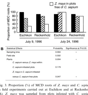
The e¡ect of C. sepium infestation on the propor-tion of MCD Z. mays roots was assessed in ¢eld experiments where adjacent plots free of C. sepium or infested with C. sepium were available. Whereas data obtained at Eschikon were similar to those from Reckenholz, sampling time had a strong in£uence on the proportions of MCD roots (Fig. 3). Apparently, calystegine-degrading microorganisms became fav-oured for colonisation of the rhizosphere of Z. mays by the second sampling, perhaps as a conse-quence of speci¢c climatic conditions or changes in exudation patterns of Z. mays roots linked to plant
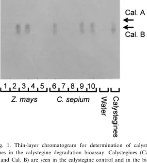
Fig. 1. Thin-layer chromatogram for determination of calyste-gines in the calystegine degradation bioassay. Calystecalyste-gines (Cal. A and Cal. B) are seen in the calystegine control and in the bio-assay carried out with root samples 2, 3 (Z. mays) and 6, 9, 10 (C. sepium). As control, the bioassay was done also without the addition of calystegines (`Water'). Root samples 1, 4, 5 (Z. mays) and 7, 8 (C. sepium) were scored as MCD as no calystegines were seen.
Fig. 2. Proportion of MCD Z. mays roots sampled at various lo-cations in the region of Zuërich, Switzerland. A plot free of C. sepium was chosen at random at each location and one root sample was assayed for each of 10 plants collected at random in each plot. Overall, the proportion of MCD Z. mays roots in the survey was 64%.
development between the two samplings. Since Z. mays is calystegine-negative, this e¡ect appears to be independent of the presence of calystegines in the rhizosphere. Overall, the proportion of MCD Z. mays roots was statistically higher in plots in-fested with the bindweed than in those without C. sepium (Fig. 3), obviously a result contributed by data from the ¢rst sampling. In C. sepium-infested plots, C. sepium and Z. mays displayed a similar proportion of MCD roots.
The presence of C. sepium plants in these plots implies that calystegines could be released into soil as part of root exudates and/or in decaying roots of C. sepium. The presence of calystegines in soil organ-ic matter may sustain the presence of calystegine-de-grading microorganisms at signi¢cant population levels in non-rhizosphere soil and contribute to their subsequent colonisation of the rhizosphere of a ca-lystegine-negative plant like Z. mays. Unfortunately, no information is available on the persistence of ca-lystegines released into soil after the death of roots. Other types of alkaloids have been shown to persist in soil and become adsorbed to soil particles [18]. The ability of bacteria to catabolise a speci¢c
com-pound present in root exudates, e.g. opine, rhizopine or £avonoids, is thought to improve their growth [19,20] or competitiveness in the rhizosphere [21^ 23]. Calystegines may have a wider signi¢cance by favouring calystegine-degrading microorganisms not only in the rhizosphere of calystegine-producing plants but perhaps also in bulk soil containing resid-ual calystegines.
3.5. Calystegine-degrading rhizosphere microorganisms are not seed-propagated
No root of C. sepium or Z. mays was MCD when autoclaved Eschikon soil was sown with seeds. Since the majority of roots of either plant species were MCD at the Eschikon ¢eld site (Fig. 3), this result indicates that the rhizosphere microorganisms re-sponsible for the ability of rhizosphere extracts to degrade calystegines do not belong to the microbiota naturally associated with the seed. Indeed, a signi¢-cant proportion of C. sepium samples were MCD when autoclaved Eschikon soil was planted with washed rhizomes of the plant (i.e. 25 þ 3%, 50 þ 3% and 35 þ 17% at 1, 7 and 21 days after planting, respectively; results pooled from three independent experiments). Furthermore, this indicates that calys-tegine-degrading microorganisms can be present in close association with the surface of the C. sepium rhizome.
3.6. High proportions of MCD roots of Z. mays and C. sepium grown in microcosms prepared with soil from ¢elds where calystegine-producing plants are present
In soil microcosms, a signi¢cant proportion of seedlings were already MCD 1 day after planting of axenic seedlings in Eschikon or Muërren soil, regard-less of whether C. sepium or Z. mays was studied (Fig. 1, Table 3). This suggests that calystegine-de-grading microorganisms were present at signi¢cant numbers in soil before the introduction of the plants, as suggested earlier for soils from Eschikon and Reckenholz (see Section 3.4). The presence of micro-organisms capable of calystegine degradation in C. sepium-free soil from Muërren (1600 m above sea lev-el) cannot be explained by earlier C. sepium infesta-tion since this plant species does not grow at this
Fig. 3. Proportion (%) of MCD roots of Z. mays and C. sepium in ¢eld experiments carried out at Eschikon and at Reckenholz (A). Z. mays was sampled from plots infested with C. sepium and from plots without bindweed. The statistical e¡ect of the ex-perimental factors were studied by Kruskal-Wallis and Mann-Whitney U statistics (B).
altitude in Switzerland. However, the Muërren soil has been cropped continuously with S. tuberosum, another calystegine-producing plant species [5].
In Muërren soil, the proportion of MCD roots re-mained similar for C. sepium and Z. mays at the other two samplings carried out (Table 3). In con-trast, the proportion of MCD Z. mays roots in Es-chikon soil decreased signi¢cantly by day 7 and did not change from days 7 to 21, whereas the propor-tion of MCD roots of C. sepium increased somewhat with time. Consequently, MCD roots were three times more frequent for C. sepium than for Z. mays at 21 days after sowing. Obviously, calyste-gine-degrading microorganisms were more competi-tive for colonisation of C. sepium than Z. mays in this soil. The reason why this did not take place in Muërren soil remains to be determined. Perhaps the amount of calystegines in a soil under continuous, intensive S. tuberosum cultivation (Muërren) was higher than that resulting from natural C. sepium infestation (Eschikon), because of the amount of ca-lystegine-containing crop residues produced in the case of potato cultivation. Consequently, in the rhi-zosphere, the calystegine status of the plants was more in£uential in Eschikon soil than in Muërren soil. 3.7. Signi¢cance of calystegines and calystegine
degradation capacity for soil microorganisms Microorganisms with the capacity to degrade
or-ganic substrates speci¢c for certain plant roots are considered to be favoured for colonisation of the rhizosphere of these plants [19^23], including C. sepium [8^10]. However, MCD roots of Z. mays, a calystegine-negative plant, were commonly found. Apparently, calystegine-degrading microorganisms were neither restricted to the rhizosphere of calyste-gine-producing plants nor impaired for colonisation of the rhizosphere of calystegine-negative plants. So far, only rhizobia and pseudomonads have been identi¢ed among rhizosphere isolates capable of ca-lystegine degradation [8,9]. Caca-lystegine degradation by bacteria other than S. meliloti Rm41 does not involve the cac genes, as in the latter strain [8,9], raising the possibility that the rhizosphere microbio-ta involved in calystegine degradation is highly di-verse in terms of both microbial taxonomy and ca-tabolic pathways.
The occurrence of MCD roots of Z. mays and C. sepium shortly (1 day) after introduction of axenic seedlings into natural soil suggests that calystegine-degrading microorganisms were present at signi¢cant population levels in soil, perhaps because of the pres-ence of calystegines in soil organic matter following the death of roots containing these alkaloids. If this were the case, it could provide an explanation for the higher proportion of MCD Z. mays roots in ¢eld plots infested with C. sepium compared with plots not infested. In this context, the ability of symbiotic bacteria such as S. meliloti Rm41 to catabolise
ca-Table 3
Proportion (%)a of roots of C. sepium and Z. mays with a microbial rhizosphere community capable of calystegine degradation (i.e.
MCD roots) in natural soil microcosms prepared with Eschikon soil (site infested with C. sepium) or Muërren soil (site cropped with S. tu-berosum)
Soil origin Plant speciesb Sampling time
Day 1 Day 7 Day 21 Eschikon C. sepium 33 þ 12 ac 47 þ 12 ab 58 þ 8 a
Z. mays 40 þ 0 a 13 þ 12 a 18 þ 17 b Muërren C. sepium 53 þ 12 a 60 þ 20 b 58 þ 22 a Z. mays 53 þ 42 a 67 þ 31 b 43 þ 12 ab Statistical e¡ects Soil origin NSd * NS
Plant species NS NS *
aMeans þ S.D. are shown.
bSeeds of C. sepium were scari¢ed with H
2SO4before surface disinfection. Seeds of Z. mays and C. sepium were surface-disinfected with 5%
NaOCl and 10% H2O2.
cAt each sampling time, the statistical relationships between the four treatments is shown with the letters a and b (P = 0.05). dNS, not signi¢cant; *, signi¢cant at P = 0.05.
lystegines would constitute not only an adaptation to alternative, non-symbiotic host plants but probably also a selective advantage for survival in soil in the absence of roots (i.e. in bulk soil).
Acknowledgments
We are indebted to D. Tepfer (INRA Versailles, France) for kindly providing calystegine standard, root culture of transformed C. sepium and strains Rm41 and Rm41-Ca1. We thank J. von Allmen for providing soil from Muërren, H.P. P¢rter for discus-sions and technical assistance, and U. Schenk and M. Hildman for technical assistance. This work was supported in part by the COST Action 816 (European cooperation in the ¢eld of scienti¢c and technical research).
References
[1] Tepfer, D., Goldmann, A., Pamboukdjian, N., Maille, M., Lepingle, A., Chevalier, D., Deènarieè, J. and Rosenberg, C. (1988) A plasmid of Rhizobium meliloti 41 encodes catabolism of two compounds from root exudate of Calystegia sepium. J. Bacteriol. 170, 1153^1161.
[2] Asano, N., Kato, A., Yokoyama, Y., Miyauchi, M., Yama-moto, M., Kizu, H. and Matsui, K. (1996) Calystegin N1, a
novel nortropane alkaloid with a bridgehead amino group from Hyoscyamus niger: structure determination and glycosi-dase inhibitory activities. Carbohydr. Res. 284, 169^178. [3] Asano, N., Oseki, K., Tomioka, E., Kizu, H. and Matsui, K.
(1994) N-containing sugars from Morus alba and their glyco-sidase inhibitory activities. Carbohydr. Res. 259, 243^255. [4] Asano, N., Kato, A., Oseki, K., Kizu, H. and Matsui, K.
(1995) Calystegines of Physalis alkekengi var. francheti (Sola-naceae): Structure determination and their glycosidase inhib-itory activities. Eur. J. Biochem. 229, 369^376.
[5] Nash, R.J., Rothschild, M., Porter, E.A., Watson, A.A., Waight, R.D. and Waterman, P.G. (1993) Calystegines in Sol-anum and Datura species and the death's-head hawk-moth (Acherontia atropus). Phytochemistry 34, 1281^1283. [6] Todd, F.G., Stermitz, F.R., Schultheis, P., Knight, A.P. and
Traub-Dargatz, J. (1995) Tropane alkaloids and toxicity of Convolvulus arvensis. Phytochemistry 39, 301^303.
[7] Molyneux, R.J., Pan, Y.T., Goldmann, A., Tepfer, D. and Elbein, D.A. (1993) Calystegines, a novel class of alkaloid glycosidase inhibitors. Arch. Biochem. Biophys. 304, 81^88. [8] Goldmann, A., Message, B., Tepfer, D., Molyneux, R.J.,
Du-clos, O., Boyer, F., Pan, Y.T. and Elbein, A. (1996) Biological activities of the nortropane alkaloid, calystegine B2and
ana-logs: structure-function relationships. J. Nat. Prod. 59, 1137^ 1142.
[9] Tepfer, D., Goldmann, A., Fleury, F., Maille, M., Message, B., Pamboukdjian, N., Boivin, C., Deènarieè, J., Rosenberg, C., Lallemand, J.Y., Descoins, C., Charpin, I. and Amarger, N. (1988) Calystegins, nutritional mediators in plant-microbe in-teractions. In: Molecular Genetics of Plant-Microbe Interac-tions (Palacios, R. and Verma, D.P.S., Eds.), pp. 139^144. APS Press, St. Paul, MN.
[10] Boivin, C., Malpica, C., Rosenberg, C., Goldmann, A., Fleury, V., Maille, M., Message, B., Pamboukdjian, N. and Tepfer, D. (1990) Catabolism of the plant secondary metabo-lites calystegins and trigonelline by Rhizobium meliloti. Sym-biosis 9, 147^154.
[11] Jung, G. and Tepfer, D. (1987) Use of genetic transformation by the Ri T-DNA of Agrobacterium rhizogenes to stimulate biomass and tropane alkaloid production in Atropa belladonna and Calystegia sepium roots grown in vitro. Plant Sci. 50, 145^ 151.
[12] Draëger, B. (1995) Identi¢cation and quanti¢cation of calyste-gines, polyhydroxyl nortropane alkaloids. Phytochem. Anal. 6, 31^37.
[13] Beringer, J.E. (1974) R-factor transfer in Rhizobium legumino-sarum. J. Gen. Microbiol. 84, 188^198.
[14] Maniatis, T., Fritsch, E.F. and Sambrook, J. (1982) Molecular Cloning: A Laboratory Manual. Cold Spring Harbor Labo-ratory, Cold Spring Harbor, NY.
[15] Deèfago, G., Berling, C.H., Henggeler, S., Hungerbuëhler, W., Kern, H., Schleppi, P., Stutz, E.W. and Zuërrer, M. (1987) Survie d'un Pseudomonas £uorescens dans le sol et protection du bleè contre des maladies d'origine fongique. Schweiz. Landw. Fo. 26, 155^160.
[16] Nievergelt, J. (1991) Die waëgbaren Lysimeter der Eidge-noëssischen Forschungsanstalt fuër landwirtschaftlichen P£an-zenbau Zuërich-Reckenholz. Landw. Schweiz 4, 535^536. [17] Oërdoëgh, F. and Szende, K. (1961) Temperate bacteriophages
isolated from Rhizobium meliloti. Acta Microbiol. Hung. 8, 65^71.
[18] Starr, R.I., Timm, R.W., Doxtader, G., Hurlbut, D.B., Volz, S.A. and Goodall, M. (1995) Sorption and aerobic biodegra-dation of strychnine alkaloid in various soil systems. J. Agric. Food Chem. 44, 1603^1608.
[19] Hartwig, U.A., Joseph, C.M. and Phillips, D.A. (1990) Fla-vonoids released naturally from alfalfa seeds enhance growth rate of Rhizobium meliloti. Plant Physiol. 95, 797^803. [20] Guyon, P., Petit, A., Tempeè, J. and Dessaux, Y. (1993)
Trans-formed plants producing opines speci¢cally promote growth of opine-degrading agrobacteria. Mol. Plant-Microbe Interact. 6, 92^98.
[21] Murphy, P.J., Wexler, W., Grzemski, W., Rao, J.P. and Gor-don, D. (1995) Rhizopines ^ their role in symbiosis and com-petition. Soil Biol. Biochem. 27, 525^529.
[22] Oger, P., Petit, A. and Dessaux, Y. (1997) Genetically engi-neered plants producing opines alter their biological environ-ment. Nature Biotechnol. 15, 369^372.
[23] Savka, M.A. and Farrand, S.K. (1997) Modi¢cation of rhizo-bacterial populations by engineering bacterium utilization of a novel plant-produced resource. Nature Biotechnol. 15, 363^ 368.