Hyperhomocysteinemia in premature
arterial disease: examination of
cystathionine p-synthase alleles at the
molecular level
Viktor Kozich
+, Eva Kraus, Raffaella de Franchis, Brian Fowler
1, Godfried H.J.Boers
2,
Ian Graham
3and Jan P.Kraus*
Departments of Pediatrics and Cellular and Structural Biology, University of Colorado School of Medicine, Denver, CO 80262, USA, department of Pediatrics, Basler Kinderspital, Basel, Switzerland, 2Department of Medicine, St Radboud Hospital, University of Nijmegen, Nijmegen, The Netherlands and 3Department of Epidemiology and Preventive Medicine, Royal College of Surgeons in Ireland, and Department of Medicine, Trinity College, Dublin, Ireland
Received January 13, 1995; Revised and Accepted January 18, 1995
Hyperhomocysteinemia occurs in approximately 30% of the patients with premature occlusive arterial disease (POAD). Some of these exhibit significantly reduced fibroblast cystathionine p-synthase (CBS) activities, suggesting that they may be heterozygous for CBS deficiency. To test this possibility, we studied cDNA derived from four well characterized patients with POAD, exhibiting hyperhomocysteinemia and reduced CBS activities, from four normal controls, and from four obligatory heterozygotes for CBS deficiency. Lysates of individual colonies of E.coli, containing full-length PCR-amplification products in the expression vector, pKK388.1, were tested for CBS activity. cDNA from at least seven of the eight possible independent POAD alleles encoded catalytically active, stable CBS which exhibited normal response to both PLP and AdoMet. The sequences of all 3-untranslated regions of all seven isolated POAD alleles were identical to the normal, 'wild-type' CBS sequences. The results of the expression studies were confirmed for one POAD patient by determining the full-length cDNA sequences for both alleles; these were entirely normal over the complete length of the cDNA. In contrast, the screening method correctly distinguished mutant from normal alleles in all four obligatory heterozygotes studied. We conclude that CBS mRNAs from POAD individuals are free from inactivating mutations, including all 33 previously identified in heterozygous carriers and homocystinuric patients.
INTRODUCTION
Mild hyperhomocysteinemia (HHC) is found in approximately one-third of cases of premature occlusive arterial disease (POAD) (1) whereas vascular complications such as stroke and thromboembolus are common among homocystinuric indi-viduals (2). As a consequence, elevated plasma levels of homocysteine are widely recognized as an independent risk factor for such vascular disorders (1). The biochemical and cellular participation of homocysteine in this condition is unclear, but may involve stimulation of clotting factors V (3) and XII (4) together with transcriptional induction of factor III (5) and inhibition of protein C conversion (6) as well as endothelial cell damage by peroxides (7).
Homocysteine is formed subsequently to transmethylation from AdoMet. Normally about half of this amino acid is resynthesized into methionine while the remainder is
trans-sulfurated to serine by cystathionine P-synthase (CBS), forming cysteine. While blocks in either pathway can lead to elevated plasma concentrations of homocysteine, CBS deficiency is acknowledged to be the most frequent cause of homocystinuria in humans (2). This enzyme is a homotetramer of 63 kDa, encoded by a gene located at human 21q22.3 (8). Heme and pyridoxal 5'-phosphate (9) are required for activity and the enzyme is regulated allosterically by AdoMet (10). Homocysti-nuria which results from CBS deficiency is inherited as an homozygous autosomal recessive trait, in which the enzyme activity is virtually extinguished (2). We have examined cDNAs from a number of homocystinuric patients and identified 33 distinct pathogenic mutations to date, using a bacterial CBS expression system which localizes the position of the mutation (11; unpublished).
*To whom correspondence should be addressed
Owing to the wide range of normal fibroblast CBS controls, it is difficult to differentiate between low normal and hetero-zygous-deficient enzyme activities; nonetheless, we were inter-ested in the potential link between heterozygous CBS deficiency and HHC/POAD. We therefore decided to employ molecular methods to differentiate between normal and mutant CBS alleles. These methods circumvent ambiguities originating from the normal/heterozygous activity overlap when assayed in fibroblast lysates.
To ascertain whether such a link existed, we examined cDNA preparations representing seven alleles which could be positively identified from four separate patients exhibiting HHC/POAD. These patients were selected from 30 candidates exhibiting POAD with accompanying HHC, exacerbated by methionine loading (12,13). Some of these also possessed persistent low levels of CBS in fibroblast cultures. A new screening method was developed in which allelic products were tested by expression of full-length cDNAs in bacteria rather than the segmental replacement method described earlier (14). Identification of synonymous mutations facilitated identi-fication of the separate alleles. Lastly, we sequenced the 3'-untranslated regions. We now report that all of the allelic products were free from pathogenic mutations and that the cDNA preparations expressed normal amounts of CBS in the bacterial expression system. It appears highly unlikely that pathogenic mutations in the coding region or the 3'-untranslated region are responsible for the HHC observed in these patients.
RESULTS
Subjects studied
Relevant biochemical and clinical findings for the patients studied in this report are listed in Table 1. Arteriosclerosis was found in all four of the HHC/POAD patients. Peak plasma homocysteine levels following a methionine load (12) were well above normal for each of the individuals, approximating the values observed for obligate heterozygotes. Fibroblast cultures from three of the four exhibited CBS activities below the normal range.
Screening strategy
We previously developed a bacterial expression method to localize pathogenic mutations in cDNA segments from homo-cystinuric patients (14). The aim of this study was to test for the presence of any inactivating mutations rather than to
localize them. Therefore, we expressed the entire coding region of each allele from each patient. The screening strategy is summarized in Figure 1.
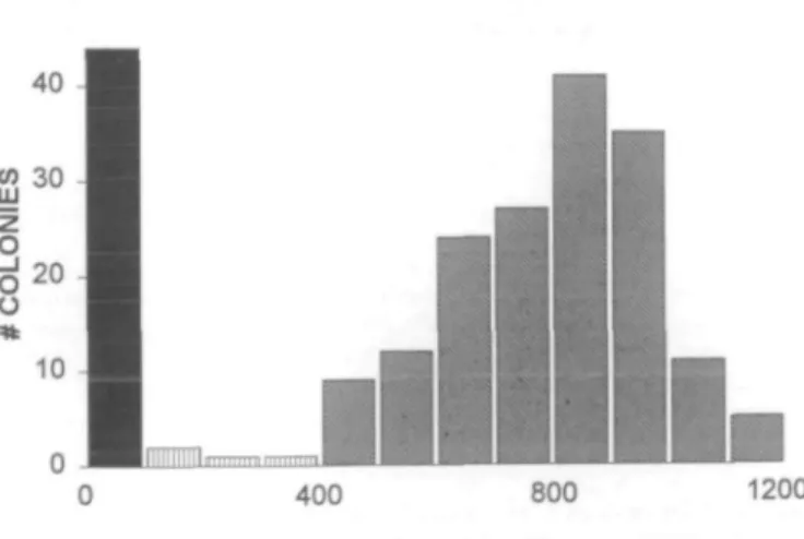
To date, 33 pathogenic mutations have been identified and expressed in E.coli. Only one of these produces appreciable CBS activity after expression, approaching 50% of the control values. The overwhelming majority, however, completely inac-tivate the enzyme. Figure 2 illustrates the wide separation in CBS activity produced by normal and mutant alleles. Coupled with Western analysis of the bacterial proteins, the method is highly sensitive; it has never failed to localize mutations in CBS from known homozygotes.
Screening for inactive CBS alleles among patients with POAD/HHC
The infidelity of 7ag-catalyzed polymerase chain reaction arises principally from the absence of proofreading in Taq DNA polymerase. Pyrococcus DNA polymerase proofreads its product; accordingly, its substitution for Taq DNA polymerase significantly improved PCR fidelity. Whereas PCR of control cDNAs with Psp polymerase produced only seven inactive clones out of 78 (9%, Fig. 3), similar experiments with Taq polymerase yielded five inactive clones out of 14. This 36% error rate was too high to allow us to use the Taq enzyme for full-length cDNA amplification, accordingly, Psp DNA polymerase was used for all experiments.
To ascertain the suitability of this method as a screen for heterozygosity, we first tested cDNA derived from known heterozygotes (Fig. 3). The frequency of inactive clones from four obligate heterozygotes (30/70; 43%) was significantly greater than for the controls (7/78; 9%), thus the method sensitively distinguishes between these groups. In contrast, the frequency of inactive CBS colonies (7/63; 11 %) from the four HHC/POAD patients was not significantly different from the controls. These functional data suggest that it is unlikely that the low CBS activities in this' group of POAD/HHC patients result from mutations in the coding region of the CBS gene, assuming that both parental alleles were equally represented in the clones which had been analyzed.
Segregation of CBS activity with allelic polymorphisms
To eliminate the possibility that only one allelic product had been tested in the expression studies, we examined the cloned cDNAs for synonymous mutations in the coding regions (15). These markers permitted us to identify both parental alleles in
Table 1. Subjects reported in this study
Subject 3014 3022 3024 3030 Obligate heterozygotes (u = Controls (n = 30) Sex F/P F/P F/P F 10)
Fasting HCY (uM)
8.9 5.7 5.3 9.1 4.2±2.8 0.0+0.9
Peak HCY (uM)
54.3 66.8 37.8 51.0 41.0±24.6 6.8±1.3 Site of arteriosclerosis
Internal carotid and anterior cerebral arteries
Renal, mesenteric and iliac arteries
Abdominal aorta and femoral arteries
Cerebral vessels
Severity of disease
Transient ischaemic attacks
Severe reno-vascular hypertension Severe dysbasia Unknown CBS U/mg 3.5 17 3.2 5.9 0.1-1 7-30
Homocysteine (HCY) levels before (fasting) and after (peak) methionine challenge (12) are listed for each of the experimental subjects as well as normal controls and obligate heterozygotes for CBS deficiency (mean ± 1 standard deviation). F/P: Premenopausal female patients. CBS activities for controls and obligate heterozygotes represent the ranges found in cultured fibroblasts by this laboratory. The majority of controls range from 20-25 U/mg protein.
individual clones exhibiting normal CBS activity. To carry out this test, we analyzed clones derived from obligate hetero-zygotes as well as from the POAD/HHC patients who were informative for these polymorphisms. The results of these experiments are summarized in Table 2.
We first tested clones from two obligate heterozygotes for silent T/C substitutions at position 699. The triplets encoded at that position (TAC/TAT) both encode tyrosine, therefore the protein sequence is unaffected. Of the 12 allelic clones tested for one patient (3056) all eight active ones exhibited 'T' alleles (i.e. T699), whereas all four inactive clones contained ' C alleles (C699). These results demonstrate that these polymorph-isms segregated with active and inactive CBS alleles for this individual. Similarly, all five active clones from 3065 exhibited ' C alleles (C699) while all five inactive clones were 'T' alleles. These results demonstrate that the PCR-based RFLP analytical method used (15) clearly identifies the segregated parental alleles.
Both parental alleles were identified among active CBS clones derived from three patients with POAD/HHC (Table 2). The T/C (699) polymorphism was found in allelic products from patients 3022 and 3030 whereas another synonymous
CDNA Clo I Kpn I Cla I J . RESTRICTION (Clal, Kpnl) C l a ' KPn '
o
T4 DNA LIGASE HUMAN CBS INSERT ATG EXPRESSION CONSTRUCT pKK 388.1 CARTRIDGEI
INDIVIDUAL E.COLI CLONES
mutation, C1080T (GCC/GCT; Ala 360), was employed to distinguish between the allelic products from patient 3014. The fourth patient, 3024, did not exhibit these polymorphisms, thus it is not certain that both parental alleles were represented in the tested clones. The presence of both these alleles in active clones unequivocally demonstrates that both parental alleles encode enzyme which is catalytically functional, there-fore neither parental CBS allele encodes protein with patho-genic mutations in these patients. These results clearly differentiate between the obligate heterozygotes whose active clones are all of the same haplotype and the POAD patients whose active clones were not.
In addition, less than 15% difference was observed in CBS activity in C and T alleles from each informative patient. The mildest pathogenic mutation studied to date, A114V (unpublished), decreases the activity of CBS by at least half when the allele is expressed in E.coli. The statistically insignificant differences between parental alleles (C and T) of each vascular patient suggest that it is quite unlikely that these individuals are carriers for any known mutation, even one so mild as A114V.
DNA sequencing
Primer portion of the cloned cDNAs
The method described in this paper effectively tests the majority of the CBS coding region, however, the sequences of the primer portions of the cDNA escape definitive scrutiny. Accordingly, we sequenced these regions to ascertain whether the abnormally low fibroblast levels of enzyme activity stemmed from cryptic mutations in the 5'- or 3'-ends of the coding sequence. These regions were first amplified by PCR.
400 800 1200
CBS EXPRESSION PLASMID DNA ANALYSES CBS ASSAY RESTRICTION ANALYSIS
WESTERN BLOT SYNONYMOUS MUTATION SCREEN DNA SEQUENCING
Figure 1. Expression system in E.coli. Patient cDNA was amplified by PCR using the primers (boxes) described in the text which contained artificial Clal and Kpnl sites. The PCR products were restricted with Clal and Kpnl, purified and ligated into pKK 388.1 cartridge. Resulting constructs contained cDNA sequence which was derived from tested individuals except of the small portion incorporated from the primers (shadowed portions). The constructs were used for E.coli transformation and individual clones were isolated. CBS was expressed after induction with IPTG, plasmid DNA of the clones was used for restriction analysis, sequencing or synonymous mutation screening.
CBS ACTIVITY (U/ml)
Figure 2. Distribution of CBS activity in separated normal and mutant alleles. A total of 211 colonies from normal, obligate heterozygotes and POAD/HHC patients were assayed for CBS. Enzyme activity (U/ml lysate) is depicted on the abscissa of the histogram; the number of colonies found in each 100 U/ ml division is indicated on the ordinate. Individual colonies were scored as inactive within the 0-100 U/ml division (black). Active colonies exhibited CBS values greater than 400 U/ml (slanted stripes). Four colonies exhibited CBS activities between 100 and 400 U/ml (vertical stripes) and were not scored. The mean activity for the 44 inactive clones, originating predominantly from the obligate heterozygotes, was IO.8±3.3 (standard error of the mean); for the 163 normal clones, 807 ±13.
In the 5'-portion, between six and eight clones derived from each patient were chosen for sequencing to limit the probability that only one parental allele was tested. The 5'-ends were sequenced from nts —1 to 40, covering the entire region occupied by the sense primer. All of the clones in all four patients possessed normal cDNA sequence in this region.
Sequencing of the 3' untranslated region
We next searched for mutations in the 3'-sequence of the cDNA using a similar strategy. To ensure that both parental alleles were represented in the clones, we tested the individual colonies for a frequent Msp I polymorphism (T2199C) in the 3'-untranslated region. Three of the patients were polymorphic, thus only two clones (each representing one allele) were chosen for DNA sequencing. The sequence of the plasmids harboring these cDNAs was indistinguishable from normal in the region homologous to the antisense primer as well as in the 3'-untranslated region. Four plasmids originating from patient 3024, who did not exhibit the Mspl polymorphism, were also sequenced through the antisense primer region further into the 3'-untranslated region and were also fully
FRACTION OF INACTIVE CLONES pHCS3 2047 2103 3077 3014 3022 3024 3030 367 3056 3065 3009 INACTIVE CLONES
•I
•
•
•
J
_Ji
ACTIVE CLONES % CONTROLS 7/78 9% HHC/POAD 7/63 1 1 % HETERO-ZYGOTES 30/70 43%Figure 3. Distribution of active and inactive E.coli clones from controls,
POAD patients, and CBS heterozygotes. Individual E.coli colonies were assayed for CBS activity and scored according to the criteria described in the legend of Figure 2. The average number of colonies examined for each patient was 18 while the range was 13-25. Each cell line is listed on the left. The percentage of positive clones is indicated by the length of the striped bar; negative clones by the black bar. The right side of the figure indicates the fractions and percentages of negative clones for each group. CBS cDNAs containing pathogenic mutations in 32/33 alleles from homocystinurics produce inactive clones.
congruent with the normal allele. These results demonstrate the absence of cryptic mutations in both the 5'- and 3'-coding regions and 3' untranslated region of CBS cDNA.
DNA sequencing of full length cDNA in patient 3014
To search for other DNA mutations which might be undetect-able in our screening system, we sequenced the entire coding region of CBS cDNA from both alleles of one patient (3014). The Ala36O synonymous mutation in the coding region was used to identify cDNA clones originating from separate alleles. The sequence of both alleles were completely normal over their entire coding regions.
Activation of patient CBS by S-adenosylmethionine
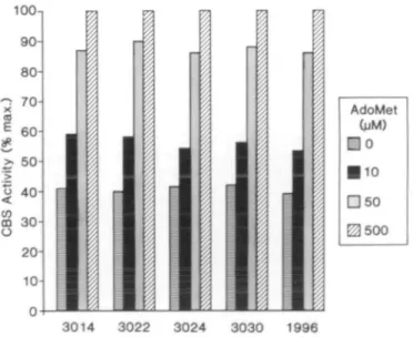
Incubation of 'wild-type' CBS with AdoMet stimulates the enzyme's activity by increasing its affinity for homocysteine (10,14). Subtle mutations in the AdoMet binding region, displacing the effector from its binding site, might reduce the enzyme activity to the levels observed in the fibroblasts from the four patients studied. These mutations would be accompanied by a lack of response of the enzyme to AdoMet. We measured CBS activity in fibroblast lysates, and in bacterial extracts, incubated with added AdoMet varying from 0-500 LtM and calculated the activation constant, Kacl, for AdoMet to determine whether such mutations might exist. Figure 4 depicts the results of the fibroblast study. Although the basal activities differed considerably, CBS in each patient cell extract responded to AdoMet similarly. Both the 15 LiM AdoMet activation constant and the 2.5-fold increase in enzyme activity were identical for the patients and controls. These results indicate that the AdoMet binding domains for CBS in all patient lines are intact and that the endogenous AdoMet concentration in the cell cultures are similar in each cell line. CBS, expressed in E.coli, behaved similarly when incubated with varying concentrations of AdoMet (data not shown). These results demonstrate unequivocally that both alleles of each patient encode CBS with identical behavior towards AdoMet; accordingly, the wide variation in the CBS activities of these patients cannot be ascribed to defective AdoMet binding.
Analysis of cloned CBS by Western blotting
Since all of the CBS clones were free from pathogenic mutations, we suspected that the low intracellular CBS activit-ies might be attributed to decreased amounts of normal enzyme subunits. The volumes of fibroblast extracts were adjusted so that each sample contained equal amounts of enzyme activity.
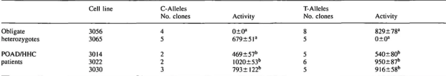
Table 2. Distribution of CBS activity between parental alleles
Cell line C-Alleles
No. clones Activity
T-Alleles
No. clones Activity
Obligate heterozygotes POAD/HHC patients 3056 3065 3014 3022 3030 010s 679±5la 469±57b 1020±53b 793±122b 829±78a 0±0a 540±80b 95O±87b 916±58b
Full-length CBS cDNAs were prepared from fibroblast mRNAs for each cell line listed in the table, cloned into pKK388.1, resolved and expressed in transformed
E.coli as described in Materials and Methods. Separate parental alleles (C-alleles or T-alleles) were identified by the presence of common synonymous mutations:
CI080T (Ala360), 3014; C699T(Tyr233), all others. No synonymous mutations were found for 3024. The activity columns refer to mean CBS activity ± I standard deviation of the mean for the number of clones indicated in the columns to the left. *C and T alleles which differ significantly by Student's f-test (p<0.001). bC and T alleles not significantly different (p>0.2).
Immunoprecipitates of these samples were analyzed by Western blotting to estimate the actual amount of enzyme in each. Comparable band intensities resulted from identical amounts of enzyme activity even though 20-fold more total cellular protein was required for the defective cell lines (Fig. 5, upper panel). We conclude that the specific activity of the pure enzyme molecules is constant in each fibroblast line and that the low CBS activity found in these lines results from decreased amounts of enzyme molecules rather than impaired catalytic activity. These conclusions were further supported by the following bacterial studies.
All seven separated alleles were examined by Western blotting of the E.coli lysates. Whereas the quantity of crude fibroblast protein required to produce a constant (2 U) amount of CBS was quite variable, bacterial lysates were far more consistent, even when the colonies expressed separate CBS alleles (Fig. 5, lower panel). These results demonstrate that the specific activity of the cloned CBS subunits does not vary in clones derived from separated parental alleles. Additionally, they reveal that the enzyme proteins encoded by each allele are equally stable in E.coli, as would be expected if each allele encoded an identical product.
DISCUSSION
The purpose of this research was to ascertain whether the hyperhomocysteinemia observed in the four patients studied originated from mutations in the CBS coding region. At least seven of the eight possible alleles were examined and did not contain mutations. We therefore conclude that the HHC observed in these four individuals does not stem from defective
CBS protein. CBS binds three cofactors in addition to its two substrates: PLP (16), AdoMet (10,14), and heme (9). All of these affect CBS activity. PLP is an active participant in the reaction (16), AdoMet stimulates the enzyme allosterically (10,14), while heme is required for both PLP binding and enzyme activity (9). Fibroblast cultures from three of the four patients exhibited low enzyme activities upon repeated assay. It is unlikely that these resulted from reduced PLP affinity since the enzyme responded normally when assayed in both the presence and absence of this cofactor (data not shown); additionally, altered PLP affinity is associated with mutations in the coding region (17) and none were found. AdoMet sensitivity, likewise, was unaffected for all of the preparations (Fig. 4). CBS specific activity depends upon its saturation with heme (9). This porphyrin binds tightly to CBS and is evidently inserted either concurrently with or shortly after synthesis. Addition of heme to enzyme incubations is without effect on either crude or pure preparations of the enzyme (9) so it is not possible to test directly the affinity of CBS for heme. Western analyses of the proteins from fibroblasts, however, demonstrated that the amount of CBS protein closely correlated to the levels of CBS activity (Fig. 5) so it is likely that the heme saturation is equal for the normal and HHC/POAD samples. These results imply that the reduced amounts of CBS in the fibroblast samples resulted from altered enzyme expression rather than a mutation in its structure.
The clear separation of activity between normal and mutant alleles in this screening method (Fig. 2) greatly simplifies identification of bacterial colonies harboring plasmids con-taining mutant CBS cDNAs. The substitution of Psp DNA polymerase, which proofreads its products, for Taq, which does not, significantly improved the sensitivity of the method. Fidelity of the screening method depends directly upon fidelity
Fibroblasts
•«• CM Cell line: £ g CO CO o ug protein, o CO o o CO§
CO CO O CO o o CO • t CO8
a Cell line: ~ co .o * « • CM r - CM o o CO COE.coli
. O O CM t CM CM O O CO CO CM O CO a O CO o co a o CO o CO 3014 3022 3024 3030 1996 CO 01 CO CO (0 ^ °? « u g p r o t e i n : t - e \ i « c < ) ' " » | O | oFigure 4. Activation of CBS in fibroblast extracts by AdoMet. Fibroblast
extracts were assayed for CBS activity in the presence of AdoMet (exogenous concentrations were 0, 10, 50 and 500 (iM). Numbers on the bottom signify the cell line examined, 1996 is a control cell line. The individually shadowed columns represent the fraction of maximum CBS activity (maximum was at 500 U.M AdoMet concentration). The maximum CBS activities obtained for each cell line were 8.6 U/mg total cellular protein for 3014; 24 for 3022; 5.4 for 3024; 19.4 for 3030; and 72.8 for 1996.
Figure 5. Western analyses. The upper panel shows 2 units of CBS activity
immunoisolated from fibroblast extracts and subjected to Western analysis. Numbers at the bottom of this panel represent the amounts of total cellular protein used to immunoisolate the identical CBS activities. The lower panel compares intensity of bands originating from each cloned parental allele (signified as letters a and b next to the cell line number) when 2 units of CBS were immunoisolated, run on SDS/PAGE and blotted. Total bacterial protein subjected to the analysis is indicated at the bottom.
of the polymerase. This was tested by PCR of normal CBS cDNA sequences. Using Taq polymerase, less than 2/3 of the PCR products encoded active CBS when a normal plasmid template was amplified. More than 90% of the Psp polymerase control products were active.
All of the known mutations associated with homocystinuria impact considerably on CBS activity expressed in transformed bacteria (11; unpublished). There was no significant difference in the frequency of inactive clones between the HHC/POAD patient and the normal control groups; although considerable variation was observed between individuals in the obligate heterozygote group. In addition, screening for synonymous mutations allowed us to show that individual parental CBS alleles were tested for pathogenic mutations in three of the patients. All of the coding regions of these sequences were free from pathogenic mutations. This was not the case for the obligate heterozygotes which were unequivocally differentiated from the controls. We subsequently separated the allelic products from the obligate heterozygotes and identified the mutations in the abnormal strand (14,21; unpublished). This method permits unequivocal identification of heterozygous carriers for homocystinuria for the first time.
Since at least three of these patients are homozygous for normal CBS, their hyperhomocysteinemias evidently stem from other factors. Homocysteine, once formed, can either serve as a substrate for transsulfuration or be resynthesized into methionine (2). Although the most frequent cause of homocystinuria is CBS deficiency (2), the condition may arise from inadequate methionine resynthesis as well (18,19). Defects in cobalamin metabolism (19) or methylenetetra-hydrofolate reductase insufficiency (18) could precipitate hyperhomocysteinemia in these subjects. Other defects could include vitamin malnutrition with or without attenuated heme biosynthesis, or concurrent low expression of key enzymes such as methylenetetrahydrofolate reductase and CBS. The rate of methionine resynthesis is currently under investigation to evaluate its contribution to these patients' condition.
Lastly, it is important to note that there is a strong overlap of the normal and heterozygote CBS activity ranges in cultured fibroblasts (20). Three of the patients studied were below the normal range while the fourth patient was well within it. Our initial intent was to evaluate whether they were heterozygous for CBS deficiency. With this publication, we demonstrate that they were all homozygous for normal CBS. In many cases, fibroblast cultures have been in passage for some time. In others, the phase of the culture may not be reliably determined. Each of these conditions can affect the level of enzyme assays, including those for CBS. Considerable variability in CBS activity may occur within cultures of apparently healthy fibroblasts as well. These considerations together with our results indicate that fibroblast CBS activities alone cannot be used to assess the CBS zygosity of a patient, even when accompanied by hyperhomocysteinemia; accordingly, the dia-gnosis of individuals suspected of being carriers of homocystin-uria must be based on analysis of their nucleic acid sequences.
MATERIALS AND METHODS
Patients and cell lines
Cell lines 3014, 3022, 3024, and 3030 from POAD/HHC patients used in these studies originated from two clinical and biochemical studies described
elsewhere (12,13). Cell lines 2047, 2103 and 3077 were from controls and 367, 3009, 3056 and 3065 were from mothers of homocystinuric patients. The mutations in these obligate heterozygotes were 1278T, E239K, G307S, and R336C, respectively (14,21; unpublished). Fibroblasts were cultured in MEM with 10% fetal calf serum, nonessential amino acids, glutamine, 100 U/ml of kanamycin and 100 Hg/ml of streptomycin.
PCR amplification of cDNA
DNA was amplified using Pyrococcus sp. GB-D DNA polymerase (Psp, Deep Vent Polymerase, New England Biolabs, Beverly, MA) or with Taq polymerase as noted in the results section. Complementary DNA was synthesized from mRNA as described previously (14). Diluted target strands were completely denatured prior to PCR by heating to 95°C for 3 min and rapidly chilling in an ice/water bath.
Each 100 nl reaction mixture for PCR with the Deep Vent enzyme contained 1 nl of cDNA, 1 X Deep Vent buffer (NEB), 300 \iM of each dNTPs, 80 pmol of both sense and antisense primers (described above), 4% dimethylsulfoxide and 8 mM MgSO4. Conditions for 7a^-mediated PCR were: 2.5 Hi cDNA,
10 mM TRIS.HC1 (pH 8.4), 50 mM KCI, 0.01% gelatin, 200 HM of each dNTPs, 1.5 mM MgCl2 and 80 pmol of both the sense and antisense primers
contained in a total of 100 |il. Primer sequences for both sets of PCRs were:
Sense primer
5'-TACGATCGATAAGGAGGTTTAAACCATGGjGJTCTG-AGACCCCCCAG-3'. The bold portion of the sense primer hybridized to the 5' coding region of CBS cDNA. The underlined codon following the initiator ATG was changed from proline to glycine to complete a Nco I site; this change coincidentally increased the activity of CBS. The nonhybridizing portion of the primer contained a prokaryotic ribosome binding site and a Cla I site; Antisense primer 5-TACGGGTACCAGCGCTCCGGACTTCACTT-CTGG-3'. The bold portion was complementary to the 3' region of the cDNA whereas the remainder contained an artificial Kpn I site.
PCR was conducted in a Techne PHC-1 thermocycler with block temperature control when the Deep Vent enzyme was used. The reaction mixture was preheated to 80°C/5 min and I U of Psp polymerase was added to each tube. The temperature was cycled 30 times (96°C/1 min; 6I°C/I min; 72°C/2.3 min) to amplify target DNA. The Techne thermocycler produced uniform results with this enzyme whereas the reactions did not proceed well with the Hybaid TR1, used for 7a<7-amplified PCR.
The Hybaid TR1 thermal reactor with tube temperature monitoring was employed for Taq DNA polymerase PCR. Enzyme was added after preheating to 80°C/5 min, then the mixture was cycled as follows: 1 cycle (94°C/3 min, 50°C/l min, 72°C/3 min), 4 cycles (94°C/1.5 min, 50°C/l min, 72°C/3 min), 24 cycles (94°C/1.5 min, 55°C/1 min, 72°C/2 min) and final cycle (94°C/1.5 min, 55°C/1 min, 72°C/4 min).
PCR products were purified, restricted with Cla I and Kpn I, gel purified, andligated in gel into similarly prepared pKK388.1 (14). Individual, ampicillin-resistant colonies of transformed DH5aFI'Q E.coli were subsequently used to express CBS or prepare plasmid DNA.
CBS expression in E.coli
Enzyme activity was measured in 20 (ll lysates of transformed cells grown from individual colonies after incubation with IPTG (isopropylthio-p"-D-galactoside, GIBCO BRL, Gaithersburg, MD) as fully described elsewhere (14). One unit of CBS activity was defined as 1 nmol of cystathionine produced in 1 h at 37°C. This radiometric assay can reliably detect enzyme activity as low as 0.03 U (100 CPM above background).
Analysis of synonymous mutations
Three synonymous mutations in the coding region of CBS cDNA and one polymorphism in the 3' untranslated region (G573A; C699T; TI080C; T2199C) were analyzed by a PCR-based RFLP method described elsewhere (15).
cDNA sequencing
A 5'- portion of CBS cDNA in all four PAD/HHC patients was amplified by PCR between nucleotides —2 and 272 using the sense primer 5'-CGTAGAATTCCAGCACCATCTGTCCGGTCCCA-3', together with the antisense primer 5'-TACGATCGATTCAGG-CCGAACTTCTTCCCAAT-3'. Reaction conditions were identical to those described for Psp PCR except that 4 mM MgSO4 was used and 30 cycles of amplification (96°C, I min;
55°C, 1 min; 75°C, 30 s) were carried out. PCR products were restricted with Cla I/Eco RI, subcloned into a pBlueScript KS(+) cartridge and the resulting plasmid preparations used for sequencing. A 3'-portion of CBS cDNA of all four patients between nucleotides 1462 and 2316 was similarly amplified with
Psp polymerase using the sense primer P5,
5'-CGTAGAATTCAAAGTCAT-CTACAAGCAGTTC-3', and the antisense primer P6, 5'-TACGATCG-ATGTGTGATTCCATTTATATG-AAATGTCC-3'. Thermocycling conditions were: 30 cycles of 96°C, 1 min; 64°C, 1 min and 72°C, 54 s. The entire CBS cDNA coding regions between nucleotides 22 and 1646 from both alleles of patient 3014 were cloned and sequenced.
Other methods
Magic MiniPrep kits (Promega, Madison, WI) were used to prepare plasmid DNA. Restriction endonucleases and T4 ligase were purchased from NEB (Beverly, MA). DNA was sequenced by the dideoxy method (23) using the CircumVent kit (NEB). Competent cells (DH5aF'IQ) were obtained from Gibco BRL.
ACKNOWLEDGMENTS
We are indebted to Ms. Rebecca B.Kucera from New England Biolabs for her help with optimizing PCR conditions using Psp polymerase, Ms. Harriet McKelvey for tissue culture work and Dr Michael D.Roper for the critical reading of the manuscript.
REFERENCES
1. Kang, S.S., Wong, P.W.K. and Malinow, M.R. (1992) Hyperhomocyst(e)inemia as a risk factor for occlusive vascular disease.
Annu. Rev. Nutr. 12, 279-288.
2. Mudd, S.H., Levy, H.L. and Skovby, F. (1989) Disorders of transsulfuration. In The Metabolic Basis Of Inherited Disease. Scriver, C.R., Beaudet, A.L, Sly, W.S. and D. Valle (eds), 6th edn. New York, Mcgraw-Hill, 693-734.
3. Rodgers, G.M. and Kane, W.H. (1986) Activation of endogenous factor V by a homocysteine-induced vascular endothelial cell activator. J. Clin.
Invest. 77, 1909-1916.
4. Ratnoff, O.D. (1968) Activation of Hageman factor by L-homocystine.
Science 162, 1007-1009.
5. Fryer, R.H., Wilson, B.D., Gubler, D.B., Fitzgerald, L.A. and Rodgers, G.M. (1993) Homocysteine, a risk factor for premature vascular disease and thrombosis, induces tissue factor activity in endothelial cells.
Arterioscler. Thromb. 13, 1327-1333.
6. Lentz, S.R. and Sadler, J.E. (1991) Inhibition of thrombomodulin surface expression and protein C activation by the thrombogenic agent homocysteine. J. Clin. Invest. 88, 1906-1914.
7. Starkebaum, G. and Harlan, J.M. (1986) Endothelial cell injury due to copper-catalyzed hydrogen peroxide generation from homocysteine.
J. Clin. Invest. 11, 1370-1376.
8. Kraus, J.P., Williamson, C.L., Firgaira, F.A., Yang-Feng, T.L., Miinke, M., Francke, U. and Rosenberg, L.E. (1986) Cloning and screening with nanogram amounts of immunopurified mRNAs: cDNA cloning and chromosomal mapping of cystathionine P-synthase and the p subunit of propionyl CoA carboxylase. Proc. Natt Acad. Sci. USA 83, 2047-2051. 9. Kery, V., Bukovska, G. and Kraus, J.P. (1994) Transsulfuration depends
on heme in addition to pyridoxal 5'-phosphate. Cystathionine P-synthase is a heme protein. J. Biol. Chem. 269, 25 283-25 288.
10. Roper, M.D. and Kraus, J. (1992) Rat cystathionine P-synthase: Expression of four alternatively spliced isoforms in transfected cultured cells. Arch.
Biochem. Biophys. 298, 514-521.
11. Kraus, J.P. (1994) Molecular basis of phenotype expression in homocystinuria. J. Inher. Metab. Dis. 17, 383-390.
12. Boers, G.H.J., Smals, A.G.H., Trijbels, F.J.M., Fowler, B., Bakkeren, J.A., Schoonderwaldt, H.C., Kleijer, WJ. and Kloppenborg, P.W. (1985) Heterozygosity for homocystinuria in premature peripheral and cerebral occlusive arterial disease. New Engl. J. Med. 313, 709-715.
13. Clarke, R., Daly, L., Robinson, K., Naughten, E., Cahalane, S. Fowler, B. and Graham, I. (1991) Hyperhomocysteinemia: an independent risk factor for vascular disease. New Engl. J. Med. 324, 1149-1155.
14. Kozich, V. and Kraus, J.P. (1992) Screening for mutations by expressing patient cDNA segments in E.coli: Homocystinuria due to cystathionine P-synthase deficiency. Hum. Mut. 1, 113-123.
15. Kraus, J.P., Le, K., Swaroop, M., Ohura, T., Tahara, T, Rosenberg, L.E., Roper, M.D. and Kozich, V. (1993) Human cystathionine P-synthase cDNA: Sequence, alternative splicing and expression in cultured cells.
Hum. Mol. Genet. 2, 1633-1638.
16. Kraus, J.P. and Rosenberg, L.E. (1983) Cystathionine P-synthase from human liver: Improved purification scheme and additional characterization of the enzyme in crude and pure form. Arch. Biochem. Biophys. 222, 44-52.
17. Lipson, M.H., Kraus, J. and Rosenberg, L.E. (1980) Affinity of cystathionine P-synthase for pyridoxal 5'-phosphate in cultured cells. A mechanism for pyridoxine-responsive homocystinuria. J. Clin. Invest. 66, 188-193.
18. Rosenblatt, D.S. (1989) Inherited disorders of folate transport and metabolism. In The Metabolic Basis Of Inherited Disease. Scriver, C.R., Beaudet, A.L., Sly, W.S. and Valle, D. (eds), 6th edn. New York, Mcgraw-Hill, 2049-2064.
19. Fenton, W.A. and Rosenberg, L.E. (1989) Inherited disorders of cobalamine transport and metabolism. In The Metabolic Basis Of Inherited
Disease. Scriver, C.R., Beaudet, A.L., Sly, W.S. and Valle, D. (eds), 6th
edn. New York, Mcgraw-Hill, 2065-2082.
20. McGill, J.J., Mettler, G., Rosenblatt, D.S. and Scriver, C.R. (1990) Detection of heterozygotes for recessive alleles. Homocyst(e)inemia: paradigm of pitfalls in phenotypes. Am. J. Med. Genet. 36, 45-52. 21. de Franchis, R., Kozich, V., Mclnnes, R.R. and Kraus, J.P. (1994)
Identical genotypes in siblings with different homocystinuric phenotypes: Identification of three mutations in cystathionine P-synthase using an improved bacterial expression system. Hum. Mol. Genet. 3, 1103-1108. 22. Ausubel, F.M., Brent, R., Kingston, R.E., Moore, D.D., Seidman, J.G.,
SmithJ.A. and Struhl, K. (1989) Short Protocols In Molecular Biology. New York, J Wiley & Sons.
23. Sanger, F., Nicklen, S. and Coulson, A.R. (1977) DNA sequencing with chain-terminating inhibitors. Proc. Nad Acad. Sci. USA 74, 5463-5467.