Double-crowned valved stents for off-pump mitral valve replacement
*Liang Ma
a, Piergiorgio Tozzi
b,*, Christoph H. Huber
b, Steven Taub
b,
Gabrielle Gerelle
b, Ludwig K. von Segesser
baCardiothoracic Department, The 1st Affiliated Hospital of Medical College, Zhejiang University, Hangzhou, China bDepartment of Cardiovascular Surgery, Centre Hospitalier Universitaire Vaudois (CHUV), Lausanne, Switzerland
Received 7 October 2004; received in revised form 11 December 2004; accepted 17 December 2004; Available online 13 June 2005
Abstract
Objective: An animal model has been designed to assess the feasibility of off-pump mitral valve replacement using valved stents. Methods: Glutaraldehyde-preserved homograft was sutured inside a prosthetic tube (Dacron). Then, two self-expandable nitinol Z-stents were sutured on the external surface of the prosthesis in such a way to create two self-expanding crowns for fixation. In adult pigs and under general anesthesia, the left atrium was exposed through a left thoracotomy and atrio-ventricular roadmapping was performed with intravascular ultrasound (IVUS) and fluoroscopy. The double-crowned valved stents were loaded into a delivery sheath. The sheath was then introduced into the left atrium and the valved stents was deployed in mitral position in such a way that the part in between the two stents was at the level of the mitral annulus. Intracardiac Unltrasound (ICUS) was used to assess the valve function. Hemodynamic parameters were gathered as well. Animal survived for no more than 3 h after the valve deployment and gross anatomy examination of the left heart was carried out. Results: The mean height of the valved stents was 29.4G0.2 mm, with an internal diameter of 20.4G1.0 mm, and an external diameter of 25.5G0.8 mm. The procedure was successfully carried out in eight animals. In vivo evaluation showed a native mitral annulus diameter of 24.9G0.6 mm, and a mean mitral valve area of 421.4G17.5 mm2. ICUS showed a mild mitral regurgitation in three out of eight animals. Mean pressure gradient across the valved stents
was 2.6G3.1 mmHg. Mean pressure gradient across the left ventricular outflow tract (LVOT) was 6.6G5.2 mmHg. The mean survival time was 97.5G56.3 min (survival time range was 40–180 min). One animal died due to the occlusion of the LVOT because of valved stents displacement. Postmortem evaluation confirmed correct positioning of the valved stent in the mitral position in seven out of eight animals. No atrial or ventricular lesions due to the valved stents were found. Conclusions: Off-pump implantation of a self-expandable valved stent in the mitral position is technically feasible. Further studies will assess if this procedure is also feasible in humans.
Q2005 Elsevier B.V. All rights reserved.
Keywords: Self-expandable valved stent; Mitral valve replacement; Off-pump cardiac surgery
1. Introduction
Since Davies H. deployed a catheter-mounted unicuspid valve above the aortic valve for the relief of aortic insufficiency in 1965 [1], much effort has been made to develop valved stent implantation. Off-bypass implantation of a valved stent in the pulmonary position is a reality[2]. Aortic valved stent can be successfully implanted without thoracotomy by using a transluminal catheter technique
[3–7]. Furthermore, percutaneous insertion of pulmonary valve has also been achieved[8–10].
Based on our previous experiences on off-pump pulmon-ary and aortic valved stent implantation [2,11,12], we decided to verify the feasibility of off-pump implantation in mitral valve position of a dedicated valved stent. After extensive bench tests, and inspired by the Ross procedure
[13–15], we choose the aortic and/or pulmonary homograft as valve of choice.
In this paper, we present the first description and preliminary results of a new self-expandable artificial heart valve designed for implantation in the mitral position without extracorporeal circulation.
2. Materials and methods
We designed and made a self-expanding valved stent specially intended for an atrio-ventricular valve. This valved stent has a shape that reminds a double opposite crowns. The groove between the two crowns is placed at the level of the mitral annulus and should secure the fixation in mitral position of all the devices.
www.elsevier.com/locate/ejcts
1010-7940/$ - see front matter Q 2005 Elsevier B.V. All rights reserved. doi:10.1016/j.ejcts.2004.12.068
*
Presented at the joint 18th Annual Meeting of the European Association for cardio-thoracic Surgery and the 12th Annual Meeting of the European Society of Thoracic Surgeons, Leipzing, Germany, September 12–15, 2004.
* Corresponding author. Address: Service de Chirurgie CardioVasculaire— BH10, Centre Hospitalier Universitaire Vaudois—CHUV, Rue du Bugnon, 46, CH-1011 Lausanne, VD, Switzerland. Tel.: C41 21 314 22 80; fax: C41 21 314 22 78.
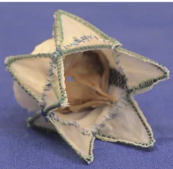
Description of valve manufacture: two glutaraldehyde-preserved aortic and six glutaraldehyde-glutaraldehyde-preserved pulmon-ary valvular homografts were sutured into a 20 mm diameter Dacron tube prior to the attachment of two self-expandable non-thermosensitive nitinol Z-stents at the center of the device, thus forming two self-expanding crowns for fixation. (Fig. 1).
The valved stent was then loaded into a custom-made Teflon sheath stent-graft delivery system with an overall diameter of 10 mm (Fig. 2).
The endpoint of the study is to asses the feasibility of the deployment procedure and verify if it can cause severe hemodynamic instability.
Study design: an acute in vivo evaluation was performed in eight pigs with a mean body weight of 46.0G4.3 kg (range 43– 56 kg). Under general anesthesia and ECG and blood pressure monitoring, both internal jugular vein were exposed, respectively, for infusion and introduction of a Swan–Ganz catheter. ECG, heart rate, mean blood pressure, cardiac output and pulmonary artery pressure were recorded before and 30 min after the valve deployment. The left femoral vein was exposed for insertion of an intracardiac ultrasonic probe. A left posterolateral thoracotomy in the fourth inter-costal space was performed and the pericardium was opened to reach the left atrium. We marked the atrio-ventricular
groove with 3–4 clips, at the level of the mitral annulus, thus regarding it as the valved stent implantation site.
Heparin (100 IU/kg) was administered intravenously. An intracardiac ultrasonic probe (ICUS) was inserted into the right atrium through the left femoral vein in order to monitor our intracardiac manipulations.
Two purse string sutures were made with 6-0 polypropy-lene thread on the left atrium. The atrium was then punctured with a needle and a guide wire was inserted into it before a short 9-F sheath was introduced.
The intravascular ultrasound (IVUS 12.5 MHz, 6F, Clearview, Boston Scientific Corporation, Sunnyvale, CA) was then inserted through the 9-F sheath, in order to measure the diameter and area of mitral valve. The position of annulus was confirmed as well under the guidance of fluoroscopy. The IVUS was then removed to allowed the introduction of the delivery system.
An incision of 1 cm was made on left atrium, controlled by the purse strings. The custom-made delivery introducer loaded with the valved stent was pushed along the guide wire, until the middle of the valved stent reached the mitral annulus previously marked with metallic clips. The sheath was pulled back while the piston was held in place, and the ventricular side of the valved stent was deployed. We gently pulled the whole introducer to ensure fixation of the valved stent before deploying the atrial side (Fig. 3).
ICUS (intracardiac ultrasound, Sequoia) was used to asses valve competence and function. The pressure gradient between left atrium and ventricle (proximal and distal to the valved stent) was directly measured with a needle.
The double-crowned valved stent function was assessed for at least 1 h after the deployment for a maximum of 3 h. Then, animals were sacrificed in order to check the adequate position of the valved stents: mitral annulus in between the two crowned stents.
All the pigs received care in compliance with the Principles of Laboratory Animals’ formulated by the National
Fig. 3. Delivering the valved stent under the guidance of fluoroscopy. The custom-made delivery introducer loaded with the valved stent was pushed along the guide wire, until the middle of the valved stent reached the mitral annulus previously marked with metallic clips. The sheath was pulled back while the piston was held in place, and the ventricular side of the valved stent was deployed. We gently pulled the whole introducer to ensure fixation of the valved stent before deploying the atrial side.
Fig. 1. The double-crowned valved stent. A porcine pulmonary valve has been sutured into a Dacron conduit. Then, two self-expandable nitinol Z-stents were sutured on the external surface of the prosthesis in such a way to create two self-expanding crowns for fixation.
Fig. 2. The self-expanding valved stent loading and half-deploying in a Teflon sheath stent-graft delivery system.
Society of Medical Research and ‘the Guide for the Care and Use of Laboratory Animals’ prepared by the Institute of Laboratory Animal Resources and published by the National Institute of Health (NIH publication 85-23, revised 1985). The protocol was approved by the Institutional Committee on Animal Research.
Data were analyzed with SPSS software (Statistical Package for the Social Sciences). Paired t tests were used. Values were reported as meanGSD.
3. Results
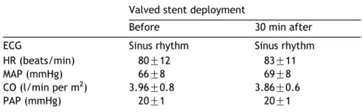
The mean height of the valved stents was 29.4G0.2 mm, with an internal diameter of 20.4G1.0 mm, and an external diameter of 25.5G0.8 mm. The procedure was successfully completed in all animals. Mean procedure time was 18G 8 min and all valves were deployed in less than 30 s. Hemodynamic data before and 30 min after valve deploy-ment are reported inTable 1. In vivo evaluation showed a native mitral annulus diameter of 24.9G0.6 mm, and a mean mitral valve area of 421.4G17.5 mm2. Comparatively, they were 19.8G0.4 mm and 238.5G15.2 mm2, respectively,
after implantation. ICUS confirmed complete valve opening and closure of the valved stents (Fig. 4) and showed a mild paravalvular regurgitation in three out of eight animals. In these animals, there was the highest degree of mismatch between mitral annulus size and stented valve size: the annulus diameter was more than 20% bigger than the stented valve diameter. Mean pressure gradient across the valved stents was 2.6G3.1 mmHg (range 0–8 mmHg). Mean pressure gradient across the left ventricular outflow tract (LVOT) was 6.6G5.2 mmHg (range 1–15 mmHg). The mean survival time was 97.5G56.3 min (survival time range was 40–180 min). One animal died due to the valve displacement 40 min after the valve deployment. Mean blood loss during the procedure was 55G25 cm3. Postmortem evaluation confirmed that the
native mitral annulus was in between the two crowned stents in seven out of eight animals. In one animal, the valved
stents migrated into the ventricle causing the complete occlusion of the LVOT. No atrial or ventricular lesions due to the valved stents were found (Fig. 5). Detailed results are reported inTables 2 and 3.
4. Discussion
4.1. Valved stents design and deployment
Because of the special anatomy of atrio-ventricular valve, the valved stent was made up of two systems: a valve system and a fixation system. The ventricular stent guarantees the fixation of the device to the mitral annulus, while the atrial stent holds in place the homograft sutured on the prosthesis. The two stents are connected in the center of the device to avoid mutual interference and in such a way to squeeze the mitral annulus in a sandwich-like technique. We chose porcine aortic or pulmonary valves as they were easily obtainable and easily sutured into a 20 mm diameter Dacron tube graft. The graft acts like an external support to the homograft, preventing its collapse during manipulation and after deployment.
Table 1
Hemodynamic data before and 30 min after the valved stent deployment Valved stent deployment
Before 30 min after ECG Sinus rhythm Sinus rhythm HR (beats/min) 80G12 83G11 MAP (mmHg) 66G8 69G8 CO (l/min per m2
) 3.96G0.8 3.86G0.6 PAP (mmHg) 20G1 20G1
Fig. 4. ICUS showed a good fixation of the valved stent in the mitral position.
Fig. 5. Postmortem evaluation confirmed good positioning of valved stents in the mitral position and no left ventricle outflow tract obstruction.
Table 2
Summary of procedural data
Procedure successfully completed 8 out of 8 animals Mean procedure time 18G8 min Valve deployment time !30 s Native mitral annulus diameter 24.9G0.6 mm Native mitral valve area 421.4G17 mm2
Double-crowned valved stents diameter 19.8G4 mm Double-crowned valved stents area 238.5G15.2 mm2
Blood loss during procedure 55G25 cm3
Complication One late valve displacement
Table 3
Echocardiography assessment of double-crowned valved stents in mitral position
Pressure gradient across the valve 2.6G3.1 mmHg Pressure gradient across the LVOT 6.6G5.2 mmHg Mild mitral regurgitation 3 out of 8 animals
The valve in its crimped position had a profile of 10 mm but, since we deployed the valve from the left atrium, the sheath diameter was not a big concern. The introducer was cut to 25–30 cm in length for easy manipulation, and its tip was kept short (!2 cm), due to space restriction in the left ventricle.
The valved stents were unloaded from the introducer system following a double stages procedure. The ventricular stent was deployed first under fluoroscopic control, by pulling the sheath. During this phase we could still adjust the position and direction of the valved stents in order to have the native mitral annulus, represented by the metallic clips, exactly in between the ventricular and atrial stent. Then the atrial stent was deployed, by pulling the sheath. This approach increased the successful rate of deployments.
The endpoint of this study was to evaluate the feasibility of the off-pump implantation of the self-made valved stent in the mitral position. In our short-term experiment, the duration of deployment was about 15–20 s and no significant hemodynamic changes were noted probably because the deployment was too quick to impair left ventricular function. All the implanted valved worked properly as the two-dimensional echocardiography has shown, and there were no significant changes in the hemodynamic parameters after the valve deployment (Table 1). However, in three animals we detected a mild paravalvular regurgitation due to the mismatch between native annulus and valve size: the annulus diameter was more than 20% bigger than the stented valve diameter. This problem was difficult to overcome just because we had a limited number of homograft available; therefore, choosing the right valve for the right annulus size was not always easy. In the next future, a wider selection of sizes will minimize the number of paravalvular leeks. In one animal, valve displacement occurred 40 min after its deployment causing sudden occlusion of the LVOT and the death of the animal. Once again, we believe the dislodgment of the valve occurred because it was undersized with respect of annulus diameter. Postmortem evaluation confirmed the good positioning of seven out of eight valved stents in the mitral position with the mitral annulus squeezed in between atrial and ventricular stents.
This preliminary study shows the feasibility of an off-pump implantation of the valved stent in the mitral position, which could be used as part of the treatment for patients with mitral regurgitation who are not candidates for open heart surgery, such as those with severe heart failure as well as hepatic and renal function failure. Also, patients with mitral stenosis could be eligible for this treatment after percutaneous dilatation of the mitral stenosis. Implantation of valved stents may protect the left ventricular function from severe regurgitation, even temporarily. The morbidity of reoperation might decrease after alleviation of left heart dysfunction by implantation of a valved stent.
4.2. Study limitations
Left ventricle outflow tract (LVOT) obstruction is one of the main drawback of this technique even if, in this study, the gradient across the LVOT was quite low, thanks to the particular geometry of the pig’s left ventricle: mitral and aortic valves lie almost on the same plane. We believe that in
the human normal heart, the ventricular portion of the valved stents can protrude into the left ventricle in such a way to push the anterior leaflet of the mitral valve towards the LVOT, miming a SAM (systolic anterior movement) effect. Moreover, the subvalvular apparatus is displaced as well and could significantly contribute to the LVOT obstruction. In order to prevent this potential complication, we are now working on a second generation of valved stent that has the ventricular stent shorter than the first one and with a asymmetric shape. All these concerns regarding the potential LVOT obstruction are, somehow, lukewarm because patients with mitral valve regurgitation have often dilated left ventricle and this condition should minimize the risk of anterior displacement of the mitral leaflet. However, there is always the possibility to incise the anterior leaflet by robotic surgery in working heart before implantation, if necessary[16].
Because only an acute study was performed, the long-term durability and stability of the valved stents are unknown.
However, all the catheter-delivered valved stents have been intended for temporary, not long-term placement and use. Our experiment also focused on short-term testing at present.
In conclusion, off-pump implantation of a self-expand-able valved stent in the mitral position is technically feasible. Further studies will assess if this procedure is also feasible in humans.
Acknowledgements
We thank Monique Augstburger, Marko Burki, Iker Mallabiabarena for their technical assistance received for and during the animal experiments, which contributed to the success of this procedure.
References
[1] Davies H. Catheter-mounted valve for temporary relief of aortic insufficiency. Lancet 1965;1:250.
[2] Zhou JQ, Corno AF, Huber CH, Tozzi P, von Segesser LK. Self-expandable valved stent of large size: off-bypass implantation in pulmonary position. Eur J Cardiothorac Surg 2003;24:212–6.
[3] Lutter G, Kuklinski D, Berg G, Von Samson P, Martin J, Handke M, Uhrmeister P, Beyersdorf F. Percutaneous aortic valve replacement:an experimental study I. Studies on implantation. J Thorac Cardiovasc Surg 2002;123(4):768–76.
[4] Andersen HR, Knudsen LL, Hasenkam JM. Transluminal implantation of artificial heart valves. Description of a new expandable aortic valve and initial results with implantation by catheter technique in closed chest pigs. Eur Heart J 1992;13(5):704–8.
[5] Boudjemline Y, Bonhoeffer P. Steps toward percutaneous aortic valve replacement. Circulation 2002;105(6):775–8.
[6] Cribier A, Eltchaninoff H, Bash A, Borenstein N, Tron C, Bauer F, Derumeaux G, Anselme F, Laborde F, Leon MB. Percutaneous transcatheter implantation of an aortic valve prosthesis for calcific aortic stenosis: first human case description. Circulation 2002;106(24): 3006–8.
[7] Sochman J, Peregrin JH, Pavcnik D, Timmermans H, Rosch J. Percuta-neous transcatheter aortic disc valve prosthesis implantation: a feasibility study. Cardiovasc Intervent Radiol 2000;23(5):384–8. [8] Bonhoeffer P, Boudjemline Y, Saliba Z, Hausse AO, Aggoun Y, Bonnet D,
Sidi D, Kachaner J. Transcatheter implantation of a bovine valve in pulmonary position: a lamb study. Circulation 2000;102:813–6.
[9] Bonhoeffer P, Boudjemline Y, Saliba Z, Merckx J, Aggoun Y, Bonnet D, Acar P, Bidois JL, Sidi D, Kachaner J. Percutaneous replacement of pulmonary valve in a right-ventrecle to pulmonary-artery prosthetic conduit with valve dyfunction. Lancet 2000;(356):1403–5.
[10] Bonhoeffer P, Boudjemline Y, Qureshi SA, Bidois JL, Iserin L, Acar P, Merckx J, Kachaner J, Sidi D. Percutaneous insertion of the pulmonary valve. J Am Coll Cardiol 2002;39:1664–9.
[11] Corno AF, Zhou JQ, Tozzi P, von Segesser LK. Off-bypass implantation of a self-expandable valved stent between inferior vena cava and right atrium. Int Cardiovac Thorac Surg 2003;2:166–9.
[12] Huber CH, Tozzi P, Corno AF, Marty B, Rucht P, Gersbach P, Nasratulla M, von Segesser LK. Do valved stents compromise coronary flow? Eur J Cardiothorac Surg 2004;25:754–9.
[13] Ross DN. Replacement of aortic and mitral valves with a pulmonary autograft. Lancet 1967;2(956):8.
[14] Halees ZA, Awad MM, Pieters F, Shahid MS, Amri MAA. Six-year follow-up of a pulmonary autograft in the mitral position: the Ross II procedure. J Thorac Cardiovasc Surg 1999;117:614–6.
[15] Kumar AS, Aggarwal S, Choudhary SK. Mitral valve replacement with the pulmonary autograft: the Ross II procedure. J Thorac Cardiovasc Surg 2001;122:378–9.
[16] Von Segesser LK, Tozzi P, Augstburger M, Corno AF. Working heart off-pump cardiac repair(OPCARE)—the next step in robotic surgery? Int Cardiovasc Thorac Surg 2003;2:120–4.
Appendix B. Editorial Comment
Off-pump mitral valve replacement: an attack on conven-tional heart surgery?
Georg Lutter*, Jochen Cremer
Department of Cardiovascular Surgery, University of Kiel, School of Medicine, 24105 Kiel, Germany
The pioneering introduction of the Starr–Edwards valve allowed complete replacement of the diseased mitral valve.
With improved cardiopulmonary bypass, myocardial protec-tion, and surgical techniques the mortality rate associated with mitral valve replacement decreased. Augmented use of mitral valve repair techniques has resulted in a substantial decrease in long-term morbidity and mortality when treating patients with mitral regurgitation. Some believed that this resulted from maintaining continuity of the mitral annular papillary muscle continuity during mitral valve repair. Additional studies have validated the positive long-term effects of maintaining the integrity of the mitral valve subvalvular apparatus[1]. Although surgical approaches to reduce mitral regurgitation associated with congestive heart failure have been reported, the high mortality rate that Appendix A. Conference discussion
Dr J. Revuelta (Santander, Spain): I have a technical question. In case that you will apply this clinically in patients, do you think that this device could be implanted through the apex with a small incision or through the intercostal groove, also with a small mini thoracotomy in the intercostal space? Are you planning to do that or just to leave it for cardiologists?
Dr Tozzi: If the question is who can do this procedure, cardiologist or surgeon, I think that anyone that can handle a hole in the left atrium without compromising his own coronary flow or his cardiac flow can do this procedure. So, in other words, this is so far a procedure that has to be done by a cardiac surgeon, because you have to do a hole in the left atrium or, if you want, in the left ventricle, but it is a surgical approach.
Dr Revuelta: But you are in favor of inter-atrial groove? There is no problem, no coronaries there, no nothing?
Dr Tozzi: No. It is not possible in the inter-atrial groove. Dr Revuelta: So you think it would be the best way?
Dr Tozzi: It will be possible, yes, with a small incision, exactly as we do in the animal.
Dr Revuelta: With thoracoscopy you can see?
Dr Tozzi: Theoretically it is feasible. We have a few technical problems, but theoretically it is feasible to do this with a thoracoscopy with a minimally invasive approach, absolutely.
Dr V. Subramanian (New York, USA): I have a theoretical question. As I believe the stent is probably going to tension the anterior leaflets in the chordae, since you still have an intact chordae and papillary muscle mechanism, will there be any left ventricular geometry changes in the short term or the long term, because all of these devices are still going to leave the chordal–papillary mechanism intact? The way it alters that tension, can that have a negative effect on the left ventricular geometry and its implications long term?
Dr Tozzi: Of course, this is a feasibility study, we just demonstrated that it is possible, and I agree with you that the ventricular side of the stent can displace the chordae and so create a modification in the left ventricular geometry, and we are working on this issue also in order to modify the geometry of the ventricular stent in order to reduce this problem and also the problem of the possible left ventricular outflow obstruction due to displacement of the anterior leaflet of the mitral valve towards the outflow tract of the left ventricle.
Dr H. Shennib (Montreal, Canada): Could you tell us about the indications that you see for this application? You have had several animals that had paravalvular leak, and one would assume that the stiffer the apparatus of the mitral valve, the annulus, the amount of calcifications and so on may determine how the stent will sit on it and the success or failure and, as a result, the risk of paravalvular leak.
On the other hand, the softer the leaflets and the softer everything is, you think those are indications for repair and not replacement.
So I am not yet clear as to your projection as to what would be the indications specifically for replacement in this group of patients.
Dr Tozzi: What I didn’t say is that the paravalvular leakage is due to a mismatch between the shape and the diameter of the mitral annulus versus the diameter and the shape of the stent. We are working on that. We are trying to create a valved stent in order to be much more flexible in order to adapt itself to the geometry of the mitral annulus.
Of course, on the other hand, we will have the problem of stented valve stability, because a very compliant valve is also a very unstable valve, so far. But this is a technical issue. I am sure there are several bioengineers that are able to give a very good answer to this problem. So I think we can overcome this technical issue. But up to now this is a problem.
Dr Shennib: The indications, which group of patients would you recommend this as you mature it?
Dr Tozzi: So far, as I said, mitral regurgitation, any kind of mitral regurgitation, theoretically there won’t be any problem to deploy a valve such as this one. Of course, a patient with mitral stenosis due to a severe calcification of the mitral annulus, we could have more problems. But once again, I think those problems can be overcome with technical modifications. So it is just a technical issue.
Dr F. Mohr (Leipzig, Germany): I think it is extremely interesting. Did you look at other stent designs, valve designs which more or less mimic the mitral valve, because you just used the rounded shape of the aortic valve design? Did you try this, yes or no?