Canine echinococcosis: genetic diversity of
Echinococcus granulosus sensu stricto (s.s.)
from definitive hosts
B. Boufana
1*
†, W. Lett
1, S. Lahmar
2, A. Griffiths
3, D.J. Jenkins
4,
I. Buishi
5, S.A. Engliez
6, M.A. Alrefadi
6, A.A. Eljaki
7,
F.M. Elmestiri
6, M.M. Reyes
8, S. Pointing
9, A. Al-Hindi
10,
P.R. Torgerson
11, M. Okamoto
12and P.S. Craig
11
Cestode Zoonoses Research Group, School of Environment and Life
Sciences, University of Salford, M5 4WT, United Kingdom:
2Parasitology
Laboratory, National School of Veterinary Medicine, Sidi Thabet, Tunisia:
3
School of Biological Sciences, University of Bristol, Life Sciences Building,
Bristol, BS8 1TQ, UK:
4School of Animal and Veterinary Sciences,
Charles Sturt University, NSW 2678, Australia:
5The University of Tripoli,
PO Box 606, Tripoli, Libya:
6Department of Zoology, University of Benghazi,
PO Box 1308, Benghazi, Libya:
7Department of Zoology, University of Omar
Almukhtar, Al Bayda, Libya:
8Alberto Hurtado School of Medicine,
Universidad Peruana Cayetano Heredia, Lima, Peru:
9Department
of Agriculture, Stanley, Falkland Islands, FIQQ 1ZZ:
10The Islamic
University of Gaza, Faculty of Health Sciences, Medical Laboratory Sciences
Department, Gaza, Palestine:
11Section of Veterinary Epidemiology,
University of Zu¨rich, Switzerland:
12Primate Research Institute, Kyoto
University, 41-2 Kanrin, Inuyama, Aichi 484-8506, Japan
(Received 8 March 2015; Accepted 24 April 2015; First Published Online 16 July 2015) Abstract
Canids, particularly dogs, constitute the major source of cystic echinococcosis (CE) infection to humans, with the majority of cases being caused by Echinococcus granulosus (G1 genotype). Canine echinococcosis is an asymptomatic disease caused by adult tapeworms of E. granulosus sensu lato (s.l.). Information on the population structure and genetic variation of adult E. granulosus is limited. Using sequenced data of the mitochondrial cytochrome c oxidase subunit 1 (cox1) we examined the genetic diversity and population structure of adult tapeworms of E. granulosus (G1 genotype) from canid definitive hosts originating from various geographical regions and compared it to that reported for the larval metacestode stage from sheep and human hosts. Echinococcus granulosus (s.s) was identified from adult tapeworm isolates from Kenya, Libya, Tunisia, Australia, China, Kazakhstan, United Kingdom and Peru, including the first known molecular confirmation from Gaza and the Falkland Islands. Haplotype analysis showed a star-shaped network with a centrally positioned common haplotype previously described for the metacestode stage from sheep and humans, and the
†Present address: Department of Zoology, University of Benghazi, PO Box 1308, Benghazi, Libya.
*E-mail: [email protected]
Journal of Helminthology (2015) 89, 689–698 doi:10.1017/S0022149X15000395
qCambridge University Press 2015
https:/www.cambridge.org/core/terms. https://doi.org/10.1017/S0022149X15000395
neutrality indices indicated population expansion. Low Fst values suggested that populations of adult E. granulosus were not genetically differentiated. Haplotype and nucleotide diversities for E. granulosus isolates from sheep and human origin were twice as high as those reported from canid hosts. This may be related to self-fertilization of E. granulosus and/or to the longevity of the parasite in the respective intermediate and definitive hosts. Improved nuclear single loci are required to investigate the discrepancies in genetic variation seen in this study.
Introduction
Cystic echinococcosis (CE) is caused by a group of cryptic species within Echinococcus granulosus sensu lato (s.l.) including E. granulosus sensu stricto (s.s.) (G1 –G3 genotypes), E. equinus (G4 genotype), E. ortleppi (G5 genotype) and E. canadensis (G6 –G10 genotypes) (Nakao et al., 2007; Thompson, 2008). Human CE due to infection with the metacestode stage of E. granulosus (s.l.) is an important zoonotic infection of major health and socio-economic impact worldwide (Budke et al., 2006; Craig et al., 2007). Conversely, canine echinococcosis is caused primarily by the adult stage of E. granulosus (s.l.) and the disease is usually asymptomatic in canid definitive hosts, even in those animals harbouring large worm burdens. Although canids and ungulates, respectively, serve as definitive and intermediate hosts of E. granulosus (s.l.), dogs and sheep are by far the most important hosts worldwide. Furthermore, infected dogs constitute the major source of CE infection to humans and are known to shed in excess of 8000 eggs per day (Gemmell, 1990). Detection of infection in dogs has traditionally relied on necropsy or arecoline purgation for assessing Echinococcus worm burdens (Eckert et al., 2001), but it also relies increasingly on the use of coproELISA for the detection of Echinococcus copro-antigens in order to determine prevalence and re-infection rates in dogs (Craig et al., 1995; Allan & Craig 2006; Moss et al., 2013).
Molecular genotypic information on Echinococcus isolates from definitive hosts is important for epidemio-logical and transmission studies, as well as for the planning and surveillance of control programmes (Craig et al., 2003). Published reports on the molecular characterization of E. granulosus (s.s.) in definitive hosts include those from Asia (Sˇtefanic´ et al., 2004; Bart et al., 2006a; Zhang et al., 2006; Ma et al., 2008; Utuk et al., 2008; Ziadinov et al., 2008), the Middle East (Al-Qaoud et al., 2003; Parsa et al., 2012), North Africa (Lahmar et al., 2009; Boufana et al., 2014), Europe (Trachsel et al., 2007; Sherifi et al., 2011; Xhaxhiu et al., 2011) and the Americas (Soriano et al., 2010; de la Rue et al., 2011). In contrast, molecular data on E. granulosus (s.s.) from definitive hosts in Sub-Saharan Africa are limited (Wachira et al., 1990, 1993a) compared to data from wild carnivores and domestic and wild herbivores (Wachira et al., 1993b; Hu¨ttner et al., 2009; Kagendo et al., 2014).
The study of the population structure of E. granulosus is important as genetic variation is thought to be related to host infectivity (Thompson & McManus, 2002). Using DNA extracted from protoscoleces and/or the germinal layer of Echinococcus hydatid cysts retrieved from
livestock and human hosts, researchers investigated the genetic diversity of E. granulosus (s.s.) from various regions (Nakao et al., 2010; Casulli et al., 2012; Yanagida et al., 2012; Konyaev et al., 2013; Boufana et al., 2014, 2015). These studies highlighted the distribution of E. granulosus haplotypes and demonstrated the existence of a single lineage for E. granulosus (s.s.) across many geographically isolated regions. In contrast, genetic variation and population structure using E. granulosus DNA derived from adult worms and/or canid faecal samples has, to date, not been investigated very widely (Boufana et al., 2014, 2015). Such studies are important in assessing the risk of infection to humans, as the majority of human CE cases are known to be caused by E. granulosus (s.s.) (G1 genotype). In a recent study, 88% of 1661 human CE isolates worldwide, were considered to be caused by E. granulosus (G1 genotype) (Alvarez Rojas et al., 2014). In the current study we examined the genetic diversity of adult stages of E. granulosus (s.s.) from canid definitive hosts originating from various geographical regions, and compared data generated with that reported for E. granulosus (s.s.) larval stage metacestodes from sheep and human hosts.
Materials and methods Sample collection
The origins of the 87 E. granulosus isolates used in this study are shown in table 1. Adult tapeworms (n ¼ 40) were retrieved at necropsy from stray and semi-stray domestic dogs and jackals (Canis aureus) from Tunisia (n ¼ 22), dingos (C. familiaris var. dingo) from Australia (n ¼ 11) and owned dogs from China (n ¼ 3) and Kazakhstan (n ¼ 4). In addition, a total of 47 coproDNA isolates were used in this study. This faecal panel included domestic dogs from Kenya (n ¼ 2), Libya (n ¼ 9), China (n ¼ 1), Gaza (Palestine) (n ¼ 3), the Falkland Islands (n ¼ 4), Lima (Peru) (n ¼ 5) (Reyes et al., 2012), as well as faeces from Australian dingos (n ¼ 5). Faecal samples from Welsh farm dogs (n ¼ 5) from a hydatid study carried out in Powys county, Wales (United Kingdom) between May 2008 and July 2010, and from farm sheep dogs (n ¼ 12) from the Welsh counties of Powys and Gwent collected between July and November 2002 (Buishi et al., 2005) were also included. A foxhound (dog) faecal sample (n ¼ 1) used in this study was collected from an anonymous Welsh hunt in 2011 (Lett, 2013). In addition, DNA extracted from sheep hydatid isolates from Benghazi (Libya) (n ¼ 13) and the Falkland Islands (n ¼ 4) were available for this study.
Molecular methods
CoproDNA was extracted from faecal samples using the QIAamp DNA stool mini kit (Qiagen, Hilden, Germany) according to the manufacturer’s instructions. Genomic DNA was extracted from ethanol-fixed tissue (adult tapeworms, protoscoleces or membranes of an individual hydatid cyst) using the Qiagen DNeasy Blood and Tissue Kit (Qiagen). DNA was used as template to amplify a fragment within the cytochrome c oxidase subunit 1 (cox1, 828 bp) mitochondrial gene, as described previously (Nakao et al., 2000). Amplified products were sequenced commercially in both forward and reverse directions (Source Bioscience, Nottingham, UK) and chromatograms were examined using FinchTV viewer (Geospiza, Seattle, Washington, USA).
Data analysis
Data analysis was carried out as described previously (Boufana et al., 2014). In brief, sequence alignments were carried out using MEGA version 6 (Tamura et al., 2013) and ClustalX2 (Larkin et al., 2007) and exported into DnaSP 5 (Librado & Rozas, 2009). Hapview (Salzburger et al., 2011) was used to generate haplotype networks. DNAML program (PHYLIP) (Felsenstein, 1989), which was run from Hapview, was used to construct maximum likelihood trees. Arelquin 3.1 (Excoffier et al., 2005) was used to calculate the number of haplotypes (hn), haplotype diversities (hd) and nucleotide diversities (pd) as well as to estimate Tajima’s D (Tajima, 1989) and Fu’s Fs (Fu, 1997) in order to examine past population demography and test for neutrality departures.
The pairwise fixation index (Fst) was generated in Arlequin to test for genetic differentiation. P values for the multiple pairwise comparisons were adjusted using Bonferroni correction.
To compare E. granulosus haplotypes from canid hosts with those found in sheep and humans, cox1 mitochon-drial nucleotide sequences publically available on the National Center for Biotechnology Information (NCBI) database (www. ncbi.nlm.nih.gov) were downloaded for inclusion (table 2). Nucleotide sequences were grouped by host (canid, sheep and human) and geographical origin according to six broad regions, Africa, Australia, Asia, Europe, the Middle East and South America. All sequences were trimmed to equal length (329 bp), giving a final dataset of 408 E. granulosus cox1 mitochondrial sequences, 95 from canids, 180 from sheep and 133 from humans. Rarefied haplotype richness, standardized to the smallest sample size, was calculated using Contrib 1.02 (Petit et al., 1998) in order to facilitate direct comparisons between hosts.
Results DNA sequence data
We amplified DNA extracted from adult tapeworms and/or faecal samples of 87 Echinococcus isolates from canids originating from Africa (Kenya, n ¼ 2; Libya, n ¼ 9; Tunisia, n ¼ 22), Australia (n ¼ 16), Asia (China, n ¼ 4; Kazakhstan, n ¼ 4), United Kingdom (n ¼ 18), the Middle East (Gaza, Palestine, n ¼ 3) and South America (Falkland Islands, n ¼ 4; Peru, n ¼ 5). A total of 827 bp within the cox1 gene were analysed for each Echinococcus canid-derived isolate. Using a BLAST search (http:www. blast.ncbi.nlm.nih.gov), E. granulosus (s.s.) was identified from adult tapeworms and faecal samples of all canid hosts included in this study. In addition, sheep hydatid cysts from Libya and the Falkland Islands had a 100% sequence identity to E. granulosus (G1 genotype) (e.g. accession numbers AB688603, AB893250).
Analysis of E. granulosus (s.s.) from canid hosts Within the 827-bp cox1 nucleotide sequences analysed there were 18 polymorphic sites, 9 were parsimony informative and 9 were singleton variable sites. The overall haplotype and nucleotide diversities for E. granulosus (s.s.) isolates from canid hosts were 0.4405 ^ 0.0674 and 0.000958 ^ 0.000777, respectively. Similarly, the overall values for Tajima’s D (2 2.26 101) and Fu’s Fs (211.8484) were significantly negative (P values # 0.0002 and # 0.0001, respectively), a feature indicative of population expansion.
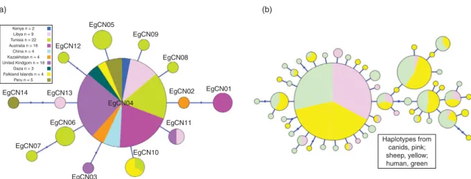
A total of 14 haplotypes (EgCN01–EgCN14) were detected within the 87 canid-derived E. granulosus (s.s.) cox1 mitochondrial sequences (fig. 1a). The frequency of the common haplotype (EgCN04) within the generated star-shaped network was (74.7%; 65/87) and it was 100% identical to the dominant E. granulosus haplotypes described from China (G01: AB491414; 789 bp) (Nakao et al., 2010), Iran (Eg01: JQ250806; 1609 bp) (Yanagida et al., 2012), Europe (EG1: JF513058; 351 bp) (Casulli et al., 2012) and Tunisia (EgTu01: KM014606; 827 bp) Table 1. Number and source of Echinococcus granulosus (s.s.)
isolates from canid hosts.
Country/location (host)
DNA source/No. of isolates Faeces Adult worms Total
Kenya 2 – 2
Libya 9
Benghazi 5 –
Tripoli 4 –
Tunisia 22
Ariana, Tozeur, Siliana – 20
Jendouba (jackals) – 2
Australia 16
Queensland – 5
Australian Capital Territory – 2
New South Wales 5 4
China 4 Qinghai 1 3 Kazakhstan – 4 4 United Kingdom 18 Wales (farm dogs) 17 – (foxhound) 1 – Palestine 3 Gaza 3 – Falkland Islands 4 – 4 Peru 5 Lima 5 – Total 47 40 87
Genetic diversity of canine echinococcosis 691
https:/www.cambridge.org/core/terms. https://doi.org/10.1017/S0022149X15000395
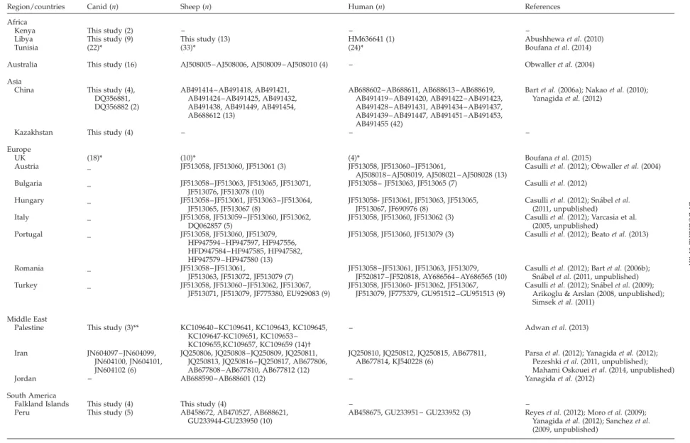
Table 2. Echinococcus granulosus (s.s.) cytochrome c oxidase subunit 1 (cox1) mitochondrial nucleotide sequences used in this study.
Region/countries Canid (n) Sheep (n) Human (n) References Africa
Kenya This study (2) – – –
Libya This study (9) This study (13) HM636641 (1) Abushhewa et al. (2010) Tunisia (22)* (33)* (24)* Boufana et al. (2014) Australia This study (16) AJ508005 – AJ508006, AJ508009 – AJ508010 (4) – Obwaller et al. (2004) Asia
China This study (4), DQ356881, DQ356882 (2)
AB491414 – AB491418, AB491421, AB491424 – AB491425, AB491432, AB491438, AB491449, AB491454, AB688612 (13)
AB688602 – AB688611, AB688613– AB688619, AB491419 – AB491420, AB491422 – AB491423, AB491428 – AB491431, AB491434 – AB491437, AB491439 – AB491447, AB491451 – AB491453, AB491455 (42)
Bart et al. (2006a); Nakao et al. (2010); Yanagida et al. (2012)
Kazakhstan This study (4) – – –
Europe
UK (18)* (10)* (4)* Boufana et al. (2015)
Austria _ JF513058, JF513060, JF513061 (3) JF513058, JF513060 – JF513061,
AJ508018 – AJ508019, AJ508021 – AJ508028 (13)
Casulli et al. (2012); Obwaller et al. (2004) Bulgaria _ JF513058 – JF513063, JF513065, JF513071, JF513076, JF513078 (10) JF513058 – JF513063, JF513065 (7) Casulli et al. (2012) Hungary _ JF513058 – JF513061, JF513063 – JF513064, JF513065, JF513067 (8) JF513058- JF513061, JF513063, JF513065, JF513067, JF690976 (8)
Casulli et al. (2012); Sna´bel et al. (2011, unpublished) Italy _ JF513058, JF513059 – JF513060, JF513062,
DQ062857 (5)
JF513058, JF513060, JF513062 (3) Casulli et al. (2012); Varcasia et al. (2005, unpublished)
Portugal _ JF513058, JF513060, JF513079, HF947594 – HF947597, HF947556, HFD947584 – HF947585, HF947582, HF947579 – HF947580 (13)
JF513058, JF513060, JF513079 (3) Casulli et al. (2012); Beato et al. (2013)
Romania _ JF513058 – JF513061,
JF513063, JF513072, JF513079 (7)
JF513058 – JF513061, JF513063, JF513079, JF520817 – JF520818, AY686564 – AY686565 (10)
Casulli et al. (2012); Bart et al. (2006b); Sna´bel et al. (2011, unpublished) Turkey _ JF513058, JF513060 – JF513062, JF513067,
JF513071, JF513079, JF775380, EU929083 (9)
JF513058, JF513060- JF513062, JF513067, JF513079, JF775379, GU951512 –GU951513 (9)
Casulli et al. (2012); Sna´bel et al. (2009); Arikoglu & Arslan (2008, unpublished); Simsek et al. (2011)
Middle East
Palestine This study (3)** KC109640 – KC109641, KC109643, KC109645, KC109647-KC109651, KC109653 – KC109655,KC109657, KC109659 (14)† – Adwan et al. (2013) Iran JN604097 – JN604099, JN604100, JN604101, JN604102 (6) JQ250806, JQ250808 –JQ250809, JQ250811, JQ250813, JQ250816 –JQ250817, AB677806, AB677808 – AB677810, AB677812 (12)
JQ250810, JQ250812, JQ250815, AB677811, AB677814, KJ540228 (6)
Parsa et al. (2012); Yanagida et al. (2012); Pezeshki et al. (2011, unpublished); Mahami Oskouei et al. (2014, unpublished) Jordan – AB688590 – AB688601 (12) – Yanagida et al. (2012)
South America
Falkland Islands This study (4) This study (4) – – Peru This study (5) AB458672, AB470527, AB688621,
GU233944-GU233950 (10)
AB458675, GU233951 – GU233952 (3) Reyes et al. (2012); Moro et al. (2009); Yanagida et al. (2012); Sanchez et al. (2009, unpublished)
n, number of nucleotide sequences.
*The original cox1 nucleotide sequences (used in the relevant reference to generate Echinococcus granulosus haplotypes from Tunisia and United Kingdom) were included in the analysis. **Gaza; †Nablus. 692 B. Boufana et al. . https://doi.org/10.1017/S0022149X15000395 https:/www.cambridge.org/core
. University of Basel Library
, on
30 May 2017 at 19:47:07
(Boufana et al., 2014). Haplotypes EgCN01–EgCN14 were deposited in the NCBI database under accession numbers KT001395–KT001408.
The diversity and neutrality indices determined for E. granulosus (s.s.) isolates from canid hosts originating from various geographical localities using the cox1 mitochondrial nucleotide sequences are shown in table 3. The haplotype diversity for the cox1 gene was highest in samples of adult E. granulosus from North Africa (including two Kenyan isolates) and lowest in samples from the United Kingdom. The values for Tajima’s D were negative for E. granulosus samples from all geographical localities (except Australia), indicating population expan-sion, but remained significant only for samples from Africa and the United Kingdom. Fu’s Fs values were also negative for all populations (except Australia and South America), which indicated further evidence of population expansion, but deviated significantly from neutrality only for samples from Africa. Tajima’s D and Fu’s Fs were positive for E. granulosus from Australian dingos which suggests that populations may have undergone a genetic bottleneck. Similar positive Fu’s Fs values were observed for South American E. granulosus canid populations. No conclusion could be drawn for the diversity and neutrality
indices of E. granulosus isolates from Gaza due to the small sample size of the Palestinian isolates (n ¼ 3).
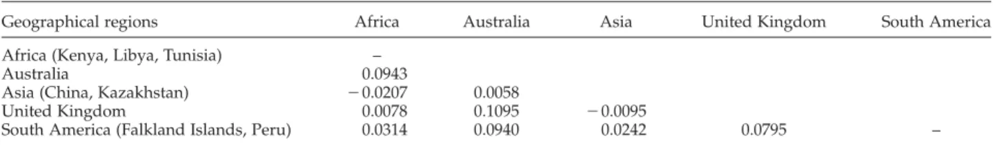
Genetic differentiation based on the analysis of the pairwise Fst values for the E. granulosus (s.s.) adult tapeworm isolates from canid hosts originating from different geographical regions are shown in table 4. Generally low, non-significant values were recorded for most pairwise comparisons (following Bonferroni correc-tion), suggesting little genetic differentiation globally.
Comparison of E. granulosus (s.s.) canid-derived haplotypes to those from sheep and human hosts
A total of 408 cox1 nucleotide sequences derived from canid, sheep and human isolates were analysed (tables 2 and 5). The overall haplotype and nucleotide diversities were 0.6631 ^ 0.0259 and 0.003618 ^ 0.002571, respectively. Within the cox1 nucleotide sequences there were 45 polymorphic sites, 20 of which were parsimony informative.
Genealogical relationships between E. granulosus (s.s.) isolates by host (canid, sheep, human) originating from Africa, Australia, Asia, Europe, the Middle East and South America are shown in fig. 1b. The generated network was
Table 3. Diversity and neutrality indices for Echinococcus granulosus (s.s.) adult tapeworm isolates from canid hosts originating from various geographical regions using nucleotide data of the cytochrome c oxidase subunit 1 (cox1) mitochondrial gene.
Geographical region (n) hn hd ^ SD pd ^ SD Tajima’s D P value Fu’s Fs P value
Africa (Kenya, Libya, Tunisia) (33) 10 0.6288 ^ 0.0944 0.001134 ^ 0.000889 22.06508 0.0037** 27.00817 0.0000***
Australia (16) 2 0.3250 ^ 0.1251 0.001183 ^ 0.000947 0.22735 0.6525 2.64424 0.8832
Asia (China, Kazakhstan) (8) 2 0.2500 ^ 0.1802 0.000303 ^ 0.000429 21.05482 0.2126 20.18197 0.1989
United Kingdom (18) 3 0.2157 ^ 0.1241 0.000404 ^ 0.000472 21.71304 0.0203* 21.02496 0.0564
South America (Falkland Islands, Peru) (9)
3 0.5556 ^ 0.1653 0.001550 ^ 0.001213 21.29379 0.1183 0.8240 0.6584
n, number of isolates, hn, number of haplotypes; hd, haplotype diversity;pd, nucleotide diversity; SD, standard deviation. Significant at *P value # 0.05, **P value # 0.01, ***P value # 0.0001.
EgCN05 EgCN09 EgCN08 (a) (b) EgCN01 Haplotypes from canids, pink; sheep, yellow; human, green EgCN02 EgCN11 EgCN10 EgCN04 EgCN03 EgCN06 EgCN13 EgCN12 Kenya n = 2 Libya n = 9 Tunisia n = 22 Australia n = 16 China n = 4 Kazakhstan n = 4 United Kindgom n = 18 Gaza n = 3 Falkland Islands n = 4 Peru n = 5 EgCN14 EgCN07
Fig. 1. Haplotype networks generated using cytochrome c oxidase subunit 1 (cox1) mitochondrial nucleotide sequences of Echinococcus granulosus (s.s.) from (a) adult tapeworm isolates derived from canid hosts from various geographic locations; and (b) from adult tapeworms and larval metacestode-stage isolates from sheep and human hosts. Circle size is relative to haplotype frequency. Small circles
indicate additional mutational steps.
Genetic diversity of canine echinococcosis 693
https:/www.cambridge.org/core/terms. https://doi.org/10.1017/S0022149X15000395
made up of 51 haplotypes, with the most common central haplotype encompassing 56.6% (231/408) of the total number of E. granulosus isolates. The percentage of canid, sheep and human isolates within the dominant haplotype were 32.9%, 41.6% and 25.5%, respectively. A BLAST search showed this dominant haplotype to be 100% identical to that of the common E. granulosus haplotype described by other researchers (Nakao et al., 2010; Casulli et al., 2012; Yanagida et al., 2012; Boufana et al., 2014). The network was also made up of a smaller number of more highly derived branches of haplotypes, distantly related to the dominant sequence. Within these, the frequency of isolates derived from canids was 7.4% (7/95), which was much lower than those from sheep (30.6%, 55/180) and humans (36.8%, 49/133). These seven canid isolates originated from Australia (n ¼ 3), Asia (China, n ¼ 1; Kazakhstan, n ¼ 1), the Middle East (Iran, n ¼ 1) and South America (Peru, n ¼ 1).
The haplotype and nucleotide diversities for E. granulosus (s.s.) isolates from sheep and human origin were at least double those in canid definitive hosts (table 5). Furthermore, the rarefied allelic richness of E. granulosus derived from canids was also approximately half that shown in sheep or humans, suggesting that these differences in genetic diversity are not purely a reflection of sample size. Tajima’s D values were significantly negative for E. granulosus (s.s.) derived from all three hosts, indicating the presence of rare polymorphic sites characteristic of population expansion. The values of Fu’s Fs for E. granulosus populations from sheep and humans were twice as high as those from canids, and deviated significantly from neutrality for all host species, also indicating population expansion. The values of the pairwise comparison for genetic differentiation (Fst) of E. granulosus (s.s.) between host species were generally low, but comparisons with canid isolates were highly signifi-cant (canid/sheep, 0.02896, P ¼ 0.0008; canid/human, 0.0359, P ¼ 0.0002; sheep/human, 2 0.0010, P ¼ 0.5186).
Discussion
In the current study we analysed cox1 mitochondrial sequences from E. granulosus (s.s.) adult tapeworm isolates recovered from dog, dingo, foxhound and jackal (C. aureus) definitive hosts (n ¼ 87) and compared them to published sequence data for the metacestode stage from sheep (n ¼ 180), humans (n ¼ 133) and dogs (n ¼ 8). The occurrence of E. granulosus (s.s.) in canid definitive hosts (dog, red fox, dingo, jackal, wolf) from many locations worldwide has been confirmed via molecular genetic analysis by several researchers (reviewed by Carmena & Cardona, 2013; Jenkins et al., 2014). However, the current study reports the first molecular confirmation of E. granulosus (s.s.) from definitive hosts originating from Gaza and the Falkland Islands. This is also the first attempt to investigate the global genetic variation and population structure of E. granulosus (s.s.) isolates from definitive hosts, although this subject has been explored extensively for the metacestode stage of E. granulosus derived from ungulate and human hosts (Nakao et al., 2010; Casulli et al., 2012; Yanagida et al., 2012; Sharma et al., 2013; Boufana et al., 2014, 2015).
The haplotype network generated using cox1 mito-chondrial sequenced data for E. granulosus (s.s.) DNA derived from definitive hosts is consistent with that previously described for the metacestode stage retrieved from sheep and human hosts (Nakao et al., 2010; Casulli et al., 2012; Yanagida et al., 2012; Boufana et al., 2014, 2015) with the dominance of the same E. granulosus centrally positioned haplotype. This finding therefore supports the worldwide common lineage of E. granulosus in canids from all geographical regions studied (i.e. Kenya, Libya, Tunisia, Australia, China, Kazakhstan, UK, Gaza, Peru and the Falkland Islands). It also confirms the absence of phylogeographic structure for E. granulosus (s.s.), as previously reported (Nakao et al., 2010). In addition, the very low Fst values observed across this study suggest Table 4. Pairwise fixation index (Fst) for Echinococcus granulosus (s.s.) adult tapeworm isolates from canid hosts originating from various geographical regions using nucleotide data of the cytochrome c oxidase subunit 1 (cox1) mitochondrial gene.
Geographical regions Africa Australia Asia United Kingdom South America
Africa (Kenya, Libya, Tunisia) –
Australia 0.0943
Asia (China, Kazakhstan) 20.0207 0.0058
United Kingdom 0.0078 0.1095 20.0095
South America (Falkland Islands, Peru) 0.0314 0.0940 0.0242 0.0795 –
Non-significant at corrected P value # 0.003.
Table 5. Diversity and neutrality indices for Echinococcus granulosus (s.s.) isolates from canid, sheep and human hosts using nucleotide data of the cytochrome c oxidase subunit 1 (cox1) mitochondrial gene.
Host (n) hn hd ^ SD pd ^ SD Rs Tajima’s D P value Fu’s Fs P value
Canid (95) 12 0.3592 ^ 0.0639 0.001654 ^ 0.001527 11.000 21.9737 0.0022* 211.5198 0.0000**
Sheep (180) 31 0.6919 ^ 0.0358 0.003892 ^ 0.002719 19.455 22.0663 0.0011* 228.7479 0.0000**
Human (133) 29 0.7787 ^ 0.0342 0.004533 ^ 0.003050 22.607 22.1295 0.0017* 227.9899 0.0000**
n, number of isolates; hn, number of haplotypes; hd, haplotype diversity;pd, nucleotide diversity; SD, standard deviation. Rs, rarefied allelic richness, standardized to the smallest sample collection (n ¼ 95). Significant at *P value # 0.01, **P value # 0.0001.
the absence of genetic differentiation between adult E. granulosus isolates, which is consistent with that reported for isolates of the metacestode stage from ungulates and humans from China (Nakao et al., 2010), Europe (Casulli et al., 2012) and the Middle East (Jordan, Iran) (Yanagida et al., 2012), but differs from that reported for Peruvian metacestode populations, which were said to be genetically differentiated from E. granulosus populations from China and the Middle East (Yanagida et al., 2012).
A combination of higher haplotype and lower nucleotide diversities, similar to those reported for cox1 nucleotide data derived from the metacestode stage of E. granulosus (Nakao et al., 2010; Casulli et al., 2012; Yanagida et al., 2012; Boufana et al., 2014, 2015), was seen in this study. The overall negative values for the neutrality indices (Tajma’s D and Fu’s Fs) observed in this study for adult E. granulosus (s.s.) were similarly reported for the larval metacestode stage, and signify populations under expansion. Conversely, the positive neutrality indices for the Australian E. granulosus samples indicate intermediate frequency of variant alleles. Additionally, comparatively low haplotypic variation was seen in this study for isolates from Australia and the UK, which may be due to the geographical isolation of these populations and would suggest moderate to limited sharing of haplotypes/alleles. In a recent study, a ‘mainland–island’ hypothesis was suggested to explain the higher genetic diversity in Echinococcus multilocularis populations from central Europe as compared to those from peripheral regions (Davidson et al., 2012). However, segregated populations may still share haplotypes as a result of their shared ancestral history, and this was observed in the current study with the occurrence of the main E. granulosus haplotype (EgCN04) in the Australian and UK populations of E. granulosus (s.s.). No similar deductions could be drawn robustly for isolates from Asia and South America, due to their small sample size. We also used publically available sequence data to compare genealogical relationships between E. granulosus by host (95 canid, 180 ungulate, 133 human). The resultant network showed the presence of the common E. granulosus haplotype and the neutrality indices were significantly negative, indicating population expansion and the presence of rare haplotypes.
The interesting finding of this work is the decreased genetic variation exemplified by the low haplotype and nucleotide diversities recorded for the cox1 mitochondrial nucleotide sequences derived from adult worms as compared to those from the metacestode stage from sheep and humans. Echinococcus granulosus is a hermaph-rodite organism with predominant self-fertilization occurring in the intestine of the definitive host (Haag et al., 1999; Nakao et al., 2010) and massive asexual reproduction in the intermediate mammalian host. Self-fertilization is known to promote homozygosity (Lymbery et al., 1997) and as a result genetic variation decreases. In this study we examined genetic variation using a maternally inherited gene (cox1) and thus the observed loss of heterozygosity may not be related to the occurrence of inbreeding, as no recombination is known to occur in mitochondrial genes. According to Lymbery et al. (1997) self-fertilization of Echinococcus occurs through autogamy and geitonogamy, and both these
processes are thought to be responsible for the increase in homozygosity within Echinococcus populations (Nakao et al., 2003). This is consistent with the low haplotype variation observed in the current study for E. granulosus (s.s.) adult tapeworms derived from canid hosts. Additionally, the discrepancies in haplotypic variation recorded in this study may be due to the relatively short lifespan of the adult worm in canid definitive hosts (,1 year) as compared to the lifespan of the metacestode stage, which is usually related to that of the host (sheep/ human) and can last for several years (Gemmell, 1990). This would suggest that the reservoirs of genetic variation within E. granulosus in wild populations are the intermediate rather than the definitive canid hosts. However, to evaluate this further the examination of a larger number of canid hosts over a longer time period is required.
In summary, the current study compared DNA sequence data for the cox1 gene from adult E. granulosus (s.s.) derived from definitive hosts and indicated a much more restricted haplotype profile compared to that published for intermediate (ungulates) and human hosts. It has also been suggested that the biphasic reproduction of E. granulosus may promote reduced genetic variability, even in nuclear genes (Nakao et al., 2010). Improved nuclear single-locus genetic markers are needed to investigate further the differences in haplotypic diversities observed in this study.
Acknowledgements
We would like to thank the following individuals for the provision of material used in this study. Anthony Bodell for UK sheep hydatid cysts (School of Environ-ment and Life Sciences, University of Salford, UK); Nick Beeching (Liverpool School of Tropical Medicine, Liver-pool, UK), Fiona Campbell (Royal Liverpool and Broad-green University Hospitals, UK) and Monica Terlizzo (Aintree University Hospital, Liverpool, UK) for the provision of pathology blocks of UK-acquired CE cases; and Wae¨l RebaI¨ and Zoubeir Ben Safta (Service de Chirurgie Ge´ne´rale, De´partement A, Hoˆpital La Rabta, 1007 Tunis, Tunisia) for hydatid cysts removed from Tunisian CE patients. The use of five farm dog faecal samples from a Welsh hydatid study funded by the Welsh Assembly Government, is gratefully acknowledged.
Financial support
We would like to acknowledge funding, in part, from a Wellcome Trust project (WT #094325/Z/10/Z).
Conflict of interest None.
References
Abushhewa, M.H., Abushhiwa, M.H., Nolan, M.J., Jex, A.R., Campbell, B.E., Jabbar, A. & Gasser, R.B.(2010) Genetic classification of Echinococcus granulosus cysts from humans, cattle and camels in Libya using
Genetic diversity of canine echinococcosis 695
https:/www.cambridge.org/core/terms. https://doi.org/10.1017/S0022149X15000395
mutation scanning-based analysis of mitochondrial loci. Molecular Cell Probes 24, 346–351.
Adwan, G., Adwan, K., Bdir, S. & Abuseir, S. (2013) Molecular characterization of Echinococcus granulosus isolated from sheep in Palestine. Experimental Para-sitology 134, 195–209.
Allan, J.C. & Craig, P.S.(2006) Coproantigens in taeniasis and echinococcosis. Parasitology International 55 (Suppl.), S75–S80.
Al-Qaoud, K.M., Abdel-Hafez, S.K. & Craig, P.S.(2003) Canine echinococcosis in northern Jordan: increased prevalence and dominance of sheep/dog strain. Parasitology Research 90, 187 –191.
Alvarez Rojas, C., Romig, T. & Lightowlers, M.W.(2014) Echinococcus granulosus sensu lato genotypes infecting humans – review of current knowledge. Internationl Journal for Parasitology 44, 9–18.
Bart, J.M., Abdukader, M., Zhang, Y.L., Lin, R.Y., Wang, Y.H., Nakao, M., Ito, A., Craig, P.S., Piarroux, R., Vuitton, D.A. & Wen, H. (2006a) Genotyping of human cystic echinococcosis in Xinjiang, PR China. Parasitology 133, 571–579. Bart, J.M., Morariu, S., Knapp, J., Ilie, M.S., Pitulescu, M.,
Anghel, A., Cosoroaba, I. & Piarroux, R. (2006b) Genetic typing of Echinococcus granulosus in Romania. Parasitology Research 98, 130 –137.
Beato, S., Parreira, R., Roque, C., Gonc¸alves, M., Silva, L., Maurelli, M.P., Cringoli, G. & Gra´cio, M.A. (2013) Echinococcus granulosus in Portugal: the first report of the G7 genotype in cattle. Veterinary Parasitology 15, 235–239.
Boufana, B., Lahmar, S., Rebaı¨, W., Ben Safta, Z., Jebabli, L., Ammar, A., Kachti, M., Aouadi, S. & Craig, P.S. (2014) Genetic variability and haplotypes of Echinococcus isolates from Tunisia. Transactions of the Royal Society of Tropical Medicine and Hygiene 108, 706–714.
Boufana, B., Lett, W.S., Lahmar, S., Buishi, I., Bodell, A.J., Varcasia, A., Casulli, A., Beeching, N.J., Campbell, F., Terlizzo, M., McManus, D.P. & Craig, P.S. (2015) Echinococcus equinus and Echinococcus granulosus sensu stricto from the United Kingdom: genetic diversity and haplotypic variation. International Journal for Parasitology 45, 161–166.
Budke, C.M., Deplazes, P. & Torgerson, P.R. (2006) Global socioeconomic impact of cystic echinococcosis. Emerging Infectious Diseases 12, 296–303.
Buishi, I., Walters, T., Guildea, Z., Craig, P. & Palmer, S. (2005) Re-emergence of canine Echinococcus granulosus infection, Wales. Emerging Infectious Diseases 11, 568–571.
Carmena, D. & Cardona, G.A.(2013) Canine echinococ-cosis: global epidemiology and genotypic diversity. Acta Tropica 128, 441–460.
Casulli, A., Interisano, M., Sreter, T., Chitimia, L., Kirkova, Z., La Rosa, G. & Pozio, E.(2012) Genetic variability of Echinococcus granulosus sensu stricto in Europe inferred by mitochondrial DNA sequences. Infection, Genetics and Evolution 12, 377–383.
Craig, P.S., Gasser, R.B., Parada, L., Cabera, P., Parietti, S., Borgues, C., Acuttis, A., Aguilla, J., Snowden, K. & Paolillo, E.(1995) Diagnosis of canine echinococcosis: comparison of coproantigen and serum antibody tests
with arecoline purgation in Uruguay. Veterinary Parasitology 56, 293–301.
Craig, P.S., Rogan, M.T. & Campos-Ponce, M. (2003) Echinococcosis: disease, detection and transmission. Parasitology 127 (Suppl.), S5 –S20.
Craig, P.S., McManus, D.P., Lightowlers, M.W., Chabal-goity, J.A., Garcia, H.H., Gavidia, C.M., Gilman, R.H., Gonzalez, A.E., Lorca, M., Naquira, C., Nieto, A. & Schantz, P.M.(2007) Prevention and control of cystic echinococcosis. The Lancet Infectious Diseases 7, 385–394.
Davidson, R.K., Romig, T., Jenkins, E., Tryland, M. & Robertson, L.J.(2012) The impact of globalisation on the distribution of Echinococcus multilocularis. Trends in Parasitology 28, 239–247.
de la Rue, M.L., Takano, K., Brochado, J.F., Costa, C.V., Soares, A.G., Yamano, K., Yagi, K., Katoh, Y. & Takahashi, K.(2011) Infection of human and animals with Echinococcus granulosus (G1 and G3 strains) and E. ortleppi in Southern Brazil. Veterinary Parasitology 177, 97–103.
Eckert, J., Gemmell, M.A., Meslin, F.X. & Pawlowski, Z.S. (2001) Manual on echinococcosis in humans and animals; a public health problem of global concern. Paris, World Organisation for Animal Health.
Excoffier, L., Laval, G. & Schneider, S.(2005) Arlequin ver. 3.0: an integrated software package for population genetics data analysis. Evolutionary Bioinformatics Online 1, 47–50.
Felsenstein, J. (1989) PHYLIP – phylogeny inference package (version 3.2). Cladistics 5, 164–166.
Fu, Y.X.(1997) Statistical tests of neutrality of mutations against population growth, hitchhiking and back-ground selection. Genetics 147, 915–925.
Gemmell, M.A.(1990) Australasian contributions to an understanding of the epidemiology and control of hydatid disease caused by Echinococcus granulosus – past, present and future. International Journal for Parasitology 20, 431–456.
Haag, K.L., Araujo, A.M., Gottstein, B., Siles-Lucas, M., Thompson, R.C.A. & Zaha, A.(1999) Breeding systems in Echinococcus granulosus (Cestoda Taeniidae): selfing or outcrossing? Parasitology 118, 63–67.
Hu¨ttner, M., Siefert, L., Mackenstedt, U. & Romig, T. (2009) A survey of Echinococcus species in wild carnivores and livestock in East Africa. International Journal for Parasitology 39, 1269–1276.
Jenkins, D.J., Lievaart, J.J., Boufana, B., Lett, W.S., Bradshaw, H. & Armua-Fernandezc, M.T. (2014) Echinococcus granulosus and other intestinal helminths: current status of prevalence and management in rural dogs of eastern Australia. Australian Veterinary Journal 92, 292–298.
Kagendo, D., Magambo, J., Agola, E.L., Njenga, S.M., Zeyhle, E., Mulinge, E., Gitonga, P., Mbae, C., Muchiri, E., Wassermann, M., Kern, P. & Romig, T. (2014) A survey for Echinococcus spp. of carnivores in six wildlife conservation areas in Kenya. Parasitology International 63, 604–611.
Konyaev, S.V., Yanagida, T., Nakao, M., Ingovatova, G.M., Shoykhet, Y.N., Bondarev, A.Y., Odnokurtsev, V.A., Loskutova, K.S., Lukmanova, G.I., Dokuchaev, N.E., Spiridonov, S., Alshinecky, M.V., Sivkova, T.N.,
Andreyanov, O.N., Abramov, S.A., Krivopalov, A.V., Karpenko, S.V., Lopatina, N.V., Dupal, T.A., Sako, Y. & Ito, A.(2013) Genetic diversity of Echinococcus spp. in Russia. Parasitology 140, 1637–1647.
Lahmar, S., Boufana, B.S., Lahmar, S., Inoubli, S., Guadraoui, M., Dhibi, M., Bradshaw, H. & Craig, P.S. (2009) Echinococcus in the wild carnivores and stray dogs of northern Tunisia: the results of a pilot survey. Annals of Tropical Medicine and Parasitology 103, 323–331.
Larkin, M.A., Blackshields, G., Brown, N.P., Chenna, R., McGettigan, P.A., McWilliam, H., Valentin, F., Wallace, I.M., Wilm, A., Lopez, R., Thompson, J.D., Gibson, T.J. & Higgins, D.G. (2007) Clustal W and Clustal X version 2.0. Bioinformatics 23, 2947–2948. Lett, W.(2013) Detection of Echinococcus granulosus and
Echinococcus equinus in dogs and epidemiology of canine echinococcosis in the UK. A thesis submitted for the partial requirement of the degree of Doctor of Philosophy (PhD), University of Salford, UK.
Librado, P. & Rozas, J.(2009) DnaDP v5: A software for comprehensive analysis of DNA polymorphism data. Bioinformatics 25, 1451 –1452.
Lymbery, A.J., Constantine, C.C. & Thompson, R.C.A. (1997) Self-fertilization without genomic or population structuring in a parasitic tapeworm. Evolution 51, 289–294.
Ma, S.M., Maillard, S., Zhao, H.L., Huang, X., Wang, H., Geng, P.L., Bart, J.M. & Piarroux, R. (2008) Assess-ment of Echinococcus granulosus polymorphism in Qinghai province, People’s Republic of China. Para-sitology Research 102, 1201–1206.
Moro, P.L., Nakao, M., Ito, A., Schantz, P.M., Cavero, C. & Cabrera, L. (2009) Molecular identification of Echinococcus isolates from Peru. Parasitology Inter-national 58, 184–186.
Moss, J.E., Chen, X., Li, T., Wang, Q., Giraudoux, P., Ito, A., Torgerson, P.R. & Craig, P.S.(2013) Reinfection studies of canine echinococcosis and role of dogs in transmission of Echinococcus multilocularis in Tibetan communities, Sichuan, China. Parasitology 140, 1685–1692.
Nakao, M., Sako, Y., Yokoyama, N., Fukunaga, M. & Ito, A. (2000) Mitochondrial genetic code in cestodes. Molecular and Biochemical Parasitology 111, 415–424. Nakao, M., Sako, Y. & Ito, A. (2003) Isolation of
polymorphic microsatellite loci from the tapeworm Echinococcus multilocularis. Infection, Genetics and Evolution 3, 159–163.
Nakao, M., McManus, D.P., Schantz, P.M., Craig, P.S. & Ito, A. (2007) A molecular phylogeny of the genus Echinococcus inferred from complete mitochondrial genomes. Parasitology 134, 713–722.
Nakao, M., Li, T., Han, X., Ma, X., Xiao, N., Qiu, J., Wang, H., Yanagida, T., Mamuti, W., Wen, H., Moro, P.L., Giraudoux, P., Craig, P.S. & Ito, A. (2010) Genetic polymorphisms of Echinococcus tapeworms in China as determined by mitochondrial and nuclear DNA sequences. International Journal for Parasitology 40, 379–385.
Obwaller, A., Schneider, R., Walochnik, J., Gollackner, B., Deutz, A., Janitschke, K., Aspo¨ck, H. & Auer, H. (2004) Echinococcus granulosus strain differentiation
based on sequence heterogeneity in mitochondrial genes of cytochrome c oxidase-1 and NADH dehydro-genase-1. Parasitology 128, 569–575.
Parsa, F., Fasihi Harandi, M., Rostami, S. & Sharbat-khori, M.(2012) Genotyping Echinococcus granulosus from dogs from Western Iran. Experimental Parasitology 132, 308–312.
Petit, R.J., El Mousadik, A. & Pons, O.(1998) Identifying populations for conservation on the basis of genetic markers. Conservation Biology 12, 844–855.
Reyes, M.M., Taramona, C.P., Saire-Mendoza, M., Gavidia, C.M., Barron, E., Boufana, B., Craig, P.S., Tello, L., Garcia, H.H. & Santivan˜ez, S.J. (2012) Human and canine Echinococcosis infection in informal, unlicensed abattoirs in Lima, Peru. PLoS Neglected Tropical Diseases 6, e1462.
Salzburger, W., Ewing, G.B. & Haeseler, A.(2011) The performance of phylogenetic algorithms in estimating haplotype genealogies with migration. Molecular Ecology 20, 1952–1963.
Sharma, M., Fomda, B.A., Mazta, S., Sehgal, R., Singh, B.B. & Malla, N.(2013) Genetic diversity and population genetic structure analysis of Echinococcus granulosus sensu stricto complex based on mitochon-drial DNA signature. PLoS One 8, e82904.
Sherifi, K., Rexhepi, A., Hamidi, A., Behluli, B., Zessin, K.H., Mathis, A. & Deplazes, P.(2011) Detection of patent infections of Echinococcus granulosus (sheep-strain, G1) in naturally infected dogs in Kosovo. Berliner und Munchener Tierarztliche Wochenschrift 124, 518–521. Simsek, S., Kaplan, M. & Ozercan, I.H. (2011) A comprehensive molecular survey of Echinococcus granulosus in formalin-fixed paraffin-embedded tissues in human isolates in Turkey. Parasitology Research 109, 411–416.
Sna´bel, V., Altintas, N., D’Amelio, S., Nakao, M., Romig, T., Yolasigmaz, A., Gunes, K., Turk, M., Busi, M., Hu¨ttner, M., Sevcova´, D., Ito, A., Altintas, N. & Dubinsky´, P.(2009) Cystic echinococcosis in Turkey: genetic variability and first record of the pig strain (G7) in the country. Parasitology Research 105, 145–154. Soriano, S.V., Pierangelli, N.B., Pianciola, L., Mazzeo, M.,
Lazzarini, L.E., Saiz, M.S., Kossman, A.V., Bergagna, H.F., Chartier, K. & Basualdo, J.A.(2010) Molecular characterization of Echinococcus isolates indicates goats as reservoir for Echinococcus canadensis G6 genotype in Neuquen, Patagonia Argentina. Parasitology International 59, 626–628.
Sˇtefanic´, S., Shaikenov, B.S., Deplazes, P., Dinkel, A., Torgerson, P.R. & Mathis, A(2004) Polymerase chain reaction for detection of patent infections of Echino-coccus granulosus (‘sheep strain’) in naturally infected dogs. Parasitology Research 92, 347–351.
Tajima, F.(1989) Statistical method for testing the neutral mutation hypothesis by DNA polymorphism. Genetics 123, 585–595.
Tamura, K., Stecher, G., Peterson, D., Filipski, A. & Kumar, S. (2013) MEGA6: Molecular Evolutionary Genetics Analysis version 6.0. Molecular Biology and Evolution 30, 2725–2729.
Thompson, R.C. (2008) The taxonomy, phlogeny and transmission of Echinococcus. Experimental Parasitology 119, 439–446.
Genetic diversity of canine echinococcosis 697
https:/www.cambridge.org/core/terms. https://doi.org/10.1017/S0022149X15000395
Thompson, R.C. & McManus, D.P. (2002) Towards a taxonomic revision of the genus Echinococcus. Trends in Parasitology 18, 452–457.
Trachsel, D., Deplazes, P. & Mathis, A. (2007) Identifi-cation of taeniid eggs in the faeces from carnivores based on multiplex PCR using targets in mitochondrial DNA. Parasitology 134, 911–920.
Utuk, A.E., Simsek, S., Koroglu, E. & McManus, D.P. (2008) Molecular genetic characterization of different isolates of Echinococcus granulosus in east and southeast regions of Turkey. Acta Tropica 107, 192–194.
Wachira, T.M., Macpherson, C.N. & Gathuma, J.M. (1990) Hydatid disease in the Turkana District of Kenya, VII: analysis of the infection pressure between definitive and intermediate hosts of Echinococcus granulosus, 1979–1988. Annals of Tropical Medicine and Parasitology 84, 361–368.
Wachira, T.M., Sattran, M., Zeyhle, E. & Njenga, M.K. (1993a) Intestinal helminths of public health import-ance in dogs in Nairobi. East African Medical Journal 70, 617–619.
Wachira, T.M., Bowles, J., Zeyhle, E. & McManus, D.P. (1993b) Molecular examination of the sympatry and distribution of sheep and camel strains of Echinococcus
granulosus in Kenya. American Journal of Tropical Medicine and Hygiene 48, 473–479.
Xhaxhiu, D., Kusi, I., Rapti, D., Kondi, E., Postoli, R., Rinaldi, L., Dimitrova, Z.M., Visser, M., Knaus, M. & Rehbein, S. (2011) Principal intestinal parasites of dogs in Tirana, Albania. Parasitology Research 108, 341–353.
Yanagida, T., Mohammadzadeh, T., Kamhawi, S., Nakao, M., Sadjjadi, S.M., Hijjawi, N., Abdel-Hafez, S.K., Sako, Y., Okamoto, M. & Ito, A.(2012) Genetic polymorphisms of Echinococcus granulosus sensu stricto in the Middle East. Parasitology Inter-national 61, 599–603.
Zhang, Y., Bart, J.M., Giraudoux, P., Craig, P., Vuitton, D. & Wen, H. (2006) Morphological and molecular characteristic of Echinococcus multilocularis and Echino-coccus granulosus mixed infection in a dog from Xinjiang. China. Veterinary Parasitology 139, 244–248. Ziadinov, I., Mathis, A., Trachsel, D.,
Rysmukhambe-tova, A., Abdyjaparov, T.A., Kuttubaev, O.T., Deplazes, P. & Torgerson, P.R. (2008) Canine echinococcosis in Kyrgyzstan; using prevalence data adjusted for measurement error to develop trans-mission dynamics models. International Journal for Parasitology 38, 1179–1190.