THE BEHAVIORAL AND BRAIN SCIENCES (1985) 8, 567-616 Printed in the United States of America
Supplementary motor area
structure and function:
Review and hypotheses
Gary Goldberg
Department of Physical Medicine and Rehabilitation, Temple University School of Medicine, Moss Rehabilitation Hospital, Philadelphia, Pa. 19141
Abstract: Though its existence has been known for well over 30 years, only recently has the supplementary motor area (SMA) and its role in the cortical organization of movement come to be examined in detail by neuroscientists. Evidence from a wide variety of investigational perspectives is reviewed in an attempt to synthesize a conceptual framework for understanding SMA function. It is suggested that the SMA has an important role to play in the intentional process whereby internal context influences the elaboration of action. It may be viewed as phylogenetically older motor cortex, derived from anterior cingulate periarchicortical limbic cortex, which, as a key part of a medial premotor system, is crucial in the "programming" and fluent execution of extended action sequences which are "projectional" in that they rely on model-based prediction. This medial system can be distinguished from a lateral premotor system postulated to have evolved over phylogeny from a different neural source. An anatomico-physiologic model of the medial premotor system is proposed which embodies the principles of cyclicity and reentrance in the process of selecting those neural components to become active in conjunction with the performance of a particular action. The postulated dynamic action of this model in the microgenesis of a discrete action is outlined. It is concluded that although there is a great deal to be learned about the SMA, a convergence of current evidence can be identified. Such evidence suggests that the SMA plays an important role in the development of the intention-to-act and the specification and elaboration of action through its mediation between medial limbic cortex and primary motor cortex.
Keywords: action programs; aphasia; apraxia; attention; command neurons; evolution; limbic system; motor system; Parkinsonism; readiness potential; supplementary motor area; voluntary behavior
1. Introduction
It has been over three decades since Penfield and Welch (1949) first reported the presence of a second "supple-mentary" motor area (SMA) situated on the mesial surface of the frontal lobe of the human brain, iiiis report and those that followed (Penfield 1954; Penfield & Jasper 1954; Penfield & Rasmussen 1950; Penfield & Welch 1951) outlined the effects on limb movement and speech produced by intraoperative electrical stimulation of the surface of this area in epileptic patients (see section 3). These findings were verified by the work of Erickson and Woolsey (1951). Further details regarding the anatomic extent, topography, and functional effects of intra-operative and chronic surface stimulation of this area in human patients have been added by the work of Talairach and Bancaud (1966), Van Buren and Fedio (1976), Wool-sey, Erickson, and Gilson (1979), and Green, Angevine, White, Edes, and Smith (1980).
Recently, evidence has been converging from neuroanatomic studies (e.g. Murray & Coulter 1981a), cortical unit studies in behaving primates (e.g. Tanji & Kurata 1982), studies of movement-associated field po-tentials (e.g. Deecke & Kornhuber 1978), clinical case reports of the behavioral effects of damage to this area
(e.g. Laplane, Talairach, Meininger, Bancaud & Orgogozo 1977), and regional cerebral blood-flow studies (e.g. Orgogozo, Larsen, Roland & Lassen 1979) to sug-gest that the SMA may play a very important role in the physiology of the generation of action. It would then appear that further elucidation of the relevant structure and physiology of this mesially located motor area and of the interaction between this area and other movement-related cortical zones and subcortical structures would be important in furthering our understanding of the genera-tion and control of acgenera-tion by the brain.
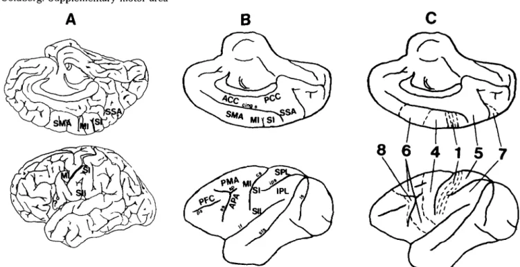
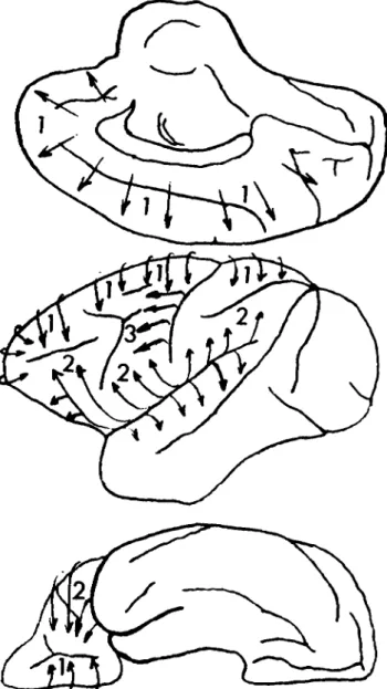
The approximate localization of the SMA on the mesial frontal surface of the primate and human brain is illus-trated in Figure 1. Also shown are approximate localiza-tions of several other cortical regions that enter into the following discussions.
Although this paper is not intended to be an exhaustive review, several different modes of investigation are exam-ined and juxtaposed in an attempt to construct an over-view of SMA structure and function based on an integra-tion of these different perspectives. Other recent reviews have examined evidence pertaining to the function of the SMA in limb control (Eccles 1982; Humphrey 1979; Wiesendanger 1981) and speech (Jonas 1981). Some of the ideas presented have been briefly dealt with
else-A
8 6 4 1 5 7
Figure 1. Functional cortical areas shown on the surface of the cerebral hemisphere of the human (A) and the primate (B) brain. The third figure in this composite (C) shows approximate localizations of Brodmann's architectonic zones. ACC: anterior cingulate cortex; APA: arcuate premotor area (infraarcuate area 6); IPL: inferior parietal lobule; MI: primary motor cortex; PCC: posterior cingulate cortex; PMA: premotor area (supraarcuate area 6); SI: primary somatosensory cortex; SII: second
somatosensory area; SMA: supplementary motor area; SPL: superior parietal lobule; SSA: supplementary sensory area; ?: possible analogue of the APA in the human brain, as: arcuate sulcus; cs: central sulcus; ps: principal sulcus; ips: intraparietal sulcus; If: lateral fissure; sts: superior temporal sulcus; cing s: cingulate sulcus; Is: lunate sulcus; sp: spur of the arcuate sulcus. Figure 1C is adapted from Bowker & Coulter 1981, p. 207.
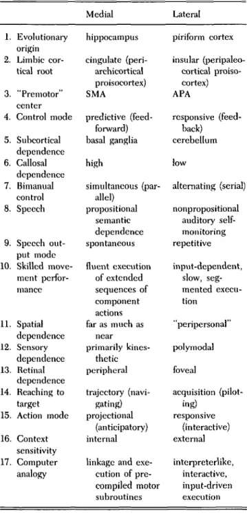
where (Goldberg 1984). In this paper, it is hypothesized that a medial bilaterally organized premotor system can be recognized in which the SMA functions as a central cortical region. This medial system is distinguished from a putative lateral premotor system in which the arcuate premotor area (Schell & Strick 1984) of the primate brain serves as the premotor cortical focus. Table 1 compares salient features of these two premotor systems.
The structural distinction between these two systems is based on a theory of the evolution of cortical architec-tonics put forward by Friedrich Sanides (1964). This theory is reviewed in section 2.1. The two systems have different anatomical relationships with the major re-entrant subcortical motor structures, the basal ganglia and the cerebellum (Schell & Strick 1984). This point is reviewed along with other recent anatomic information regarding the connectivity of the SMA in section 2.2. The functional significance of the distinction between these two systems is examined through an analysis of the reported effects of lesions of the SMA in humans and primates (section 4) as well as through an examination of the physiologic approaches to understanding SMA func-tion through cortical unit studies in animals (secfunc-tion 5), regional cerebral blood-flow studies (section 6), and event-related brain potential studies (section 7). A special section deals with the role of these two motor program-ming systems in bimanual coordination (section 4.3). Some of the conclusions of these discussions are summa-rized in Table 1. In particular, it is postulated that there are two complementary systems within the cerebral hemisphere, a medial one which derives from the hippo-campal formation and a lateral one which derives from the
piriform cortex. The medial system operates in "projec-tional" action or action that is driven forward by predic-tion derived from an internal model of the world com-posed from previous experience which permits the creation of a probabilistic model of the future (Bernstein 1967). The lateral system is part of a system responsible for recognizing and associating motivational significance with external objects and, in relation to action, operates in a responsive mode in which each action is dependent upon an explicit external input. These hypotheses are developed in more detail in the following discussions. Finally, a new anatomico-physiologic model of the medial premotor system is presented. In this model, the SMA is considered to be a key element in a medial, bilaterally organized system which operates in concert with a variety of other cortical and subcortical structures to perform context dependent selection, linkage, initia-tion, and anticipatory control of a set of "precompiled" motor subroutines each of which corresponds to a particu-lar component perceptual-motor strategy or schema of the complete action. The operation of this system in the performance of a discrete action is outlined. Action is assumed to be initiated through a developmental sequence in which increasing amounts of detail are spec-ified as the time the action is to be expressed overtly is approached. This microgenetic process of action specifi-cation underlying the formation of an action recapitulates the evolutionary process of phylogenetic development of the relevant structures, with each participating structure manifesting its involvement through a component feature of the complete act (Brown 1977). In this context, the SMA is viewed as a crucial link within a widely
Goldberg: Supplementary motor area
Table 1. A comparison of features of the putative medial and
lateral motor programming systems
1. Evolutionary origin 2. Limbic cor-tical root 3. "Premotor" center 4. Control mode 5. Subcortical dependence 6. Callosal dependence 7. Bimanual control 8. Speech 9. Speech out-put mode 10. Skilled move-ment perfor-mance 11. Spatial dependence 12. Sensory dependence 13. Retinal dependence 14. Reaching to target 15. Action mode 16. Context sensitivity 17. Computer analogy Medial hippocampus cingulate (peri-archicortical proisocortex) SMA predictive (feed-forward) basal ganglia high simultaneous (par-allel) propositional semantic dependence spontaneous fluent execution of extended sequences of component actions far as much as near primarily kines-thetic peripheral trajectory (navi-gating) projectional (anticipatory) internal
linkage and exe-cution of pre-compiled motor subroutines Lateral piriform cortex insular (peripaleo-cortical proiso-cortex) APA responsive (feed-back) cerebellum low alternating (serial) nonpropositional auditory self-monitoring repetitive input-dependent, slow, seg-mented execu-tion "peripersonal" polymodal foveal acquisition (pilot-ing) responsive (interactive) external interpreterlike, interactive, input-driven execution
tributed layered system of structures involved in the generation of action, rather than a particular site from which voluntary movements are initiated (Eccles 1982). The proposed scheme should be able to provide a frame-work for the study of the preparatory processes preceding movement, the way such a system participates in the microgenesis of a motor act, and the processes associated with the acquisition of a new skill. Possible experimental approaches to the testing of these hypotheses and further clarification of our understanding of SMA function are suggested.
2. Neuroanatomic considerations
2.1. Architectonics. Architectonics is the study of the histological structure of brain tissues. Campbell (1905)
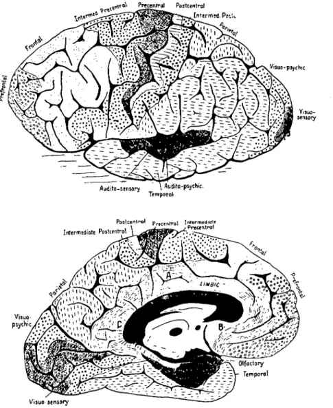
completed one of the first architectonic analyses of human cerebral cortex by examining serial sections stained for nerve cell bodies and myelin (see Figure 2). He identified a zone of cortex extending anteriorly from the precentral gyrus on the ventrolateral, dorsolateral, and medial as-pects of the frontal lobe, which he called the "intermedi-ate precentral" area. Campbell's map indic"intermedi-ates that he felt this zone to be a uniform architectonic field. He identified it as being associated with the highest level of the motor hierarchy postulated by Hughlings Jackson 20 years earlier (Jackson 1884).
G. E. Smith (1907), examining the gross appearance of fresh-cut autopsy specimens, was able to subdivide the human cerebral cortex on the basis of local variations in the whitish stripes of intracortical myelinated fibers called the bands of Baillarger. He found that Campbell's intermediate precentral area could be clearly subdivided into several architectonic fields, including an anterior superior frontal field located dorsally and medially, an intermediate frontal field located dorsolaterally, and posterior inferior and inferior frontal fields located ven-trolaterally (see Figure 3).
In the most famous of architectonic maps, that of Brodmann (1908), the differentiation of a superomedial frontal field corresponding to the SMA is not as clear as it appears on Smith's map. This was later clarified by the subdivision of Brodmann's area 6 by the Vogts (Vogt and Vogt 1919).
In a recent architectonic analysis of the human telen-cephalic cortex, Braak (1980) has clearly separated a dorsomedial part of the premotor cortex from a ventrolateral component (see Figure 4). He has identified these two major zones anterior to the paraganglionic belt of the precentral area:
a. the inferofrontal magnopyramidal region in front of the ventrolateral part of the paraganglionic belt (corre-sponding roughly to Smith's inferior frontal field B) and b. the superofrontal magnopyramidal region located anterior to the superior and medial aspects of the para-ganglionic belt (corresponding roughly to Smith's superi-or and anterisuperi-or superisuperi-or frontal fields)
Although one can conjecture that the former zone is the human equivalent of the primate arcuate premotor area (see Schell & Strick 1984) and may possibly correspond to Broca's area, it is the latter architectonically defined cortical zone that probably comes closest to being congruent with the SMA. Braak (1980) himself suggests the correspondence of this field with the area activated in various motor tasks and identified as the SMA in regional cerebral blood-flow studies (e.g., Roland, Larsen, Lassen & Skinh0j 1980; see further discussion in section 6).
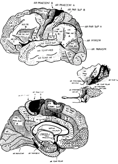
Friedrich Sanides (1964; 1970; 1972), in a careful exam-ination of the patterns of distribution of the architectonic fields of the human frontal lobe, and in an attempt to link these patterns with functional fields revealed by neurophysiologic investigations, has proposed a theory of the evolution of the structure of the cerebral cortex and its phylogenetic differentiation. This work extended a hy-pothesis initially put forward by Dart (1934) following detailed studies of reptilian brain structure. This concept was further supported by the work of Abbie (1940) on the primitive mammalian brain of the monotremes. The same concept has been recently extended to the human par-ietal lobe (Eidelberg & Galaburda 1984).
^TCX Preccntral Postcentral *4t x _ .<'J'Ji!?.«»»w. Viauo-psychic-Audito-sensory \Auiiito-psychic. Temporal
Postcentral precentral Inttrmediate Intermdiatc Postcentral - ' - y
reantro
Visuo- / psychic.
Visuo-sensory
Figure 2. Architectonic map of Campbell (1905). Note his "intermediate pre-central" area with both a dorsomedial and a ventrolateral expansion.
This theory views the cerebral cortex as a dynamic, fluid biologic entity whose architectonic structure has evolved across phylogeny. New architectonic fields arise from phylogenetically older regions carrying with them structural likenesses from their predecessors. Pro-gressive systematic sequences of architectonic differ-entiation, referred to as "protogradations" (Sanides 1964) or "ur-trends" (Sanides 1970), can thus be recognized beginning in the oldest cortical areas and moving through progressively more recent fields. Sanides (1964, p. 280) summarized his theory with the following statement: "The gradations originating from phylogenetically older cortices determine the structure of more recent cortices. Therefore they should be recognized at the same time as evolutional directions of differentiation."
In applying this theory to the architectonic structure of the human frontal lobe, Sanides identified three "pro-togradations" (see Figure 5). One originates in the peripaleocortical proisocortex of the insular region and gives rise to the second somatosensory area (SII) and the ventrolateral part of the premotor cortex - the inferofron-tal magnopyramidal region of Braak (1980) in the human brain - corresponding to the arcuate premotor area of the primate (Sanides 1972). The second protogradation origi-nates in the periarchicortical proisocortex of the medial
wall of the frontal lobe, the anterior cingulate area. This medial protogradation then proceeds superiorly on the medial surface and laterally over to the external surface of the hemisphere. The two protogradations interact on the lateral aspect of the hemisphere at the level of the principal sulcus in the primate brain and the inferior frontal sulcus in the human brain. On the inferior surface of the frontal lobe, the two protogradations meet in the area of the prefrontal orbital region. The SMA can thus be considered part of the medial protogradation, a paralim-bic "protomotor" zone derived from the anterior cingu-late cortex (Sanides 1964). Similarly, the arcuate pre-motor area, part of the parainsular protogradation, may be considered a lateral "protomotor" zone (Sanides 1964;
1972). Sanides states:
considering our ur-trends of differentiation in evolu-tion from archicortex via the cingulate gyms medially and from paleocortex via the insula laterally, it was conceived that the supplementary motor representa-tion . . . is an earlier stage of motor control, and the second somatic sensory representation is an earlier stage of sensory control than the respective classic representations. (Sanides 1970, p. 163)
It is thus tempting to speculate that the fundamental dualism of architectonic evolution created by these two 570 THE BEHAVIORAL AND BRAIN SCIENCES (1985) 8:4
AR PRAECENT 8
AR PRAECENT A
AR PAR SUP B
- - A R PAR SUP A
- - A R PFRISTR
Figure 3. Architectonic map of Elliot Smith (1907). Note the separate dor-somedial (Ar. front, sup. and Ar. fr. sup. ant.) and ventrolateral (Ar. fr. inf.) fields extending rostrally from the precentral area.
simultaneously developing coronal protogradations gives rise to a dualism of limbic-cortical systems and thus dual channels through which sensory input can be neocor-tically processed and associated with limbic structures (Bear 1983; Ungerleider & Mishkin 1982), and through which action can likewise be generated (see also Trevarthen 1968). It is this idea, as it relates to the regions of the brain that organize action, that is elaborated into the major hypothesis developed in this paper.
The architectonic concept of evolution of cortical struc-tures from separate sources has some interesting implica-tions for connectivity which appear in section 2.2. Sanides (1970; 1972), for example, recognized the rela-tionship between his theory and the dual nature of prefrontal connectivity noted by Nauta (1964). In the primate brain, the region of prefrontal cortex medial and superior to the principal sulcus tends to project medially into the cingulum whereas the region ventrolateral to the principal sulcus tends to project with fibers from the orbital prefrontal system toward the anterior temporal lobe. Furthermore, connections between the prefrontal-premotor regions and parietal cortex seem to be
orga-nized in such a way that the areas ventrolateral to the intraparietal sulcus are most heavily connected with the areas ventrolateral to the principal sulcus in the frontal lobe, whereas the areas medial and dorsal to the intra-parietal sulcus tend to project most strongly to the frontal areas dorsal and medial to the principal sulcus (Petrides & Pandya 1983).
There is a third and more recent protogradation identi-fied by Sanides which begins with the classic primary motor cortex (MI) and then proceeds poleward in the anterior direction. Thus, MI is considered to have appeared much later in the evolution of frontal lobe architectonic structure than the previous two protograda-tions. Sanides (1964) postulated that the primary motor cortex is a relatively recent cortical structure which has evolved for the purpose of controlling discrete frac-tionated distal contralateral movements, a function ap-pearing late in the evolution of mammalian species and best developed in humans. Sanides (1970, p. 202) sug-gested that the paralimbic-parainsular neocortical growth rings of the SMA and the ventrolateral premotor areas "serve a general tetrapod function," whereas the
Figure 4. Architectonic map of Braak (1980). Note two sepa-rate frontal fields anterior to the paraganglionic belt of the precentral area: the inferofrontal magnopyramidal region (A) and the superofrontal magnopyramidal region (B). Adapted from Braak (1980).
new sensorimotor representation of the classic areas is related to further adaptation to terrestrial life which required the limbs - particularly the forelimb - to be released from "compulsory tetrapody" in order to serve independent adaptive functions, for example, in feeding and in using tools.
It might be argued that the appearance of a phylogenetically more recent primary motor cortex may have relegated the medial "protomotor" SMA to a ves-tigial role. It would appear more likely, however, that the primary motor cortex arose as a necessary extension of the more rudimentary SMA in response to evolutionary "pressure" pushing toward the development of in-creasingly lateralized distal dexterity and coordinated, prehensile skills. The dolphin, clearly not subject to the evolutionary pressure peculiar to terrestrial life, has a brain that, despite an advanced gyral folding pattern of the cortical mantle, has no evidence of the development of a hypergranular core - the architectonic marker of MI - on the convexity of the hemisphere; instead, the cor-tical development appears to have been arrested at the parainsular-paralimbic stage of development (Morgane,
Figure 5. The protogradations of Sanides (1964; 1970). The three directions of neocortical differentiation within the frontal lobe are noted on this diagram of the primate cerebral hemi-sphere: (1) the medial protogradation taking origin in the cingu-late cortex; (2) the cingu-lateral protogradation taking origin in the insular cortex. (3) the most recent protogradation beginning in the ganglionic core of the precentral area and proceeding poleward.
Galaburda & Jacobs 1983). Viewed as an extension of more medial protomotor cortex, which, through its con-centration of large pyramidal cells, provides a direct, multiply-parallel, refined capability for phasic control of extremity musculature, MI would be necessary for the performance of more complex and phylogenetically more recent motor behaviors in animals with developed dex-trous limb function. A more basic infrastructure of action in which the aspects controlled by MI are embedded, may be conveyed via the medial (and lateral) protomotor cortex.
What is the functional nature of such a basis for move-ment for which the medial and lateral protomotor areas may be responsible? A major clue lies in the recognition of a fundamental separation of function based on the
structural derivation of the two protomotor areas from different brain sources - namely, the hippocampus as the source from which the medial protomotor area (SMA) developed, and the piriform cortex as the source from which the lateral protomotor area (APA) was derived. 2.2. Connectivity. If we define the term "premotor" to mean the areas of frontal cortex rostral to the primary motor cortex that contain a substantial proportion of cells projecting monosynaptically to the primary cortex in a topographically organized fashion, then the SMA can be considered a "premotor" cortex. Horseradish peroxidase (HRP) retrograde transport studies have examined the nature of the projection pattern to the primary motor cortex in the primate brain (Matsumura & Kubota 1979; Muakkassa & Strick 1979; Pieper, Goldring, Jenny & McMahon 1980). These HRP studies demonstrate that the major ipsilateral projection to the primary motor cortex arises from the SMA. Less intense projections to the primary motor cortex appear from the ventrolateral premotor area in the posterior bank of the arcuate sulcus, labeled the "arcuate premotor area" (APA) by Strick and his coworkers. These findings have recently been studied in an elegant anatomic study using three different retro-grade fluorescent tracers injected into the face, hand, and foot areas of MI (Godschalk, Lemon, Kuypers & Ronday 1984). This study clearly demonstrates the two routes of access to MI: from the SMA and the APA. It further shows that whereas the SMA projects to all three injected zones, the APA projects only to face and hand areas of MI, with those neurons projecting to the hand area being located in the posterior bank of the inferior limb of the arcuate sulcus and around the arcuate spur more rostral and dorsal to the adjacent APA area on the ventrolateral aspect of the precentral gyrus projecting to the face zone. Some projections to MI arise from dorsolateral area 6, dorsal to the upper branch of the arcuate sulcus, but these tend to come from caudal zones contiguous with MI. This, along with evidence from architectonic studies, patterns of connectivity, and stimulation mapping stud-ies, has led Wiesendanger (1981) to propose that dor-solateral area 6 consists of structurally and functionally distinct rostral and caudal subdivisions. The caudal sub-division can be considered a rostral microexcitable exten-sion of primary motor cortex which relates to proximal and axial muscle activation (Murphy, Kwan, Mackay & Wong 1978) with corresponding corticospinal projections (Murray & Coulter 1981a). The rostral extent can be considered motor association cortex with sparse direct connection to motor cortex and prefrontallike connec-tivity (Kiinzle 1978) and physiologic acconnec-tivity {Sakai 1978; Weinrich & Wise 1982). This zone can be clearly distinguished from the SMA by its relative lack of direct connections to MI (Matsumura & Kubota 1979; Muak-kassa & Strick 1979). It may represent a dorsolateral elaboration of the SMA with convergent connections to the SMA (Kiinzle 1978), which is particularly activated with movements into extrapersonal space (Roland, Skinh0j, Lassen, & Larsen 1980).
It is of interest to consider how patterns of anatomic connection may help to differentiate the SMA from the APA in the primate brain. Kiinzle (1978) found that, although both areas project to MI, SMA tended to relate
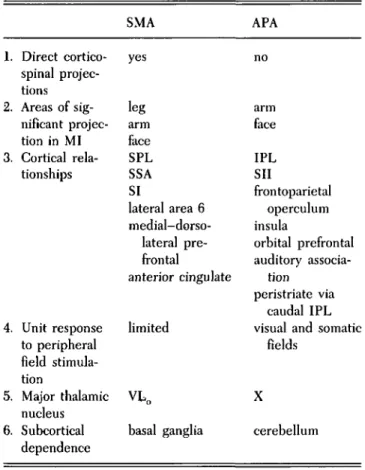
to the medial prefrontal area in front of it whereas the infraarcuate part of area 6, roughly corresponding to an area of cortex around the APA, related to ventrolateral prefrontal cortex below the principal sulcus and the orbital prefrontal cortex. Furthermore, this ventrolateral premotor area was connected with the insular and inferior temporal cortex. The SMA has significant corticospinal projections, whereas the APA does not (Kiinzle 1978). Furthermore, relationships with parietal cortex are quite different (Petrides & Pandya 1983). Although the SMA has major connections to area 5 of the superior parietal lobule (Bowker & Coulter 1981), the APA receives somatosensory projections from SII and combined visual and somatic inputs from the inferior parietal lobule (Godschalk et al. 1984; Petrides & Pandya 1983) as well as projections from auditory association cortex (Galaburda & Pandya 1982). Clearly, the APA has access to a wealth of processed polymodal sensory information, a point con-sistent with the hypothesis developed in this paper and with the physiologic response characteristics of APA (Rizzolatti, Scandolara, Matelli & Gentilucci 1981a; 1981b). Thus the SMA and the APA may be rather unambiguously differentiated on the basis of their pat-terns of connectivity (see Table 2 and Figures 6 and 7). Evidence has recently been accumulating to confirm the suggestion that the SMA projects directly into the corticospinal tract (Biber, Kneisley &: La Vail 1978; Jones & Wise 1977; Macpherson, Marangoz, Miles & Wiesen-danger 1982; Murray & Coulter 1981a). Murray and Coulter (1981a) have reported finding a significant direct projection (7.5% of all projecting neurons) from the SMA into the corticospinal tract using HRP injections into different levels of the spinal cord. SMA projections, along
Table 2. Two routes to primary motor cortex (MI)
1. 2. 3. 4. 5. 6. Direct cortico-spinal projec-tions Areas of sig-nificant projec-tion in MI Cortical rela-tionships Unit response to peripheral field stimula-tion Major thalamic nucleus Subcortical dependence SMA yes leg arm face SPL SSA SI lateral area 6 medial-dorso-lateral pre-frontal anterior cingulate limited VLO basal ganglia APA no arm face IPL SII frontoparietal operculum insula orbital prefrontal auditory associa-tion peristriate via caudal IPL visual and somatic
fields
X
with those from MI, terminated predominantly in the gray matter of the ventral horn, in contrast to corticospinal projections from the sensory cortices which were directed predominantly to the dorsal horn.
A report by Macpherson et al. (1982) confirms anatom-ically and functionally that the SMA is coupled directly to spinal centers. These authors injected HRP into cervical and lumbosacral levels of macaca fascicularis after mapping the extent of the SMA functionally by intracor-tical microstimulation (ICMS). HRP-labeled cells were subsequently found in the microexcitable parts of the SMA.
In a study of the corticocortical relationships of the SMA, Bowker and Coulter (1981) found a major re-ciprocal relationship between the SMA and the medial extension of area 5 on the mesial face of the parietal lobe, an area corresponding to the "supplementary sensory area" (SSA) identified by Penfield and Jasper (1954) in the human brain and more recently examined by Murray and Coulter (1981b). Reciprocal interconnections with lateral area 5, MI, and the more lateral parts of area 6 were identified. The SMA also received a nonreciprocal pro-jection from the primary somatosensory cortex (SI).
The significant relationship with the SSA is of some interest. It is possible that with the successful application of Sanides's architectonic theory to the parietal lobe (Eidelberg & Galaburda 1984), the SSA can be viewed as a focal paralimbic region which is part of the periarchicor-tical medial protogradation developing from posterior cingulate cortex at the base of the parietal lobe. By applying the same logic Sanides used to analyze the frontal lobe, one can consider the SSA as a parietal analog of the SMA with which it coevolved. The pattern of connectivity of the SSA is very similar to that of the SMA (Murray & Coulter 1981b); in addition the two areas strongly interconnect (Bowker & Coulter 1981). The function of the SSA remains obscure.
Some very important clues about the organization of these structures may be obtained through a detailed analysis of the corticothalamic relations of these areas since both sensory inputs to the cortex and reentrant input from the basal ganglia and the cerebellum are linked to the cortex through thalamic connections. Recent evidence regarding the connections of the "motor" thalamus forms an important basis for the con-struction of the anatomico-physiologic model of the medi-al motor programming system presented in section 9. It is becoming apparent from anatomic and elec-trophysiologic investigation that pallidal and cerebellar motor reentrant inputs as well as lemniscal inputs are directed to anatomically distinct thalamic targets (Hen-dry, Jones & Graham 1979; Jones 1981) and that these target zones then project differentially to the cortex. This has recently been shown with electrophysiologic tech-niques (Yamamoto, Hassler, Huber, Wagner & Sasaki 1983).
Kalil (1978) studied the afferent and efferent linkages of the ventral thalamic nuclei using radioactive tracers in rhesus monkeys. The SMA and the mesial prefrontal cortex rostral to it were found to be major cortical projec-tion zones of the VLO and VA nuclei. MI received its
thalamic connections from VLC and rostral VPLO.
Projec-tions from VLC tended to go to more rostral aspects of area
4 and contiguous caudal parts of area 6. Efferent fibers
from the deep cerebellar nuclei were found to terminate in VPLO, VLC, and VLO, whereas lemniscal efferents
terminated primarily in VPLC. Basal ganglia outflow via
projections from the internal pallidal segment has been found to be directed to thalamic nuclei VA and VLO
(DeVito & Anderson 1982). Tracey, Asanuma, Jones, and Porter (1980) examined the relationship of different parts of the ventral thalamus with sensorimotor structures. HRP injections into VPLO and VLC were found to label
cells in the deep cerebellar nuclei whereas injections into VLO led to retrograde labeling of cells only in the internal
palhdal segment. Schell and Strick (1984) have recently examined the thalamic connections of the SMA, the arcuate premotor area, and MI, using HRP injections into these areas of the Macaca mulatto. Little or no overlap between thalamic projection zones was found. Whereas the SMA was connected most densely with the VLO, an
area of the thalamus receiving projections almost exclusively from the globus pallidus, the APA was con-nected most intensively with the medial thalamic nucleus X, an area that receives input primarily from the caudal parts of the deep cerebellar nuclei (Kalil 1981). MI was primarily connected with the VPLO, a thalamic nucleus
that also receives primarily cerebellar input. It is thus apparent that the two premotor areas, the SMA and the APA, can be distinguished on the basis of subcortical dependence: The SMA receives its reentrant drive from outflow from the basal ganglia via the internal pallidal segment connections to the thalamus; the APA (along with MI) receives its reentrant drive from outflow from the deep cerebellar nuclei via their thalamic projections. These anatomic studies of pallidothalamic and cere-bellothalamic relationships and the corresponding thalamocortical relationships have been examined re-cently using electrophysiologic techniques in primates (Yamamoto et al. 1983). This study indicated that inhib-itory inputs from the globus pallidus reached the lateral-dorsal-rostral part of the primate motor thalamus, whereas the stimulation of the deep cerebellar nuclei produced responses in the medial-ventral-caudal re-gion. Convergence of cerebellar and pallidal projection to single thalamic cells was seen only rarely. These results have been recently confirmed in another laboratory (Huffman, Felpel & Lum 1984). Uno, Ozawa, and Yamamoto (1978) reported that, in the cat, those thalamic cells that showed evidence of inhibitory input from the entopeduncular nucleus - the feline equivalent of the internal pallidal segment - could be activated anti-dromically by stimulating the medial precruciate cortex, a zone that may correspond to the SMA. A similar electrophysiologic examination in the primate would be very important and could add critical support to the evidence suggesting that the SMA interacts directly with that part of the thalamus being inhibited by pallidal projections whereas the APA interacts with cerebellar-dependent thalamic neurons (Schell & Strick 1984).
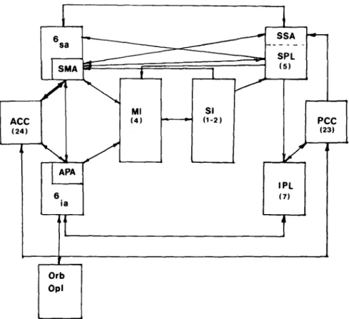
Some of the cortical connectivity pattern between the SMA, the APA, and related cortical zones within the hemisphere is shown in Figure 7.
Neuroanatomic and functional evidence is accumulat-ing to support the notion that the caccumulat-ingulate cortex, long felt to be a major site of interaction between the limbic system and the rest of the cerebral cortex (Papez 1937), may be considered to have distinct, but reciprocally
THALAMUS
Goldberg: Supplementary motor area
CORTEX
R M S
GP
DCbN
Figure 6. Connections between subcortical structures, thalainic nuclei, and functional sen-sorimotor cortical areas. The first tier shows the cortical areas and their interconnections, the second the thalainic nuclei found to relate anatomically to the cortical areas (see Baleydier & Mauguiere 1980; Kalil 1981; Schell & Strick 1984), and the third the connections of the reentrant motor subsystems (RMS) and the medial lemniscus (ML). Abbreviations as in Figure 1 with the addition of GP^ globus pallidus, internal segment; DCbN: deep cerebellar nuclei. Note that the reentrant input from the GP; interacts with ACC and SMA whereas that from the DCbN relates
to areas of the ventral thalamus connected to the APA and MI. Inputs from DCbN and GPj connect with anatomically distinct zones of the thalamus which, in turn, relate to distinct cortical areas. MI is subdivided here into rostral (r) and caudal (c) components.
interconnected anterior and posterior subdivisions (Baleydier & Mauguiere 1980; Vogt, Rosene & Pandya 1979).
The anterior zone of the cingulate cortex would appear to be an area of high-level efferent integration linked with cortical and subcortical regions (Baleydier & Mauguiere 1980). Stimulation of this region in human epileptic patients produced various forms of highly integrated but isolated motor fragments somewhat reminiscent of those produced with stimulation of the SMA (Talairach, Bancaud, Geier, Bordas-Ferrer, Bonis, Szikla & Rusu 1973). The elicited movements appeared to be movement "primitives" or "subroutines" that could be linked to-gether in context to perform complete movement se-quences. With stimulation of the anterior cingulate it was noted that when a movement was elicited it was associ-ated with a spread of excitation to the adjacent frontal medial cortex - most probably including the SMA - and along the cingulate cortex of the same hemisphere as well as across to the corresponding zone of the contralateral cingulate gyms. With this spread of excitation and the appearance of a motor response there developed a steady 3-8 Hz rhythm in the EEG which was maximal at the vertex (Talairach et al. 1973).
The rostral extension of the anterior cingulate cortex has been studied to determine its role in the control of vocalization in the primate (Aitken 1981; Jiirgens &
Miiller-Preuss 1977; Jiirgens & von Cramon 1982). Jiirgens and Muller-Preuss (1977) found that all cortical areas from which vocalization in the primate could be elicited through electrical stimulation received direct inputs from the anterior cingulate cortex. They suggested that the anterior cingulate cortex may not be responsible for the actual specifics of the vocalization but rather maintained thresholds for a particular vocalization across the network, thus controlling the activation of specific behaviors. Jiirgens and von Cramon (1982, p. 246) postu-lated that "anterior cingulate cortex seems to function as a drive-controlling mechanism which determines by its activity the readiness to phonate as well as the intensity." It could be argued that this is the general role of the anterior cingulate cortex as it relates to all action (e.g. Gray 1982a), that is, the control of drive-dependent thresholds and a generalized specification of intensity or "amplitude" of a voluntary act. The major importance of the SMA would then be the intermediary role it could play between the generalized internal drive control manifested through the anterior cingulate cortex and the selection and execution of specific action sequences or motor strategies performed downstream from this point, by virtue of its anatomic position between the anterior cingulate cortex on the one hand and the primary motor cortex on the other.
para-ACC (24) 6 sa
s
SMA / APA 6 ia • • • Ml (4) SI (1-2) •** SSA SPL PC (2 IPL (7) Orb Opl 1 -C 3)Figure 7. Some of the major cortical relationships of the SMA and related areas shown schematically. Abbreviations as in Figure 1 with the addition of 6sa:
supraarcu-ate component of area 6; 6ia: infraarcuate component of area 6; Orb: orbital frontal
cortex; Opl: frontal opercular and insular cortex. Adapted from Jones (1983). limbic cortex (see section 2.1; Brown 1977; Sanides 1964)
located at the confluence of anterior cingulate, superior mesial prefrontal, dorsolateral area 6, and mesial primary motor cortices. It is reciprocally interconnected with the anterior cingulate cortex (Damasio, Van Hoesen & Vil-ensky 1981) and, as discussed in the previous section, may have "evolved" from ventrally adjacent limbic periarchicortical proisocortex (Sanides 1970). It is part of an efference-synthesizing corticolimbic-reticular system (Watson, Miller & Heilman 1978) which focuses limbic outflow onto motor executive regions (Damasio et al. 1981), thus linking intention formation to the program-ming and execution of specific actions. Disorders of this system have been shown to lead to a neglect syndrome based, not on a perceptual inability, but rather on a response deficiency (Watson, Heilman, Cauthen & King 1973; Watson et al. 1978). Intimately associated with this system is an afferent-regulating corticolimbic-reticular network that controls the rostral flow of sensory informa-tion and thus atteninforma-tional and perceptual processes, in part through the selective subcortical "gating" of afferent flow and, in part, through the modulation of the re-sponses of the sensory association areas to their afferences (Heilman & Valenstein 1979; Mesulam 1981; Robinson, Goldberg & Stanton 1978; Roland 1981; 1982; Skinner & Yingling 1977; Watson, Valenstein & Heilman 1981). On the basis of differences in connectivity patterns (Bal-eydier & Mauguiere 1980; Vogt et al. 1979), anterior cingulate cortex may be more closely related to the efference-synthesizing system, whereas posterior cingu-late cortex may be more directly involved in the afferent-regulating network (see also Watson et al. 1981). Through
multiple linkages and convergences at many different levels between these two systems, an action-perception cycle (Neisser 1976) is formed. One of the major sites for such integration is the prefrontal cortex.
Fuster (1980) has proposed that the prefrontal cortex is necessary for formulating temporally integrated, context-dependent behavioral structures for goal-oriented action, particularly in novel or complex circumstances. Not only does it operate to maintain temporal contiguity, it also acts to suppress interferences or competing tendencies so that goal orientation can be maintained. The orbital prefrontal cortex is particularly important in response control - the suppression or inhibition of interfering tendencies to respond to external inputs when such responses would disrupt purposive behavior. It is part of a system that includes the temporal cortex and the amyg-dala, wliich forms a "neural complex essential for the appraisal of the motivational significance of objects" (Fuster 1980, p. 70). It can be argued on the basis of information reviewed in the previous section that the APA should be considered a node in this particular network whereas the SMA is not. It might also be postu-lated that the component elements of this functional network have evolved from the piriform-derived paleocortical root and, as such, would be part of the lateral protogradation. Similarly, the dorsomedial pre-frontal cortex above the principal sulcus of the primate brain may be considered part of the medial protograda-tion and thus a separate funcprotograda-tional network derived from the hippocampal-derived archicortical root. This part of prefrontal cortex is particularly involved in the "integra-tion of spatially and temporally discontiguous elements in
cognition" (Fuster 1980, p. 56) and thus has an important cognitive-perceptual role to play in the context-depen-dent performance of learned and instinctual behaviors. The SMA, along with the supraarcuate part of area 6, is associated with the dorsal and medial prefrontal system. This system tends to project medially into the cingulum toward retrosplenial and parahippocampal regions as op-posed to the ventral and lateral areas of the prefrontal cortex which tend to project into the temporal lobe (Nauta 1964). Thus, what Nauta (1964, p. 405) noted as "a certain dualism in the prefrontolimbic association," may translate into a dualism in functional systems in the hemisphere and a related dualism in the premotor re-gions, all of which may relate to the manner in which the neocortex has evolved.
To summarize, the SMA is an area of cortical convergence receiving projections from primary and sec-ondary somatosensory areas as well as from parietal asso-ciation cortex. It lacks extrastriate inputs (Pandya & Kuypers 1969) which do reach the arcuate premotor area via the inferior parietal lobule (area 7). Such visual inputs may also distinguish the SMA from the APA and may be important for certain functions of the APA in visually dependent behaviors (Godschalk, Lemon, Nijs & Kuypers 1981; Kubota & Hamada 1978). The SMA is a zone of internal convergence within the premotor regions (Kiinzle 1978). It is linked reciprocally with the anterior cingulate part of "limbic" cortex and would appear to be a major site at the cortical level through which limbic outflow may influence cortical and subcortical motor structures (Damasio et al. 1981). Each SMA receives input from MI as well as other parts of premotor and prefrontal cortex and then projects bilaterally back to MI (though more densely to ipsilateral MI), to the con-tralateral SMA, and to various subcortical structures. It sends projections bilaterally to the striatum and also projects strongly to the cerebellar cortex via the pontine nuclei. The SMA also sends direct projections to the spinal cord. Through these projections the SMA could potentially participate in the coordination of both axial and distal musculature, ipsilaterally as well as con-tralaterally. Its pattern of inputs would indicate that the SMA has available to it sensory data conveying informa-tion about the external environment and the body schema (though without the visual input available to the APA) required for setting up and adapting motor programs, as well as limbic-mediated inputs via its reciprocal connec-tions with ventrally adjacent anterior cingulate cortex, conveying motivational-behavioral influences concern-ing internal needs and drives (Orgogozo & Larsen 1979). Recent data (Schell & Strick 1984) demonstrate that the SMA is a major cortical target of basal ganglia outflow, suggesting that it may preferentially participate in the execution of learned motor sequences (Marsden 1982). The SMA would appear to be a major cortical site mediat-ing the interaction between cortical limbic outflow via anterior cingulate cortex, the context-sensitive, goal-setting functions of the prefrontal cortex (whose outputs, along with those of many other associational areas of the cortex, are integrated and refocused onto the SMA via the basal ganglia reentrant circuit), the sensory analysis func-tions of the association cortex of the superior parietal lobule, and the executive components of the motor system.
An examination of some of the important issues in brain architectonics and connectivity that relate to the struc-ture of the SMA has been presented. This information may provide important clues about how the SMA might be expected to function physiologically. The most impor-tant information may be derived from a careful considera-tion of the evoluconsidera-tionary perspective and the work of Sanides dealt with in section 2.1.
3. Effects of electrical stimulation
Penfield and Welch (1949; 1951) defined the "supple-mentary" motor area through surface stimulation of the cortex in conscious human epileptic patients. It should be recognized that there are numerous difficulties with the interpretation of this type of data, particularly as it relates to the implied physiologic function of a part of the cortex. A detailed discussion of these problems is beyond the scope of this review.
Penfield and Welch (1951, p. 316) noted a zone of cortex "situated almost altogether within the median longitudinal fissure and anterior to the primary motor foot area" which, when stimulated, produced one of a group of clinical observations, including the following:
a. Inhibition and transient arrest of ongoing voluntary activity: Following the completion of a period of stimula-tion, the patient often expressed puzzlement at the in-ability to execute a.voluntary act as it had been intended while the stimulation was applied.
b. At higher levels of stimulation, the assumption of a posture, most commonly elevation of the contralateral arm with abduction and external rotation of the shoulder. Vocalization was also produced at some sites, with perseverated syllables heard (e.g. Woolsey et al. 1979). Talairach and Bancaud (1966) observed that a behavior may develop sequentially with continued stimulation at one point.
Woolsey and his colleagues (Woolsey, Settlage, Meyer, Sencer, Hamuy & Travis 1952) found a somatotopic arrangement in SMA in the primate, with responses produced in the face found more anteriorly than those of the upper limb, trunk, and lower limb. In comparing the SMA to MI, they found that the SMA became more readily inexcitable with anesthesia than MI and that thresholds for responses tended to be generally higher in the SMA. Whereas MI rapidly habituated to repetitive stimuli, the SMA responded best to prolonged trains of stimulation. Thus although stimulation of MI produced transient, phasic movements with rapid habitu-ation, the SMA stimulation was found to produce sus-tained influences on behavior, which did not habituate readily. Similar observations in man led Talairach and Bancaud (1966, p. 341) to postulate that the SMA organizes "postural movements and . . . tonic motility." When the SMA is stimulated following excision of MI, the movements elicited tend to be more bilateral and proximal (Penfield & Welch 1951; Wiesendanger, Sequin & Kiinzle 1973). It has been questioned whether with stimulation of the cortical surface current spreads from the stimulating site to activate nearby MI directly or SMA projections to MI modulate MI activity transsynaptically (Wiesendanger et al. 1973; for review, see Humphrey 1979). However, it has recently been demonstrated that
movements of both contralateral proximal and distal limb joints can be produced with intracortical microstimula-tion of the SMA (Macpherson et al. 1982). This finding led the authors to suggest "the possibility of a close coupling between SMA and spinal motor nuclei" (Macpherson et al. 1982, p. 415), although the coupling was not felt to be as "tight" as that between MI and the segmental motor circuits.
4. Effects of lesions
4.1. The effects of lesions in primates. There is little agreement in the literature regarding the effects of le-sions of the SMA in subhuman primates (see Humphrey 1979; Wiesendanger 1981). Horsley and Schafer (1888) noted extensive paralysis which followed bilateral abla-tion of the marginal gyri. They were impressed by the severity of the deficit compared to the effects of removing equal amounts of tissue from the lateral convexity. Travis (1955) found that, whereas unilateral SMA lesions had negligible lasting effects, simultaneous bilateral SMA damage produced a flexion posturing of the limbs. When lesions of MI were extended to include the SMA, changes in postural tone and reflex hyperexcitability appeared. This led Travis to postulate that the SMA played an important role in the development of "spasticity." Coxe and Landau (1965) were not able to reproduce the deficit that Travis reported.
Unilateral SMA ablation, generally, appears to involve more transient and less evident deficits than bilateral ablations. Although there is a transient appearance of both forced grasping (Seyffarth & Denny-Brown 1948) in the contralateral hand of the primate (A. M. Smith 1979; Smith, Bourbonnais & Blanchette 1981) and a mild bilat-eral apraxia of fractionated distal movement of the upper extremities (C. Brinkman 1982; J. Brinkman 1981), the lasting effect of unilateral SMA ablation in both monkeys and man appears to be an impairment of bimanual coordi-nation (C. Brinkman 1982; J. Brinkman 1981; see section 4.3).
The effect of SMA ablation on the organization of voluntary movements in monkeys has received little attention. Moll and Kuypers (1977) found that premotor ablation that included the SMA but was extended to the lateral premotor areas impaired the ability of a monkey to adapt a trained, visually directed reaching movement of the contralateral limb to reaching around a transparent barrier for a piece of food. The authors postulated that the animal was not able to inhibit a subcortically driven reaching movement evoked by the visual image of the food seen through the barrier. It is unclear, however, to what extent this observation is related to SMA as opposed to APA injury since both areas were damaged in this study. This type of released visually dependent approach behavior, termed "magnetic apraxia" (Denny-Brown 1958) when unilateral or "utilization behavior" (Lher-mitte 1983) when bilateral, has been observed in animals conditioned to reach toward visual targets in extraper-sonal space (Deuel & Dunlop 1979; Stepien 1974) and in human patients presented with familiar objects (Lher-mitte 1983), after damage involving dorsal premotor or adjacent dorsolateral prefrontal cortex. In these situa-tions, behaviors that have been conditioned prior to the lesion become inflexibly linked to presentation of the
visual cue regardless of the context as a result of the damage to areas of the frontal cortex. The way that damage to different subdivisions of the premotor cortex participates in producing such effects remains to be clearly determined.
4.2. Effects of lesions of the SMA in humans. Damage to the SMA in patients has been reported to produce effects on speech as well as on limb movement (Alexander & Schmitt 1980; Goldberg, Mayer & Toglia 1981; Jonas 1981; Kornyey 1975; Masdeu, Schoene & Funkenstein 1978; Racy, Janotta & Lehner 1979; Rubens 1975). The effects on speech are usually observed with damage to the dominant left hemisphere although an exception has been reported (Brust, Plank, Burke, Guobadia & Healton 1982). The syndrome usually consists of a lack of spon-taneous conversational speech with a well-preserved abil-ity to repeat phrases. There is difficulty in initiating speech, with struggle and hesitation. Some argue that the impairment cannot be considered a true aphasia since the structure of the speech produced is intact; they call it instead a "partial mutism" (Brown 1977; Damasio & Van Hoesen 1980). With unilateral SMA damage, the impair-ment of speech is often transient and the prognosis for recovery appears good (Rubens 1975). This may result from bilateral participation of the SMA in the generation of speech (Larsen, Skinh0j & Lassen 1978).
In a recent paper, McCarthy and Warrington (1984) have proposed a dual mechanism in the speech-produc-tion system to account for the striking dissociaspeech-produc-tions seen with certain lesions between the impairment of repeated, nonpropositional speech as opposed to spontaneous, propositional speech, such as that seen with SMA lesions. They propose that speech output can be obtained through a route involving semantic analysis as well as through a direct route between "auditory/phonologic transcoding" and articulatory output. It could be suggested from the hypothesis developed in this paper that the semantic-analysis route involves the SMA and the related compo-nents of the medial premotor system that are primarily responsible for the generation of propositional speech, that is, speech that conveys semantic content and emanates from an endogenous source (see also Jonas 1981). On the other hand, the lateral premotor system including the APA (possibly Broca's area of the human brain) and the inferior parietal lobule, may include the auditory-phonologic transcoding "feedback loop postu-lated by Lichtheim (1885) to be a direct "route between the auditory images of words and the motor images of words" (McCarthy & Warrington 1984, p. 464). The speech output mode of the medial system would thus be spontaneous voluntary output of propositional speech, whereas that of the lateral system would be repetition (see Table 1, line 9).
Luria (1966) has described, in great detail, patient "Ch," who almost certainly sustained injury to the SMA following a midline frontal depressed skull fracture. The patient exhibited what Luria referred to as "de-auto-matization" of speech and limb movements. Luria partic-ularly noted a sharp contrast between the relative fluency of speech in dialogue and the considerable difficulty with independent extended speech. The patient was able to reply quickly and directly to questions that required brief, direct responses but was unable to elaborate his 578 THE BEHAVIORAL AND BRAIN SCIENCES (1985) 8:4
ideas beyond such short, abrupt answers. With regard to this apparent lack of spontaneity, the patient remarked that "thoughts do not enter my head" (Luria, 1966, p. 226). Limb movements were characterized by a distur-bance in organizing smooth, integrated sequences. A gross disturbance was noted in the ability to coordinate bimanual acts. The patient would tend to perform a bimanual task by successive acts with each hand sepa-rately, particularly when the task required simultaneous asymmetric or reciprocal action. There was a strong tendency, when bimanual simultaneous movement was performed, to produce symmetric mirror movements (see section 4.3). The patient recognized these problems in his own performance and would remark: "it seems as if my hands do not belong to me . . . something holds them back, and they do not do what they should (Luria 1966, p. 230), suggesting that although the image of the intended act was intact and the intent-to-act verbally expressable, the transformation into successful coordinated perfor-mance was impaired. This led the patient to interpret his own behavior in terms of an exogenously produced dissociation between intent and action (Bogen 1979; Goldberg et al. 1981). With unilateral movements, the patient's greatest difficulty occurred in the assembly of component movements into a smooth and continuous sequence. Movement elements in a sequences were performed in isolation, but the formation of a "kinetic melody" governing the smooth and automatic execution of the sequence did not occur even after months of practice. This difficulty was labeled "a disturbance of the formation of motor dynamic stereotypes" (Luria 1966, p. 231). Luria postulated that the disturbance involved a defect in the selective gating of kinesthetic impulses responsible for the rapid and smooth transition from one motor link or "subschema" (Arbib 1981) to the next in a serially organized movement. Similarly, the intention to express a thought spontaneously in words did not lead to a smooth unfolding of a complete propositional sentence. Laplane et al. (1977) reported observations on three patients in whom unilateral corticectomies of the SMA had been performed to ameliorate intractable epilepsy with SMA foci. They found that, in addition to the partial mutism noted above, the initial effect of the lesion was a severe decrease in spontaneous movement of the limbs which was particularly pronounced contralaterally. Actions not produced spontaneously could be elicited in response to strong spoken commands by the examiner. A contralateral facial paralysis was noted with spontaneous smiling which disappeared when the patient smiled to command. Grasp automatisms were not seen. Several months after the surgery, recovery was almost complete. Patients continued, however, to have difficulty with bi-manual coordination (see section 4.3).
Laplane et al. (1977) proposed that the SMA acts to initiate and sustain spontaneous motor activity and that damage to the SMA thus produces more severe impair-ment of "intentional" action arising primarily out of internal context as opposed to environmentally contingent "responsive" action. Damasio and Van Hoesen (1980) came to similar conclusions in their report of three patients with SMA damage. They hypothesized (p. 359) that the SMA provides the internal "'drive' for willed movement" but is not necessary for the "eventual realization " of such movement, presumably because it
was possible to demonstrate that simple movements could be performed when cued from external context. Goldberg et al. (1981) have reported two cases of left medial frontal cortex infarction involving the SMA in which organized goal-directed movements of the contralateral hand and arm appeared in apparently extra-volitional fashion; that is, they arose in conflict with the verbalized internal context. This type of striking dis-sociative disturbance has been labeled the "alien hand sign" (Bogen 1979; Brion & Jedynak 1972). Although it has been thought that this disturbance results from a deterioration of interhemispheric communication, it has been suggested that its appearance may be related to dysfunction of cortical structures on the mesial wall of the contralateral frontal lobe (Goldberg et al. 1981).
The appearance of extravolitional, goal-directed actions suggests that the SMA may function normally to inhibit such actions unless they are specifically addressed by a volitionally generated signal reflecting internal con-text. It is possible that the alien hand sign may follow from a disconnection between limbic outflow and the motor executive areas. Just as it may appear from the examina-tion of the dissociative sensory phenomena seen with temporal lobe seizures that a neocortically-elaborated percept requires limbic association to assume "experien-tial immediacy" (Gloor, Olivier, Quesney, Andermann & Horowitz 1982), so action may require limbic participa-tion to assume its voliparticipa-tional or self-referenced origin. If the neural substrate mediating the coupling of limbic drive to the executive motor areas at the cortical level is impaired, one might expect an impoverishment of spon-taneous intentional action arising primarily out of internal context, and, possibly, the appearance of relatively auto-matic behaviors occurring extravolitionally in response to a particular external context. This might be expected if the lesion resulted in an abnormal imbalance favoring the initiation of environmentally contingent "automatic" ac-tions, which are responsive in nature, rather than inter-nally generated actions, which are anticipatory in nature (see Figure 8).
Jonas (1981) has reported on an extensive survey of the clinical literature describing speech disturbances associ-ated with SMA lesions. He noted that during the recov-ery from lesions of the SMA, "nonpropositional 'auto-matic' speech may be initiated easily, even involuntarily, while initiation of propositional speech is still difficult or impossible" (Jonas 1981, p. 349; emphasis added). The hypothesis formulated by Jonas to account for these observations was that the intact SMA acts to facilitate and control the initiation of propositional speech, whereas it "plays a role in the suppression of the emission of nonpropositional 'automatic' speech" (Jonas 1981, p. 369). It could be argued that this hypothesis can be extended in an analogous fashion to the role of the SMA in voluntary action in general.
4.3. Effects of lesions of the SMA on bimanual coordina-tion: Medial and lateral motor programming systems. C.
Brinkman (1982) and J. Brinkman (1981) have noted that primates with unilateral SMA lesions have difficulty with bimanual coordination tasks requiring independent movements of the two hands. The animals tended to produce mirror-symmetric movements in such situa-tions, as did the patients with SMA lesions. When
Figure 8. The separation of feed-forward (projectional) and feedback (responsive) control mechanisms in the organization of action (adapted from D. M. MacKay 1978). F: sensorimotor field of action; R: receptor system; E: effector system; If: information submitted to comparator (C) for evaluation against current criteria (I ); M: running indication of match/mismatch between Ifand Ig; S: supervisory system which deter-mines current criteria and supervises and monitors the organizing subsystems Offand On) whose task it is to "prescribe a running selection from E's repertoire calculated to bring or keep Irin line with Ig" (D. M. MacKay 1978, p. 54). O^: subsystem within the organizing system which implements projectional feed-forward control under the direction of S and uses recognition of salient task-relevant features of the current sensory context for anticipation. On): subsystem within the organizing system which implements action in response to a mismatch signal from C. This conceptualization thus enables us to "distinguish between two basically different ways in which action can originate" (D. M. MacKay 1978, p. 55).
Brinkman subsequently sectioned the corpus callosum in the lesioned animals, they were once again able to perform the bimanual task successfully (C. Brinkman 1982). How can these findings be understood?
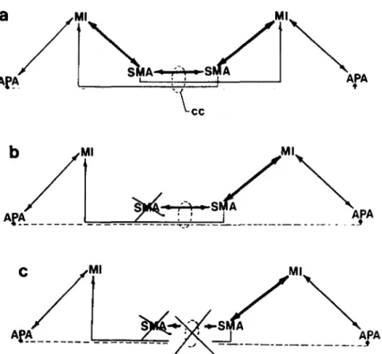
As has been noted, the SMA of each hemisphere projects to both the ipsilateral and the contralateral primary motor cortices. One can hypothesize that there are two potentially independent but necessarily interac-tive motor programming channels, each having access to
the executive apparatus for both sides of the body. If we
assume that each SMA is connected to the Mis in such a way that homologous muscles of the two extremities are activated together then one SMA acting alone could very well tend to produce mirror-symmetric movement. Un-der normal conditions for independent bimanual control it would be necessary for each "active" SMA to establish a dominant role in control of the contralateral limb through its projections to ipsilateral MI and to suppress the other SMA's potential influence through its callosal connec-tions to the contralateral SMA and MI. It is known that synchronous bilateral movements using homologous muscles (i.e. mirror-symmetric movements) are per-formed with much greater agility than those using non-homologous muscles (Wyke 1969). For bilateral move-ments requiring synchronous activation of nonhomo-logous muscles, both hemispheric programming chan-nels would require activation and each SMA would then operate via its connections to ipsilateral MI. To maintain appropriate phase relationships between the two chan-nels so the hands could work together in a complemen-tary, cooperative fashion, communication between the two SMAs via the corpus callosum would be critical. This coordination may be necessary to establish an overall temporal structure for the task that ensures predictable
parallel phase relationships between the two simul-taneous programs controlling the two hands (Kelso, Southard & Goodman 1979). However, an alternative mode of bimanual control could be obtained through slower visual guidance mechanisms mediated intra-hemispherically by corticocortical extrastriate connec-tions to the more lateral arcuate premotor cortex (Jones, Coulter & Hendry 1978; Pandya & Kuypers 1969). Thus, in corpus callosectomy, the long-term impairment of complex intermanual coordination (Zaidel & Sperry 1977) may be due to an inability to maintain appropriate phas-ing between two independently operatphas-ing intra-hemispheric programming channels, though this may be partially compensated through visual feedback.
One could then hypothesize the presence of two sepa-rate intrahemispheric motor programming systems which follow from the earlier discussions regarding a duality of premotor projections: a medial motor system including the S MA (medial paralimbic protomotor cortex) and the closely associated basal ganglia (Schell & Strick 1984), and a lateral system including ventrolateral arcuate premotor cortex. Such a hypothesis is based on the idea that the SMA is part of a dorsomedial hippocampally derived system which is concerned with perception and representation of space and is necessary for extended, internally dependent, predictive or projectional action. This system is more concerned with the general problem of navigating the limb through space than with the more focal problem of accurately acquiring identified objects in "peripersonal" (Rizzolatti, Matelli & Pavesi 1983) or reachable space. The lateral protomotor area, the APA, is part of a ventrolateral piriform-derived system which is concerned with perceiving and recognizing external inputs and investing them with motivational significance, 5 8 0 THE BEHAVIORAL AND BRAIN SCIENCES (1985) 8:4
Goldberg: Supplementary motor area Ml APA ML
tyirfr
SMA J AW Figure 9. Bihemispheric relationships of the motor areas (see also C. Brinkman 1982; J. Brinkman 1981). Abbreviations as in text with the addition of cc: corpus callosum. (a) Normal pattern of connection. Each SMA is connected to both Mis though more strongly connected ipsilaterally. fl also receives predominantly ipsilateral input from the APA. Callosal connections between SMAs are more intensive than those between APAs. (b) With unilateral damage to the SMA, the contralateral remaining SMA predomi-nates in both ipsilateral and contralateral limb control through its projections to the Mis of both hemispheres. This impairs bimanual coordination by increasing the tendency for movements to be mirror symmetric. This tendency is dependent on callosally mediated input from the intact SMA to the contralateral MI and is also responsible for the persisting impairment of simultaneous reciprocal action (see text), (c) With an additional lesion of the corpus callosum, the fibers from the intact SMA to the contralateral MI are disrupted, thus releasing the MI on the left side of the diagram from any medial system control. Bimanual coordination may be improved to some extent (C. Brinkman 1982), but now there is no interhemispheric coordina-tion. The MI on the left is under exclusively lateral system (APA) influence whereas that on the right still has medial (SMA) and lateral (APA) system inputs. This difference in system dependence between the two hemispheres under such conditions could represent the basis for observation of the alien hand phenomenon in human patients with such damage (Goldberg et al. 1981).an operation depending, for visual recognition, on foveal information (Bear 1983; Ungerleider & Mishkin 1982; see also Trevarthen 1968). It is hypothesized that the ven-trolateral premotor system, which has a more direct association with this object analysis and recognition sys-tem, is used in the production of interactive, externally contingent responsive action driven by the presence and identified nature of specific objects in the organism's immediate environment (Paillard 1982b; Rizzolatti et al. 1983).
Task performance depends on two internal representa-tions of the world: a categoric model of the world at present based on an integration of past and current experience of it, and a probabilistic model of the future which drives the action forward by permitting anticipato-ry interpolation between the current sensed state and a
future predicted state (Bernstein 1967; Requin, Semjen & Bonnet 1984). Action can occur in response to the world as it is sensed in the present, or it can be guided by projections about how the world will be at a point in the future. The former mode of control would occur through the lateral system and the latter through the medial system, according to the present proposal.
In the more bilaterally organized medial system, the programming of unilateral and bilateral movements requires close callosally mediated interaction between the two hemispheres. Bihemispheric activation of the SMAs and the basal ganglia is seen even with unilateral movement when MI activation is only contralateral (Roland, Meyer, Shibasaki, Yamamoto & Thompson 1982). The medial system predominates when rapid, well learned, "skilled" movement sequences are executed