HAL Id: hal-02116773
https://hal-amu.archives-ouvertes.fr/hal-02116773
Submitted on 1 May 2019
HAL is a multi-disciplinary open access archive for the deposit and dissemination of sci-entific research documents, whether they are pub-lished or not. The documents may come from teaching and research institutions in France or abroad, or from public or private research centers.
L’archive ouverte pluridisciplinaire HAL, est destinée au dépôt et à la diffusion de documents scientifiques de niveau recherche, publiés ou non, émanant des établissements d’enseignement et de recherche français ou étrangers, des laboratoires publics ou privés.
Ultrafiltration membrane fouling characterization by
multi-scale methodology
Morgane Le Hir, Yvan Wyart, Gaelle Georges, Laure Siozade, Michelle
Sergent, Philippe Moulin
To cite this version:
Morgane Le Hir, Yvan Wyart, Gaelle Georges, Laure Siozade, Michelle Sergent, et al.. Ultrafiltra-tion membrane fouling characterizaUltrafiltra-tion by multi-scale methodology. Euromembrane 2018, Jul 2018, Valence, Spain. �hal-02116773�
Ultrafiltration membrane fouling characterization by multi-scale
methodology
M. Le Hir*, Y. Wyart*, G. Georges**, L. Siozade**, M. Sergent***, P. Moulin* * Aix Marseille Université, CNRS, Centrale Marseille, M2P2 UMR 7340, Equipe Procédés
Membranaires (EPM), Europôle de l’Arbois, BP80, Pavillon Laennec, Hall C, 13545 Aix en Provence Cedex, France
** Aix Marseille Univ, CNRS, Centrale Marseille, Institut Fresnel, Marseille, France
*** Aix Marseille Université, LISA EA4672, Avenue Escadrille Normandie Niemen, 13397 Marseille, France
Keywords: Characterization; multi-scale; fouling; nanoparticles; fluorescence
Introduction
Membrane characterization by tracer retention is widely used to determine the retention potential and integrity of membranes by quantification of tracers passed through this one. The use of fluorescent tracers makes this retention quantification quick and reliable. Moreover, additional information like the location of retained tracers (Wu et al. 2014; Marroquin et al. 2014) and the composition of membrane fouling, with the use of complementary techniques, can be obtained, allowing to characterize membrane morphology and fouling. This study presents a multiscale characterization methodology to clearly determine the retention potential of ultrafiltration membrane regarding fluorescent nanoparticles (NPs) with calibrated size. Location of each NPs size in or/and on the membrane after filtration run allows to identify with precision the penetration profiles of NPs in membrane structure (skin and support) and to determine fouling mechanism(s) which can be correlated with macroscopic flux data observed during the filtration.
Material and Methods
Ultrafiltration (UF) membranes tested are PES multichannel hollow fiber with nominal pore size of 20 nm. Ideal suspensions of fluorescent calibrated NPs with size of 1,5, 10 and 100 nm were filtered. The size of the NPs used was chosen regarding the membrane pore size (smaller, close to and bigger than the membrane pore size). Zeta potential of NPs suspension and membrane were measured to ensure that no aggregation or adsorption occurs during filtration. The efficiency of the membrane was determined by NPs concentrations measurement of filtration fluxes by fluorimetry and Nanoparticle Tracking Analysis (NTA). The use of Nanosight NS300 (using NTA) allowed to obtain concentrations in number of particles per mL allowing to estimate the number of NPs remaining blocked in and/or on the membrane. Location of NPs fluorescent signal in and/or on the membrane was visualized by confocal laser scanning microscopy (CLSM) and penetration profiles of NPs have been determined. In the case of a NPs deposit on membrane surface, its thickness was measured with scanning electron microscopy (SEM) and CLSM. The use of NPs with different fluorescent characteristics allowed to distinguish each size of NPs in membrane material. Location of NPs fluorescent signals have been correlated with macroscopic permeate flux data to validate fouling mechanism(s) operating during the filtration experiment. Influence of Transmembrane Pressure (TMP) and Volume Concentration Factor (VCF) on NPs retention, fouling establishment and location was treated by experimental design.
Results and Discussion
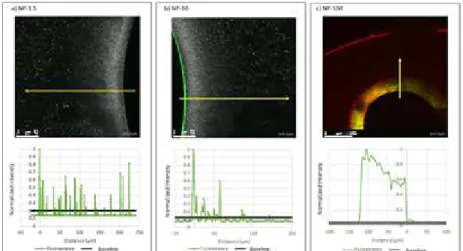
Ultrafiltration of ideal monodisperse and polydisperse NPs suspensions was conducted. Membrane shows interesting retention rates (≥ 50% for all NPs size and for each condition tested) and a low recovery in the retentate reflecting a retention of NP by blocking at the membrane. Fouled membranes were imaged by CLSM on their section allowing to enjoy the lateral resolution of CSLM (about 500 nm), better than the axial one used in a previous study (Wu et al. 2014). Analysis in reflective mode allowed to visualize membrane and another in fluorescent mode allowed to identify NPs. MCBL visualization showed that 100 nm NPs were essentially retained on membrane surface, 10 nm NPs were stopped in the membrane skin (thickness ≈ 50 µm) and on membrane surface and 1.5 nm NPs are located in the entire material (Figure 1). These locations reflect respectively fouling models of cake filtration (100 nm), standard blocking followed by cake filtration (10 nm) and standard blocking (1.5 nm) evaluated with experimental data of permeate flux.
Figure 1. Penetration profile of a) 1.5 nm NP, b) 10 nm NP and c) 100 nm NP in membrane material
Conclusions
Multi-scale characterization techniques were used to determine UF membrane efficiency towards NPs, fouling establishment and its location in or/and on the membrane. The complementary techniques used allow to obtain concordant results of interest showing the reliability of characterization realized. It is the first time that the penetration profile of NP in UF membrane was determined with high precision (skin vs. support). Moreover, the number of NP in the permeate (i.e. in the drinking water) was evaluated against to answer at a societal point of view.
Acknowledgment
"The project leading to this publication has received funding from Excellence Initiative of Aix-Marseille University - A*MIDEX, a French "Investissements d'Avenir" programme. It has been carried out in the framework of the Labex MEC." The authors are grateful to Aquasource for providing the new generation membranes used during this work.
References
Hermia, J. 1982. “Constant Pressure Blocking Filtration Laws - Application to Power-Law Non-Newtonian Fluids.” Trans IChemE.
Marroquin, Milagro, Anh Vu, Terri Bruce, Rhonda Powell, S. Ranil Wickramasinghe, and Scott M. Husson. 2014. “Location and Quantification of Biological Foulants in a Wet Membrane Structure by Cross-Sectional Confocal Laser Scanning Microscopy.” Journal of Membrane Science 453 (March): 282–91.
Wu, N., Y. Wyart, L. Siozade, G. Georges, and P. Moulin. 2014. “Characterization of Ultrafiltration Membranes Fouled by Quantum Dots by Confocal Laser Scanning Microscopy.” Journal of Membrane Science 470 (November):40–51.