HAL Id: tel-01557752
https://tel.archives-ouvertes.fr/tel-01557752
Submitted on 6 Jul 2017
HAL is a multi-disciplinary open access
archive for the deposit and dissemination of sci-entific research documents, whether they are pub-lished or not. The documents may come from teaching and research institutions in France or abroad, or from public or private research centers.
L’archive ouverte pluridisciplinaire HAL, est destinée au dépôt et à la diffusion de documents scientifiques de niveau recherche, publiés ou non, émanant des établissements d’enseignement et de recherche français ou étrangers, des laboratoires publics ou privés.
positioning and activation in mammals
Nina Danielle Kirstein
To cite this version:
Nina Danielle Kirstein. Chromatin-dependent pre-replication complex positioning and activation in mammals. Human health and pathology. Université Montpellier; Ludwig-Maximilians Universität (Munich, Allemagne), 2017. English. �NNT : 2017MONTT005�. �tel-01557752�
Délivré par l’Université de Montpellier et l’Université
Ludwig Maximilian de Munich
Préparée au sein de l’école doctorale CBS2
Et de l’unité de recherche
Institute of Regenerative
Medicine and Biotherapy et Helmholtz Zentrum München
Spécialité : Biologie/ Santé
Présentée par Nina Danielle KIRSTEIN
Soutenue le 8. Juin 2017 devant le jury composé de
Dr. María GOMEZ, Centro de Biologia Molecular (ES) Rapporteur Dr. Torsten KRUDE, Univ. of Cambridge (UK) Rapporteur Prof. Dr. Axel IMHOF, Univ. Ludwig Maximilian (DE) Président du jury Dr. Philippe PASERO, IGH (FR) Examinateur Prof. Dr. Gunnar SCHOTTA, Univ. Ludwig Maximilian (DE) Examinateur Prof. Dr. John DE VOS, IRMB (FR) Examinateur Dr. Jean-Marc LEMAITRE, IRMB (FR) Codirecteur de thèse Dr. Aloys SCHEPERS, Helmholtz Zentrum München (DE) Directeur de thèse
Chromatin-Dependent Pre-Replication
Complex Positioning and Activation in
2 |
R
ESUME
Chaque division cellulaire requiert une duplication précise du génome. Des dizaines de milliers de sites d’initiation de la réplication d’ADN (origines de réplication) sont impliqués dans la réplication complète du génome humain. L’activation des origines de réplication est régulée précisément et des études génomiques extensives ont démontré la présence de caractéristiques génomiques associées à l’activation des origines de réplication. Le complexe de pré-réplication (pre-RC) est la base de l’initiation de la réplication et consiste en deux sous-complexes majeurs : l’ « origin recognition complex » (ORC) qui interagit directement avec l'ADN et est nécessaire pour recruter le second sous-complexe, les hélicases Mcm2-7, qui sont responsables de l'initiation de la réplication. La régulation de l’assemblage du pre-RC est bien étudiée, mais les caractéristiques de la chromatine qui déterminent le positionnement du pre-RC sur le génome restent peu connues. Les études génomiques par immuno-précipitation de la chromatine et séquençage à haut débit (ChIP-seq) des pre-RCs sont rares et jusqu’à aujourd’hui seulement disponibles pour ORC. Du fait que Mcm2-7 migre de son site de chargement initial, il est crucial d'obtenir des informations sur le positionnement des Mcm2-7 pour la compréhension complète de la régulation de la réplication.
Ce travail présente la première analyse génomique par méthode ChIP-seq des deux sous-unités majeures du pre-RC, ORC et Mcm2-7, dans la lignée cellulaire de lymphome de Burkitt Raji infectée par le virus d’Epstein-Barr (EBV). La présence du génome d’EBV permet d'avoir un contrôle interne de la qualité de nos expériences, en comparant les positions de pre-RC déterminées avec des positions du pre-RC précédemment publiées. Sur le génome humain, les résultats de séquençage du pre-RC corrèlent bien avec des zones de réplication active. De façon intéressante, les zones de terminaison de la réplication étaient spécifiquement bas en pre-RC, spécialement en Mcm2-7. La localisation des sites d'initiation de la réplication identifiés est généralement bien corrélée avec les sites de transcription active. En effet, des sites d’assemblage du pre-RC de haute affinité sont localisés préférentiellement en voisinage de sites de transcription active, ce qui est possiblement dû à l’accessibilité de la
3 |
chromatine dans ces régions. La fixation de Mcm2-7 fluctuait de façon dépendante du cycle cellulaire, ce qui suggère des translocations de Mcm2-7 en G1, probablement dépendantes de la machinerie active de la transcription. Ces résultats indiquent que les positions de ORC et Mcm2-7 sont principalement dépendantes de l’accessibilité de la chromatine avec un accès privilégié dans la chromatine active et Mcm2-7 étant le déterminant majeur de l’initiation de la réplication.
Au sein de l'hétérochromatine, ORC était enrichi dans des zones associées avec l'histone modifié H4K20me3. Cependant, cet enrichissement était moins important pour les Mcm2-7. En utilisant un système de réplication basé sur des plasmides, nous avons démontré que l’association d'ORC et H4K20me3 favorise l’assemblage du pre-RC et l’initiation de la réplication. Cette observation suggère que l’interaction ORC-chromatine est le déterminant majeur de la régulation de la réplication d’ADN au sein de l’hétérochromatine. En conclusion, cette étude propose deux mécanismes différents de la régulation de l'assemblage du pre-RC dépendants de l’environnement de la chromatine.
Mots clés : Réplication d’ADN, pre-RC, chromatine, PR-Set7, Suv4-20h1/h2, H4K20 méthylation
4 |
Z
USAMMENFASSUNG
Mit jeder Zellteilung muss das Genom exakt dupliziert werden. Zehntausende Replikationsinitiationsstellen sind bei der Replikation des gesamten humanen Genoms beteiligt. Die Aktivierung der Initiationsstellen ist präzise reguliert und umfassende Genom-weite Studien haben verschiedene genomische Faktoren identifiziert, die die Aktivierung der Replikationsinitiationsstellen beeinflussen. Der Prä-Replikationskomplex (pre-RC) bildet die Grundlage der Replikationsinitiation und besteht aus zwei Hauptuntereinheiten: der „origin recognition complex“ (ORC) bindet DNS und wird zum Laden der zweiten Untereinheit, den Mcm2-7 Helikasen benötigt, die die eigentliche Replikationsinitiation veranlassen. Während die Regulation des pre-RC Aufbaus vielfach untersucht wurde und mittlerweile gut verstanden wird, sind die Chromatinkomponenten, welche die Positionierung der pre-RCs regulieren, weitgehend unbekannt. Die wenigen Genom-weiten pre-RC Chromatin Immunopräzipitations- und Sequenzierungsstudien (ChIP-seq), behandeln bis heute nur ORC. Da sich Mcm2-7 allerdings von seiner initialen Ladeposition fortbewegen kann, werden vor allem die Genom-weiten Positionen von Mcm2-7 benötigt, um die Regulation der DNS Replikation vollständig zu verstehen.
Diese Arbeit umfasst die erste Genom-weite pre-RC ChIP-seq Analyse der zwei pre-RC Hauptkomponenten ORC und Mcm2-7 in der Epstein-Barr Virus (EBV) infizierten Burkitt-Lymphom Zelllinie Raji. Als Qualitätskontrolle für erfolgreiche ChIPs wurden die aus den vorliegenden Experimenten bestimmten pre-RC Positionen auf dem EBV-Genom mit bereits bekannten pre-RC Positionen verglichen. Auf dem humanen Genom korrelierten die pre-RC ChIP Ergebnisse mit aktiven Replikationszonen, während Replikationsterminationszonen eine spezifische Abnahme der pre-RC Komponenten, besonders von Mcm2-7, aufzeigten. Es ist bereits bekannt, dass aktive Replikation mit aktiver Transkription korreliert. Starke pre-RC Bindung war in der Tat hauptsächlich an Regulationsstellen der aktiven Transkription zu finden, was vermutlich durch die Zugänglichkeit des Chromatins determiniert wird. Starke Mcm2-7 Bindung variierte dabei in Abhängigkeit des Zellzyklus, was für Mcm2-7 Translokationen während der G1 Phase spricht, die vermutlich von der aktiven Transkriptionsmaschinerie
5 |
beeinflusst werden. Diese Ergebnisse deuten darauf hin, dass ORC und Mcm2-7 Positionen in der aktiven Chromatinumgebung hauptsächlich von der Zugänglichkeit des Chromatins abhängen und Mcm2-7 die Hauptkomponente der Bestimmung der Replikationsinitiationsstellen darstellt.
In Heterochromatin assoziierte vorwiegend ORC mit der heterochromatischen Histonmodifikation H4K20me3, während Mcm2-7 weniger Anreicherung zeigte. Unter Verwendung eines Plasmid-basierten Replikationssystems wurde bestätigt, dass diese ORC-Chromatin Assoziation einen essentiellen Einfluss auf die Regulation der pre-RC Positionierung und Aktivierung ausübt. Dieses Ergebnis zeigt, dass ORC-Chromatin Interaktionen einen entscheidenden Faktor für die Regulation der Replikation in Heterochromatin darstellen. Zusammenfassed schlägt diese Studie zwei verschiedene Modi der pre-RC Positionierung vor, welche jeweils vom Chromatinkontext abhängen.
Schlagwörter : DNA Replikation, pre-RC, Chromatin, PR-Set7, Suv4-20h1/h2, H4K20 Methylierung
6 |
A
BSTRACT
With every cell division, the genome needs to be faithfully duplicated. Tens of thousands of DNA replication initiation sites (origins of replication) are involved in replicating the human genome. Origin activation is precisely regulated and extensive genome-wide studies found association of origin activation to several different genomic features. The pre-replication complex (pre-RC) is the basis for replication initiation and consists of two major subcomponents: the origin recognition complex (ORC) binds DNA and is required for loading of the second component, Mcm2-7 helicases, which initiate DNA replication. Regulation of pre-RC assembly is well studied, however, chromatin features driving pre-RC positioning on the human genome remain largely unknown. Genome-wide pre-RC chromatin immunoprecipitation experiments followed by sequencing (ChIP-seq) studies are rare and so far only performed for ORC. As Mcm2-7 can translocate from their initial loading site, information about Mcm2-7 positioning are required for full understanding of DNA replication regulation. This work presents the first genome-wide ChIP-seq analysis of the two major pre-RC subcomponents ORC and Mcm2-7 in the Epstein-Barr virus (EBV) infected Burkitt’s lymphoma cell line Raji. Successful ChIPs were validated on the EBV genome by comparing obtained pre-RC positions with already existing pre-RC ChIP-on chip data. On the human genome, pre-RC sequencing results nicely correlated with zones of active replication. Interestingly, zones of replication termination were specifically depleted from pre-RC components, especially from Mcm2-7. Active DNA replication is known to correlate with active transcription. Indeed, strong pre-RC assembly preferentially occurred at sites of active transcriptional regulation, presumably determined by chromatin accessibility. Strong Mcm2-7 binding thereby fluctuated cell cycle-dependently, arguing for Mcm2-7 translocations during G1, possibly depending on the active transcriptional machinery. These results indicate ORC and Mcm2-7 positions being mainly dependent on chromatin accessibility in active chromatin, with Mcm2-7 being the major determinant of replication initiation.
In heterochromatin, ORC was enriched at H4K20me3 sites, while Mcm2-7 enrichment was less prominent. Employing a plasmid-based replication
7 |
system, ORC association to H4K20me3 was proven to promote successful pre-RC assembly and replication initiation, situating direct ORC-chromatin interactions being the major determinant for DNA replication regulation in heterochromatin. Taken together, this study proposes two different modes of pre-RC assembly regulation depending on chromatin environment.
Keywords: DNA replication, origin licensing, pre-RC, chromatin, PR-Set7, Suv4-20h1/h2, H4K20methylation
8 |
T
ABLE OF
C
ONTENTS
RESUME ___________________________________________________ 2 ZUSAMMENFASSUNG_________________________________________ 4 ABSTRACT _________________________________________________ 6 TABLE OF CONTENTS ________________________________________ 8 1. INTRODUCTION _________________________________________ 11 DNAREPLICATION IS TIGHTLY REGULATED – AND STOCHASTIC 11 1.1.1 ASSEMBLY OF THE PRE-REPLICATION COMPLEX IN LATE MITOSIS/ EARLY G112 1.1.2 REPLICATION ACTIVATION IN S-PHASE: FROM PRE-REPLICATION COMPLEX TO
PRE-INITIATION COMPLEX 13
1.1.3 TERMINATING DNA REPLICATION 13
1.1.4 CELL CYCLE REGULATION OF DNA REPLICATION:PREVENTING RE
-REPLICATION AND INCOMPLETE -REPLICATION 13
1.1.5 METAZOAN ORIGIN LICENSING AND ACTIVATION IS MAINLY STOCHASTIC 15
DNAREPLICATION REGULATION THROUGH CHROMATIN 17 1.2.1 HISTONE MODIFICATIONS REGULATING DNA REPLICATION 18
1.2.2 HISTONE 4LYSINE 20 METHYLATION AFFECTS DNA REPLICATION 18
DNAREPLICATION DEPENDS ON NUCLEAR CHROMATIN ORGANIZATION 19
MAPPING OF DNAREPLICATION INITIATION EVENTS 21 1.4.1 SINGLE MOLECULE DNA COMBING 21
1.4.2 BUBBLE-SEQUENCING AND SHORT NASCENT STRAND SEQUENCING 21
1.4.3 OKAZAKI-FRAGMENT SEQUENCING 22
1.4.4 CHIP-SEQUENCING OF PRE-RC COMPONENTS 23
1.4.5 EPSTEIN-BARR VIRUS GENOME AS MODEL OF HUMAN DNA REPLICATION 24
2. AIM OF THE THESIS ______________________________________ 25 3. MATERIAL AND METHODS _________________________________ 26 MEDIA,MATERIAL,DEVICES,CHEMICALS AND AGENTS 26
BIOLOGICAL METHODS 29
3.2.1 CELL CULTURE 29
9 |
3.2.3 FLUORESCENCE ACTIVATED CELL SORTING (FACS) 32
3.2.4 IMMUNOBLOT 32
3.2.5 PLASMID ABUNDANCE ASSAY 34
3.2.6 CROSS-LINK 35
3.2.7 CHROMATIN IMMUNOPRECIPITATION (CHIP) 35
3.2.8 QUANTITATIVE PCR(QPCR) 38
3.2.9 SEQUENCING 39
BIOINFORMATICS 39
3.3.1 BOWTIE MAPPING OF SEQUENCING READS AGAINST THE GENOME 40
3.3.2 GENERATION OF PILEUP PROFILES 40
3.3.3 MACS2,HOMER, AND T-PIC PEAK-CALLING 40
3.3.4 DEFINITION OF ORC,MCM2-7, AND PRE-RC 41
3.3.5 PEAK COMPARISONS:JACCARD INDEX AND OVERLAPS 41
3.3.6 CALCULATING GENOMIC DISTRIBUTIONS WITH CEAS 41
4. RESULTS AND DISCUSSION ________________________________ 42 SETTING UP CHIP-SEQ EXPERIMENTS IN HUMAN CELLS 42 4.1.1 CONSIDERATIONS TO OPTIMIZE PRE-RCCHIP-SEQ EXPERIMENTS 42
4.1.2 PERFORMING CHIP-SEQ EXPERIMENTS IN RAJI CELLS 44
4.1.3 PERFORMING CHIP-SEQ EXPERIMENTS IN HUMAN EMBRYONIC STEM (HES)
CELLS 56
PRE-RCCOMPONENTS ARE ENRICHED WITHIN REPLICATION UNITS 60 4.2.1 COMPARING T-PIC VS.HOMER PEAK-CALLING ALGORITHMS AND DEFINING
ORC,MCM2-7, AND PRE-RC ON THE HUMAN GENOME 60
4.2.2 PRE-RCS CORRELATE WITH ACTIVE REPLICATION UNITS 65
STABLE ORIGIN LICENSING IS ASSOCIATED TO REGULATION OF ACTIVE
TRANSCRIPTION 72
4.3.1 PRE-RC COVERAGE WAS INCREASED AT ACTIVE TRANSCRIPTION START SITES
72 4.3.2 STRONG HOMER-DEFINED ORC,MCM2-7 AND PRE-RC ASSOCIATED WITH
ACTIVE TRANSCRIPTION START SITES 74
4.3.3 PRE-RC COVERAGE WAS ENHANCED AT H3K4ME3 PEAKS 76
4.3.4 STRONG HOMER-DEFINED MCM2-7 AND PRE-RC ENTIRELY ASSOCIATE WITH
H3K4ME3 78
MCM2-7HELICASES APPROXIMATE ORC IN S/G2 82 4.4.1 COMPLEX NUMBER AND SIZES DO NOT CHANGE IN S/G2 82
4.4.2 MCM2-7 COVERAGE AT AS AND DEPLETION FROM DS ARE LESS PRONOUNCED
85 4.4.3 PRE-RC COVERAGE AT TSS AND H3K4ME3 WAS INCREASED IN S/G2 87
4.4.4 MCM2-7 HELICASES APPROXIMATE ORC POSITIONS IN S/G2 90
ORC ASSOCIATES WITH H4K20ME3 IN HETEROCHROMATIN 94 4.5.1 H4K20ME1 IS PRESENT IN ACTIVE CHROMATIN WHILE H4K20ME3
LOCALIZES TO HETEROCHROMATIN REGIONS 94
4.5.2 ORC PREFERENTIALLY ASSOCIATES WITH H4K20ME3 95
4.5.3 NO DISTRIBUTIONAL CHANGE WAS OBSERVED IN S/G2 99
H4K20ME3 IS NECESSARY FOR ORIGIN LICENSING AND ACTIVATION IN
HETEROCHROMATIN 100
4.6.1 SUV4-20H1 TARGETING UNSPECIFICALLY INDUCES H4K20ME3 102
4.6.2 PR-SET7 TARGETING LEADS TO INDUCTION OF H4K20ME1, CONVERSION INTO H4K20ME3 AND PRE-RC FORMATION 103
10 |
4.6.4 PR-SET7 TARGETING INDUCES PLASMID REPLICATION 106
4.6.5 REPLICATION LICENSING AND ACTIVATION DEPEND ON H4K20ME3 107
5. CONCLUSION __________________________________________ 114 CHROMATIN ACCESSIBILITY IS THE MAIN DETERMINANT OF PRE-RC
POSITIONING IN ACTIVE CHROMATIN 114
DIRECT ORC-CHROMATIN INTERACTIONS REGULATE PRE-RCPOSITIONING IN
HETEROCHROMATIN 117 APPENDIX _______________________________________________ 119 BIBLIOGRAPHY ___________________________________________ 149 TABLE OF FIGURES ________________________________________ 159 TABLE OF TABLES _________________________________________ 163 ABBREVIATIONS __________________________________________ 164 RESUME SUBSTANTIEL _____________________________________ 166 ACKNOWLEDGEMENTS _____________________________________ 174
11 |
1. I
NTRODUCTION
DNA replication is the process of precisely copying the complete genetic information, which assures faithful inheritance of the genome during each cell division. Thereby, DNA replication initiates only once per cell cycle. Misregulation during this process leads to genetic instability, which might have fatal consequences on organs and tissues and can cause cancer or other genetic disorders in humans (Abbas, Keaton, and Dutta 2013). Sites of DNA replication initiation are called origins of replication. While bacteria initiate genome replication at one specific origin (Mott and Berger 2007), replication regulation in Saccharomyces cerevisiae is already more complex. In S. cerevisiae, DNA replication origins were initially identified as autonomous replication sequences (ARS), which contain an 11-17 bp AT-rich consensus sequence. However, only a small part of all ARS consensus sequences are used as replication origins, suggesting that other features also contribute to origin recognition and activation (Fragkos et al. 2015). In higher eukaryotes, origins do not exhibit sequence specificities and DNA replication can initiate at any DNA sequence (Vashee et al. 2003). In humans, 30000-50000 origins are activated in each cell at each cell cycle. Numerous studies during the recent years revealed that origins exhibit a preference for specific features, although none of these features alone are predictive for replication origins (Méchali 2010). In the following, I will detail the regulation of DNA replication in higher eukaryotes and the impact of chromatin, histone modifications, and structural arrangements within the nucleus.
DNA
R
EPLICATION IS
T
IGHTLY
R
EGULATED
–
AND
S
TOCHASTIC
DNA replication is spatio-temporally separated to ensure correct duplication of the genome. During each S-phase of the cell cycle, 30000-50000 origins are activated per cell. These origins are chosen from an exceeding pool of possible origins. For an origin to be activated, proper loading of the pre-replication complex (pre-RC) during preceding G1 is required.
12 |
1.1.1 A
SSEMBLY OF THE PRE-
REPLICATION COMPLEX IN LATE MITOSIS/
EARLYG1
The process of pre-RC assembly is called origin licensing and is restricted from late mitosis to the restriction point in G1-phase of the cell cycle. All possible origins need to be properly licensed, while only a subset is activated during S-phase. Origin licensing consists of the sequential loading of pre-RC proteins and starts with the binding of the origin recognition complex (ORC, Figure 1.1).
FIGURE 1.1: SCHEMATIC REPRESENTATION OF SEQUENTIAL PRE-RC ASSEMBLY. Licensing consists of the sequential loading of pre-RC components and is restricted to G1-phase. First ORC binds DNA as hexameric complex. Cdc6 recruitment traps DNA in the center of the circle and initiates Cdt1-dependent Mcm2-7 helicase loading. Cdt1 dissociates from Mcm2-7 after loading. Another Mcm2-7 hexamer is subsequently loaded in head-to-head conformation (Model template from Bleichert, Botchan, and Berger 2015).
ORC is a hexameric complex with ATPase activity and ATP-dependent DNA contact (Bleichert, Botchan, and Berger 2015). After ORC binding, the ATPase Cdc6 is recruited and DNA-bound ORC-Cdc6 initiates Cdt1 association. Cdt1 chaperones Mcm2-7 (for Minichromosome Maintenance) loading, the ring-structured core of the replicative helicase (Remus et al. 2009; Bell and Kaguni 2013). Cdc6 hydrolyses ATP only when bound to ORC and is required for proper Mcm2-7 loading. Mcm2-7 helicases are sequentially loaded as head-to-head double-hexamer (Remus et al. 2009). Interestingly, once Mcm2-7 double-hexamers are loaded, ORC, Cdc6, and Cdt1 are no longer required for replication initiation (Hua and Newport 1998; Yeeles et al. 2015). Pre-RC assembly is restricted to G1-phase when cyclin-dependent kinase (CDK) activity is low. Licensed origins are then competent for replication activation.
13 |
1.1.2 R
EPLICATION ACTIVATION INS-
PHASE:
FROM PRE-
REPLICATION COMPLEX TO PRE-
INITIATION COMPLEXWith the onset of DBF4-dependent kinase (DDK) and CDK activity during the G1/S-phase transition (for kinase activities, see also Figure 1.2, chapter 1.1.4, p. 124), pre-RCs are converted into the pre-initiation complexes (pre-ICs). Pre-IC formation involves the binding of further proteins, such as Mcm10, Cdc45, Dbp11 and the GINS (Sld5, Psf1, Psf2, Psf3) complex (Gambus et al. 2006). During origin activation, several pre-IC proteins, including Mcm2-7, are phosphorylated by CDK and DDK to initiate replication (Francis et al. 2009). Cdc45-Mcm2-7-GINS (CMG) represent the active replicative helicase, which unwinds the DNA and DNA polymerase loads on single-stranded DNA (Ilves et al. 2010). Replication activation involves the dissociation of the Mcm2-7 double-hexamer into two active hexamers that originate the two replisomes consisting of about 150 proteins replicating DNA bidirectionally (Herrera et al. 2015).
Bidirectional DNA replication involves one strand to be replicated continuously (leading strand), while the other strand is synthesized in discontinuous fragments (Okazaki fragments), which are subsequently ligated (lagging strand). RNA primase de novo generates RNA primers, which are extended by DNA polymerases in 5’ to 3’ direction and removed during a maturation process (see also Figure 1.6, p. 22). DNA polymerases duplicate several hundreds of kb before replication forks collapse (Fragkos
et al. 2015; Petryk et al. 2016). Replication termination happens e.g. when
two replisomes converge.
1.1.3 T
ERMINATINGDNA
REPLICATIONAlthough initiation of DNA replication is well characterized, replication termination remains poorly understood. Replication termination mainly occurs when two opposite replication forks (from two adjacent origins of replication) collide. Also, termination needs to be tightly regulated to prevent premature termination without completing replication. The process involves the disassembly of converging replisomes and resolution of replication-induced DNA catenated intertwines by topoisomerases (Fachinetti et al. 2010). Recent work has shown that the polyubiquitination of the Mcm2-7 subunit Mcm7 at the end of S-phase leads to CMG disassembly (Moreno et al. 2014; Maric et al. 2014). However, this ubiquitination is necessary but not sufficient, suggesting additional factors being involved in replication termination (Lengronne and Pasero 2014).
1.1.4 C
ELL CYCLE REGULATION OFDNA
REPLICATION:
P
REVENTING RE -REPLICATION AND INCOMPLETE -REPLICATIONIt is crucial for genetic stability, that the genome is completely replicated only once per cell cycle. The proteins that regulate cell cycle progression (amongst others cyclins, CDKs, and ubiquitin ligases) are also tightly linked with the control of DNA replication (DePamphilis et al. 2006). Origin
14 |
licensing only happens in absence of DDK and CDKs (Figure 1.2). Existing pre-RCs are inactivated during S, G2 and M, preventing repeated licensing and activation (Blow and Dutta 2005; DePamphilis 2005). As already mentioned in chapter 1.1.2, DDK and CDK phosphorylate several members of pre-RC and pre-IC, which leads to replication activation, but also inhibits re-licensing of replication origins.
FIGURE 1.2: CELL CYCLE KINASE ACTIVITIES DETERMINE PRE-RC FORMATION AND ACTIVATION. Pre-RC assembly happens in absence of kinase activities. With onset of CDKs (red, blue) and DDK (green), replication is activated and re-licensing prevented (Adapted from PINES 1999). The dashed grey line represents expression H4K20 monomethyltransferase PR-Set7 (introduced in chapter 1.2.2, p. 18.)
In metazoans, Cyclin A-Cdk2 phosphorylates Cdt1 and Orc1 (Depamphilis
et al. 2012). Phosphorylation of the Mcm2-7 chaperone Cdt1 in S-phase,
leads to its ubiquitination, export to the cytoplasm, and subsequent degradation. This process ensures Cdt1 protein levels only being present in the nucleus in late mitosis/early G1. Additionally, Cdt1 activity is also repressed by Geminin, a specific Cdt1 inhibitor exclusively existing in metazoans. Geminin is active from S-phase on and degraded with the onset of mitosis. Interestingly, Cdt1 recruits Geminin to DNA and their binding stabilizes Cdt1, probably securing the availability of Cdt1 for the next G1-phase (Ballabeni et al. 2004).
In metazoan cells, ORC subunits are phosphorylated to prevent re-licensing during and after S phase (DePamphilis 2005; DePamphilis et al. 2006). Orc1 phosphorylation reduces binding affinities to chromatin is followed by ubiquitination and degradation (Méndez et al. 2002). Orc2 phosphorylation also dissociates ORC subunits 2-5 from DNA (Lee et al. 2012). Orc1 re-associates to chromatin during mitosis to G1 transition, followed by origin licensing (DePamphilis 2005).
15 |
Replication forks encountering obstacles (such as DNA secondary structures or lesions), or reduced deoxyribonucleotide triphosphate (dNTP) pools within the cell can lead to replication stress and induce replication fork stalling (Yekezare, Gómez-González, and Diffley 2013). For the cell to complete DNA replication, it is crucial to activate another licensed origin that has not been activated so far (dormant origin). Consequently, activation of additional origins decreases inter-origin distances and S-phase length remains constant (Blow, Ge, and Jackson 2011). Existence of dormant origins requires an excess of origin licensing during G1. Only a small proportion of licensed origins is actually activated during S-phase, while the vast majority remains dormant (Musiałek and Rybaczek 2015) and are evicted during passive replication (Kuipers et al. 2011). The choice of which origin to activate is thereby mainly stochastic.
FIGURE 1.3: ORIGIN USAGE FREQUENCY DEFINES ORIGIN EFFICIENCY. Pre-RCs are assembled in G1. Only a subset is stochastically activated during S-phase, with each cell using a different cohort. The frequency of origin usage defines origin efficiency. Origin efficiency is represented schematically as result of SNS-seq (explained in chapter 1.4.2).
1.1.5 M
ETAZOAN ORIGIN LICENSING AND ACTIVATION IS MAINLY STOCHASTICOnly a subset of licensed origins is activated with the onset of S-phase and each cell uses a different cohort of replication origins. This observation led to the definition of origin efficiency as the frequency of a specific origin to be activated in a given cell during a given cell cycle (Méchali 2010) (Figure 1.3).
16 |
There are many features identified so far that influence origin licensing and efficiency (Figure 1.4). While AT-richness defines ARS elements in yeasts, there is a clear preference for CG-rich regions in mammals (Cayrou et al. 2015). Interestingly, not only CG-content, but also potentially resulting three-dimensional G-quadruplex structures (G4) impact on origin efficiency (Besnard et al. 2012; Valton et al. 2014; Langley et al. 2016). Thereby it remains controversial whether G4s actually positively or negatively influence DNA replication (Valton and Prioleau 2016). DNA accessibility, as well as specific histone modifications have been linked to active replication (introduced in detail in chapter 1.2, p. 17), as well as transcriptional activity and enhancer functions. Origins are often more concentrated and active in promoter regions, most probably due to open chromatin configurations, as direct interactions of transcription factors and replication factors have not been found so far (Fragkos et al. 2015). However, while all these different features contribute to origin licensing and activation on different levels, none of them is sufficient on its own and it is most likely a random combination of these different features that defines an origin (Méchali 2010).
FIGURE 1.4: DIFFERENT FEATURES INFLUENCE REPLICATION ORIGINS. From DNA sequence to structure over chromatin to functional organization, all features have been linked to regulation of replication, while none of these features alone define an origin. DNA: AT-rich sequences might facilitate replication activation due to lower melting temperatures. Replication and GC-richness seems to be mainly linked on the structural level, as G4-structures are enriched at active initiation sites. Whether G4-structures facilitate pre-RC binding or replication initiation remains unclear so far. Structure: Bent DNA or loop formation might also facilitate pre-RC loading; MAR: matrix attachment regions. Local chromatin: nucleosome positioning/ nucleosome-free regions promote pre-RC binding, specific histone modification might directly interact with pre-RC components, targeting factors (EBNA1, HMGA1a) interact with ORC and direct replication. Functional organization: Active transcription/transcriptional regulation through enhancers/promoters have been described to influence DNA replication. Direct interactions between replication origin factors and transcription factors have not been reported. (Modified from Méchali 2010).
Recent work on the sequencing of Okazaki fragments led to the identification of broad (10-100 kb) initiation zones (Petryk et al. 2016, introduced in detail in chapter 1.4, p21). The authors claim that broad initiation zones represent replication units, within which replication preferentially initiates from multiple inefficient origins but only one single origin stochastically fires per cell. This observation also implies the necessity of broadly distributed licensed replication origins within such replication units.
17 |
It has been demonstrated, that the number of chromatin-bound Mcm2-7 helicases exceeds the number of active origins of ORC by a factor of 10 to 50 (Donovan et al. 1997; Powell et al .2015; Hyrien 2016). This can be achieved by either i) ORC loading several Mcm2-7 double-hexamers which spread from their binding site (Powell et al. 2015, Drosophila) or ii) ORC loading only one Mcm2-7 double-hexamer at a time, associating and dissociating quickly from DNA (Sonneville et al. 2012, C. elegans). There is evidence for either mechanism and further investigations will be required to conclusively resolve this question.
Finally, these recent findings evoked the model of the Mcm2-7 double-hexamer excess influencing organization of replication timing (Das and Rhind 2016; Hyrien 2016). Replication timing describes the time-point of origin activation during S-phase (early, middle, or late). Given that Mcm2-7 helicase activation can occur without the presence of ORC, higher densities of Mcm2-7 proteins could define early replicating regions, as the probability of early origin firing is simply higher than in late replicating domains containing less Mcm2-7 double-hexamers loaded on DNA. The organization of replication timing domains is described in chapter 1.3.
DNA
R
EPLICATION
R
EGULATION THROUGH
C
HROMATIN
Chromatin is a combination of DNA and proteins and ensures DNA compaction in the cell. The core unit of chromatin is the nucleosome – 147 bp DNA wrapped 1.7 times around a histone octamer consisting of two copies of H2A, H2B, H3 and H4. The most accessible chromatin level is the 10 nm fiber, often also referred to as “beads on a string” while higher order compacted structures constitute condensed chromatin (Soshnev, Josefowicz, and Allis 2016). Two main chromatin states exist: i) the compact and transcriptionally inactive state of chromatin is called heterochromatin, while ii) euchromatin represents accessible, transcriptionally active chromatin. Specific modifications of histone tails regulate chromatin states. Activating histone marks often involve acetylation, methylation (e.g. H3K4me1/2/3), and ubiquitination (Kouzarides 2007). Main repressing histone modifications associated to gene silencing are H3K9me3 and H3K27me3. Histone modifications are recognized by specific chromatin readers, which recruit other chromatin modifiers or remodelers, establishing chromatin as a very dynamic structure, being able to rapidly respond to environmental cues and requirements. However, not only histone modifications regulate chromatin structure, also nucleosome positioning impacts on chromatin and gene expression. Promoters and enhancers frequently exhibit accessible chromatin conformations, to allow binding of transcriptional regulators (Kouzarides 2007). Chromatin accessibility is not only a feature of transcriptional regulations, also pre-RC proteins preferentially bind accessible DNA regions (Méchali 2010; Miotto, Ji, and Struhl 2016), rendering chromatin conformation also important for regulation of DNA replication.
18 |
1.2.1 H
ISTONE MODIFICATIONS REGULATINGDNA
REPLICATIONNucleosome depleted regions (NDRs) mark origins of replication in yeast (Field et al. 2008), Drosophila (Ding and MacAlpine 2011), Epstein-Barr-Virus (Papior et al. 2012) and humans (Miotto, Ji, and Struhl 2016). Nucleosome depletion might also be the link between DNA replication and G4 structures, as G4s exclude nucleosomes, thereby eventually favoring pre-RC formation (Fenouil et al. 2012; Valton and Prioleau 2016).
Transcriptionally active chromatin is marked by active histone modifications, typically acetylations of lysine residues of histone H3 and H4 (H3ac, H4ac), and H3K4me1/2/3. Genome-wide studies often correlate active chromatin modifications with DNA replication (Cadoret et al. 2008; Sequeira-Mendes et al. 2009; Valenzuela et al. 2011; Martin et al. 2011; Picard et al. 2014; Smith et al. 2016; Miotto, Ji, and Struhl 2016). However, most of these correlations result from the general association of replication with accessible chromatin. The only evidence of direct interactions between pre-RC components and chromatin regulators is the histone acetyltransferase HBO1 (histone acetyltransferase binding to Orc1, (Iizuka
et al. 2009; Miotto and Struhl 2010). HBO1 interacts with Orc1 and Cdt1,
and acetylates H4K5 and H4K12 in G1, which leads to chromatin decondensation and is necessary for Mcm2-7 loading.
In silent chromatin, origin licensing seems to be differently organized. Compacted chromatin has no accessible regions for pre-RCs to bind. Peptide-binding assays reported H3K9me3 and H3K27me3 peptides to interact with ORC components (Vermeulen et al. 2010). It has been shown very recently that pre-RC proteins directly interact with H3K9me3 demethylase Kdm4d, which presumably removes H3K9me3 to prepare favorable chromatin environment for origin firing (R. Wu et al. 2016). However, the question remains of how pre-RC assembly occurs in heterochromatin in the first place. One candidate is H4K20me3, which also localizes in silent chromatin and has been shown to directly interact with ORC (Vermeulen et al. 2010; Kuo et al. 2012; Beck et al. 2012).
1.2.2 H
ISTONE4
L
YSINE20
METHYLATIONA
FFECTSDNA
REPLICATIONHistone 4 lysine 20 methylation (H4K20me) is for several reasons the most promising histone methylation to be directly involved in regulation of DNA replication. First, the H4K20 monomethyltransferase PR-Set7 (also known as Set8, SetD8 or KMT5A) is cell cycle-dependently regulated, with low protein levels in G1 and complete absence in S-phase, while expression increases in G2 and peaks in mitosis (Figure 1.2, grey dashed line; S. Wu
et al. 2010; S. Wu and Rice 2011). Second, both stabilization of PR-Set7
expression and depletion of PR-Set7 have severe effects on DNA replication and S-phase progression. Cell cycle regulation of PR-Set7 occurs mainly through the CRL4Cdt2 E3 ubiquitin ligase complex that uses PR-Set7 as direct
substrate and targets it for proteasomal degradation during S-phase (Abbas
et al. 2010; Centore et al. 2010; Oda et al. 2010). Expression of a
19 |
repeated origin licensing and activation (Tardat et al. 2010; Beck et al. 2012). Additional mutation of the SET-domain of PR-Set7, responsible for methylation activity, rescues this re-replication phenotype. Complete absence of PR-Set7 impairs cell cycle progression and is embryonic lethal at early stages of mouse development (Oda et al. 2010; S. Wu and Rice 2011; Beck et al. 2012). PR-Set7 is the only enzyme known to catalyze H4K20me1, whereas Suv4-20h1 and –h2 further convert H4K20me1 to H4K20me2 and –me3 (Schotta et al. 2004; Schotta et al. 2008; Brustel et
al. 2011). Interestingly, loss of Suv4-20h1/h2 and H4K20me2/3 results in
a less severe phenotype than PR-Set7 knock-out (Schotta et al. 2008), strengthening the importance for H4K20 methylation states during cell cycle regulation.
Artificial targeting of PR-Set7 to an integrated targeting site in the genome leads to induction of H4K20me1, conversion to H4K20me2/3 by endogenous Suv4-20h1/2 enzymes and to pre-RC assembly at the targeting site (Tardat et al. 2010). However, while Beck et al. claim necessity of conversion to H4K20me2/3 for efficient origin licensing (Beck et al. 2012), half of all detected active origins are found associated to H4K20me1 (Picard
et al. 2014). Consequently, the exact role of PR-Set7/H4K20 methylation in
origin licensing and/or activation remains to be uncovered.
DNA
R
EPLICATION
D
EPENDS ON
N
UCLEAR
C
HROMATIN
O
RGANIZATION
DNA replication is organized on three different spatial levels. The first organizational level was described in detail in the chapter 1.1, consisting in pre-RC formation and single origin activation. The second level is composed in replication units (50-120 kb in size), which contain several origins from which only one is flexibly activated per cell. The third level consists of synchronously firing clusters of active replication units, the replication domains (400 kb – 1 Mb), which depend on nuclear compartmentalization (Fragkos et al. 2015).
Replication domains are mainly characterized by their replication timing program. Replication timing is established at one precise point during G1 (timing decision point), which coincides with chromatin anchorage after mitosis (Pope and Gilbert 2013). Concordantly, replication domains accord with topologically associating domains (TADs) (Pope et al. 2014). TADs were described by recent Hi-C (evolution of 3C: chromosome confirmation capture technique) experiments and are units of chromatin in spatial proximity, which are separated by distinct conserved boundaries. These boundaries are defined at the molecular level by CCCTC-binding factor (CTCF) and cohesins (Ciabrelli and Cavalli 2015). Interestingly, TADs are inherited to daughter cells after cell division. Furthermore, they are often conserved across cell types and species (Gonzalez-Sandoval and Gasser 2016) and are only reorganized during lineage specification in embryonic stem cells (Wilson et al. 2016). TADs contacting the nuclear lamina are also
20 |
called LADs (lamin associated domains). LADs represent repressive chromatin with low transcriptional activities (Guelen et al. 2008). In general, LADs and also TADs with low gene densities and heterochromatin marks are replicated late in S-phase, while early replicating TADs are gene-rich, transcriptionally active with active histone modifications (Fragkos et
al. 2015) (Figure 1.5). With the compartmentalization of the genome into
replication domains, which are further subdivided into replication units, cells reduce the complexity of replication origin distribution. Also, replication fork stalling can be resolved at a local level and avoids involving the whole cellular organization (Rivera-Mulia and Gilbert 2016b).
In conclusion, replication timing is largely imposed by the nuclear compartmentalization of chromatin in TADs and their transcriptional activity. Thus, chromatin state within these TADs also defines origin activities. Boundaries of these domains are precisely defined by insulators (CTCF, cohesins) and are also hypersensitive to DNaseI, indicating a nucleosome- and protein-free chromatin environment.
FIGURE 1.5: REPLICATION DOMAIN MODEL. Upper panel: Active chromatin TADs replicate early in S-phase and locate towards nuclear interior, while heterochromatin TADs replicate late during S-S-phase and locate towards the nuclear periphery (these lamin-associated TADs are also called LADs). Lower panel: Replication domains correspond to TADs. TADs are generally identified using Hi-C techniques and interactions between regions are plotted as Hi-C interaction heatmaps with dark-red regions strongly interacting and light-red regions only showing weak interaction (from Rivera-Mulia and Gilbert 2016a).
21 |
M
APPING OF
DNA
R
EPLICATION
I
NITIATION
E
VENTS
Several different approaches have been developed to detect active replication initiation events in metazoans. Strategies vary from detection of single, defined replication origins to revelation of whole replication units.
1.4.1 S
INGLE MOLECULEDNA
COMBINGSingle molecule DNA combing consists in pulse-labeling replicating DNA with the thymidine analogues CldU and IdU and detecting progress of replication forks by fluorescent microscopy. This technique requires uniform spreading of DNA molecules on a cover-slip and allows obtaining precise information about replication fork speed, fork asymmetry and inter-origin distances within the same DNA molecule during the same S-phase. Combining this technique with fluorescence in situ hybridization (FISH) allows to map precise loci of interest (Urban et al. 2015). However, technical limitations permit only detecting a defined set of single origins and efforts are currently made to establish high-throughput origin detection by molecular combing (De Carli et al. 2016).
1.4.2 B
UBBLE-S
EQUENCING AND SHORT NASCENT STRAND SEQUENCINGAlternative methods taking advantage of high-throughput sequencing are bubble trap or the purification of short nascent strands (SNS) followed by sequencing. Bubble trap uses the circular nature of replication bubbles to trap restriction fragments containing replication forks in gelling agarose. Purification and subsequent sequencing revealed 35000 to 40000 bubble-containing fragments per experiment (Mesner et al. 2013). However, although bubble trap was shown to contain few false positives (Mesner, Crawford, and Hamlin 2006), use of single restriction fragments does not allow to recover all possible genomic origins and resolution is limited (Urban
et al. 2015).
For SNS-seq, short DNA fragments originating from leading strands of active replication forks are isolated and sequenced. Application of elevated sequencing depth revealed more than 200000 activated origins from bulk cells, representing different cohorts of potential origins being used per cell (Besnard et al. 2012). In short, this method relies on isolation of 500 - 2500 bp fragments by size fractionation on a sucrose gradient. Exploiting the RNA-primer used to initiate DNA replication, λ-exonuclease selectively digests all contaminating DNA fragments which are not protected by an RNA-primer from the 5’ end. The resulting DNA population represents nascent leading strands adjacent to replication origins (Figure 1.6). As λ-exonuclease digest remains debated in the field for possible biases (favoring detection of GC-rich/G4 DNA), nascent DNA can be alternatively labeled with the thymidine analogue BrdU and precipitated by immunoprecipitation (Fu et al. 2014). However, both methods have been shown to be highly concordant (Smith et al. 2016) and the detection of G4-structures at origins has been recently confirmed by an independent approach (Langley et al.
22 |
2016). Isolation of SNS followed by sequencing has been performed on several different organisms, including mouse (Cayrou et al. 2015) and human (Besnard et al. 2012; Picard et al. 2014; Cayrou et al. 2015; Smith
et al. 2016). Thereby, origin efficiency can be deduced from the
accumulation of sequencing reads per origin (schematically represented in Figure 1.3).
FIGURE 1.6: BIDERECTIONAL DNA REPLICATION REQUIRES LEADING AND LAGGING STRAND SYNTHESIS. RNA primer allow DNA polymerase to start synthesis. DNA polymerase only synthesizes in 5‘ à 3‘ direction, which leads to the continuously synthesized leading strand and the discontinuous Okazaki fragments.
1.4.3 O
KAZAKI-
FRAGMENT SEQUENCINGIsolation and sequencing of Okazaki fragments (OK-seq) not only represents an alternative method to detect replication origins, it also allows determining replication fork directionality (Petryk et al. 2016). Principal of OK-seq is pulse-labeling of Okazaki-fragments with the Thymidine analogue EdU and subsequent size fractionation of these fragments (< 200 bp). After biotinylation, adapter ligation, and precipitation, Okazaki fragments are sequenced and specific adapters allow to distinguish between Watson (leftward moving fork) and Crick (rightward moving fork) Okazaki fragments. Calculation of replication fork directionality (RFD = (!"#$%&')*+,-.
(!"#$%/')*+,-.)
results in ascending (AS), descending (DS), and flat segments of different sizes and slopes (see also Figure 4.17 A). AS represent zones of preferential replication initiation, while DS are zones of preferential replication termination. The amplitude thereby reflects initiation efficiency. A broad initiation zone represents a zone of preferential replication initiation with multiple inefficient initiation sites but only one single origin firing per cell, corresponding to replication units (chapter 1.1.5, p. 15).
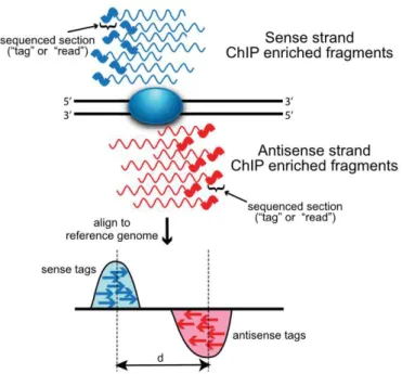
Interestingly, these different methods of replication origin detection show little concordance. SNS-seq studies already show poor overlap between each other, which might result from insufficient sequencing saturation (Urban et al. 2015). Also comparison of bubble-seq and SNS-seq only results in little agreement on the local level of single origins (Mesner et al. 2013). By contrast, initiation zones detected by OK-seq better align to bubbles than to SNS (Petryk et al. 2016). These little concordances between different approaches might originate from divergent replication events
23 |
considered. SNS-seq focuses on narrow sites of defined active replication events, while bubble-seq and OK-seq both detect broader regions which might contain several origins, but only one origin firing per cell. Especially OK-seq reveals zones of preferential origin activation within initiation zones and preferential replication termination in termination zones. However, this does not limit origins to initiation zones, as inefficient origins might also be present (and detected by SNS-seq) in termination zones.
Information about pre-RC sites would help in this context, to bridge the discrepancies of the different detection methods. To date, two attempts have been made to perform ChIP against ORC components in human cells and will be introduced in the following.
1.4.4 C
HIP-
SEQUENCING OF PRE-RC
COMPONENTSLittle is known about genome-wide pre-RC positioning in humans. Major difficulty is thereby the low enrichment over background that hampers precise pre-RC detection (Schepers and Papior 2010). Due to the low sequence specificity and potentially high on-and-off rates, chromatin association of metazoan ORC is dispersed, especially in comparison to other nuclear factors (such as transcription factors). To date, only two genome-wide studies are available that target either Orc1 (Dellino et al. 2013) or Orc2 (Miotto, Ji, and Struhl 2016) in human cells. Dellino et al. overcame the high background problem by performing Orc1 ChIP from low-density chromatin. Thereby, they detected a prominent association of ORC to transcriptional start sites (TSS). However, chromatin selection prior to ChIP also introduced a bias towards Orc1 sites belonging to early replication origins (Dellino et al. 2013). Furthermore, 44% of all Orc1 sites overlap with replication origins identified by SNS-seq (Picard et al. 2014) and Orc1 was rather found at borders of replication initiation zones determined by OK-seq (Petryk et al. 2016). Miotto et al. performed Orc2 ChIP-seq experiments in unfractionated chromatin. Orc2 peaks showed moderate concordance with replication origins detected from SNS-seq (13% of all SNS sites located within 1 kb distance of Orc2 binding sites, 41% within 10 kb distance, Miotto, Ji, and Struhl 2016). Comparing Orc2 positions with several chromatin features, the authors concluded that ORC positioning majorly depends on chromatin accessibility.
Taken together, these results reveal that while ORC ChIP-seq already shows a certain level of concordance with active replication data, the overall picture remains incomplete. As ORC does not bind any consensus sequence in humans, but seems to solely depend on chromatin accessibility, ORC positions likely vary from cell to cell, resulting in a scattered ChIP-seq profile. This phenomenon might be even more pronounced for Mcm2-7, as there is evidence for multiple Mcm2-7 helicases at individual origins, which might even translocate from their original loading site (Das and Rhind 2016; Hyrien 2016). Consequently, Mcm2-7 ChIP-seq would result in a broad Mcm2-7 distribution, mostly lacking clear peaks. This is presumably the
24 |
reason why human Mcm2-7 positions have not been assessed so far. However, as Mcm2-7 helicases can activate replication without spatial proximity to ORC, Mcm2-7 positions might be the missing link to conclusively connect replication initiation SNS-seq and ORC ChIP-seq results.
1.4.5 E
PSTEIN-B
ARR VIRUS GENOME AS MODEL OF HUMANDNA
REPLICATIONUntil now, genome-wide pre-RC ChIP experiments in humans have been unsuccessful. My laboratory used Epstein-Barr virus (EBV) as model system to study the relation between pre-RC formation and origin activation. EBV infects human B-cells and establishes a persistent latent infection. During latency, the EBV genome is maintained autonomously in proliferating cells and is thereby replicated in synchrony with the host cell genome by the cellular replication machinery (J. L. Yates and Guan 1991). For autonomous maintenance of the EBV episome, the cis-acting element oriP is required.
OriP consists of two distinct elements: the family of repeats (FR-element)
and the dyad symmetry (DS) element. Both elements contain binding arrays for the viral transactivator EBNA1 (Epstein-Barr virus nuclear antigen 1). By binding of EBNA1 to the FR-element, the EBV genome is tethered to the host chromatin during chromosome segregation (Marechal et al. 1999; Sears et al. 2003; Sears et al. 2004). EBNA1 binding to DS targets ORC to
oriP, designating oriP as exceptional origin, since origin licensing depends
on direct interaction between DNA, a targeting factor and ORC (Schepers et
al. 2001; Ritzi et al. 2003). Thus, oriP represents a very strong origin
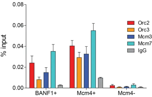
licensing site, however, this is not necessarily accompanied by efficient origin activation (Papior et al. 2012). My laboratory took advantage of full chromatinization of the EBV genome (166 kbp in size), and high EBV copy-numbers in the Burkitt’s lymphoma cell line Raji. Performing ChIP against the pre-RC components Orc2 and Mcm3, SNS isolation and micrococcal nuclease (MNase) digest in Raji cells, followed by hybridization against a designated microarray, allowed extensive analysis of the relation between origin licensing, activation and nucleosome positioning in a cell cycle resolved manner. They found 64 pre-RC sites, which also highly correlated with origin activation and S-phase-specific MNase sensitivities. Origin activation efficiencies were moderately influenced by AT-richness, however pre-RC sites themselves were independent of any specific primary motifs (Papior et al. 2012).
EBV employs cellular replication machinery to duplicate its genome and correlation of pre-RC and replication initiation sites provided insights in our understanding of replication origin organization in mammalian cells. It also showed that ChIP of human pre-RC components is technically feasible and sensibilized for need of careful controls. The next step will be to perform similar analysis on the human genome to answer the question of similar pre-RC organization genome-wide.
25 |
2. A
IM OF THE
T
HESIS
While the regulation of replication initiation in humans is already extensively studied and led to the identification of several contributing features, full understanding of the regulation of DNA replication can only be provided by combining replication initiation with the regulation of pre-RC positioning. Especially Mcm2-7 positioning – which has not been assessed so far – is expected to bridge the discrepancies between current ORC ChIP-seq and replication initiation data. In my laboratory, human pre-RC ChIP has been technically established using the EBV genome as model system. Intent of my thesis was to adopt this pre-RC ChIP technique for genome-wide ChIP-seq in Raji cells, by targeting the two major pre-RC subunits ORC and Mcm2-7. Containing the EBV genome as internal reference, Raji cells present the perfect control for ChIP-seq result quality. Once established, ChIP-seq will be also performed on embryonic stem (hES) cells, in collaboration with my co-supervisor Dr. Jean-Marc Lemaitre (Genome and Stem Cell Plasticity in Development and Ageing, IRMB, Montpellier, France). When compared to replication initiation, information about pre-RC positions and features that drive this positioning will contribute to the fundamental understanding of the relation between origin licensing and activation. Furthermore, pre-RC positions will be attributed to chromatin features, such as histone modifications. There is functional evidence for H4K20 methylation to be involved in DNA replication, however, we lack molecular understanding. Thus, ChIP-seq will also be performed for H4K20me1 and – me3, in order to directly conclude for possible implications between either H4K20 methylation and origin licensing. Possible candidates will be validated using an EBV-based plasmid system established in our laboratory, to functionally confirm the relation between H4K20 methylation and replication.
Consequently, I aim to combine genome-wide pre-RC positioning with the regulation of replication initiation. Simultaneously, I will evaluate the implication of H4K20 methylation as a promising histone modification candidate for regulation of DNA replication licensing and activation both by genome-wide correlation studies and by functional approaches.
26 |
3. M
ATERIAL AND
M
ETHODS
M
EDIA
,
M
ATERIAL
,
D
EVICES
,
C
HEMICALS AND
A
GENTS
In the following, cell culture media and supplements (Table 3.1), chemicals and agents (Table 3.2), Enzymes (Table 3.3), Kits (Table 3.4), Material (Table 3.5), and devices (Table 3.6) used in this work are listed.
Table 3.1 lists all media, supplements, antibiotics, and other substances that were used for cell culture.
TABLE 3.1: CELL CULTURE MEDIA, SUPPLEMENTS, ANTIBIOTICS AND AGENTS.
Cell culture media and supplements
Distributor BD Matrigel BD Biosciences CryoStor CS10 STEMCELL, Canada
Dimethylsulfoxide (DMSO) Carl Roth GmbH, Germany DMEM, high glucose, L-Glutamine Gibco, Thermo Fisher, USA
Essential 8 Basal Medium + supplement
Gibco, Thermo Fisher, USA
Fetal calf serum (FCS) Lot BS225160.5, Bio&SELL,
Germany
G418 Carl Roth GmbH, Germany
L-Glutamin Gibco, Thermo Fisher, USA Lipofectamine 2000 Invitrogen, Germany
MEM non-essential amino acids Gibco, Thermo Fisher, USA PBS Dulbecco, pH 7.2 Biochrom AG, Berlin
Penicillin Streptomycin Gibco, Thermo Fisher, USA RPMI 1640 Gibco, Thermo Fisher, USA Sodium pyruvate Gibco, Thermo Fisher, USA
TripLE Select Life technologies, USA Trypsin-EDTA Gibco, Thermo Fisher, USA Versene solution Gibco, Thermo Fisher, USA
27 |
In the following Table 3.2, all chemicals and agents used during this work are specified.
TABLE 3.2: CHEMICALS AND AGENTS.
Chemicals and agents Distributor
Ampicillin sodium salt Carl Roth GmbH, Germany ATX Ponceau S red staining
solution
Fluka Analytical, Germany
Bovine serum albumin Sigma-Aldrich, Germany Bradford reagent, Bio-Rad protein
assay
Bio-Rad, Germany
Chloroform Merck-Eurolab GmbH, Germany Deoxycholic acid (DOC) Sigma-Aldrich, Germany
Dithiothreitol (DTT) Sigma-Aldrich, Germany EGTA, Titriplex® Merck Millipore, Germany
Ethanol Merck-Eurolab GmbH, Germany
Ethylenediaminetetraacetic acid (EDTA)
Carl Roth GmbH, Germany
Formaldehyde, MeOH-free Thermo Scientific, USA
Glycerol AppliChem GmbH, Germany
Glycine Carl Roth GmbH, Germany
HEPES Sigma-Aldrich, Germany
Isoamyl alcohol Merck-Eurolab GmbH, Germany Methanol Merck-Eurolab GmbH, Germany NP-40 (Igepal CA-630) Sigma-Aldrich, Germany
Phenol Carl Roth GmbH, Germany
Polyacrylamide Carl Roth GmbH, Germany Propidium iodide Sigma-Aldrich, Germany
Salmon sperm Invitrogen, Germany
Sodium chloride Merck-Eurolab GmbH, Germany Sodium dodecylsulfate (SDS) Serva Electrophoresis GmbH,
Germany
Sodium lauroyl sarcosinate (Sarkosyl)
Sigma-Aldrich, Germany
Tris AppliChem GmbH, Germany
Triton-X-100 Sigma-Aldrich, Germany
28 |
Table 3.3 shows all enzymes employed during the study.
TABLE 3.3: ENZYMES.
Enzymes Distributor
Benzonase Sigma-Aldrich, Germany
DNase Roche, Germany
DpnI New England Biolabs, USA
Protease inhibitor complete Roche, Germany Proteinase K Roche, Germany RNase, DNase-free Roche, Germany
Kits used during this work are listed in Table 3.4.
TABLE 3.4: KITS.
Kits Distributor BD StemflowTM Human and Mouse
Pluripotent Stem Cell Analysis Kit
BD Biosciences, Germany
NTB buffer Macherey-Nagel, Germany NucleoSpin Extract II Kit Macherey-Nagel, Germany
Qubit HS dsDNA Invitrogen, Germany SYBR Green I Master Roche, Germany
In Table 3.5, all material needed for the experiments are specified.
TABLE 3.5: MATERIAL.
Material Distributor AFA Fiber & Cap tubes (12x12 mm) Covaris Inc., UK
Amersham Hybond ECL GE Healthcare, Germany CEA Blue Sensitive X-ray films Agfa Healthcare, Germany
Cell culture dishes and 6-well plates
Nunc GmbH, Germany
Cell strainer, 40 µm, 100 µm Corning Inc., USA
CoolCell LX Freezing Container Sigma-Aldrich, Germany
Cryotubes Nunc GmbH, Germany
Nalgen Nunc Cryo 1°C freezing container
29 |
Table 3.6 names the devices used during this work.
TABLE 3.6: DEVICES.
Devices Distributor Beckman JE-5.0 rotor with a large
separation chamber
Beckman-Coulter, Germany
Cole-Parmer Masterflex pump Cole-Parmer, USA
FACSCaliburTM BD Biosciences, Germany
semiDry blotting system Hoefer Scientific Instruments, USA Covaris S220 Covaris Inc., Germany
NanoDrop ND-1000 Spectrometer ThermoScientific, USA Qubit fluorometer Invitrogen, Germany Roche LightCycler 480 System Roche, Germany
Optimax X-ray film processor Rotec GmbH, Germany Electroporation system
Gene-Pulser II
Bio-Rad Laboratories, USA
B
IOLOGICAL
M
ETHODS
3.2.1 C
ELL CULTURECells with corresponding AGV-internal identification number, a short description and the respective media are listed in Table 3.7. More detailed cultivation information is given in the following.
RAJI CELLS
Raji cells (ATCC) were cultivated at 37°C and 5% CO2 in RPMI 1640 (Gibco,
Thermo Fisher, USA) supplemented with 8% FCS (Lot BS225160.5, Bio&SELL, Germany), 100 Units/ml Penicillin/ 100 µg/ml Streptomycin (Gibco, Thermo Fisher, USA), 1x MEM non-essential amino acids (Gibco, Thermo Fisher, USA), 2 mM L-Glutamin (Gibco, Thermo Fisher, USA), and 1 mM Sodium pyruvate (Gibco, Thermo Fisher, USA). Cells were routinely diluted to 2x10⁵ cells/ml and maximally grown to a density of 5x10⁵.
ADHERENT HEK293 CELLS
HEK293 EBNA1+ cells were cultivated at 37°C and 5% CO2 in DMEM (Gibco,
Thermo Fisher, USA) supplemented with 8% FCS (Lot BS225160.5, Bio&SELL, Germany), 100 Units/ml Penicillin/ 100 µg/ml Streptomycin (Gibco, Thermo Fisher, USA), and 220 µg/ml G418 (Carl Roth GmbH, Germany). Cells were grown to 80% confluence and routinely split 1:4. Therefore, cells were washed with PBS, treated with 0.25% Trypsin-EDTA (Thermo Fisher, USA) for 2 min at 37°C, carefully resuspended in new medium and seeded on a new culture dish.
30 |
GENERATING STABLE HEK293 CELL LINES
Cells were seeded in a 6-well (Nunc GmbH, Germany) to a density of 2x10⁵ and transfected with 3 µg linearized expression plasmid (see also Table 3.7) using Lipofectamine2000 according manufacturer’s instructions (Invitrogen, Germany). Transfected cells from one 6-well were plated in medium with 20 µg/ml Zeocine (Invitrogen, Germany) on three 150 mm culture dishes (Nunc GmbH, Germany) the next day. After two to three weeks, single colonies were selected and expanded in 6-well plates. Expression of the protein of interest was verified by immunoblot. Positive clones were expanded and frozen.
HES CELLS H9
HES cells were cultivated in standard conditions (Ludwig et al. 2006) at 37°C, 5% CO2. The hES cell line H9 was cultivated on BD Matrigel
(Basement membrane matrix, BD Biosciences) covered dishes. From BD Matrigel stocks, 72 µl aliquots were routinely prepared in chilled 2 ml Eppendorf tubes and stored at -20°C. For BD Matrigel preparation, one aliquot was thawed, resuspended in 1.5 ml Essential 8 Basal Medium (Gibco, Thermo Fisher, USA) and mixed with 4.5 ml medium (6 ml total). Of this mixture, 1 ml was distributed per 35 mm dish and incubated for at least 30 min at 37°C. For larger plates, surfaces and volumes were upscaled accordingly.
For passaging, cells were washed twice with PBS, 1 ml of Versene solution (Gibco, Thermo Fisher, USA) was added per 35 mm dish. Before cells detached, Versene solution was removed, new medium added and cells were gently detached from the dish. Cells were routinely diluted 1:6 once per week. Medium was changed every day.
CRYOPRESERVATION
HEK293 and Raji cells were concentrated by centrifugation (200g, 7 min, room temperature) and the cell pellet was resuspended in FCS 10% DMSO to a concentration of ~ 5x10⁷ cells/ml (HEK293) and 5x10⁶ cells/ml (Raji
cells). 1 ml cell suspension was aliquoted in 2 ml cryotubes (Nunc GmbH, Germany) and slowly cooled at a rate of -1°C/ min in a Nalgen Nunc Cryo 1°C freezing container (Nunc GmbH, Germany) at -80°C. After few days, cells were transferred in liquid nitrogen for long term storage. For thawing, cells were incubated at 37°C in water bath, washed with 30 ml fresh pre-warmed medium and plated accordingly.
HES cells were detached as previously described and concentrated by centrifugation (300g, 5 min, room temperature). 106 cells were
resuspended in 1 ml CryoStor CS10 (STEMCELL, Canada), transferred to 2 ml cryotubes, placed in CoolCell LX Freezing Container (Sigma, Germany) at -80°C. After few days, cells were transferred in liquid nitrogen.
31 | TABLE 3.7: ESSENTIAL CELL LINE INFORMATION.
Cell lines (AGV identification)
Description Medium
Raji (#1577) EBV-containing
Burkitt’s lymphoma cell line RPMI 1640, 8% FCS, 1% Penicillin/ Streptomycin, 2 mM Glutamin, 1% MEM Non-essential amino acids, 1 mM Sodium Pyruvate HEK293 EBNA1+(#1803) Human embryonic kidney cells, stably expressing EBNA1
DMEM, 8% FCS, 1% Penicillin/
Streptomycin, 220 µg/ml G418
HEK293 EBNA1+ Gal4 (#2116)
Generated from cell
line #1803 by integrating expression plasmid p5237 DMEM, 8% FCS, 1% Penicillin/ Streptomycin, 220 µg/ml G418, 20 µg/ml Zeocine
HEK293 EBNA1+ Gal4-Suv4-20h1 (#2680)
Generated from cell
line #1803 by integrating expression plasmid p5572 DMEM, 8% FCS, 1% Penicillin/ Streptomycin, 220 µg/ml G418, 20 µg/ml Zeocine
HEK293 EBNA1+ Gal4-PR-Set7 (#2113)
Generated from cell
line #1803 by integrating expression plasmid p5235 DMEM, 8% FCS, 1% Penicillin/ Streptomycin, 220 µg/ml G418, 20 µg/ml Zeocine
HEK293 EBNA1+ Gal4-PR-Set7SETmut (#2188)
Generated from cell
line #1803 by integrating expression plasmid p5236 DMEM, 8% FCS, 1% Penicillin/ Streptomycin, 220 µg/ml G418, 20 µg/ml Zeocine hES cells H9 (cultivated at the IRMB, Montpellier) Human embryonic stem cell line, cultivated from passage 15 to 24
Essential 8 Basal Medium + Essential 8 Supplement
3.2.2 C
ELL CYCLE FRACTIONATION BY CENTRIFUGAL ELUTRIATIONFor centrifugal elutriation, 5x10⁹ exponentially growing Raji cells were
harvested, washed with PBS and resuspended in 50 ml RPMI 1640/ 8% FCS/ 1mM EDTA/ 0.25 U/ml DNaseI (Roche, Germany). Concentrated cell suspension was passed through 40 µm cell strainer and injected in a Beckman JE-5.0 rotor with a large separation chamber turning at 1500 rpm and a flow rate of 30 ml/min controlled by a Cole-Parmer Masterflex pump.